Tag Archives: dentistry

Nanocoating to reduce dental implant failures

Scientists at Plymouth University (UK) have developed a nanocoating that could reduce the number of dental implant failures. From a March 24, 2017 news item on Nanowerk (Note: A link has been removed),

According to the American Academy of Implant Dentistry (AAID), 15 million Americans have crown or bridge replacements and three million have dental implants — with this latter number rising by 500,000 a year. The AAID estimates that the value of the American and European market for dental implants will rise to $4.2 billion by 2022.

Dental implants are a successful form of treatment for patients, yet according to a study published in 2005, five to 10 per cent of all dental implants fail.

The reasons for this failure are several-fold – mechanical problems, poor connection to the bones in which they are implanted, infection or rejection. When failure occurs the dental implant must be removed.

The main reason for dental implant failure is peri-implantitis. This is the destructive inflammatory process affecting the soft and hard tissues surrounding dental implants. This occurs when pathogenic microbes in the mouth and oral cavity develop into biofilms, which protects them and encourages growth. Peri-implantitis is caused when the biofilms develop on dental implants.

A research team comprising scientists from the School of Biological Sciences, Peninsula Schools of Medicine and Dentistry and the School of Engineering at the University of Plymouth, have joined forces to develop and evaluate the effectiveness of a new nanocoating for dental implants to reduce the risk of peri-implantitis.

The results of their work are published in the journal Nanotoxicology (“Antibacterial activity and biofilm inhibition by surface modified titanium alloy medical implants following application of silver, titanium dioxide and hydroxyapatite nanocoatings”).

A March 27, 2017 Plymouth University press release, which originated the news item, gives more details about the research,

In the study, the research team created a new approach using a combination of silver, titanium oxide and hydroxyapatite nanocoatings.

The application of the combination to the surface of titanium alloy implants successfully inhibited bacterial growth and reduced the formation of bacterial biofilm on the surface of the implants by 97.5 per cent.

Not only did the combination result in the effective eradication of infection, it created a surface with anti-biofilm properties which supported successful integration into surrounding bone and accelerated bone healing.

Professor Christopher Tredwin, Head of Plymouth University Peninsula School of Dentistry, commented:

“In this cross-Faculty study we have identified the means to protect dental implants against the most common cause of their failure. The potential of our work for increased patient comfort and satisfaction, and reduced costs, is great and we look forward to translating our findings into clinical practice.”

The University of Plymouth was the first university in the UK to secure Research Council Funding in Nanoscience and this project is the latest in a long line of projects investigating nanotechnology and human health.

Nanoscience activity at the University of Plymouth is led by Professor Richard Handy, who has represented the UK on matters relating to the Environmental Safety and Human Health of Nanomaterials at the Organisation for Economic Cooperation and Development (OECD). He commented:

“As yet there are no nano-specific guidelines in dental or medical implant legislation and we are, with colleagues elsewhere, guiding the way in this area. The EU recognises that medical devices and implants must: perform as expected for its intended use, and be better than similar items in the market; be safe for the intended use or safer than an existing item, and; be biocompatible or have negligible toxicity.”

He added:

“Our work has been about proving these criteria which we have done in vitro. The next step would be to demonstrate the effectiveness of our discovery, perhaps with animal models and then human volunteers.”

Dr Alexandros Besinis, Lecturer in Mechanical Engineering at the School of Engineering, University of Plymouth, led the research team. He commented:

“Current strategies to render the surface of dental implants antibacterial with the aim to prevent infection and peri-implantitis development, include application of antimicrobial coatings loaded with antibiotics or chlorhexidine. However, such approaches are usually effective only in the short-term, and the use of chlorhexidine has also been reported to be toxic to human cells. The significance of our new study is that we have successfully applied a dual-layered silver-hydroxyapatite nanocoating to titanium alloy medical implants which helps to overcome these risks.”

Dr Besinis has been an Honorary Teaching Fellow at the Peninsula School of Dentistry since 2011 and has recently joined the School of Engineering. His research interests focus on advanced engineering materials and the use of nanotechnology to build novel biomaterials and medical implants with improved mechanical, physical and antibacterial properties.

Here’s a link to and a citation for the paper,

Antibacterial activity and biofilm inhibition by surface modified titanium alloy medical implants following application of silver, titanium dioxide and hydroxyapatite nanocoatings by A. Besinis, S. D. Hadi, H. R. Le, C. Tredwin & R. D. Handy.  Nanotoxicology Volume 11, 2017 – Issue 3  Pages 327-338  http://dx.doi.org/10.1080/17435390.2017.1299890 Published online: 17 Mar 2017

This paper appears to be open access.
At the root of nanotechnology: advances in dentistry

I couldn’t resist the dental wordplay in my headline. Strictly speaking this posting features a research paper that is looking into dentistry’s nanotechnology-enabled future. From an Oct. 19, 2015 news item on phys.org,

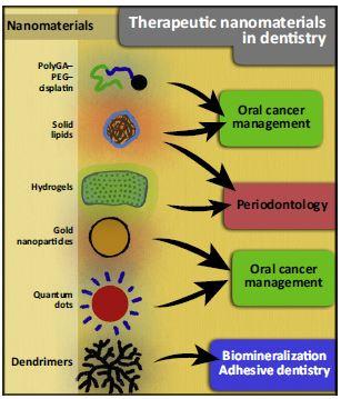
Have a cavity? Ask your dentist about filling it with a mixture of nanoparticles including silica and zirconia. These white fillings (known as nano-composite resins) resemble teeth better than their metal alternatives and are less likely to come loose or fracture teeth. This is just the beginning argue Brazilian scientists in a review of “nanodentistry,” published October 19 [2015] in Trends in Biotechnology. Next-generation dental materials incorporating nanotechnology aim to help teeth self-heal, rebuild enamel, and protect against bacterial infections.

An Oct. 19, 2015 Cell Press news release on EurekAlert, which originated the news item, expands on the theme,

“Nanotechnology can be faced sometimes as a paradigm that promised a lot and delivered very little,” says senior author Nelson Durán of the Universidade Estadual de Campinas. “The evolution of dental materials though nanotechnology is real and remarkable, reflecting on a billionaire market. In this way, dentistry was in fact one of the most benefited areas from the development of nanotechnology.”

Since the introduction of nano-composite resins a decade ago, engineers have been exploring how else nanotechnology can safely be used in the dentist’s office. Products could include antimicrobial adhesives made up of carbon nanotubes–creating a kind of wearable toothpaste–or quantum dots combined with cancer-specific antibodies that can be applied inside the mouth, emitting light if they detect any troublesome cells.

“The remineralization of enamel and dentin with the use of nanoparticles (incorporated in different vehicles), a key issue for improving the quality and longevity of resin restorations, is being currently investigated,” says co-author Amauri Jardim de Paula of Universidade Federal do Ceará. “A future perspective is that nanoparticles incorporated in dental materials will prevent and/or control oral diseases through their long-term release and action.”

Although nanodental technologies have evolved quickly, safety and cost will be barriers to getting them on the market. Some nanomaterials might be toxic to healthy cells, so any new nanomaterials to be used for dentistry would need formal pre-clinical and clinical trials before they can receive approval. Patients will also need to be told that a treatment will use materials in the nanometer size range and should be aware of any possible side effects. This new technology could also be expensive, and insurance companies may not want to foot the bill if treatments could be considered cosmetic; composite resins, for example, are still an out-of-pocket cost.

The review authors believe these hurdles can be overcome, however, and that new nanodental products should be available within a few years.

The research has been illustrated,

Caption: This schematic represents the current use and perspectives on the use of nanomaterials on therapeutic dentistry. Credit: Padovania et al./Trends in Biotechnology 2015

Caption: This schematic represents the current use and perspectives on the use of nanomaterials on therapeutic dentistry.
Credit: Padovania et al./Trends in Biotechnology 2015

Here’s a link to and a citation for the paper,

Advances in Dental Materials through Nanotechnology: Facts, Perspectives and Toxicological Aspects by Gislaine C. Padovani, Victor P. Feitosa, Salvatore Sauro, Franklin R. Tay, Gabriela Durán, Amauri J. Paula, & Nelson Durán. Trends in Biotechnology – Cell DOI: http://dx.doi.org/10.1016/j.tibtech.2015.09.005 Publication stage: In Press Corrected Proof Published online Oct. 19, 2015

The paper appears to be open access.

Bone implants and restorative dentistry at the University of Malaya

The research into biomedical implants at the University of Malaya is part of an international effort and is in response to a demographic reality, hugely increased populations of the aged. From a Sept. 18, 2014 news item on ScienceDaily,

A major success in developing new biomedical implants with the ability to accelerate bone healing has been reported by a group of scientists from the Department of Restorative Dentistry, University of Malaya. This stems from a project partly funded by HIR [High Impact Research] and also involves Mr. Alireza Yaghoubi, HIR Young Scientist.

According to WHO (World Health Organization), between 2000 and 2050, the world’s population over 60 years is expected to increase from 605 million to more than 2 billion. This trend is particularly more prominent in Asia and Europe where in some countries by 2050, the majority of people will be older than 50. That is why in recent years, regenerative medicine has been among the most active and well-funded research areas in many developing nations.

As part of this global effort to realize better treatments for age-related conditions, a group of scientists from the department of restorative dentistry, University of Malaya and four other universities in the US have recently reported a major success in developing new biomedical implants with the ability to accelerate bone healing.

Two studies were published according to the Sept.15, 2014 University of Malaya news release, which originated the news item,

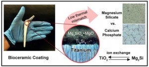
The two studies funded by the National Science Fund (NSF) in the US and the High Impact Research (HIR) program in Malaysia tackled the issue of bone-implant integration from different angles. In the first study appearing on the front cover of the July issue of Applied Surface Science, researchers demonstrated a mechanically superior bioactive coating based on magnesium silicates rather than the commercially available calcium phosphate which develops microcracks during preparation and delaminates under pressure. The new material owing to its lower thermal mismatch with titanium can prolong the durability of load-bearing orthopedic implants and reduce chances of post-surgery complications.

The other study published in the American Chemical Society’s Applied Materials & Interfaces reported a method for fabricating titanium implants with special surface topographies which double the chance of cell viability in early stages. The new technique is also much simpler as compared to the existing ones and therefore enables the preparation of personalized implants at the fraction of time and cost while offering a higher mechanical reliability.

Alireza Yaghoubi, the corresponding author of both studies believes that we are moving toward a future of personalized products. “It is very much like your taste in music and TV shows. People are different and the new trend in biotechnology is to make personalized medicine that matches the patient’s needs” Yaghoubi said. He continued “With regard to implants, we have the problem of variations in bone density in patients with osteoporosis and in some cases, even healthy individuals. Finding ways to integrate the implants with bone tissues can be challenging. There are also problems with the long-term performance of implants, such as release of debris from bioactive films which can potentially lead to osteolysis and chronic inflammation”.

The new technique employed by the scientists to create titanium implants with desirable surface properties uses microwave heating to create a porosity gradient on top of a dense core. The principles are very similar to a kitchen microwave and how it can make cooking easier, however apparently the fast heating capability is not only useful in cooking but it has numerous industrial applications. Prof. Bhaduri, the Director of Multi-functional materials laboratory at University of Toledo says that they have been using microwave for years to simplify fabrication of complex metallic components. “We needed a way to streamline the process and microwave sintering was a natural fit. With our new method, making the implant from titanium powder in custom sizes and with specific surface topographies is achieved through one easy step.” Bhaduri elaborated.

Researchers are hoping to carry out the clinical trial for this new generation of implants in order to make them available to the market soon. Dr. Kutty, one of the lead authors suggests that there is still room for improvement. Kutty concluded that “Roughened surfaces and bioceramics have desirable effects on osseointegration, but we are not stopping there. We are now developing new ways to use peptides for enhancing the performance of implants even further.”

This image provides an illustration of the proposed new material for implants,

The artwork appeared on the front cover of Applied Surface Science summarizes the benefits of a new bioceramic coating versus the commercially available Calcium Phosphate which develops microcracks during processing and may later cause osteolysis in load-bearing orthopedic implants. Courtesy: University of Malaya

The artwork appeared on the front cover of Applied Surface Science summarizes the benefits of a new bioceramic coating versus the commercially available Calcium Phosphate which develops microcracks during processing and may later cause osteolysis in load-bearing orthopedic implants. Courtesy: University of Malaya

Here are links to and citations for the papers,

Electrophoretic deposition of magnesium silicates on titanium implants: Ion migration and silicide interfaces by M. Afshar-Mohajer, A. Yaghoubi, S. Ramesh, A.R. Bushroa, K.M.C. Chin, C.C. Tin, and W.S. Chiu.  Applied Surface Science (2014) , Volume 307, 15 July 2014, Pages 1–6, DOI: 10.1016/j.apsusc.2014.04.033

Microwave-assisted Fabrication of Titanium Implants with Controlled Surface Topography for Rapid Bone Healing by Muralithran G. Kutty, Alok De, Sarit B. Bhaduri, and Alireza Yaghoubi. ACS Appl. Mater. Interfaces, 2014, 6 (16), pp 13587–13593 DOI: 10.1021/am502967n Publication Date (Web): August 6, 2014

Copyright © 2014 American Chemical Society

Both of these papers are behind paywalls.

Diamonds in your teeth—for health reasons

Scientists at the University of California at Los Angeles (UCLA) in collaboration with their colleagues at the NanoCarbon Research Institute (Japan) are investigating the possibility of using nanodiamonds to promote bone growth that supports dental implants. From the Sept.18, 2013 news item on ScienceDaily,

UCLA researchers have discovered that diamonds on a much, much smaller scale than those used in jewelry could be used to promote bone growth and the durability of dental implants.

Nanodiamonds, which are created as byproducts of conventional mining and refining operations, are approximately four to five nanometers in diameter and are shaped like tiny soccer balls. Scientists from the UCLA School of Dentistry, the UCLA Department of Bioengineering and Northwestern University, along with collaborators at the NanoCarbon Research Institute in Japan, may have found a way to use them to improve bone growth and combat osteonecrosis, a potentially debilitating disease in which bones break down due to reduced blood flow.

The Sept. 17,2013 UCLA news release by Brianna Deane (also on EurekAlert), which originated the news item, describes how osteonecrosis affects bones and the impact that this new technique using nanodiamonds could have on applications for regenerative medicine (Note: A link has been removed),

When osteonecrosis affects the jaw, it can prevent people from eating and speaking; when it occurs near joints, it can restrict or preclude movement. Bone loss also occurs next to implants such as prosthetic joints or teeth, which leads to the implants becoming loose — or failing.
Implant failures necessitate additional procedures, which can be painful and expensive, and can jeopardize the function the patient had gained with an implant. These challenges are exacerbated when the disease occurs in the mouth, where there is a limited supply of local bone that can be used to secure the prosthetic tooth, a key consideration for both functional and aesthetic reasons.
….
During bone repair operations, which are typically costly and time-consuming, doctors insert a sponge through invasive surgery to locally administer proteins that promote bone growth, such as bone morphogenic protein.
Ho’s team discovered that using nanodiamonds to deliver these proteins has the potential to be more effective than the conventional approaches. The study found that nanodiamonds, which are invisible to the human eye, bind rapidly to both bone morphogenetic protein  and fibroblast growth factor, demonstrating that the proteins can be simultaneously delivered using one vehicle. The unique surface of the diamonds allows the proteins to be delivered more slowly, which may allow the affected area to be treated for a longer period of time. Furthermore, the nanodiamonds can be administered non-invasively, such as by an injection or an oral rinse.
“We’ve conducted several comprehensive studies, in both cells and animal models, looking at the safety of the nanodiamond particles,” said Laura Moore, the first author of the study and an M.D.-Ph.D. student at Northwestern University under the mentorship of Dr. Ho. “Initial studies indicate that they are well tolerated, which further increases their potential in dental and bone repair applications.”
“Nanodiamonds are versatile platforms,” said Ho, who is also professor of bioengineering and a member of the Jonsson Comprehensive Cancer Center and the California NanoSystems Institute. “Because they are useful for delivering such a broad range of therapies, nanodiamonds have the potential to impact several other facets of oral, maxillofacial and orthopedic surgery, as well as regenerative medicine.”
Ho’s team previously showed that nanodiamonds in preclinical models were effective at treating multiple forms of cancer. Because osteonecrosis can be a side effect of chemotherapy, the group decided to examine whether nanodiamonds might help treat the bone loss as well. Results from the new study could open the door for this versatile material to be used to address multiple challenges in drug delivery, regenerative medicine and other fields.

Here’s a citation for and a link to the researchers’ published paper,

Multi-protein Delivery by Nanodiamonds Promotes Bone Formation by L. Moore, M. Gatica, H. Kim, E. Osawa, & D. Ho. Published online before print September 17, 2013, doi: 10.1177/0022034513504952 JDR September 17, 2013 0022034513504952

This paper is behind a paywall.

Dental fillings that improve your teeth

If you have lousy teeth, this is exciting news. From the May 2, 2012 news item on Nanowerk (I have removed a link),

Scientists using nanotechology at the University of Maryland School of Dentistry have created the first cavity-filling composite that kills harmful bacteria and regenerates tooth structure lost to bacterial decay. [emphasis mine]

Rather than just limiting decay with conventional fillings, the new composite is a revolutionary dental weapon to control harmful bacteria, which co-exist in the natural colony of microorganisms in the mouth, says professor Huakun (Hockin) Xu, PhD, MS. [emphasis mine]

While the possibilities are promising, I find the idea of a weapon in my mouth disconcerting. (They might want to check out their metaphors a little more closely.) Moving on, there’s a little more detail about this new composite  (from the news item),

Fillings made from the School of Dentistry’s new nanocomposite, with antibacterial primer and antibacterial adhesive, should last longer than the typical five to 10 years, though the scientists have not thoroughly tested longevity. Xu says a key component of the new nanocomposite and nano-structured adhesive is calcium phosphate nanoparticles that regenerate tooth minerals. The antibacterial component has a base of quaternary ammonium and silver nanoparticles along with a high pH. The alkaline pH limits acid production by tooth bacteria.

“The bottom line is we are continuing to improve these materials and making them stronger in their antibacterial and remineralizing capacities as well as increasing their longevity,” Xu says.

The new products have been laboratory tested using biofilms from saliva of volunteers. The Xu team is planning to next test its products in animal teeth and in human volunteers in collaboration with the Federal University of Ceara in Brazil.

The folks at the enewsparkforest blog are not quite so sanguine about this dental development as per their May 3, 2012 posting on the topic (I have removed llinks),

A study conducted in 2008 and confirmed by another study in 2009 shows that washing nano-silver textiles releases substantial amounts of the nanosilver into the laundry discharge water, which will ultimately reach natural waterways and potentially poison fish and other aquatic organisms. A study found nanosilver to cause malformations and to be lethal to small fish at various stages of development since they are able to cross the egg membranes and move into the fish embryos. A 2010 study by scientists at Oregon State University and in the European Union highlights the major regulatory and educational issues that they believe should be considered before nanoparticles are used in pesticides.

As Dexter Johnson in his May 3, 2012 posting on his Nanoclast blog (on the Institute for Electrical and Electronics Engineers website) notes,

The researchers are continuing with their animal and human testing with the nanocomposite. Given that some sectors of the public are concerned about the potential risks of silver nanoparticles, they should probably take a look at the issue as part of their research.

This is not unreasonable especially in light of the concern some folks have had over mercury in dental fillings. Sufficient concern by the way to occasion this cautionary note from Health Canada (from the Mercury and Human Health webpage on their website),

Minimizing Your Risk

Elemental mercury from dental fillings does not generally pose a health risk. There is, however, a fairly small number of people who are hypersensitive to mercury. While Health Canada does not recommend that you replace existing mercury dental fillings, it does suggest that when the fillings need to be repaired, you may want to consider using a product that does not contain mercury.

Pregnant women, people allergic to mercury and those with impaired kidney function should avoid mercury fillings. Whenever possible, amalgam fillings should not be removed when you are pregnant because the removal may expose you to mercury vapour. When appropriate, the primary teeth of children should be filled with non-mercury materials.

Side note: I find it interesting that while Health Canada has not banned the use of mercury in fillings, it does advise against adding more mercury-laced fillings to your mouth and/or using them in your children’s primary teeth, if possible.

Getting back to silver nanoparticles in our mouths, I reiterate Dexter’s suggestion.

Printing bones

Apparently all you need is an inkjet printer and some researchers from Washington State University (WSU) at Pullman to create new bone. From the Nov. 29, 2011 news item (written by Eric Sorenson of WSU) on Nanowerk,

Washington State University researchers have used a 3D printer to create a bone-like material and structure that can be used in orthopedic procedures, dental work and to deliver medicine for treating osteoporosis. Paired with actual bone, it acts as a scaffold for new bone to grow on and ultimately dissolves with no apparent ill effects. [emphasis mine]

The authors report on successful in vitro tests in the journal Dental Materials (“Effects of silica and zinc oxide doping on mechanical and biological properties of 3D printed tricalcium phosphate tissue engineering scaffolds” [behind a paywall]) and say they’re already seeing promising results with in vivo tests on rats and rabbits. It’s possible that doctors will be able to custom order replacement bone tissue in a few years, said Susmita Bose, co-author and professor in WSU’s School of Mechanical and Materials Engineering.

The printer works by having an inkjet spray a plastic binder over a bed of powder in layers of 20 microns, about half the width of a human hair. Following a computer’s directions, it creates a channeled cylinder the size of a pencil eraser.

After just a week in a medium with immature human bone cells, the scaffold was supporting a network of new bone cells.

Here’s a video of Dr. Bose discussing the inkjet printer that produces bone-like material,

The Nov. 30, 2011 news item about the bone scaffolding work on BBC News adds more detail,

Prof Bose’s team have spent four years developing the bone-like substance.

Their breakthrough came when they discovered a way to double the strength of the main ceramic powder – calcium phosphate – by adding silica and zinc oxide.

To create the scaffold shapes they customised a printer which had originally been designed to make three-dimensional metal objects.

It sprayed a plastic binder over the loose powder in layers half as thick as the width of a human hair.

The process was repeated layer by layer until completed, at which point the scaffold was dried, cleaned and then baked for two hours at 1250C (2282F).

Earlier this year I highlighted a story about a trachea transplant where they used scaffolding to grow trachea cells in much the same way the WSU team is using a scaffolding to grow bone cells. Here are the posts about the trachea transplant and scaffolding from the first to the last,

Body parts nano style

Making nanotechnology-enabled body parts

More on synthetic windpipe; Swedes and Italians talk about nanoscience and medicine

Nano dental technique promises a world without root canals

Unfortunately, it’s not here yet but according to a news item on Nanowerk,

Scientists are reporting an advance toward the next big treatment revolution in dentistry — the era in which root canal therapy brings diseased teeth back to life, rather than leaving a “non-vital” or dead tooth in the mouth. In a report in the monthly journal ACS Nano, they describe a first-of-its-kind, nano-sized dental film that shows early promise for achieving this long-sought goal (“Nanostructured Assemblies for Dental Application”).

I gather from further reading into the news item that we’re a long way from clinical trials (drat!),

The scientists are reporting development of a multilayered, nano-sized film — only 1/50,000th the thickness of a human hair containing a substance that could help regenerate dental pulp. Previous studies show that the substance, called alpha melanocyte stimulating hormone, or alpha-MSH, has anti-inflammatory properties. The scientists showed in laboratory tests alpha-MSH combined with a widely-used polymer produced a material that fights inflammation in dental pulp fibroblasts. Fibroblasts are the main type of cell found in dental pulp. Nano-films containing alpha-MSH also increased the number of these cells. This could help revitalize damaged teeth and reduce the need for a root canal procedure, the scientists suggest.

Sometimes I just like to focus on the benefits that nanotechnology offers and this would be a major benefit for a lot of people around the world.