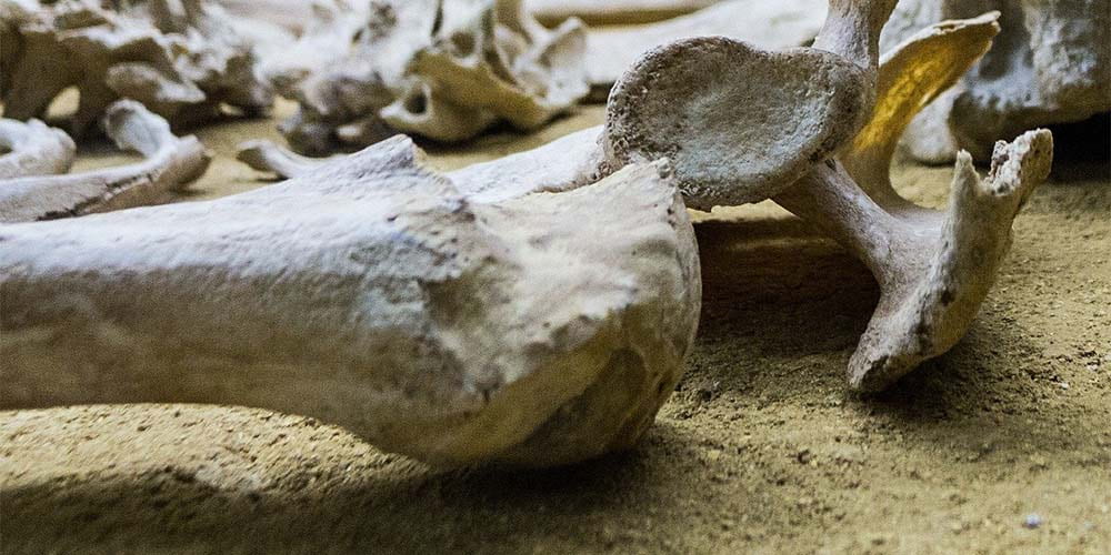
What a great image of bones! This December 3, 2020 University of Arkansas news release (also on EurekAlert) by Matt McGowan features research focused on bone material looks exciting. The date for the second study citation and link that I have listed (at the end of this posting) suggests the more recent study may have been initially overlooked in the deluge of COVID-19 research we are experiencing,
University of Arkansas researchers Marco Fielder and Arun Nair have conducted the first study of the combined nanoscale effects of water and mineral content on the deformation mechanisms and thermal properties of collagen, the essence of bone material.
The researchers also compared the results to the same properties of non-mineralized collagen reinforced with carbon nanotubes, which have shown promise as a reinforcing material for bio-composites. This research aids in the development of synthetic materials to mimic bone.
Using molecular dynamics — in this case a computer simulation of the physical movements of atoms and molecules — Nair and Fielder examined the mechanics and thermal properties of collagen-based bio-composites containing different weight percentages of minerals, water and carbon nanotubes when subjected to external loads.
They found that variations of water and mineral content had a strong impact on the mechanical behavior and properties of the bio-composites, the structure of which mimics nanoscale bone composition. With increased hydration, the bio-composites became more vulnerable to stress. Additionally, Nair and Fielder found that the presence of carbon nanotubes in non-mineralized collagen reduced the deformation of the gap regions.
The researchers also tested stiffness, which is the standard measurement of a material’s resistance to deformation. Both mineralized and non-mineralized collagen bio-composites demonstrated less stability with greater water content. Composites with 40% mineralization were twice as strong as those without minerals, regardless of the amount of water content. Stiffness of composites with carbon nanotubes was comparable to that of the mineralized collagen.
“As the degree of mineralization or carbon nanotube content of the collagenous bio-composites increased, the effect of water to change the magnitude of deformation decreased,” Fielder said.
The bio-composites made of collagen and carbon nanotubes were also found to have a higher specific heat than the studied mineralized collagen bio-composites, making them more likely to be resistant to thermal damage that could occur during implantation or functional use of the composite. Like most biological materials, bone is a hierarchical – with different structures at different length scales. At the microscale level, bone is made of collagen fibers, composed of smaller nanofibers called fibrils, which are a composite of collagen proteins, mineralized crystals called apatite and water. Collagen fibrils overlap each other in some areas and are separated by gaps in other areas.
“Though several studies have characterized the mechanics of fibrils, the effects of variation and distribution of water and mineral content in fibril gap and overlap regions are unexplored,” said Nair, who is an associate professor of mechanical engineering. “Exploring these regions builds an understanding of the structure of bone, which is important for uncovering its material properties. If we understand these properties, we can design and build better bio-inspired materials and bio-composites.”
Here are links and citations for both papers mentioned in the news release,
Two research groups are working to the same end where bone marrow is concerned, encourage bone cell growth, but they are using different strategies.
University of British Columbia and McMaster University (Canada)
Caption: Researchers treated nanocrystals derived from plant cellulose so that they can link up and form a strong but lightweight sponge (an aerogel) that can compress or expand as needed to completely fill out a bone cavity. Credit: Clare Kiernan, UBC
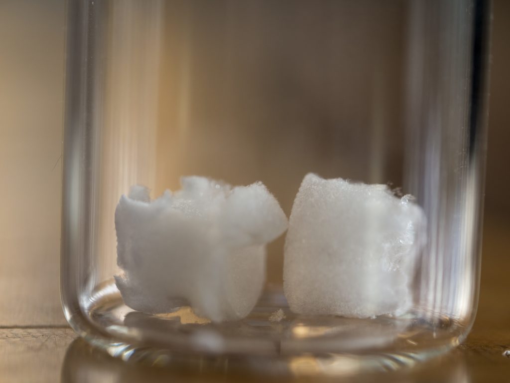
The samples look a little like teeth, don’t they?
Before diving into the research news, there’s a terminology issue that should be noted as you’ll see when you read the news/press releases. Nanocrystal cellulose/nanocrystalline cellulose (NCC) is a term coined by Canadian researchers. Since those early day, most researchers, internationally, have adopted the term cellulose nanocrystals (CNC) as the standard term. It fits better with the naming conventions for other nnanocellulose materials such as cellulose nanofibrils, etc. By the way, a Canadian company (CelluForce) that produces CNC retained the term nanocrystalline cellulose (NCC) as a trademark for the product, CelluForce NCC®.
For anyone not familiar with aerogels, what the University of British Columbia (UBC) and McMaster University researchers are developing, are also popularly known known as ‘frozen smoke’ (see the Aerogel Wikipedia entry for more).
Researchers from the University of British Columbia and McMaster University have developed what could be the bone implant material of the future: an airy, foamlike substance that can be injected into the body and provide scaffolding for the growth of new bone.
It’s made by treating nanocrystals derived from plant cellulose so that they link up and form a strong but lightweight sponge — technically speaking, an aerogel — that can compress or expand as needed to completely fill out a bone cavity.
“Most bone graft or implants are made of hard, brittle ceramic that doesn’t always conform to the shape of the hole, and those gaps can lead to poor growth of the bone and implant failure,” said study author Daniel Osorio, a PhD student in chemical engineering at McMaster. “We created this cellulose nanocrystal aerogel as a more effective alternative to these synthetic materials.”
For their research, the team worked with two groups of rats, with the first group receiving the aerogel implants and the second group receiving none. Results showed that the group with implants saw 33 per cent more bone growth at the three-week mark and 50 per cent more bone growth at the 12-week mark, compared to the controls.
“These findings show, for the first time in a lab setting, that a cellulose nanocrystal aerogel can support new bone growth,” said study co-author Emily Cranston, a professor of wood science and chemical and biological engineering who holds the President’s Excellence Chair in Forest Bio-products at UBC. She added that the implant should break down into non-toxic components in the body as the bone starts to heal.
The innovation can potentially fill a niche in the $2-billion bone graft market in North America, said study co-author Kathryn Grandfield, a professor of materials science and engineering, and biomedical engineering at McMaster who supervised the work.
“We can see this aerogel being used for a number of applications including dental implants and spinal and joint replacement surgeries,” said Grandfield. “And it will be economical because the raw material, the nanocellulose, is already being produced in commercial quantities.”
The researchers say it will be some time before the aerogel makes it out of the lab and into the operating room.
“This summer, we will study the mechanisms between the bone and implant that lead to bone growth,” said Grandfield. “We’ll also look at how the implant degrades using advanced microscopes. After that, more biological testing will be required before it is ready for clinical trials.”
Here’s a link to and a citation for the paper,
Cross-linked cellulose nanocrystal aerogels as viable bone tissue scaffolds by Daniel A. Osorio, Bryan E. J. Lee, Jacek M. Kwiecien, Xiaoyue Wang, Iflah Shahid, Ariana L. Hurley, Emily D. Cranston and Kathryn Grandfield. Acta Biomaterialia Volume 87, 15 March 2019, Pages 152-165 DOI: https://doi.org/10.1016/j.actbio.2019.01.049
This paper is behind a paywall
Now for the Russian team.
National University of Science and Technology “MISIS” (formerly part of the Moscow Mining Academy)
These scientists have adopted a different strategy as you’ll see in the March 19, 2019 news item on Nanwerk, which, coincidentally, was published on the same day as the Canadian research,
Scientists from the National University of Science and Technology “MISIS” developed a nanomaterial, which will be able to rstore the internal structure of bones damaged due to osteoporosis and osteomyelitis. A special bioactive coating of the material helped to increase the rate of division of bone cells by 3 times. In the future, it can allow to abandon bone marrow transplantation and patients will no longer need to wait for suitable donor material.
Such diseases as osteoporosis and osteomyelitis cause irreversible degenerative changes in the bone structure. Such diseases require serious complex treatment and surgery and transplantation of the destroyed bone marrow in severe stages. Donor material should have a number of compatibility indicators and even close relationship with the donor cannot guarantee full compatibility.
Research group from the National University of Science and Technology “MISIS” (NUST MISIS), led by Anton Manakhov (Laboratory for Inorganic Nanomaterials) developed material that will allow to restore damaged internal bone structure without bone marrow transplantation. It is based on nanofibers of polycaprolactone, which is biocompatible self-dissolvable material. Earlier, the same research group has already worked with this material: by adding antibiotics to the nanofibers, scientists have managed to create non-changeable healing bandages.
“If we want the implant to take, not only biocompatibility is needed, but also activation of the natural cell growth on the surface of the material. Polycaprolactone as such is a hydrophobic material, meaning, and cells feel uncomfortable on its surface. They gather on the smooth surface and divide extremely slow”, Elizaveta Permyakova, one of the co-authors and researcher at NUST MISIS Laboratory for Inorganic Nanomaterials, explains.
To increase the hydrophilicity of the material, a thin layer of bioactive film consisting of titanium, calcium, phosphorus, carbon, oxygen and nitrogen (TiCaPCON) was deposited on it. The structure of nanofibers identical to the cell surface was preserved. These films, when immersed in a special salt medium, which chemical composition is identical to human blood plasma, are able to form on its surface a special layer of calcium and phosphorus, which in natural conditions forms the main part of the bone. Due to the chemical similarity and the structure of nanofibers, new bone tissue begins to grow rapidly on this layer. Most importantly, polycaprolactone nanofibers dissolve, having fulfilled their functions. Only new “native” tissue remains in the bone.
In the experimental part of the study, the researchers compared the rate of division of osteoblastic bone cells on the surface of the modified and unmodified material. It was found that the modified material TiCaPCON has a high hydrophilicity. In contrast to the unmodified material, the cells on its surface felt clearly more comfortable, and divided three times faster.
According to scientists, such results open up great prospects for further work with modified polycaprolactone nanofibers as an alternative to bone marrow transplantation.
Here’s a link to and a citation for the paper,
Bioactive TiCaPCON-coated PCL nanofibers as a promising material for bone tissue engineering by Anton Manakhov, Elizaveta S. Permyakova, Sergey Ershov, Alexander Sheveyko, Andrey Kovalskii, Josef Polčák, Irina Y. Zhitnyak, Natalia A. Gloushankova, Lenka Zajíčková, Dmitry V. Shtansky. Applied Surface Science Volume 479, 15 June 2019, Pages 796-802 DOI: https://doi.org/10.1016/j.apsusc.2019.02.163
A July 3, 2018 Canadian Light Source news release by Colleen MacPherson describes an investigation into how dental implants and bones interact with the hope of making dental implantation safer and more certain,
Research carried out recently at the Canadian Light Source (CLS) [also known as a synchrotron] in Saskatoon [Saskatchewan, Canada] has revealed promising information about how to build a better dental implant, one that integrates more readily with bone to reduce the risk of failure.
“There are millions of dental and orthopedic implants placed every year in North America and a certain number of them always fail, even in healthy people with healthy bone,” said Kathryn Grandfield, assistant professor in the Department of Materials Science and Engineering at McMaster University in Hamilton [Ontario, Canada].
A dental implant restores function after a tooth is lost or removed. It is usually a screw shaped implant that is placed in the jaw bone and acts as the tooth roots, while an artificial tooth is placed on top. The implant portion is the artificial root that holds an artificial tooth in place.
Grandfield led a study that showed altering the surface of a titanium implant improved its connection to the surrounding bone. It is a finding that may well be applicable to other kinds of metal implants, including engineered knees and hips, and even plates used to secure bone fractures.
About three million people in North America receive dental implants annually. While the failure rate is only one to two percent, “one or two percent of three million is a lot,” she said. Orthopedic implants fail up to five per cent of the time within the first 10 years; the expected life of these devices is about 20 to 25 years, she added.
“What we’re trying to discover is why they fail, and why the implants that are successful work. Our goal is to understand the bone-implant interface in order to improve the design of implants.”
Grandfield’s research team, which included post-doctoral fellow Xiaoyue Wang and McMaster colleague Adam Hitchcock from the Department of Chemistry and Chemical Biology. The team members used the soft X-ray spectromicroscopy beamline at the CLS as well as facilities at the Canadian Centre for Electron Microscopy in Hamilton to examine a failed dental implant that had to be removed, along with a small amount of surrounding bone, from a patient. Prior to implantation, a laser beam was used to alter the implant, to roughen the surface, creating what looked like “little volcanoes” on the surface. After removal from the patient, the point of connection between bone and metal was then carefully studied to understand how the implant behaved.
“What we found was that the surface modification changed the chemistry of the implant. The modification created an oxide layer, but not a bad oxide layer like rust but a better, more beneficial layer that helps integrate with bone material.”
The research results were published in Advanced Materials Interfaces in May [2018], ensuring the findings are available “to implant companies interested in using nanotechnology to change the structure of the implants they produce,” said Grandfield.
The next steps in the research will be to apply the surface modification technique to other types of implants “to be able to understand fully how they function.” Grandfield added the research done at the CLS involved healthy bone “so I’d be really interested in seeing the response when bone is a bit more compromised by age or disease, like osteoporosis. We need to find the best surface modifications … because the technology we have today to treat patients with healthier bone may not be sufficient with compromised bone.”
Here’s a link to (even though it’s in the news release text) and a citation for the paper,
The University of British Columbia’s (UBC) Peter Wall Institute for Advanced Studies (PWIAS) is hosting along with local biotech firm, Aspect Biosystems, a May 3 -5, 2017 international research roundtable known as ‘Printing the Future of Therapeutics in 3D‘.
A May 1, 2017 UBC news release (received via email) offers some insight into the field of bioprinting from one of the roundtable organizers,
This week, global experts will gather [4] at the University of British
Columbia to discuss the latest trends in 3D bioprinting—a technology
used to create living tissues and organs.
In this Q&A, UBC chemical and biological engineering professor
Vikramaditya Yadav [5], who is also with the Regenerative Medicine
Cluster Initiative in B.C., explains how bioprinting could potentially
transform healthcare and drug development, and highlights Canadian
innovations in this field.
WHY IS 3D BIOPRINTING SIGNIFICANT?
Bioprinted tissues or organs could allow scientists to predict
beforehand how a drug will interact within the body. For every
life-saving therapeutic drug that makes its way into our medicine
cabinets, Health Canada blocks the entry of nine drugs because they are
proven unsafe or ineffective. Eliminating poor-quality drug candidates
to reduce development costs—and therefore the cost to consumers—has
never been more urgent.
In Canada alone, nearly 4,500 individuals are waiting to be matched with
organ donors. If and when bioprinters evolve to the point where they can
manufacture implantable organs, the concept of an organ transplant
waiting list would cease to exist. And bioprinted tissues and organs
from a patient’s own healthy cells could potentially reduce the risk
of transplant rejection and related challenges.
HOW IS THIS TECHNOLOGY CURRENTLY BEING USED?
Skin, cartilage and bone, and blood vessels are some of the tissue types
that have been successfully constructed using bioprinting. Two of the
most active players are the Wake Forest Institute for Regenerative
Medicine in North Carolina, which reports that its bioprinters can make
enough replacement skin to cover a burn with 10 times less healthy
tissue than is usually needed, and California-based Organovo, which
makes its kidney and liver tissue commercially available to
pharmaceutical companies for drug testing.
Beyond medicine, bioprinting has already been commercialized to print
meat and artificial leather. It’s been estimated that the global
bioprinting market will hit $2 billion by 2021.
HOW IS CANADA INVOLVED IN THIS FIELD?
Canada is home to some of the most innovative research clusters and
start-up companies in the field. The UBC spin-off Aspect Biosystems [6]
has pioneered a bioprinting paradigm that rapidly prints on-demand
tissues. It has successfully generated tissues found in human lungs.
Many initiatives at Canadian universities are laying strong foundations
for the translation of bioprinting and tissue engineering into
mainstream medical technologies. These include the Regenerative Medicine
Cluster Initiative in B.C., which is headed by UBC, and the University
of Toronto’s Institute of Biomaterials and Biomedical Engineering.
WHAT ETHICAL ISSUES DOES BIOPRINTING CREATE?
There are concerns about the quality of the printed tissues. It’s
important to note that the U.S. Food and Drug Administration and Health
Canada are dedicating entire divisions to regulation of biomanufactured
products and biomedical devices, and the FDA also has a special division
that focuses on regulation of additive manufacturing – another name
for 3D printing.
These regulatory bodies have an impressive track record that should
assuage concerns about the marketing of substandard tissue. But cost and
pricing are arguably much more complex issues.
Some ethicists have also raised questions about whether society is not
too far away from creating Replicants, à la _Blade Runner_. The idea is
fascinating, scary and ethically grey. In theory, if one could replace
the extracellular matrix of bones and muscles with a stronger substitute
and use cells that are viable for longer, it is not too far-fetched to
create bones or muscles that are stronger and more durable than their
natural counterparts.
WILL DOCTORS BE PRINTING REPLACEMENT BODY PARTS IN 20 YEARS’ TIME?
This is still some way off. Optimistically, patients could see the
technology in certain clinical environments within the next decade.
However, some technical challenges must be addressed in order for this
to occur, beginning with faithful replication of the correct 3D
architecture and vascularity of tissues and organs. The bioprinters
themselves need to be improved in order to increase cell viability after
printing.
These developments are happening as we speak. Regulation, though, will
be the biggest challenge for the field in the coming years.
Imagine a world where drugs are developed without the use of animals, where doctors know how a patient will react to a drug before prescribing it and where patients can have a replacement organ 3D-printed using their own cells, without dealing with long donor waiting lists or organ rejection. 3D bioprinting could enable this world. Join us for lively discussion and dessert as experts in the field discuss the exciting potential of 3D bioprinting and the ethical issues raised when you can print human tissues on demand. This is also a rare opportunity to see a bioprinter live in action!
Open Session
Friday, May 5, 2017
Peter Wall Institute for Advanced Studies 2:00 – 7:00pm
A Scientific Discussion on the Promise of 3D Bioprinting
The medical industry is struggling to keep our ageing population healthy. Developing effective and safe drugs is too expensive and time-consuming to continue unchanged. We cannot meet the current demand for transplant organs, and people are dying on the donor waiting list every day. We invite you to join an open session where four of the most influential academic and industry professionals in the field discuss how 3D bioprinting is being used to shape the future of health and what ethical challenges may be involved if you are able to print your own organs.
ROUNDTABLE INFORMATION
The University of British Columbia and the award-winning bioprinting company Aspect Biosystems, are proud to be co-organizing the first “Printing the Future of Therapeutics in 3D” International Research Roundtable. This event will congregate global leaders in tissue engineering research and pharmaceutical industry experts to discuss the rapidly emerging and potentially game-changing technology of 3D-printing living human tissues (bioprinting). The goals are to:
Highlight the state-of-the-art in 3D bioprinting research
Ideate on disruptive innovations that will transform bioprinting from a novel research tool to a broadly adopted systematic practice
Formulate an actionable strategy for industry engagement, clinical translation and societal impact
Present in a public forum, key messages to educate and stimulate discussion on the promises of bioprinting technology
The Roundtable will bring together a unique collection of industry experts and academic leaders to define a guiding vision to efficiently deploy bioprinting technology for the discovery and development of new therapeutics. As the novel technology of 3D bioprinting is more broadly adopted, we envision this Roundtable will become a key annual meeting to help guide the development of the technology both in Canada and globally.
We thank you for your involvement in this ground-breaking event and look forward to you all joining us in Vancouver for this unique research roundtable.
Kind Regards,
The Organizing Committee Christian Naus, Professor, Cellular & Physiological Sciences, UBC Vikram Yadav, Assistant Professor, Chemical & Biological Engineering, UBC Tamer Mohamed, CEO, Aspect Biosystems Sam Wadsworth, CSO, Aspect Biosystems Natalie Korenic, Business Coordinator, Aspect Biosystems
I’m glad to see this event is taking place—and with public events too! (Wish I’d seen the Café Scientifique announcement earlier when I first checked for tickets yesterday. I was hoping there’d been some cancellations today.) Finally, for the interested, you can find Aspect Biosystems here.
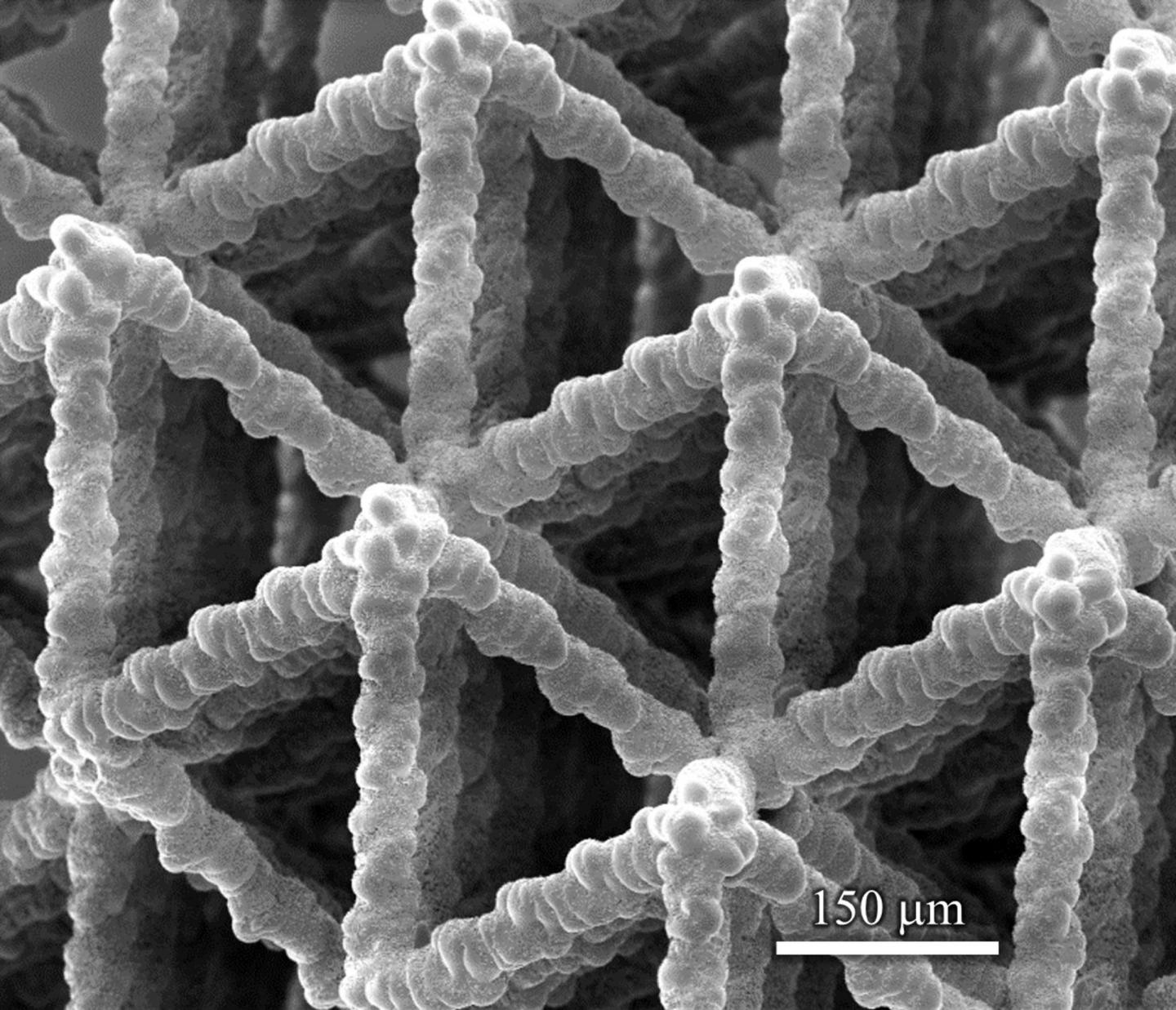
Caption: Microstructures like this one developed at Washington State University could be used in batteries, lightweight ultrastrong materials, catalytic converters, supercapacitors and biological scaffolds. Credit: Washington State University
A March 3, 2017 news item on Nanowerk features a new 3D manufacturing technique for creating biolike materials, (Note: A link has been removed)
Washington State University nanotechnology researchers have developed a unique, 3-D manufacturing method that for the first time rapidly creates and precisely controls a material’s architecture from the nanoscale to centimeters. The results closely mimic the intricate architecture of natural materials like wood and bone.
They report on their work in the journal Science Advances (“Three-dimensional microarchitected materials and devices using nanoparticle assembly by pointwise spatial printing”) and have filed for a patent.
“This is a groundbreaking advance in the 3-D architecturing of materials at nano- to macroscales with applications in batteries, lightweight ultrastrong materials, catalytic converters, supercapacitors and biological scaffolds,” said Rahul Panat, associate professor in the School of Mechanical and Materials Engineering, who led the research. “This technique can fill a lot of critical gaps for the realization of these technologies.”
The WSU research team used a 3-D printing method to create foglike microdroplets that contain nanoparticles of silver and to deposit them at specific locations. As the liquid in the fog evaporated, the nanoparticles remained, creating delicate structures. The tiny structures, which look similar to Tinkertoy constructions, are porous, have an extremely large surface area and are very strong.
Silver was used because it is easy to work with. However, Panat said, the method can be extended to any other material that can be crushed into nanoparticles – and almost all materials can be.
The researchers created several intricate and beautiful structures, including microscaffolds that contain solid truss members like a bridge, spirals, electronic connections that resemble accordion bellows or doughnut-shaped pillars.
The manufacturing method itself is similar to a rare, natural process in which tiny fog droplets that contain sulfur evaporate over the hot western Africa deserts and give rise to crystalline flower-like structures called “desert roses.”
Because it uses 3-D printing technology, the new method is highly efficient, creates minimal waste and allows for fast and large-scale manufacturing.
The researchers would like to use such nanoscale and porous metal structures for a number of industrial applications; for instance, the team is developing finely detailed, porous anodes and cathodes for batteries rather than the solid structures that are now used. This advance could transform the industry by significantly increasing battery speed and capacity and allowing the use of new and higher energy materials.
Researchers at Australia’s University of New South of Wales (UNSW) have devised a means of ‘weaving’ a material that mimics *bone tissue, periosteum according to a Jan. 11, 2017 news item on ScienceDaily,
For the first time, UNSW [University of New South Wales] biomedical engineers have woven a ‘smart’ fabric that mimics the sophisticated and complex properties of one nature’s ingenious materials, the bone tissue periosteum.
Having achieved proof of concept, the researchers are now ready to produce fabric prototypes for a range of advanced functional materials that could transform the medical, safety and transport sectors. Patents for the innovation are pending in Australia, the United States and Europe.
Potential future applications range from protective suits that stiffen under high impact for skiers, racing-car drivers and astronauts, through to ‘intelligent’ compression bandages for deep-vein thrombosis that respond to the wearer’s movement and safer steel-belt radial tyres.
Many animal and plant tissues exhibit ‘smart’ and adaptive properties. One such material is the periosteum, a soft tissue sleeve that envelops most bony surfaces in the body. The complex arrangement of collagen, elastin and other structural proteins gives periosteum amazing resilience and provides bones with added strength under high impact loads.
Until now, a lack of scalable ‘bottom-up’ approaches by researchers has stymied their ability to use smart tissues to create advanced functional materials.
UNSW’s Paul Trainor Chair of Biomedical Engineering, Professor Melissa Knothe Tate, said her team had for the first time mapped the complex tissue architectures of the periosteum, visualised them in 3D on a computer, scaled up the key components and produced prototypes using weaving loom technology.
“The result is a series of textile swatch prototypes that mimic periosteum’s smart stress-strain properties. We have also demonstrated the feasibility of using this technique to test other fibres to produce a whole range of new textiles,” Professor Knothe Tate said.
In order to understand the functional capacity of the periosteum, the team used an incredibly high fidelity imaging system to investigate and map its architecture.
“We then tested the feasibility of rendering periosteum’s natural tissue weaves using computer-aided design software,” Professor Knothe Tate said.
The computer modelling allowed the researchers to scale up nature’s architectural patterns to weave periosteum-inspired, multidimensional fabrics using a state-of-the-art computer-controlled jacquard loom. The loom is known as the original rudimentary computer, first unveiled in 1801.
“The challenge with using collagen and elastin is their fibres, that are too small to fit into the loom. So we used elastic material that mimics elastin and silk that mimics collagen,” Professor Knothe Tate said.
In a first test of the scaled-up tissue weaving concept, a series of textile swatch prototypes were woven, using specific combinations of collagen and elastin in a twill pattern designed to mirror periosteum’s weave. Mechanical testing of the swatches showed they exhibited similar properties found in periosteum’s natural collagen and elastin weave.
First author and biomedical engineering PhD candidate, Joanna Ng, said the technique had significant implications for the development of next-generation advanced materials and mechanically functional textiles.
While the materials produced by the jacquard loom have potential manufacturing applications – one tyremaker believes a titanium weave could spawn a new generation of thinner, stronger and safer steel-belt radials – the UNSW team is ultimately focused on the machine’s human potential.
“Our longer term goal is to weave biological tissues – essentially human body parts – in the lab to replace and repair our failing joints that reflect the biology, architecture and mechanical properties of the periosteum,” Ms Ng said.
An NHMRC development grant received in November [2016] will allow the team to take its research to the next phase. The researchers will work with the Cleveland Clinic and the University of Sydney’s Professor Tony Weiss to develop and commercialise prototype bone implants for pre-clinical research, using the ‘smart’ technology, within three years.
In searching for more information about this work, I found a Winter 2015 article (PDF; pp. 8-11) by Amy Coopes and Steve Offner for UNSW Magazine about Knothe Tate and her work (Note: In Australia, winter would be what we in the Northern Hemisphere consider summer),
Tucked away in a small room in UNSW’s Graduate School of Biomedical Engineering sits a 19th century–era weaver’s wooden loom. Operated by punch cards and hooks, the machine was the first rudimentary computer when it was unveiled in 1801. While on the surface it looks like a standard Jacquard loom, it has been enhanced with motherboards integrated into each of the loom’s five hook modules and connected to a computer. This state-of-the-art technology means complex algorithms control each of the 5,000 feed-in fibres with incredible precision.
That capacity means the loom can weave with an extraordinary variety of substances, from glass and titanium to rayon and silk, a development that has attracted industry attention around the world.
The interest lies in the natural advantage woven materials have over other manufactured substances. Instead of manipulating material to create new shades or hues as in traditional weaving, the fabrics’ mechanical properties can be modulated, to be stiff at one end, for example, and more flexible at the other.
“Instead of a pattern of colours we get a pattern of mechanical properties,” says Melissa Knothe Tate, UNSW’s Paul Trainor Chair of Biomedical Engineering. “Think of a rope; it’s uniquely good in tension and in bending. Weaving is naturally strong in that way.”
…
The interface of mechanics and physiology is the focus of Knothe Tate’s work. In March [2015], she travelled to the United States to present another aspect of her work at a meeting of the international Orthopedic Research Society in Las Vegas. That project – which has been dubbed “Google Maps for the body” – explores the interaction between cells and their environment in osteoporosis and other degenerative musculoskeletal conditions such as osteoarthritis.
Using previously top-secret semiconductor technology developed by optics giant Zeiss, and the same approach used by Google Maps to locate users with pinpoint accuracy, Knothe Tate and her team have created “zoomable” anatomical maps from the scale of a human joint down to a single cell.
She has also spearheaded a groundbreaking partnership that includes the Cleveland Clinic, and Brown and Stanford universities to help crunch terabytes of data gathered from human hip studies – all processed with the Google technology. Analysis that once took 25 years can now be done in a matter of weeks, bringing researchers ever closer to a set of laws that govern biological behaviour. [p. 9]
I gather she was recruited from the US to work at the University of New South Wales and this article was to highlight why they recruited her and to promote the university’s biomedical engineering department, which she chairs.
Getting back to 2017, here’s a link to and citation for the paper,
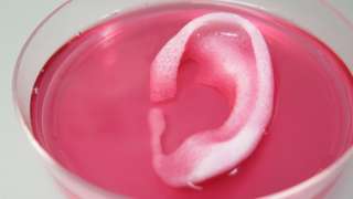
Over ten years ago I attended a show at the Vancouver (Canada) Art Gallery titled ‘Massive Change’ where I saw part of a nose or ear being grown in a petri dish (the work was from an Israeli laboratory) and that was my introduction to tissue engineering. For anyone who’s been following the tissue engineering story, 3D printers have sped up the growth process considerably. More recently, researchers at Wake Forest Baptist Medical Center (North Carolina, US) have announced another step forward for growing organs and body parts, from a Feb. 15, 2016 Wake Forest Baptist Medical Center news release on EurekAlert,
Using a sophisticated, custom-designed 3D printer, regenerative medicine scientists at Wake Forest Baptist Medical Center have proved that it is feasible to print living tissue structures to replace injured or diseased tissue in patients.
Reporting in Nature Biotechnology, the scientists said they printed ear, bone and muscle structures. When implanted in animals, the structures matured into functional tissue and developed a system of blood vessels. Most importantly, these early results indicate that the structures have the right size, strength and function for use in humans.
“This novel tissue and organ printer is an important advance in our quest to make replacement tissue for patients,” said Anthony Atala, M.D., director of the Wake Forest Institute for Regenerative Medicine (WFIRM) and senior author on the study. “It can fabricate stable, human-scale tissue of any shape. With further development, this technology could potentially be used to print living tissue and organ structures for surgical implantation.”
With funding from the Armed Forces Institute of Regenerative Medicine, a federally funded effort to apply regenerative medicine to battlefield injuries, Atala’s team aims to implant bioprinted muscle, cartilage and bone in patients in the future.
Tissue engineering is a science that aims to grow replacement tissues and organs in the laboratory to help solve the shortage of donated tissue available for transplants. The precision of 3D printing makes it a promising method for replicating the body’s complex tissues and organs. However, current printers based on jetting, extrusion and laser-induced forward transfer cannot produce structures with sufficient size or strength to implant in the body.
The Integrated Tissue and Organ Printing System (ITOP), developed over a 10-year period by scientists at the Institute for Regenerative Medicine, overcomes these challenges. The system deposits both bio-degradable, plastic-like materials to form the tissue “shape” and water-based gels that contain the cells. In addition, a strong, temporary outer structure is formed. The printing process does not harm the cells.
A major challenge of tissue engineering is ensuring that implanted structures live long enough to integrate with the body. The Wake Forest Baptist scientists addressed this in two ways. They optimized the water-based “ink” that holds the cells so that it promotes cell health and growth and they printed a lattice of micro-channels throughout the structures. These channels allow nutrients and oxygen from the body to diffuse into the structures and keep them live while they develop a system of blood vessels.
It has been previously shown that tissue structures without ready-made blood vessels must be smaller than 200 microns (0.007 inches) for cells to survive. In these studies, a baby-sized ear structure (1.5 inches) survived and showed signs of vascularization at one and two months after implantation.
“Our results indicate that the bio-ink combination we used, combined with the micro-channels, provides the right environment to keep the cells alive and to support cell and tissue growth,” said Atala.
Another advantage of the ITOP system is its ability to use data from CT and MRI scans to “tailor-make” tissue for patients. For a patient missing an ear, for example, the system could print a matching structure.
Several proof-of-concept experiments demonstrated the capabilities of ITOP. To show that ITOP can generate complex 3D structures, printed, human-sized external ears were implanted under the skin of mice. Two months later, the shape of the implanted ear was well-maintained and cartilage tissue and blood vessels had formed.
To demonstrate the ITOP can generate organized soft tissue structures, printed muscle tissue was implanted in rats. After two weeks, tests confirmed that the muscle was robust enough to maintain its structural characteristics, become vascularized and induce nerve formation.
And, to show that construction of a human-sized bone structure, jaw bone fragments were printed using human stem cells. The fragments were the size and shape needed for facial reconstruction in humans. To study the maturation of bioprinted bone in the body, printed segments of skull bone were implanted in rats. After five months, the bioprinted structures had formed vascularized bone tissue.
Ongoing studies will measure longer-term outcomes.
###
The research was supported, in part, by grants from the Armed Forces Institute of Regenerative Medicine (W81XWH-08-2-0032), the Telemedicine and Advanced Technology Research Center at the U.S. Army Medical Research and Material Command (W81XWH-07-1-0718) and the Defense Threat Reduction Agency (N66001-13-C-2027).
(Sometimes the information about the funding agencies is almost as interesting as the research.) Here’s a link to and a citation for the paper,
For the first time, the US Food and Drug Administration (FDA) has approved a nanotechnology-enabled interbody spinal fusion implant, according to a Nov. 12, 2014 news item on Azonano,
Titan Spine, a medical device surface technology company focused on developing innovative spinal interbody fusion implants, today announced that it has received 510(k) clearance from the U.S. Food and Drug Administration (FDA) to market its Endoskeleton® line of interbody fusion implants featuring its next-generation nanoLOCKTM surface technology.
This clearance marks Titan’s line of Endoskeleton® spinal implants as the first FDA-approved interbody fusion devices to feature nanotechnology.
A Nov. 22, 2014 news item on Today’s Medical Developments.com provides more detail about the implants,
Titan’s new nanoLOCK surface technology enhances the company’s line of Endoskeleton devices with an increased amount of nano-scaled textures to up-regulate a statistically significant greater amount of the osteogenic and angiogenic growth factors that are critical for bone growth and fusion when compared to PEEK and the company’s current surface.
Barbara Boyan, Ph.D., dean of the School of Engineering at Virginia Commonwealth University, and an investigator in various Titan Spine studies, said, “This new surface technology further enhances Titan’s current surface and is the result of extensive research in how to create a significantly greater amount of nano-scaled textures that we have shown to be important for the osteogenic response necessary for fusion. The nanoLOCK surface topography is far different than what is found on titanium-coated PEEK implants. In addition, the nanoLOCK surface is not created by applying a coating, but rather is formed by a reductive process of the titanium itself. This eliminates the potential for delamination, which is a concern for products with a PEEK-titanium interface. My team is proud to collaborate with Titan Spine to help develop such a differentiated technology that is truly designed to benefit both patients and surgeons.”
Titan’s nanoLOCK surface is a significant advancement of the company’s first-generation surface. The patented nanoLOCK manufacturing process creates additional textures at the critical nano level. However, there are no changes to the device indications for use, design, dimensions, or materials. Additionally, mechanical testing demonstrated that the strength of the company’s line of Endoskeletonimplants are unaffected by the new surface treatment.
Titan Spine, a medical device surface technology company focused on developing innovative spinal interbody fusion implants, today announced that it has received clearance from the U.S. Food and Drug Administration (FDA) to commercially release its Endoskeleton® TL system, a spinal fusion system utilizing a lateral approach. The Endoskeleton® TL represents the first lateral fusion device to feature surface technology that is designed to participate in the fusion process by creating an osteogenic response to the implant’s topography.
The Endoskeleton® TL device utilizes Titan’s proprietary roughened titanium surface technology which has been shown to upregulate the production of osteogenic and angiogenic factors that are critical for bone growth and fusion. In addition, the design of the TL device incorporates large windows and large internal volumes to allow for significant bone graft packing, clear CT and MRI imaging, desired bone graft loading, and the ability to pack additional bone graft material within the device following implantation. Members of the TL design team include Kade Huntsman, M.D., Orthopedic Spine Surgeon with the Salt Lake Orthopaedic Clinic in Salt Lake City, Utah; Andy Kranenburg, M.D., Co-Medical Director of the Providence Medford Medical Center Spine Institute in Medford, OR; Axel Reinhardt, M.D., Head of the Department of Spinal Surgery at the Specialized Orthopaedic Hospital in Potsdam, Germany; and Paul Slosar, M.D., Chief Medical Officer for Titan Spine.
Dr. Huntsman performed the first surgeries utilizing the Endoskeleton® TL on July 9th, 2014 at St. Mark’s Hospital in Salt Lake City, Utah. …
“The Endoskeleton® TL device is the first application of surface technology to the lateral approach,” commented Dr. Slosar. “The ability to orchestrate cellular behavior and promote bone growth in response to an interbody device has not been in the lateral surgeon’s armamentarium until now. The TL is the byproduct of a unique collaboration between academic biomaterial scientists, spine surgeons, and industry experts to create a truly differentiated lateral interbody device that is designed to benefit both patients and surgeons. With the addition of the TL device, Titan Spine now offers its surface technology and complete line of titanium devices for virtually all interbody fusion spine surgery procedures in the cervical and lumbar spine.”
The full line of Endoskeleton® devices features Titan Spine’s proprietary implant surface technology, consisting of a unique combination of roughened topographies at the macro, micro, and cellular levels. [emphasis mine] This combination of surface topographies is designed to create an optimal host-bone response and actively participate in the fusion process by promoting new bone growth, encouraging natural production of bone morphogenetic proteins (BMP’s) and creating the potential for a faster and more robust fusion.
It would seem the implant used in the July 2014 surgery is not nanotechnology-enabled, which suggests nanoLOCK is a next-generation implant being marketed only a few months after the first generation was made available. Unfortunately, the Titan Spine website is still partially (‘surface technology’ tab) under construction so I was not able to find more details about the technology. In any event, that’s quite a development pace.
It’s time to finally publish this which has been languishing in drafts folder: from a Sept. 16, 2014 news item on Nanowerk (Note: A link has been removed),
Murdoch University [Australia] nanotechnology researchers have successfully engineered synthetic materials which encouraged bone formation in sheep (“The synthesis, characterisation and in vivo study of a bioceramic for potential tissue regeneration applications”).
The advancement means the successful use of synthetic materials in bone grafts for human patients is a step closer. The material could also have potential future applications in fracture repair and reconstructive surgery.
Currently the patient’s own bone, donated bone or artificial materials are used for bone grafts but limitations with all these options have prompted researchers to investigate how synthetic materials can be enhanced.
Dr Eddy Poinern and his team from the Murdoch Applied Nanotechnology Research Group worked with powdered forms of the bio ceramic hydroxyapatite (HAP) to form pellets with a sponge-like structure which were then successfully implanted behind the shoulders of four sheep by collaborators from the School of Veterinary and Life Sciences at Murdoch University.
HAP is already being used in a number of biomedical applications such as bone augmentation in dentistry because of its similarity to the inorganic mineral component of human bone. But treatments of HAP so that it can be successfully used in a bone graft have yet to be developed because of the complexities involved with compatibility and HAP’s load bearing limitations.
The news release goes on to provide a few technical details,
Dr Poinern and his team prepared pellets with varying density and porosity using a variety of chemical methods including sintering, ultrasound and microwaves. Four pellets were implanted into muscles in each of the sheep, later demonstrating good bio-compatibility, including mixed cell colonisation after four weeks and even new bone formation 12 weeks after the surgery.
“Using synthetic materials in this way is difficult and complicated because they need to be engineered to be porous and to replicate the various physical, chemical and mechanical properties found in natural bone tissue,” explained Dr Poinern.
“They also need to be non-toxic and have a degradation rate which will allow for cells from the host to steadily recolonize the area and permit the formation of blood vessels necessary for the delivery of nutrients to the forming bone tissues.
“We already knew that synthetic HAP was a good material to study for possible use in bone-related medicine, but we needed to find out if the pellets we’d engineered were bio-compatible.
“Our results were very positive – our pellets acted as a scaffold for the growth of bone material, made possible because of its porous properties allowing cells to infiltrate.
“The pellets were also very cost effective to make.”
Although the study was small scale and originally intended to test the bio-compatibility of the HAP pellets, the bone growth was beyond what the interdisciplinary team expected.
Associate Professor Martin Cake, who surgically implanted the pellets into the sheep, described the results as “stunning” and said they boded well for the use of engineered HAP in bone implants.
“This material begins as a powder that can be theoretically moulded to any shape, or perhaps one day even 3D printed, then sintered to harden it,” he said.
…
Dr Poinern said he was hoping to improve and match the physical and mechanical properties of the pellets with those of natural bone tissue in a new study.
“Once these properties have been achieved, further implantation studies will be carried out to establish the feasibility of using this scaffold for bone grafts,” he said.
This news release included information of a type I haven’t previously seen included,
The implantation study was carried out in non pregnant Merino ewes with the approval of Murdoch University’s Animal Ethics Committee and all experiments were conducted in accordance with the Australian National Health and Medical Research Council’s (NHMRC) Code of Practice for the care and use of animals for scientific purposes.
In accordance with the ethical principles of the Code, the sheep were simultaneously used in an unrelated trial involving surgery of the stifle joints.
After the pellets were removed, the sheep were humanely euthanased.
I’m glad to see the information and hope more research groups follow suit.
One final note, Murdoch University, Eddy Poinern, and Dereck Fawcett have been mentioned here before in an Aug. 1, 2014 posting about ‘green’ chemistry involving eucalyptus leaves, and gold nanoparticles.
* ‘encourage’ corrected to ‘encourages’ on Oct. 7, 2014 at 1315 hours PDT.
Mammals of all kind have a horror disfigurement and will avoid members of their group who are disfigured. This horror is one of the themes to be found in the novel Frankenstein by Mary Shelley. Despite the difficulties, Roger Ebert (film critic) continued to make public appearances after cancer surgeries that changed his appearance (from a June 27, 2012 article by Ronni Gordon for Cancer Today),
Facing the Critics
Roger Ebert finds peace with his appearance following disfiguring cancer surgery
“Today I look like an exhibit in the Texas Chainsaw Museum,” he muses in his 2011 memoir, Life Itself. But Ebert decided he wasn’t going to hide the way he looks. In 2007, before attending his annual Overlooked Film Festival, now referred to as Ebertfest, at the University of Illinois at Urbana-Champaign, Ebert and his wife, Chaz, decided that a photograph of him should accompany a story he wrote for the Sun-Times. Later, he posed for a full-page photo that appeared in Esquire in March 2010.
“No point in denying it,” he wrote about his appearance in Life Itself. “No way to hide it. Better for it to be out there.”
Given the difficulties most people experience, researchers are eager to find solutions. An Aug. 13, 2014 American Chemical Society (ACS) news release (also on EurekAlert) describes a presentation at the ACS 284h meeting about shape-shifting material that could be used to ameliorate bone defects,
Injuries, birth defects (such as cleft palates) or surgery to remove a tumor can create gaps in bone that are too large to heal naturally. And when they occur in the head, face or jaw, these bone defects can dramatically alter a person’s appearance. Researchers will report today that they have developed a “self-fitting” material that expands with warm salt water to precisely fill bone defects, and also acts as a scaffold for new bone growth.
…
Currently, the most common method for filling bone defects in the head, face or jaw (known as the cranio-maxillofacial area) is autografting. That is a process in which surgeons harvest bone from elsewhere in the body, such as the hip bone, and then try to shape it to fit the bone defect.
“The problem is that the autograft is a rigid material that is very difficult to shape into these irregular defects,” says Melissa Grunlan, Ph.D., leader of the study. Also, harvesting bone for the autograft can itself create complications at the place where the bone was taken.
Another approach is to use bone putty or cement to plug gaps. However, these materials aren’t ideal. They become very brittle when they harden, and they lack pores, or small holes, that would allow new bone cells to move in and rebuild the damaged tissue.
To develop a better material, Grunlan and her colleagues at Texas A&M University made a shape-memory polymer (SMP) that molds itself precisely to the shape of the bone defect without being brittle. It also supports the growth of new bone tissue.
SMPs are materials whose geometry changes in response to heat. The team made a porous SMP foam by linking together molecules of poly(ε-caprolactone), an elastic, biodegradable substance that is already used in some medical implants. The resulting material resembled a stiff sponge, with many interconnected pores to allow bone cells to migrate in and grow.
Upon heating to 140 degrees Fahrenheit, the SMP becomes very soft and malleable. So, during surgery to repair a bone defect, a surgeon could warm the SMP to that temperature and fill in the defect with the softened material. Then, as the SMP is cooled to body temperature (98.6 degrees Fahrenheit), it would resume its former stiff texture and “lock” into place.
The researchers also coated the SMPs with polydopamine, a sticky substance that helps lock the polymer into place by inducing formation of a mineral that is found in bone. It may also help osteoblasts, the cells that produce bone, to adhere and spread throughout the polymer. The SMP is biodegradable, so that eventually the scaffold will disappear, leaving only new bone tissue behind.
To test whether the SMP scaffold could support bone cell growth, the researchers seeded the polymer with human osteoblasts. After three days, the polydopamine-coated SMPs had grown about five times more osteoblasts than those without a coating. Furthermore, the osteoblasts produced more of the two proteins, runX2 and osteopontin, that are critical for new bone formation.
Grunlan says that the next step will be to test the SMP’s ability to heal cranio-maxillofacial bone defects in animals. “The work we’ve done in vitro is very encouraging,” she says. “Now we’d like to move this into preclinical and, hopefully, clinical studies.”
The researchers acknowledge funding from the Texas A&M Engineering Experiment Station.
It sounds like there’s still quite a long way to go before this research makes its way out of the laboratory. I wish the researchers all the best.