Tag Archives: femtosecond laser

Non-viral ocular gene therapy with gold nanoparticles and femtosecond lasers

I love the stylistic choice the writer made (pay special attention to the second paragraph) when producing this November 19, 2018 Polytechnique Montréal news release (also on EurekAlert),

A scientific breakthrough by Professor Michel Meunier of Polytechnique Montréal and his collaborators offers hope for people with glaucoma, retinitis or macular degeneration.

In January 2009, the life of engineer Michel Meunier, a professor at Polytechnique Montréal, changed dramatically. Like others, he had observed that the extremely short pulse of a femtosecond laser (0.000000000000001 second) could make nanometre-sized holes appear in silicon when it was covered by gold nanoparticles. But this researcher, recognized internationally for his skills in laser and nanotechnology, decided to go a step further with what was then just a laboratory curiosity. He wondered if it was possible to go from silicon to living matter, from inorganic to organic. Could the gold nanoparticles and the femtosecond laser, this “light scalpel,” reproduce the same phenomenon with living cells?

A very pretty image illustrating the work,
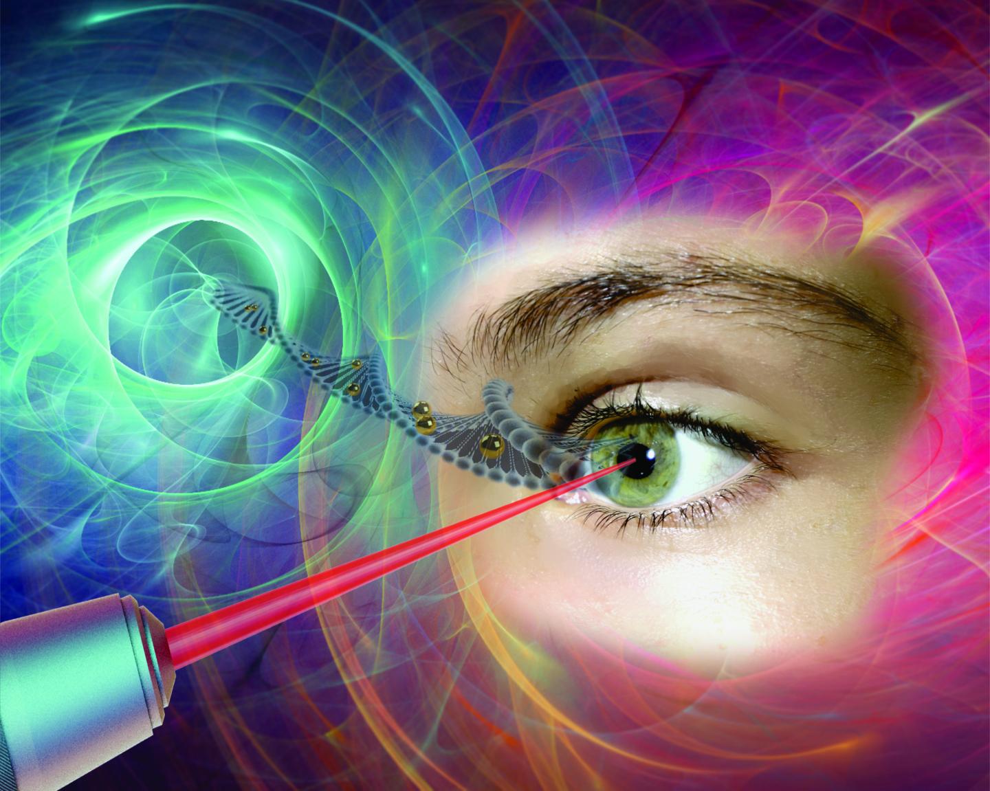
Caption: Gold nanoparticles, which act like “nanolenses,” concentrate the energy produced by the extremely short pulse of a femtosecond laser to create a nanoscale incision on the surface of the eye’s retina cells. This technology, which preserves cell integrity, can be used to effectively inject drugs or genes into specific areas of the eye, offering new hope to people with glaucoma, retinitis or macular degeneration. Credit and Copyright: Polytechnique Montréal

The news release goes on to describe the technology in more detail,

Professor Meunier started working on cells in vitro in his Polytechnique laboratory. The challenge was to make a nanometric incision in the cells’ extracellular membrane without damaging it. Using gold nanoparticles that acted as “nanolenses,” Professor Meunier realized that it was possible to concentrate the light energy coming from the laser at a wavelength of 800 nanometres. Since there is very little energy absorption by the cells at this wavelength, their integrity is preserved. Mission accomplished!

Based on this finding, Professor Meunier decided to work on cells in vivo, cells that are part of a complex living cell structure, such as the eye for example.

The eye and the light scalpel

In April 2012, Professor Meunier met Przemyslaw Sapieha, an internationally renowned eye specialist, particularly recognized for his work on the retina. “Mike”, as he goes by, is a professor in the Department of Ophthalmology at Université de Montréal and a researcher at Centre intégré universitaire de santé et de services sociaux (CIUSSS) de l’Est-de-l’Île-de-Montréal. He immediately saw the potential of this new technology and everything that could be done in the eye if you could block the ripple effect that occurs following a trigger that leads to glaucoma or macular degeneration, for example, by injecting drugs, proteins or even genes.

Using a femtosecond laser to treat the eye–a highly specialized and fragile organ–is very complex, however. The eye is part of the central nervous system, and therefore many of the cells or families of cells that compose it are neurons. And when a neuron dies, it does not regenerate like other cells do. Mike Sapieha’s first task was therefore to ensure that a femtosecond laser could be used on one or several neurons without affecting them. This is what is referred to as “proof of concept.”

Proof of concept

Mike and Michel called on biochemistry researcher Ariel Wilson, an expert in eye structures and vision mechanisms, as well as Professor Santiago Costantino and his team from the Department of Ophthalmology at Université de Montréal and the CIUSSS de l’Est-de-l’Île-de-Montréal for their expertise in biophotonics. The team first decided to work on healthy cells, because they are better understood than sick cells. They injected gold nanoparticles combined with antibodies to target specific neuronal cells in the eye, and then waited for the nanoparticles to settle around the various neurons or families of neurons, such as the retina. Following the bright flash generated by the femtosecond laser, the expected phenomenon occurred: small holes appeared in the cells of the eye’s retina, making it possible to effectively inject drugs or genes in specific areas of the eye. It was another victory for Michel Meunier and his collaborators, with these conclusive results now opening the path to new treatments.

The key feature of the technology developed by the researchers from Polytechnique and CIUSSS de l’Est-de-l’Île-de-Montréal is its extreme precision. With the use of functionalized gold nanoparticles, the light scalpel makes it possible to precisely locate the family of cells where the doctor will have to intervene.

Having successfully demonstrated proof of concept, Professor Meunier and his team filed a patent application in the United States. This tremendous work was also the subject of a paper reviewed by an impressive reading committee and published in the renowned journal Nano Letters in October 2018.

While there is still a lot of research to be done–at least 10 years’ worth, first on animals and then on humans–this technology could make all the difference in an aging population suffering from eye deterioration for which there are still no effective long-term treatments. It also has the advantage of avoiding the use of viruses commonly employed in gene therapy. These researchers are looking at applications of this technology in all eye diseases, but more particularly in glaucoma, retinitis and macular degeneration.

This light scalpel is unprecedented.

Here’s a link to and a citation for the paper,

In Vivo Laser-Mediated Retinal Ganglion Cell Optoporation Using KV1.1 Conjugated Gold Nanoparticles by Ariel M. Wilson, Javier Mazzaferri, Éric Bergeron, Sergiy Patskovsky, Paule Marcoux-Valiquette, Santiago Costantino, Przemyslaw Sapieha, Michel Meunier. Nano Lett.201818116981-6988 DOI: https://doi.org/10.1021/acs.nanolett.8b02896 Publication Date: October 4, 2018  Copyright © 2018 American Chemical Society

This paper is behind a paywall.

Archivists, rejoice! Fused quartz stores data for millions of years at the University of Southampton (UK)

There’s a July 9,  2013 news item on Nanowerk touting nanostructured glass device which is being compared to Superman’s memory crystal (see this Wikipedia essay on Superman’s Fortress of Solitude for a description of Superman’s memory crystals),

Using nanostructured glass, scientists at the University of Southampton have, for the first time, experimentally demonstrated the recording and retrieval processes of five dimensional digital data by femtosecond laser writing. The storage allows unprecedented parameters including 360 TB/disc data capacity, thermal stability up to 1000°C and practically unlimited lifetime.

Coined as the ‘Superman’ memory crystal, as the glass memory has been compared to the “memory crystals” used in the Superman films, the data is recorded via self-assembled nanostructures created in fused quartz, which is able to store vast quantities of data for over a million years. The information encoding is realised in five dimensions: the size and orientation in addition to the three dimensional position of these nanostructures. [emphases mine]

The July 9, 2013 University of Southampton news release, which originated the news item, provides more details,

A 300 kb digital copy of a text file was successfully recorded in 5D using ultrafast laser, producing extremely short and intense pulses of light. The file is written in three layers of nanostructured dots separated by five micrometres (one millionth of a metre).

The self-assembled nanostructures change the way light travels through glass, modifying polarisation of light that can then be read by combination of optical microscope and a polariser, similar to that found in Polaroid sunglasses.

The research is led by Jingyu Zhang from the University’s Optoelectronics Research Centre (ORC) and conducted under a joint project with Eindhoven University of Technology.

“We are developing a very stable and safe form of portable memory using glass, which could be highly useful for organisations with big archives. At the moment companies have to back up their archives every five to ten years because hard-drive memory has a relatively short lifespan,” says Jingyu. [emphasis mine]

“Museums who want to preserve information or places like the national archives where they have huge numbers of documents, would really benefit.”

This work was presented at the CLEO 2013 (Conference on Lasers and Electro-Optics in San Jose [US]). Here’s a link to and a citation for the 2 pp presentation paper,

5D Data Storage by Ultrafast Laser Nanostructuring in Glass by Jingyu Zhang, Mindaugas Gecevičius, Martynas Beresna, and Peter G. Kazansky. Presentation paper for CLEO 2013

© 2013 Optical Society of America OCIS codes (140.3390) Laser materials processing, (210.0210) Optical data storage

This research was conducted as part of the European Union’s Femtoprint project, which is funded under the Framework Programme 7 initiative. Here’s more about Femtoprint from the homepage,

FEMTOPRINT is to develop a printer for microsystems with nano-scale features fabricated out of glass. Our ultimate goal is to provide a large pool of users from industry, research and universities with the capability of producing their own micro-systems, in a rapid-manner without the need for expensive infrastructures and specific expertise. Recent researches have shown that one can form three-dimensional patterns in glass material using low-power femtosecond laser beam. This simple process opens interesting new opportunities for a broad variety of microsystems with feature sizes down to the nano-scale. These patterns can be used to form integrated optics components or be ‘developed’ by chemically etching to form three-dimensional structures like fluidic channels and micro-mechanical components. Worth noticing, sub-micron resolution can be achieved and sub-pattern smaller than the laser wavelength can be formed. Thanks to the low-energy required to pattern the glass, femtosecond laser consisting simply of an oscillator are sufficient to produce such micro- and nano- systems.

These systems are nowadays table-top and cost a fraction of conventional clean-room equipments. It is highly foreseeable that within 3 to 5 years such laser systems will fit in a shoe-box. The project specific objectives are:

1/ Develop a femtosecond laser suitable for glass micro-/nano- manufacturing that fits in a shoe-box

2/ Integrate the laser in a machine similar to a printer that can position and manipulate glass sheets of various thicknesses

3/ Demonstrate the use of the printer to fabricate a variety of micro-/nano-systems with optical, mechanical and fluid-handling capabilities. A clear and measurable outcome of Femtoprint will be to be in a situation to commercialize the ‘femtoprinter’ through the setting-up of a consortium spin-off. The potential economical impact is large and is expected in various industrial sectors.

I think any archivist hearing about data storage that can last a million years will be thrilled although I suspect it’s going to be a long, long time before these 5D ‘memory’ crystals are going to be storing any data for anyone. In the meantime, there are efforts such as the Council of Canadian Academies’ (CCA) Memory Institutions and the Digital Revolution assessment (mentioned 2/3 of the way down in my June 5, 2013 posting).

Nanosurgery in Montréal (Canada)

When I was typing up charts for home nursing care (nurses visiting patients in their home after a hospital procedure), I routinely asked if a patient whose cancer had metastasized would require palliative care even though the answer would be yes. In over 3 years and after hundreds of charts, I only had one ‘No’. So it is with some interest I read about Michel Meunier and his team’s work at the Polytechnique Montréal (Québec, Canada). From the Feb. 16, 2012 news item on Nanowerk,

The unique method developed by Professor Michel Meunier and his team uses a femtosecond laser (a laser with ultra-short pulses) along with gold nanoparticles. Deposited on the cells, these nanoparticles concentrate the laser’s energy and make it possible to perform nanometric-scale surgery in an extremely precise and non-invasive fashion. The technique allows to change the expression of genes in the cancer cells and could be used to slow their migration and prevent the formation of metastases.

The technique perfected by Professor Meunier and his colleagues is a promising alternative to conventional cellular transfection methods, such as lipofection. The experiment, carried out in Montréal laboratories on malignant human melanoma cells, demonstrated 70% optoporation effectiveness, as well as a transfection performance three times higher than lipofection treatment. In addition, unlike conventional treatment, which destroys the physical integrity of the cells, the new method assures cellular viability, with a toxicity of less than 1%.

The Polytechnique’s Feb. 16, 2012 press release is here and you can find out more about Meunier and his lab here (in English and en français). For those eager to read the article, it was published in Biomaterials (vol. 33, no. 7, March 2012, pp. 2345-50) is titled, Off-resonance plasmonic enhanced femtosecond laser optoporation and transfection of cancer cells and is behind a paywall.