Tag Archives: bioinspired

Squeezing out ‘polymer opals’ for smart clothing and more

Researchers at the University of Cambridge have developed a technology for producing ‘polymer opals’ on industrial scales according to a June 3, 2016 news item on Nanowerk (Note: A link has been removed),

Using a new method called Bend-Induced-Oscillatory-Shearing (BIOS), the researchers are now able to produce hundreds of metres of these materials, known as ‘polymer opals’, on a roll-to-roll process. The results are reported in the journal Nature Communications (“Large-scale ordering of nanoparticles using viscoelastic shear processing”).

A June 3, 2016 University of Cambridge press release (also on EurekAlert), which originated the news item, provides more detail (Note: Links have been removed),

Researchers have devised a new method for stacking microscopic marbles into regular layers, producing intriguing materials which scatter light into intense colours, and which change colour when twisted or stretched.

Some of the brightest colours in nature can be found in opal gemstones, butterfly wings and beetles. These materials get their colour not from dyes or pigments, but from the systematically-ordered microstructures they contain.

The team behind the current research, based at Cambridge’s Cavendish Laboratory, have been working on methods of artificially recreating this ‘structural colour’ for several years, but to date, it has been difficult to make these materials using techniques that are cheap enough to allow their widespread use.

In order to make the polymer opals, the team starts by growing vats of transparent plastic nano-spheres. Each tiny sphere is solid in the middle but sticky on the outside. The spheres are then dried out into a congealed mass. By bending sheets containing a sandwich of these spheres around successive rollers the balls are magically forced into perfectly arranged stacks, by which stage they have intense colour.

By changing the sizes of the starting nano-spheres, different colours (or wavelengths) of light are reflected. And since the material has a rubber-like consistency, when it is twisted and stretched, the spacing between the spheres changes, causing the material to change colour. When stretched, the material shifts into the blue range of the spectrum, and when compressed, the colour shifts towards red. When released, the material returns to its original colour. Such chameleon materials could find their way into colour-changing wallpapers, or building coatings that reflect away infrared thermal radiation.

I always like it when there are quotes which seem spontaneous (from the press release),

“Finding a way to coax objects a billionth of a metre across into perfect formation over kilometre scales is a miracle [emphasis mine],” said Professor Jeremy Baumberg, the paper’s senior author. “But spheres are only the first step, as it should be applicable to more complex architectures on tiny scales.”

In order to make polymer opals in large quantities, the team first needed to understand their internal structure so that it could be replicated. Using a variety of techniques, including electron microscopy, x-ray scattering, rheology and optical spectroscopy, the researchers were able to see the three-dimensional position of the spheres within the material, measure how the spheres slide past each other, and how the colours change.

“It’s wonderful [emphasis mine] to finally understand the secrets of these attractive films,” said PhD student Qibin Zhao, the paper’s lead author.

There’s also the commercialization aspect to this work (from the press release),

Cambridge Enterprise, the University’s commercialisation arm which is helping to commercialise the material, has been contacted by more than 100 companies interested in using polymer opals, and a new spin-out Phomera Technologies has been founded. Phomera will look at ways of scaling up production of polymer opals, as well as selling the material to potential buyers. Possible applications the company is considering include coatings for buildings to reflect heat, smart clothing and footwear, or for banknote security [emphasis mine] and packaging applications.

There is a Canadian company already selling its anti-counterfeiting (banknote security) bioinspired technology. It’s called Opalux and it’s not the only bioinspired anti-counterfeiting Canadian technology company, there’s also NanoTech Security which takes its inspiration from a butterfly (Blue Morpho) wing.

Getting back to Cambridge, here’s a link to and a citation for the research team’s paper,

Large-scale ordering of nanoparticles using viscoelastic shear processing by Qibin Zhao, Chris E. Finlayson, David R. E. Snoswell, Andrew Haines, Christian Schäfer, Peter Spahn, Goetz P. Hellmann, Andrei V. Petukhov, Lars Herrmann, Pierre Burdet, Paul A. Midgley, Simon Butler, Malcolm Mackley, Qixin Guo, & Jeremy J. Baumberg. Nature Communications 7, Article number: 11661  doi:10.1038/ncomms11661 Published 03 June 2016

This paper is open access.

There is a video demonstrating the stretchability of their ‘polymer opal’ film

It was posted on YouTube three years ago when the researchers were first successful. It’s nice to see they’ve been successful at getting the technology to the commercialization stage.

Green Hairstreak butterfly could make computer screens more brilliant

When talking about mimicking the nanostructures on butterfly wings, the butterfly in question is the Blue Morpho, usually. In a change of pace, researchers at Australia’s Swinburne University of Technology have focused their attention on the Green Hairstreak butterfly,

Nanostructures on the wings of the Callophrys Rubi or Green Hairstreak butterfly have inspired the design of an artificial material that could be used in photonics and optics technologies. Courtesy: Swinburne University of Technology

Nanostructures on the wings of the Callophrys Rubi or Green Hairstreak butterfly have inspired the design of an artificial material that could be used in photonics and optics technologies. Courtesy: Swinburne University of Technology

A June 1, 2016 Swinburne University of Technology press release describes the work,

Inspired by the intricate structure of a butterfly wing, Swinburne researchers have developed a technique that could be used to make more brilliant computer screens.

The researchers used a special printing technique to create tiny structures similar to those found in the wings of the Callophrys Rubi butterfly, also known as the Green Hairstreak.

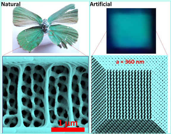
In some iridescent butterfly wings, such as the Green Hairstreak, the wing is made up of a pattern of intertwining and curved surfaces, known as a gyroid structure.  This gyroid structure has amazing properties when it comes to its interactions with light.

The researchers used two-beams of light to print at a super-resolution, creating gyroid structures that are three-dimensional and mechanically strong.

Lead author Dr Zongsong Gan from Swinburne’s Centre for Micro-Photonics says that materials made from these artificial gyroids should respond to light at ultrafast speeds making them ideal for high-speed switches.

He says the technique has two significant advantages.

“The first is that it has improved resolution and the second is that the materials fabricated with this technique have better mechanical strength.

“These new gyroid structures could help make more compact light based electronics because, thanks to their smaller size, larger numbers of devices can be integrated onto a single chip.

“However, for three-dimensional devices, smaller and more compact also means there is a higher risk of structure collapse because of weaker mechanical strength.

“Our fabrication technique allows us to make stronger architectures to overcome this problem,” Dr Gan says.

Here’s an image illustrating the work,

Comparison of natural gyroid structure with artificial structure. Courtesy: Swinburne University of Technology

Comparison of natural gyroid structure with artificial structure. Courtesy: Swinburne University of Technology

Here’s a link to and a citation for the paper,

Biomimetic gyroid nanostructures exceeding their natural origins by Zongsong Gan, Mark D. Turner, and Min Gu. Science Advances  13 May 2016: Vol. 2, no. 5, e1600084
DOI: 10.1126/sciadv.1600084

This appears to be an open access paper,

Making better concrete by looking to nature for inspiration

Researchers from the Masssachusetts Institute of Technology (MIT) are working on a new formula for concrete based on bones, shells, and other such natural materials. From a May 25, 2016 news item on Nanowerk (Note: A link has been removed),

Researchers at MIT are seeking to redesign concrete — the most widely used human-made material in the world — by following nature’s blueprints.

In a paper published online in the journal Construction and Building Materials (“Roadmap across the mesoscale for durable and sustainable cement paste – A bioinspired approach”), the team contrasts cement paste — concrete’s binding ingredient — with the structure and properties of natural materials such as bones, shells, and deep-sea sponges. As the researchers observed, these biological materials are exceptionally strong and durable, thanks in part to their precise assembly of structures at multiple length scales, from the molecular to the macro, or visible, level.

A May 26, 2016 MIT news release (also on EurekAlert), which originated the news item, provides more detail,

From their observations, the team, led by Oral Buyukozturk, a professor in MIT’s Department of Civil and Environmental Engineering (CEE), proposed a new bioinspired, “bottom-up” approach for designing cement paste.

“These materials are assembled in a fascinating fashion, with simple constituents arranging in complex geometric configurations that are beautiful to observe,” Buyukozturk says. “We want to see what kinds of micromechanisms exist within them that provide such superior properties, and how we can adopt a similar building-block-based approach for concrete.”

Ultimately, the team hopes to identify materials in nature that may be used as sustainable and longer-lasting alternatives to Portland cement, which requires a huge amount of energy to manufacture.

“If we can replace cement, partially or totally, with some other materials that may be readily and amply available in nature, we can meet our objectives for sustainability,” Buyukozturk says.

“The merger of theory, computation, new synthesis, and characterization methods have enabled a paradigm shift that will likely change the way we produce this ubiquitous material, forever,” Buehler says. “It could lead to more durable roads, bridges, structures, reduce the carbon and energy footprint, and even enable us to sequester carbon dioxide as the material is made. Implementing nanotechnology in concrete is one powerful example [of how] to scale up the power of nanoscience to solve grand engineering challenges.”

From molecules to bridges

Today’s concrete is a random assemblage of crushed rocks and stones, bound together by a cement paste. Concrete’s strength and durability depends partly on its internal structure and configuration of pores. For example, the more porous the material, the more vulnerable it is to cracking. However, there are no techniques available to precisely control concrete’s internal structure and overall properties.

“It’s mostly guesswork,” Buyukozturk says. “We want to change the culture and start controlling the material at the mesoscale.”

As Buyukozturk describes it, the “mesoscale” represents the connection between microscale structures and macroscale properties. For instance, how does cement’s microscopic arrangement affect the overall strength and durability of a tall building or a long bridge? Understanding this connection would help engineers identify features at various length scales that would improve concrete’s overall performance.

“We’re dealing with molecules on the one hand, and building a structure that’s on the order of kilometers in length on the other,” Buyukozturk says. “How do we connect the information we develop at the very small scale, to the information at the large scale? This is the riddle.”

Building from the bottom, up

To start to understand this connection, he and his colleagues looked to biological materials such as bone, deep sea sponges, and nacre (an inner shell layer of mollusks), which have all been studied extensively for their mechanical and microscopic properties. They looked through the scientific literature for information on each biomaterial, and compared their structures and behavior, at the nano-, micro-, and macroscales, with that of cement paste.

They looked for connections between a material’s structure and its mechanical properties. For instance, the researchers found that a deep sea sponge’s onion-like structure of silica layers provides a mechanism for preventing cracks. Nacre has a “brick-and-mortar” arrangement of minerals that generates a strong bond between the mineral layers, making the material extremely tough.

“In this context, there is a wide range of multiscale characterization and computational modeling techniques that are well established for studying the complexities of biological and biomimetic materials, which can be easily translated into the cement community,” says Masic.

Applying the information they learned from investigating biological materials, as well as knowledge they gathered on existing cement paste design tools, the team developed a general, bioinspired framework, or methodology, for engineers to design cement, “from the bottom up.”

The framework is essentially a set of guidelines that engineers can follow, in order to determine how certain additives or ingredients of interest will impact cement’s overall strength and durability. For instance, in a related line of research, Buyukozturk is looking into volcanic ash [emphasis mine] as a cement additive or substitute. To see whether volcanic ash would improve cement paste’s properties, engineers, following the group’s framework, would first use existing experimental techniques, such as nuclear magnetic resonance, scanning electron microscopy, and X-ray diffraction to characterize volcanic ash’s solid and pore configurations over time.

Researchers could then plug these measurements into models that simulate concrete’s long-term evolution, to identify mesoscale relationships between, say, the properties of volcanic ash and the material’s contribution to the strength and durability of an ash-containing concrete bridge. These simulations can then be validated with conventional compression and nanoindentation experiments, to test actual samples of volcanic ash-based concrete.

Ultimately, the researchers hope the framework will help engineers identify ingredients that are structured and evolve in a way, similar to biomaterials, that may improve concrete’s performance and longevity.

“Hopefully this will lead us to some sort of recipe for more sustainable concrete,” Buyukozturk says. “Typically, buildings and bridges are given a certain design life. Can we extend that design life maybe twice or three times? That’s what we aim for. Our framework puts it all on paper, in a very concrete way, for engineers to use.”

This is not the only team looking at new methods for producing the material, my Dec. 24, 2012 posting features a number of ‘concrete’ research projects.

Also, I highlighted the reference to ‘volcanic ash’ as it reminded me of Roman concrete which has lasted for over 2000 years and includes volcanic sand and volcanic rock.  You can read more about it in a Dec. 18, 2014 article by Mark Miller for Ancient Origins where he describes the wonders of the material and what was then a recent discovery of the Romans’ recipe.

I have two links and citations, first, the MIT paper, then the paper on Roman concrete.

Roadmap across the mesoscale for durable and sustainable cement paste – A bioinspired approach by Steven D. Palkovic, Dieter B. Brommer, Kunal Kupwade-Patil, Admir Masic, Markus J. Buehler, Oral Büyüköztürk.Construction and Building Materials Volume 115, 15 July 2016, Pages 13–31.  doi:10.1016/j.conbuildmat.2016.04.020

Mechanical resilience and cementitious processes in Imperial Roman architectural mortar by Marie D. Jackson, Eric N. Landis, Philip F. Brune, Massimo Vitti, Heng Chen, Qinfei Li, Martin Kunz, Hans-Rudolf Wenk, Paulo J. M. Monteiro, and Anthony R. Ingraffea. Proceedings of the National Academy of Sciences  vol. 111 no. 52 18484–18489, doi: 10.1073/pnas.1417456111

The first paper is behind a paywall but the second one appears to be open access.

Spider webs inspire liquid wire

Courtesy University of Oxford

Courtesy University of Oxford

Usually, when science talk runs to spider webs the focus is on strength but this research from the UK and France is all about resilience. From a May 16, 2016 news item on phys.org,

Why doesn’t a spider’s web sag in the wind or catapult flies back out like a trampoline? The answer, according to new research by an international team of scientists, lies in the physics behind a ‘hybrid’ material produced by spiders for their webs.

Pulling on a sticky thread in a garden spider’s orb web and letting it snap back reveals that the thread never sags but always stays taut—even when stretched to many times its original length. This is because any loose thread is immediately spooled inside the tiny droplets of watery glue that coat and surround the core gossamer fibres of the web’s capture spiral.

This phenomenon is described in the journal PNAS by scientists from the University of Oxford, UK and the Université Pierre et Marie Curie, Paris, France.

The researchers studied the details of this ‘liquid wire’ technique in spiders’ webs and used it to create composite fibres in the laboratory which, just like the spider’s capture silk, extend like a solid and compress like a liquid. These novel insights may lead to new bio-inspired technology.

A May 16, 2016 University of Oxford press release (also on EurekAlert), which originated the news item, provides more detail,

Professor Fritz Vollrath of the Oxford Silk Group in the Department of Zoology at Oxford University said: ‘The thousands of tiny droplets of glue that cover the capture spiral of the spider’s orb web do much more than make the silk sticky and catch the fly. Surprisingly, each drop packs enough punch in its watery skins to reel in loose bits of thread. And this winching behaviour is used to excellent effect to keep the threads tight at all times, as we can all observe and test in the webs in our gardens.’

The novel properties observed and analysed by the scientists rely on a subtle balance between fibre elasticity and droplet surface tension. Importantly, the team was also able to recreate this technique in the laboratory using oil droplets on a plastic filament. And this artificial system behaved just like the spider’s natural winch silk, with spools of filament reeling and unreeling inside the oil droplets as the thread extended and contracted.

Dr Hervé Elettro, the first author and a doctoral researcher at Institut Jean Le Rond D’Alembert, Université Pierre et Marie Curie, Paris, said: ‘Spider silk has been known to be an extraordinary material for around 40 years, but it continues to amaze us. While the web is simply a high-tech trap from the spider’s point of view, its properties have a huge amount to offer the worlds of materials, engineering and medicine.

‘Our bio-inspired hybrid threads could be manufactured from virtually any components. These new insights could lead to a wide range of applications, such as microfabrication of complex structures, reversible micro-motors, or self-tensioned stretchable systems.’

Here’s a link to and a citation for the paper,

In-drop capillary spooling of spider capture thread inspires hybrid fibers with mixed solid–liquid mechanical properties by Hervé Elettro, Sébastien Neukirch, Fritz Vollrath, and Arnaud Antkowiak. PNAS doi: 10.1073/pnas.1602451113

This paper appears to be open access.

‘Beleafing’ in magic; a new type of battery

A Jan. 28, 2016 news item on ScienceDaily announces the ‘beleaf’,

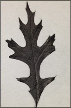
Scientists have a new recipe for batteries: Bake a leaf, and add sodium. They used a carbonized oak leaf, pumped full of sodium, as a demonstration battery’s negative terminal, or anode, according to a paper published yesterday in the journal ACS Applied Materials Interfaces.

Scientists baked a leaf to demonstrate a battery. Credit: Image courtesy of Maryland NanoCenter

Scientists baked a leaf to demonstrate a battery.
Credit: Image courtesy of Maryland NanoCenter

A Jan. ??, 2016 Maryland NanoCenter (University of Maryland) news release, which originated the news item, provides more information about the nature (pun intended) of the research,

“Leaves are so abundant. All we had to do was pick one up off the ground here on campus,” said Hongbian Li, a visiting professor at the University of Maryland’s department of materials science and engineering and one of the main authors of the paper. Li is a member of the faculty at the National Center for Nanoscience and Technology in Beijing, China.

Other studies have shown that melon skin, banana peels and peat moss can be used in this way, but a leaf needs less preparation.

The scientists are trying to make a battery using sodium where most rechargeable batteries sold today use lithium. Sodium would hold more charge, but can’t handle as many charge-and-discharge cycles as lithium can.

One of the roadblocks has been finding an anode material that is compatible with sodium, which is slightly larger than lithium. Some scientists have explored graphene, dotted with various materials to attract and retain the sodium, but these are time consuming and expensive to produce.  In this case, they simply heated the leaf for an hour at 1,000 degrees C (don’t try this at home) to burn off all but the underlying carbon structure.

The lower side of the maple [?] leaf is studded with pores for the leaf to absorb water. In this new design, the pores absorb the sodium electrolyte. At the top, the layers of carbon that made the leaf tough become sheets of nanostructured carbon to absorb the sodium that carries the charge.

“The natural shape of a leaf already matches a battery’s needs: a low surface area, which decreases defects; a lot of small structures packed closely together, which maximizes space; and internal structures of the right size and shape to be used with sodium electrolyte,” said Fei Shen, a visiting student in the department of materials science and engineering and the other main author of the paper.

“We have tried other natural materials, such as wood fiber, to make a battery,” said Liangbing Hu, an assistant professor of materials science and engineering. “A leaf is designed by nature to store energy for later use, and using leaves in this way could make large-scale storage environmentally friendly.”

The next step, Hu said, is “to investigate different types of leaves to find the best thickness, structure and flexibility” for electrical energy storage.  The researchers have no plans to commercialize at this time.

Here’s a link to and a citation for the paper,

Carbonized-leaf Membrane with Anisotropic Surfaces for Sodium-ion Battery by Hongbian Li, Fei Shen, Wei Luo, Jiaqi Dai, Xiaogang Han, Yanan Chen, Yonggang Yao, Hongli Zhu, Kun Fu, Emily Hitz, and Liangbing Hu. ACS Appl. Mater. Interfaces, 2016, 8 (3), pp 2204–2210 DOI: 10.1021/acsami.5b10875 Publication Date (Web): January 4, 2016

Copyright © 2016 American Chemical Society

This paper is behind a paywall.

Nanotechnology-enabled flame retardant coating

This is a pretty remarkable demonstration made more so when you find out the flame retardant is naturally derived and nontoxic. From an Oct. 5, 2015 news item on Nanowerk,

Inspired by a naturally occurring material found in marine mussels, researchers at The University of Texas at Austin have created a new flame retardant to replace commercial additives that are often toxic and can accumulate over time in the environment and living animals, including humans.

An Oct. 5, 2015 University of Texas news release, which originated the news item, describes the situation with regard to standard flame retardants and what makes this new flame retardant technology so compelling,

Flame retardants are added to foams found in mattresses, sofas, car upholstery and many other consumer products. Once incorporated into foam, these chemicals can migrate out of the products over time, releasing toxic substances into the air and environment. Throughout the United States, there is pressure on state legislatures to ban flame retardants, especially those containing brominated compounds (BRFs), a mix of human-made chemicals thought to pose a risk to public health.

A team led by Cockrell School of Engineering associate professor Christopher Ellison found that a synthetic coating of polydopamine — derived from the natural compound dopamine — can be used as a highly effective, water-applied flame retardant for polyurethane foam. Dopamine is a chemical compound found in humans and animals that helps in the transmission of signals in the brain and other vital areas. The researchers believe their dopamine-based nanocoating could be used in lieu of conventional flame retardants.

“Since polydopamine is natural and already present in animals, this question of toxicity immediately goes away,” Ellison said. “We believe polydopamine could cheaply and easily replace the flame retardants found in many of the products that we use every day, making these products safer for both children and adults.”

Using far less polydopamine by weight than typical of conventional flame retardant additives, the UT Austin team found that the polydopamine coating on foams leads to a 67 percent reduction in peak heat release rate, a measure of fire intensity and imminent danger to building occupants or firefighters. The polydopamine flame retardant’s ability to reduce the fire’s intensity is about 20 percent better than existing flame retardants commonly used today.

Researchers have studied the use of synthetic polydopamaine for a number of health-related applications, including cancer drug delivery and implantable biomedical devices. However, the UT Austin team is thought to be one of the first to pursue the use of polydopamine as a flame retardant. To the research team’s surprise, they did not have to change the structure of the polydopamine from its natural form to use it as a flame retardant. The polydopamine was coated onto the interior and exterior surfaces of the polyurethane foam by simply dipping it into a water solution of dopamine for several days.

Ellison said he and his team were drawn to polydopamine because of its ability to adhere to surfaces as demonstrated by marine mussels who use the compound to stick to virtually any surface, including Teflon, the material used in nonstick cookware. Polydopamine also contains a dihydroxy-ring structure linked with an amine group that can be used to scavenge or remove free radicals. Free radicals are produced during the fire cycle as a polymer degrades, and their removal is critical to stopping the fire from continuing to spread. Polydopamine also produces a protective coating called char, which blocks fire’s access to its fuel source — the polymer. The synergistic combination of both these processes makes polydopamine an attractive and powerful flame retardant.

Ellison and his team are now testing to see whether they can shorten the nanocoating treatment process or develop a more convenient application process.

“We believe this alternative to flame retardants can prove very useful to removing potential hazards from products that children and adults use every day,” Ellison said. “We weren’t expecting to find a flame retardant in nature, but it was a serendipitous discovery.”

Here’s a link to and a citation for the paper,

Bioinspired Catecholic Flame Retardant Nanocoating for Flexible Polyurethane Foams by Joon Hee Cho, Vivek Vasagar, Kadhiravan Shanmuganathan, Amanda R. Jones, Sergei Nazarenko, and Christopher J. Ellison. Chem. Mater., 2015, 27 (19), pp 6784–6790 DOI: 10.1021/acs.chemmater.5b03013
Publication Date (Web): September 9, 2015
Copyright © 2015 American Chemical Society

This paper is behind a paywall. It should be noted that researchers from the University of Southern Mississippi and the Council of Scientific & Industrial Research (CSIR)-National Chemical Laboratory in Pune, India were also involved in this work.

The challenges of wet adhesion and how nature solves the problem

I usually post about dry adhesion, that is, sticking to dry surfaces in the way a gecko might. This particular piece concerns wet adhesion, a matter of particular interest in medicine where you want bandages and such to stick to a wet surface and in marine circles where they want barnacles and such to stop adhering to boat and ship hulls.

An Aug. 6, 2015 news item on ScienceDaily touts ‘wet adhesion’ research focused on medicinal matters,

Wet adhesion is a true engineering challenge. Marine animals such as mussels, oysters and barnacles are naturally equipped with the means to adhere to rock, buoys and other underwater structures and remain in place no matter how strong the waves and currents.

Synthetic wet adhesive materials, on the other hand, are a different story.

Taking their cue from Mother Nature and the chemical composition of mussel foot proteins, the Alison Butler Lab at UC [University of California] Santa Barbara [UCSB] decided to improve a small molecule called the siderophore cyclic trichrysobactin (CTC) that they had previously discovered. They modified the molecule and then tested its adhesive strength in aqueous environments. The result: a compound that rivals the staying power of mussel glue.

An Aug. 6, 2015 UCSB news release by Julie Cohen, which originated the news item, describes some of the reasons for the research, the interdisciplinary aspect of the collaboration, and technical details of the work (Note: Links have been removed),

“There’s real need in a lot of environments, including medicine, to be able to have glues that would work in an aqueous environment,” said co-author Butler, a professor in UCSB’s Department of Chemistry and Biochemistry. “So now we have the basis of what we might try to develop from here.”

Also part of the interdisciplinary effort were Jacob Israelachvili’s Interfacial Sciences Lab in UCSB’s Department of Chemical Engineering and J. Herbert Waite, a professor in the Department of Molecular, Cellular and Developmental Biology, whose own work focuses on wet adhesion.

“We just happened to see a visual similarity between compounds in the siderophore CTC and in mussel foot proteins,” Butler explained. Siderophores are molecules that bind and transport iron in microorganisms such as bacteria. “We specifically looked at the synergy between the role of the amino acid lysine and catechol,” she added. “Both are present in mussel foot proteins and in CTC.”
Mussel foot proteins contain similar amounts of lysine and the catechol dopa. Catechols are chemical compounds used in such biological functions as neurotransmission. However, certain proteins have adopted dopa for adhesive purposes.

From discussions with Waite, Butler realized that CTC contained not only lysine but also a compound similar to dopa. Further, CTC paired its catechol with lysine, just like mussel foot proteins do.

“We developed a better, more stable molecule than the actual CTC,” Butler explained. “Then we modified it to tease out the importance of the contributions from either lysine or the catechol.”

Co-lead author Greg Maier, a graduate student in the Butler Lab, created six different compounds with varying amounts of lysine and catechol. The Israelachvili lab tested each compound for its surface and adhesion characteristics. Co-lead author Michael Rapp used a surface force apparatus developed in the lab to measure the interactions between mica surfaces in a saline solution.

Only the two compounds containing a cationic amine, such as lysine, and catechol exhibited adhesive strength and a reduced intervening film thickness, which measures the amount two surfaces can be squeezed together. Compounds without catechol had greatly diminished adhesion levels but a similarly reduced film thickness. Without lysine, the compounds displayed neither characteristic. “Our tests showed that lysine was key, helping to remove salt ions from the surface to allow the glue to get to the underlying surface,” Maier said.

“By looking at a different biosystem that has similar characteristics to some of the best-performing mussel glues, we were able to deduce that these two small components work together synergistically to create a favorable environment at surfaces to promote adherence,” explained Rapp, a chemical engineering graduate student. “Our results demonstrate that these two molecular groups not only prime the surface but also work collectively to build better adhesives that stick to surfaces.”

“In a nutshell, our discovery is that you need lysine and you need the catechol,” Butler concluded. “There’s a one-two punch: the lysine clears and primes the surface and the catechol comes down and hydrogen bonds to the mica surface. This is an unprecedented insight about what needs to happen during wet adhesion.”

Here’s a link to and a citation for the paper,

Adaptive synergy between catechol and lysine promotes wet adhesion by surface salt displacement by Greg P. Maier, Michael V. Rapp, J. Herbert Waite, Jacob N. Israelachvili, and Alison Butler. Science 7 August 2015: Vol. 349 no. 6248 pp. 628-632 DOI: 10.1126/science.aab055

This paper is behind a paywall.

I have previously written about mussels and wet adhesion in a Dec. 13, 2012 posting regarding some research at the University of British Columbia (Canada). As for dry adhesion, there’s my June 11, 2014 posting titled: Climb like a gecko (in DARPA’s [US Defense Advanced Research Projects Agency] Z-Man program) amongst others.

Building nanocastles in the sand

Scientists have taken inspiration from sandcastles to build robots made of nanoparticles. From an Aug. 5, 2015 news item on ScienceDaily,

If you want to form very flexible chains of nanoparticles in liquid in order to build tiny robots with flexible joints or make magnetically self-healing gels, you need to revert to childhood and think about sandcastles.

In a paper published this week in Nature Materials, researchers from North Carolina State University and the University of North Carolina-Chapel Hill show that magnetic nanoparticles encased in oily liquid shells can bind together in water, much like sand particles mixed with the right amount of water can form sandcastles.

An Aug. 5, 2015 North Carolina State University (NCSU) news release (also on EurekAlert) by Mick Kulikowski, which originated the news item, expands on the theme,

“Because oil and water don’t mix, the oil wets the particles and creates capillary bridges between them so that the particles stick together on contact,” said Orlin Velev, INVISTA Professor of Chemical and Biomolecular Engineering at NC State and the corresponding author of the paper.

“We then add a magnetic field to arrange the nanoparticle chains and provide directionality,” said Bhuvnesh Bharti, research assistant professor of chemical and biomolecular engineering at NC State and first author of the paper.

Chilling the oil is like drying the sandcastle. Reducing the temperature from 45 degrees Celsius to 15 degrees Celsius freezes the oil and makes the bridges fragile, leading to breaking and fragmentation of the nanoparticle chains. Yet the broken nanoparticles chains will re-form if the temperature is raised, the oil liquefies and an external magnetic field is applied to the particles.

“In other words, this material is temperature responsive, and these soft and flexible structures can be pulled apart and rearranged,” Velev said. “And there are no other chemicals necessary.”

The paper is also co-authored by Anne-Laure Fameau, a visiting researcher from INRA [French National Institute for Agricultural Research or Institut National de la Recherche Agronomique], France. …

Here’s a link to and a citation for the paper,

Nanocapillarity-mediated magnetic assembly of nanoparticles into ultraflexible filaments and reconfigurable networks by Bhuvnesh Bharti, Anne-Laure Fameau, Michael Rubinstein, & Orlin D. Velev. Nature Materials (2015) doi:10.1038/nmat4364 Published online 03 August 2015

This paper is behind a paywall.

Cleaning antennae—ant style

The University of Cambridge (UK) has produced research that could lead to cleaning at the microscale and nanoscale and it’s all due to ants. From a July 28, 2015 news item on Nanowerk (Note: A link has been removed),

For an insect, grooming is a serious business. If the incredibly sensitive hairs on their antennae get too dirty, they are unable to smell food, follow pheromone trails or communicate. So insects spend a significant proportion of their time just keeping themselves clean. Until now, however, no-one has really investigated the mechanics of how they actually go about this.

In a study published in Open Science (“Functional morphology and efficiency of the antenna cleaner in Camponotus rufifemur ants”), Alexander Hackmann and colleagues from the Department of Zoology [University of Cambridge] have undertaken the first biomechanical investigation of how ants use different types of hairs in their cleaning apparatus to clear away dirt from their antennae.

A July 27, 2015 University of Cambridge press release, which originated the news item, expands on the theme,

“Insects have developed ingenious ways of cleaning very small, sensitive structures, so finding out exactly how they work could have fascinating applications for nanotechnology – where contamination of small things, especially electronic devices, is a big problem. Different insects have all kinds of different cleaning devices, but no-one has really looked at their mechanical function in detail before,” explains Hackmann.

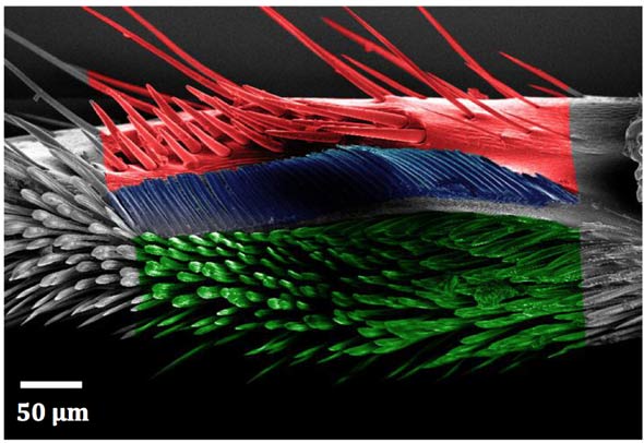
Camponotus rufifemur ants possess a specialised cleaning structure on their front legs that is actively used to groom their antennae. A notch and spur covered in different types of hairs form a cleaning device similar in shape to a tiny lobster claw. During a cleaning movement, the antenna is pulled through the device which clears away dirt particles using ‘bristles’, a ‘comb’ and a ‘brush’.

To investigate how the different hairs work, Hackmann painstakingly constructed an experimental mechanism to mimic the ant’s movements and pull antennae through the cleaning structure under a powerful microscope. This allowed him to film the process in extreme close up and to measure the cleaning efficiency of the hairs using fluorescent particles.

What he discovered was that the three clusters of hairs perform a different function in the cleaning process. The dirty antenna surface first comes into contact with the ‘bristles’ (shown in the image in red) which scratch away the largest particles. It is then drawn past the ‘comb’ (shown in the image in blue) which removes smaller particles that get trapped between the comb hairs. Finally, it is drawn through the ‘brush’ (shown in the image in green) which removes the smallest particles.

Scanning electron micrograph of the tarsal notch (Alexander Hackmann). Courtesy: University of Cambridge

Scanning electron micrograph of the tarsal notch (Alexander Hackmann). Courtesy: University of Cambridge

The news release offers more detail about the ‘notch’s’ cleaning properties,

“While the ‘bristles’ and the ‘comb’ scrape off larger particles mechanically, the ‘brush’ seems to attract smaller dirt particles from the antenna by adhesion,” says Hackmann, who works in the laboratory of Dr Walter Federle.

Where the ‘bristles’ and ‘comb’ are rounded and fairly rigid, the ‘brush’ hairs are flat, bendy and covered in ridges – this increases the surface area for contact with the dirt particles, which stick to the hairs. Researchers do not yet know what makes the ‘brush’ hairs sticky – whether it is due to electrostatic forces, sticky secretions, or a combination of factors.

“The arrangement of ‘bristles’, ‘combs’ and ‘brush’ lets the cleaning structure work as a particle filter that can clean different sized dirt particles with a single cleaning stroke,” says Hackmann. “Modern nanofabrication techniques face similar problems with surface contamination, and as a result the fabrication of micron-scale devices requires very expensive cleanroom technology. We hope that understanding the biological system will lead to building bioinspired devices for cleaning on micro and nano scales.”

If you want to see the a video of the ‘cleaning action’, you can check either Nanowerk’s July 28, 2015 news item or the University of Cambridge’s July 27, 2015 press release.

Here’s a link to and citation for the paper,

Functional morphology and efficiency of the antenna cleaner in Camponotus rufifemur ants by Alexander Hackmann, Henry Delacave, Adam Robinson, David Labonte, and Walter Federle. Royal Society Open Science DOI: 10.1098/rsos.150129  Published 22 July 2015

As you may have guessed from the journal’s title, this is an open access paper.