Tag Archives: UCSD

Do-it-yourself sensors possible with biocatalytic pen technology

The engineers at the University of California at San Diego (UCSD) are envisioning a future where anyone can create a biosensor anywhere. From a March 3, 2015 news item on Azonano,

A new simple tool developed by nanoengineers at the University of California, San Diego, is opening the door to an era when anyone will be able to build sensors, anywhere, including physicians in the clinic, patients in their home and soldiers in the field.

The team from the University of California, San Diego, developed high-tech bio-inks that react with several chemicals, including glucose. They filled off-the-shelf ballpoint pens with the inks and were able to draw sensors to measure glucose directly on the skin and sensors to measure pollution on leaves.

A March 2, 2015 UCSD news release by Ioana Patringenaru, which originated the news item, describes the researchers’ hopes for this technology,

Skin and leaves aren’t the only media on which the pens could be used. Researchers envision sensors drawn directly on smart phones for personalized and inexpensive health monitoring or on external building walls for monitoring of toxic gas pollutants. The sensors also could be used on the battlefield to detect explosives and nerve agents.

The team, led by Joseph Wang, the chairman of the Department of NanoEngineering at the University of California, San Diego, published their findings in the Feb. 26 [2015] issue of Advanced Healthcare Materials. Wang also directs the Center for Wearable Sensors at UC San Diego.

“Our new biocatalytic pen technology, based on novel enzymatic inks, holds considerable promise for a broad range of applications on site and in the field,” Wang said.

The news release goes on to describe one of the key concerns with developing the ink,

The biggest challenge the researchers faced was making inks from chemicals and biochemicals that aren’t harmful to humans or plants; could function as the sensors’ electrodes; and retain their properties over long periods in storage and in various conditions. Researchers turned to biocompatible polyethylene glycol, which is used in several drug delivery applications, as a binder. To make the inks conductive to electric current they used graphite powder. They also added chitosan, an antibacterial agent which is used in bandages to reduce bleeding, to make sure the ink adhered to any surfaces it was used on. The inks’ recipe also includes xylitol, a sugar substitute, which helps stabilize enzymes that react with several chemicals the do-it-yourself sensors are designed to monitor.

There’s a backstory to this research,

Wang’s team has been investigating how to make glucose testing for diabetics easier for several years. The same team of engineers recently developed non-invasive glucose sensors in the form of temporary tattoos. In this study, they used pens, loaded with an ink that reacts to glucose, to draw reusable glucose-measuring sensors on a pattern printed on a transparent, flexible material which includes an electrode. Researchers then pricked a subject’s finger and put the blood sample on the sensor. The enzymatic ink reacted with glucose and the electrode recorded the measurement, which was transmitted to a glucose-measuring device. Researchers then wiped the pattern clean and drew on it again to take another measurement after the subject had eaten.

Researchers estimate that one pen contains enough ink to draw the equivalent of 500 high-fidelity glucose sensor strips. Nanoengineers also demonstrated that the sensors could be drawn directly on the skin and that they could communicate with a Bluetooth-enabled electronic device that controls electrodes called a potentiostat, to gather data.

As mentioned earlier, there are more applications being considered (from the news release),

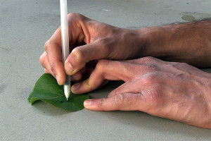
The pens would also allow users to draw sensors that detect pollutants and potentially harmful chemicals sensors on the spot. Researchers demonstrated that this was possible by drawing a sensor on a leaf with an ink loaded with enzymes that react with phenol, an industrial chemical, which can also be found in cosmetics, including sunscreen. The leaf was then dipped in a solution of water and phenol and the sensor was connected to a pollution detector. The sensors could be modified to react with many pollutants, including heavy metals or pesticides.

Next steps include connecting the sensors wirelessly to monitoring devices and investigating how the sensors perform in difficult conditions, including extreme temperatures, varying humidity and extended exposure to sunlight.

The researchers’ have provided a picture of the pen and a leaf,

Researchers drew sensors capable of detecting pollutants on a leaf. Courtesy: University of California at San Diego

Researchers drew sensors capable of detecting pollutants on a leaf. Courtesy: University of California at San Diego

Here’s a link to and a citation for the paper,

Biocompatible Enzymatic Roller Pens for Direct Writing of Biocatalytic Materials: “Do-it-Yourself” Electrochemical Biosensors by Amay J. Bandodkar, Wenzhao Jia, Julian Ramírez, and Joseph Wang. Advanced Healthcare Materials DOI: 10.1002/adhm.201400808 Article first published online: 26 FEB 2015

© 2015 WILEY-VCH Verlag GmbH & Co. KGaA, Weinheim

This article is behind a paywall.

Tattoos that detect glucose levels

Temporary tattoos with a biomedical function are a popular topic and one of the latest detects glucose levels without subjecting a person with diabetes to pin pricks. From a Jan. 14, 2015 news item on ScienceDaily,

Scientists have developed the first ultra-thin, flexible device that sticks to skin like a rub-on tattoo and can detect a person’s glucose levels. The sensor, reported in a proof-of-concept study in the ACS [American Chemical Society] journal Analytical Chemistry, has the potential to eliminate finger-pricking for many people with diabetes.

A Jan. 14, 2015 ACS news release on EurekAlert, which originated the news item, describes the current approaches to testing glucose and the new painless technique,

Joseph Wang and colleagues in San Diego note that diabetes affects hundreds of millions of people worldwide. Many of these patients are instructed to monitor closely their blood glucose levels to manage the disease. But the standard way of checking glucose requires a prick to the finger to draw blood for testing. The pain associated with this technique can discourage people from keeping tabs on their glucose regularly. A glucose sensing wristband had been introduced to patients, but it caused skin irritation and was discontinued. Wang’s team wanted to find a better approach.

The researchers made a wearable, non-irritating platform that can detect glucose in the fluid just under the skin based on integrating glucose extraction and electrochemical biosensing. Preliminary testing on seven healthy volunteers showed it was able to accurately determine glucose levels. The researchers conclude that the device could potentially be used for diabetes management and for other conditions such as kidney disease.

There is a Jan. 14, 2015 University of California at San Diego news release (also on EurekAlert) describing the work in more detail,

Nanoengineers at the University of California, San Diego have tested a temporary tattoo that both extracts and measures the level of glucose in the fluid in between skin cells. …

The sensor was developed and tested by graduate student Amay Bandodkar and colleagues in Professor Joseph Wang’s laboratory at the NanoEngineering Department and the Center for Wearable Sensors at the Jacobs School of Engineering at UC San Diego. Bandodkar said this “proof-of-concept” tattoo could pave the way for the Center to explore other uses of the device, such as detecting other important metabolites in the body or delivering medicines through the skin.

At the moment, the tattoo doesn’t provide the kind of numerical readout that a patient would need to monitor his or her own glucose. But this type of readout is being developed by electrical and computer engineering researchers in the Center for Wearable Sensors. “The readout instrument will also eventually have Bluetooth capabilities to send this information directly to the patient’s doctor in real-time or store data in the cloud,” said Bandodkar.

The research team is also working on ways to make the tattoo last longer while keeping its overall cost down, he noted. “Presently the tattoo sensor can easily survive for a day. These are extremely inexpensive—a few cents—and hence can be replaced without much financial burden on the patient.”

The Center “envisions using these glucose tattoo sensors to continuously monitor glucose levels of large populations as a function of their dietary habits,” Bandodkar said. Data from this wider population could help researchers learn more about the causes and potential prevention of diabetes, which affects hundreds of millions of people and is one of the leading causes of death and disability worldwide.

People with diabetes often must test their glucose levels multiple times per day, using devices that use a tiny needle to extract a small blood sample from a fingertip. Patients who avoid this testing because they find it unpleasant or difficult to perform are at a higher risk for poor health, so researchers have been searching for less invasive ways to monitor glucose.

In their report in the journal Analytical Chemistry, Wang and his co-workers describe their flexible device, which consists of carefully patterned electrodes printed on temporary tattoo paper. A very mild electrical current applied to the skin for 10 minutes forces sodium ions in the fluid between skin cells to migrate toward the tattoo’s electrodes. These ions carry glucose molecules that are also found in the fluid. A sensor built into the tattoo then measures the strength of the electrical charge produced by the glucose to determine a person’s overall glucose levels.

“The concentration of glucose extracted by the non-invasive tattoo device is almost hundred times lower than the corresponding level in the human blood,” Bandodkar explained. “Thus we had to develop a highly sensitive glucose sensor that could detect such low levels of glucose with high selectivity.”

A similar device called GlucoWatch from Cygnus Inc. was marketed in 2002, but the device was discontinued because it caused skin irritation, the UC San Diego researchers note. Their proof-of-concept tattoo sensor avoids this irritation by using a lower electrical current to extract the glucose.

Wang and colleagues applied the tattoo to seven men and women between the ages of 20 and 40 with no history of diabetes. None of the volunteers reported feeling discomfort during the tattoo test, and only a few people reported feeling a mild tingling in the first 10 seconds of the test.

To test how well the tattoo picked up the spike in glucose levels after a meal, the volunteers ate a carb-rich meal of a sandwich and soda in the lab. The device performed just as well at detecting this glucose spike as a traditional finger-stick monitor.

The researchers say the device could be used to measure other important chemicals such as lactate, a metabolite analyzed in athletes to monitor their fitness. The tattoo might also someday be used to test how well a medication is working by monitoring certain protein products in the intercellular fluid, or to detect alcohol or illegal drug consumption.

This reminds me a little of the Google moonshot project concerning health diagnostics. Announced in Oct. 2014, that project involved swallowing a pill containing nanoparticles that would circulate through your body monitoring your health and recongregating at your wrist so a band worn there could display your health status (Oct. 30, 2014 article by Signe Brewster for GigaOm). Experts welcomed the funding while warning the expectations seemed unrealistic given the current state of research and technology. This temporary tattoo seems much better grounded in terms of the technology used and achievable results.

Here’s a link to and a citation for the paper,

Tattoo-Based Noninvasive Glucose Monitoring: A Proof-of-Concept Study by Amay J. Bandodkar, Wenzhao Jia, Ceren Yardımcı, Xuan Wang, Julian Ramirez, and Joseph Wang. Anal. Chem., 2015, 87 (1), pp 394–398 DOI: 10.1021/ac504300n Publication Date (Web): December 12, 2014

Copyright © 2014 American Chemical Society

This appears to be an open access paper.

My latest posting posting on medical tattoos (prior to this) is an Aug. 13, 2014 post about a wearable biobattery.

RNA interference: a Tekmira deal and a new technique births Solstice Biologics

I have two news items concerning ribonucleic acid interference (RNAi). The first item features Tekmira Pharmaceuticals Corporation (a Canadian company located in the Vancouver area) and a licencing deal with Dicerna Pharmaceuticals (Massachusetts, US), according to a Nov. 18, 2014 news item on Azonano,

Tekmira Pharmaceuticals Corporation a leading developer of RNA interference (RNAi) therapeutics, today announces a licensing and collaboration agreement with Dicerna Pharmaceuticals, Inc. Tekmira has licensed its proprietary lipid nanoparticle (LNP) delivery technology for exclusive use in Dicerna’s primary hyperoxaluria type 1 (PH1) development program.

Under the agreement, Dicerna will pay Tekmira $2.5 million upfront and payments of $22 million in aggregate development milestones, plus a mid-single-digit royalty on future PH1 sales. This new partnership also includes a supply agreement with Tekmira providing clinical drug supply and regulatory support in the rapid advancement of the product candidate.

The agreement announced today follows the successful testing and demonstration of positive results combining Tekmira’s LNP technology with DCR-PH1 in pre-clinical animal models.

I don’t entirely understand what they mean by “pre-clinical animal models” as I’ve not noticed the term “pre-clinical” applied to animal testing before this. It’s possible they mean they’ve run tests on animals (in vivo) and are now proceeding to human clinical trials or it could mean they’ve run in silico (computer modeling) or in vitro (test tube/test slide) tests and are now proceeding to animal tests. If anyone should have some insights, please do share them with me in the comments section.

A Nov. 17, 2014 Tekmira news release, which originated the news item, describes the deal in more detail,

Dicerna will use Tekmira’s third generation LNP technology for delivery of DCR-PH1, Dicerna’s Dicer substrate RNA (DsiRNA) molecule, for the treatment of PH1, a rare, inherited liver disorder that often results in kidney failure and for which there are no approved therapies.

“This new agreement validates our leadership position in RNAi delivery with LNP technology, and it underscores the significant value we can bring to partners who leverage our technology. Our LNP technology is enabling the most advanced applications of RNAi therapeutics in the clinic, and it continues to do so. We are excited to be working with Dicerna to be able to advance a needed therapeutic for the treatment of PH1,” said Dr. Mark J. Murray, Tekmira’s President and CEO.

“As a core pillar of our business strategy, we continue to engage in partnerships where our technology improves the risk profile and accelerates the development programs of our collaborators and provides meaningful non-dilutive financing to TKMR,” added Dr. Murray.

“Dicerna is focused on realizing the full clinical potential of our proprietary pipeline of highly targeted RNAi therapies by applying proven technologies,” said Douglas Fambrough, Ph.D., Chief Executive Officer of Dicerna. “By drawing on Tekmira’s extensive and deep experience with lipid nanoparticle delivery to the liver, the agreement will streamline the development path for DCR-PH1. We look forward to initiating Phase 1 trials of DCR-PH1 in 2015, aiming to fill a high unmet medical need for patients with PH1.”

The news release also provides a high level description of the various technologies being researched and brought to market and a bit more information about the liver disorder being addressed by this research,

About RNAi

RNAi therapeutics have the potential to treat a number of human diseases by “silencing” disease-causing genes. The discoverers of RNAi, a gene silencing mechanism used by all cells, were awarded the 2006 Nobel Prize for Physiology or Medicine. RNAi trigger molecules often require delivery technology to be effective as therapeutics.

AboutTekmira’s LNP Technology

Tekmira believes its LNP technology represents the most widely adopted delivery technology for the systemic delivery of RNAi triggers. Tekmira’s LNP platform is being utilized in multiple clinical trials by Tekmira and its partners. Tekmira’s LNP technology (formerly referred to as stable nucleic acid-lipid particles, or SNALP) encapsulates RNAi triggers with high efficiency in uniform lipid nanoparticles that are effective in delivering these therapeutic compounds to disease sites. Tekmira’s LNP formulations are manufactured by a proprietary method which is robust, scalable and highly reproducible, and LNP-based products have been reviewed by multiple regulatory agencies for use in clinical trials. LNP formulations comprise several lipid components that can be adjusted to suit the specific application.

About Primary Hyperoxaluria Type 1 ( PH1)

PH1 is a rare, inherited liver disorder that often results in severe damage to the kidneys. The disease can be fatal unless the patient undergoes a liver-kidney transplant, a major surgical procedure that is often difficult to perform due to the lack of donors and the threat of organ rejection. In the event of a successful transplant, the patient must live the rest of his or her life on immunosuppressant drugs, which have substantial associated risks. Currently, there are no FDA approved treatments for PH1.

PH1 is characterized by a genetic deficiency of the liver enzyme alanine:glyoxalate-aminotransferase (AGT), which is encoded by the AGXT gene. AGT deficiency induces overproduction of oxalate by the liver, resulting in the formation of crystals of calcium oxalate in the kidneys. Oxalate crystal formation often leads to chronic and painful cases of kidney stones and subsequent fibrosis (scarring), which is known as nephrocalcinosis. Many patients progress to end-stage renal disease (ESRD) and require dialysis or transplant. Aside from having to endure frequent dialysis, PH1 patients with ESRD may experience a build-up of oxalate in the bone, skin, heart and retina, with concomitant debilitating complications. While the true prevalence of primary hyperoxaluria is unknown, it is estimated to be one to three cases per one million people.1 Fifty percent of patients with PH1 reach ESRD by their mid-30s.2

About DCR-PH1

Dicerna is developing DCR-PH1, which is in preclinical development, for the treatment of PH1. DCR-PH1 is engineered to address the pathology of PH1 by targeting and destroying the messenger RNA (mRNA) produced by HAO1, a gene implicated in the pathogenesis of PH1. HAO1 encodes glycolate oxidase, a protein involved in producing oxalate. By reducing oxalate production, this approach is designed to prevent the complications of PH1. In preclinical studies, DCR-PH1 has been shown to induce potent and long-term inhibition of HAO1 and to significantly reduce levels of urinary oxalate, while demonstrating long-term efficacy and tolerability in animal models of PH1.

About Dicerna’s Dicer Substrate Technology

Dicerna’s proprietary RNAi molecules are known as Dicer substrates, or DsiRNAs, so called because they are processed by the Dicer enzyme, which is the initiation point for RNAi in the human cell cytoplasm. Dicerna’s discovery approach is believed to maximize RNAi potency because the DsiRNAs are structured to be ideal for processing by Dicer. Dicer processing enables the preferential use of the correct RNA strand of the DsiRNA, which may increase the efficacy of the RNAi mechanism, as well as the potency of the DsiRNA molecules relative to other molecules used to induce RNAi.

You can find more information about Tekmira here and about Dicerna here. I mentioned Tekmira previously in a Sept. 28, 2014 post about Ebola and treatments.

Further south at the University of California at San Diego (UCSD), researcher and founder of Solstice Biologics, Dr Steven Dowdy has developed and patented a new technique for delivering RNAi drugs into cells according to a Nov. 18, 2014 news item on Azonano,

Small pieces of synthetic RNA trigger a RNA interference (RNAi) response that holds great therapeutic potential to treat a number of diseases, especially cancer and pandemic viruses. The problem is delivery — it is extremely difficult to get RNAi drugs inside the cells in which they are needed. To overcome this hurdle, researchers at University of California, San Diego School of Medicine have developed a way to chemically disguise RNAi drugs so that they are able to enter cells. Once inside, cellular machinery converts these disguised drug precursors — called siRNNs — into active RNAi drugs. …

A Nov. 17, 2014 UCSD news release (also on EurekAlert) by Heather Buschman, which originated the news item, describes the issues with delivering RNAi drugs to cells and the new technique,

“Many current approaches use nanoparticles to deliver RNAi drugs into cells,” said Steven F. Dowdy, PhD, professor in the Department of Cellular and Molecular Medicine and the study’s principal investigator. “While nanotechnology protects the RNAi drug, from a molecular perspective nanoparticles are huge, some 5,000 times larger than the RNAi drug itself. Think of delivering a package into your house by having an 18-wheeler truck drive it through your living room wall — that’s nanoparticles carrying standard RNAi drugs. Now think of a package being slipped through the mail slot — that’s siRNNs.”

The beauty of RNAi is that it selectively blocks production of target proteins in a cell, a finding that garnered a Nobel Prize in 2006. While this is a normal process that all cells use, researchers have taken advantage of RNAi to inhibit specific proteins that cause disease when overproduced or mutated, such as in cancer. First, researchers generate RNAi drugs with a sequence that corresponds to the gene blueprint for the disease protein and then delivers them into cells. Once inside the cell, the RNAi drug is loaded into an enzyme that specifically slices the messenger RNA encoding the target protein in half. This way, no protein is produced.

As cancer and viral genes mutate, RNAi drugs can be easily evolved to target them. This allows RNAi therapy to keep pace with the genetics of the disease — something that no other type of therapy can do. Unfortunately, due to their size and negatively charged chemical groups (phosphates) on their backbone, RNAi drugs are repelled by the cellular membrane and cannot be delivered into cells without a special delivery agent.

It took Dowdy and his team, including Bryan Meade, PhD, Khirud Gogoi, PhD, and Alexander S. Hamil, eight years to find a way to mask RNAi’s negative phosphates in such a way that gets them into cells, but is still capable of inducing an RNAi response once inside.

In the end, the team added a chemical tag called a phosphotriester group. The phosphotriester neutralizes and protects the RNA backbone — converting the ribonucleic acid (RNA) to ribonucleic neutral (RNN), and thus giving the name siRNN. The neutral (uncharged) nature of siRNNs allows them to pass into the cell much more efficiently. Once inside the cell, enzymes cleave off the neutral phosphotriester group to expose a charged RNAi drug that shuts down production of the target disease protein. siRNNs represent a transformational next-generation RNAi drug.

“siRNNs are precursor drugs, or prodrugs, with no activity. It’s like having a tool still in the box, it won’t work until you take it out,” Dowdy said. “Only when the packaging — the phosphotriester groups — is removed inside the cells do you have an active tool or RNAi drug.”

The findings held up in a mouse model, too. There, Dowdy’s team found that siRNNs were significantly more effective at blocking target protein production than typical RNAi drugs — demonstrating that once siRNNs get inside a cell they can do a better job.

“There remains a lot of work ahead to get this into the clinics. But, in theory, the therapeutic potential of siRNNs is endless,” Dowdy said. “Particularly for cancer, viral infections and genetic diseases.”

The siRNN technology forms the basis for Solstice Biologics, a biotech company in La Jolla, Calif. that is now taking the technique to the next level. Dowdy is a co-founder of Solstice Biologics and serves as a Board Director.

Here’s a link to and a citation for the research paper,

Efficient delivery of RNAi prodrugs containing reversible charge-neutralizing phosphotriester backbone modifications by Bryan R Meade, Khirud Gogoi, Alexander S Hamil, Caroline Palm-Apergi, Arjen van den Berg, Jonathan C Hagopian, Aaron D Springer, Akiko Eguchi, Apollo D Kacsinta, Connor F Dowdy, Asaf Presente, Peter Lönn, Manuel Kaulich, Naohisa Yoshioka, Edwige Gros, Xian-Shu Cui, & Steven F Dowdy. Nature Biotechnology (2014) doi:10.1038/nbt.3078 Published online 17 November 2014

This paper is behind a paywall.

I have not been able to locate a website for Solstice Biologics but did find a rather curious item about Dr. Dowdy and a shooting incident last year. From a Sept. 18, 2013 news article by Kat Robinson for thewire.sheknows.com,

A wealthy San Diego community is shaken after a man opens fire on his former neighborhood early Wednesday morning. Police say Hans Petersen, a 48-year-old man, is the prime suspect in the shooting of Steven Dowdy and Michael Fletcher.

There’s also a Nov. 8, 2013 article about the incident by Lucas Laursen for Nature magazine,

On September 18 [2013], former Traversa Therapeutics CEO Hans Petersen went on a shooting spree. One of two people wounded was molecular biologist Steven Dowdy, a professor at University of California San Diego (UCSD) School of Medicine, in La Jolla, and cofounder of Traversa, according to a San Diego police report.…

The rest of the article is behind a paywall.
A tattoo that’s a biobattery and a sensor?

It’s going to be an American Chemical Society (ACS) 248th meeting kind of week as yet another interesting piece of scientific research is bruited (spread) about the internet. This time it’s all about sweat, exercise, and biobatteries. From an Aug. 13, 2014 news item on Nanowerk,

In the future, working up a sweat by exercising may not only be good for your health, but it could also power your small electronic devices. Researchers will report today that they have designed a sensor in the form of a temporary tattoo that can both monitor a person’s progress during exercise and produce power from their perspiration.

An Aug. 13, 2014 ACS news release on EurekAlert, which originated the news item, describes the inspiration (as opposed to perspiration) for this technology,

The device works by detecting and responding to lactate, which is naturally present in sweat. “Lactate is a very important indicator of how you are doing during exercise,” says Wenzhao Jia, Ph.D.

In general, the more intense the exercise, the more lactate the body produces. During strenuous physical activity, the body needs to generate more energy, so it activates a process called glycolysis. Glycolysis produces energy and lactate, the latter of which scientists can detect in the blood.

Professional athletes monitor their lactate levels during performance testing as a way to evaluate their fitness and training program. In addition, doctors measure lactate during exercise testing of patients for conditions marked by abnormally high lactate levels, such as heart or lung disease. Currently, lactate testing is inconvenient and intrusive because blood samples must be collected from the person at different times during the exercise regime and then analyzed.

The news release goes on to describe the research process which resulted in a temporary tattoo that could be used to power small scale electronics,

Jia, a postdoctoral student in the lab of Joseph Wang, D.Sc., at the University of California San Diego, and her colleagues developed a faster, easier and more comfortable way to measure lactate during exercise. They imprinted a flexible lactate sensor onto temporary tattoo paper. The sensor contained an enzyme that strips electrons from lactate, generating a weak electrical current. The researchers applied the tattoo to the upper arms of 10 healthy volunteers. Then the team measured the electrical current produced as the volunteers exercised at increasing resistance levels on a stationary bicycle for 30 minutes. In this way, they could continuously monitor sweat lactate levels over time and with changes in exercise intensity.

The team then went a step further, building on these findings to make a sweat-powered biobattery. Batteries produce energy by passing current, in the form of electrons, from an anode to a cathode. In this case, the anode contained the enzyme that removes electrons from lactate, and the cathode contained a molecule that accepts the electrons.

When 15 volunteers wore the tattoo biobatteries while exercising on a stationary bike, they produced different amounts of power. Interestingly, people who were less fit (exercising fewer than once a week) produced more power than those who were moderately fit (exercising one to three times per week). Enthusiasts who worked out more than three times per week produced the least amount of power. The researchers say that this is probably because the less-fit people became fatigued sooner, causing glycolysis to kick in earlier, forming more lactate. The maximum amount of energy produced by a person in the low-fitness group was 70 microWatts per cm2 of skin.

“The current produced is not that high, but we are working on enhancing it so that eventually we could power some small electronic devices,” Jia says. “Right now, we can get a maximum of 70 microWatts per cm2, but our electrodes are only 2 by 3 millimeters in size and generate about 4 microWatts — a bit small to generate enough power to run a watch, for example, which requires at least 10 microWatts. So besides working to get higher power, we also need to leverage electronics to store the generated current and make it sufficient for these requirements.”

Biobatteries offer certain advantages over conventional batteries: They recharge more quickly, use renewable energy sources (in this case, sweat), and are safer because they do not explode or leak toxic chemicals.

“These represent the first examples of epidermal electrochemical biosensing and biofuel cells that could potentially be used for a wide range of future applications,” Wang says.

The ACS has made a video about this work available,

It seems to me this tattoo battery could be used as a self-powered monitoring device in a medical application for heart or lung disease.

Synthetic Aesthetics: a book and an event (UK’s Victoria & Albert Museum) about synthetic biology and design

Sadly, I found out about the event after it took place (April 25, 2014) but I’m including it here as I think it serves a primer on putting together an imaginative art/science (art/sci) event, as well, synthetic biology is a topic I’ve covered here many times.

First, the book. Happily, it’s not too late to publicize it and, after all, that was at least one of the goals for the event. Here’s more about the book, from the UK’s Engineering and Physical Sciences Research Council April 25, 2014 news release (also on EurekAlert),

The emerging field of synthetic biology crosses the boundary between science and design, in order to design and manufacture biologically based parts, devices and systems that do not exist in the natural world, as well as the redesign of existing, natural biological systems.

This new technology has the potential to create new organisms for a variety of applications from materials to machines. What role can artists and designers play in our biological future?

This Friday [April 25, 2014], the Victoria & Albert Museum’s Friday Late turns the V&A into a living laboratory, bringing science and design together for one night of events, workshops and installations.

It will also feature the official launch of a new EPSRC-funded book ‘Synthetic Aesthetics: Investigating Synthetic Biology’s Designs on Nature’.

The book, by Alexandra Daisy Ginsberg, Jane Calvert, Pablo Schyfter, Alistair Elfick and Drew Endy, emerged from a research project ‘Sandpit: Synthetic aesthetics: connecting synthetic biology and creative design’ which was funded by the UK’s Engineering and Physical Sciences Research Council (EPSRC) and the National Science Foundation in the US.

Kedar Pandya, EPSRC’s Head of Engineering, said: “This event and the Synthetic Aesthetics book will act as a catalyst to spark informed debates and future research into how we develop and apply synthetic biology. Engineers and scientists are not divorced from the rest of society; ethical, moral and artistic questions need to be considered as we explore new science and technologies.”

The EPSRC project aimed to:

  • bring together scientists and engineers working in synthetic biology with artists and designers working in the creative industries, to develop long-lasting relationships which could help to improve their work
  • ensure aesthetic concerns and questions are reflected in the lifecycle of research projects and implementation of products, and enable inclusive and responsive technology development
  • produce new social scientific research that analyses and reflects on these interactions
  • initiate a new and expanded curriculum across both engineering and design disciplines to lead to new forms of engineering and new schools of art
  • improve synthetic biological projects, products and thus the world
  • engage and enable the full diversity of civilization’s creative resources to work with the synthetic biology community as full partners in creating and stewarding a beautifully integrated natural and engineered living world

Weirdly, the news release offered no link to the book.  Here’s the Synthetic Aesthetics: Investigating Synthetic Biology’s Designs on Nature page on the MIT Press website,

In this book, synthetic biologists, artists, designers, and social scientists investigate synthetic biology and design. After chapters that introduce the science and set the terms of the discussion, the book follows six boundary-crossing collaborations between artists and designers and synthetic biologists from around the world, helping us understand what it might mean to ‘design nature.’ These collaborations have resulted in biological computers that calculate form; speculative packaging that builds its own contents; algae that feeds on circuit boards; and a sampling of human cheeses. They raise intriguing questions about the scientific process, the delegation of creativity, our relationship to designed matter, and, the importance of critical engagement. Should these projects be considered art, design, synthetic biology, or something else altogether?

Synthetic biology is driven by its potential; some of these projects are fictions, beyond the current capabilities of the technology. Yet even as fictions, they help illuminate, question, and even shape the future of the field.

About the Authors

Alexandra Daisy Ginsberg is a London-based artist, designer, and writer.

Jane Calvert is a social scientist based in Science, Technology and Innovation Studies at the University of Edinburgh.

Pablo Schyfter is a social scientist based in Science, Technology and Innovation Studies at the University of Edinburgh.

Alistair Elfick is Codirector of the SynthSys Centre at the University of Edinburgh.

Drew Endy is a bioengineer at Stanford University and President of the BioBrick

Now for the event description from the Victoria and Albert Museum’s Friday Late series, the April 25,2014  event Synthetic Aesthetics webpage,

Synthetic Aesthetics

Friday 25 April, 18.30-22.00

Can we design life itself? The emerging field of synthetic biology crosses the boundary between science and design to manipulate the stuff of life. These new designers use life as a programmable material, creating new organisms with radical applications from materials to machines. Friday Late turns the V&A into a living laboratory, bringing science and design together for one night of events, workshops and installations, each exploring our biological future.

The evening will feature the book launch of Synthetic Aesthetics: Investigating Synthetic Biology’s Designs on Nature (MIT Press). The book marks an important point in the development of the emerging discipline of synthetic biology, sitting at the intersection between design and science. The book is a result of research funded by the UK’s Engineering and Physical Sciences Research Council and the National Science Foundation in the US.

All events are free and places are designated on a first come, first served basis, unless stated otherwise. Filming and photography will be taking place at this event.

Please note, if the Museum reaches capacity we will allow access on a one-in-one-out basis.

#FridayLate

ALL EVENING (18.30 – 21.30)

Live Lab

Spotlight Space, Grand Entrance
A functioning synthetic biology lab in the grand entrance places this experimental field front and centre within the historic home of the V&A. Conducting experiments and answering questions from visitors, the lab will be run by synthetic biologists from Imperial College London’s EPSRC National Centre for Synthetic Biology & Innovation and SynbiCITE UK Innovation and Knowledge Centre for Synthetic Biology.

No Straight Line, No True Circle

Medieval & Renaissance, Room 50a
Young artists from the Royal College of Art’s Visual Communication course explore synthetic biology through projections on the walls of the galleries. Each one takes its inspiration from the sculptures around it in a series of site-specific installations.

Xylinum Cones

Lunchroom (access via staircase L, follow signs)
What would it mean for our daily lives if we could grow our objects? Xylinum Cones presents an experimental production line that uses bacteria to grow geometric forms. Meet designers Jannis Huelsen and Stefan Schwabe and learn how they are developing a renewable cellulose composite for future industrial uses.

Selfmade

Poynter Room, Café
This film tells the story of how biologist Christina Agapakis and smell provocateur Sissel Tolaas produce human cheese. Using swabs from hands, feet, noses and armpits as starter cultures, they produce unique smelling fresh cheeses as unusual portraits of our biological lives.

Grow Your Own Ink

Lunchroom (access via staircase L, follow signs)
A workshop led by scientist Thomas Landrain and designer Marie-Sarah Adenis showing how to ‘grow your own ink’. Try out some of the steps, from the culturing of bacteria to the extraction and purification of biological pigments. Discover the marvellous properties of this one-of-a-kind ink.

Bio Logic

Architecture Landing, Room 127 (access via staircase P, follow signs)
Take a trip into the Petri dish, where microchips meet microbes, cells become computers and all is not quite as it seems. Bio Computation, a short film by David Benjamin and Hy-Fi by The Living demonstrate revolutionary design using new composite building materials at the intersection of synthetic biology, architecture, and computation.

Zero Park

Bottom of NAL staircase (staircase L) Where is the line between the natural and the artificial? Somewhere in the midst of Zero Park. Sascha Pohflepp’s installation leads you through a synthetic landscape, which poses questions about human agency in natural ecosystems.

Faber Futures: The Rhizosphere Pigment Lab

Tapestries, Room 94 (access via staircase L)
Bacteria are no longer the bane, but the birth of tapestries! Natsai Audrey Chieza creates a gallery of futurist scarves for which bacteria are the sole agent of colour transformation. In collaboration with John Ward, professor of Structural Molecular Biology, University College London.

Living Things

Fashion, Room 40
Breathing, living, ‘second skins’ change their shape and appearance as you approach. Silicon-like smart-fabrics show movement and moving patterns. The Cyborg project – led by Carlos Olguin, with Autodesk Research – explores possibilities of new software to create materials with their own ‘life’.

The Opera of Prehistoric Creatures

Raphael Gallery, Room 48a
‘Lucy’, the extinct hominid Autralopithecus Afarensis, performs an opera just for you. Marguerite Humeau recreates her vocal tract and cords to bring you the lost voice of this prehistoric creature.

Electro Magnetic Signals from Bacterial DNA

Cast Courts, Room 46a
Can we imagine what it sounds like inside the molecular structure of a DNA helix? This composition is inspired by theoretical speculation on bacteria’s ability to transmit EMF signals, played amongst the V&A’s cast collection.

Living Among Living Things

The Edwin and Susan Davies Galleries, Room 87 (access via staircase L, follow signs)
Will Carey explores how living things will replace the products and foods we use today: from packaging that produces its own drink to skincare products secreted from bespoke microbial cultures. This series of images show exotic commodities that could be normal to future generations.

Neo-Nature

Lunchroom (access via staircase L, follow signs)
Join this workshop to create your own synthetic corals and contribute to the V&A’s very own coral reef. Michail Vanis invites you to bring seemingly impossible scenarios to life and discuss their scientific and ethical implications.

Synthetic Aesthetics on Film

The Lydia and Manfred Gorvey Lecture Theatre (access via staircase L, follow signs)
18.30 – 19.00 & 20.00 – 21.45
DNA replication, Bjork, swallowable perfume… these eight films demonstrate a myriad of cultural crossovers; synthetic biology at its aesthetic finest.
Dunne & Raby – Future Foragers (2009)
Tobias Revell – New Mumbai (2012)
Lucy McRae – Swallowable Parfum (2013)
UCSD – Biopixels (2011)
Zeitguised – Comme des Organismes (2014)
Drew Berry for Bjork – Hollow (2011)
Alexandra Daisy Ginsberg and James King – E. chromi (2009)
Neri Oxman – Silk Pavilion (2013)

FROM 19.00

Synthetic Aesthetics Authors’ Panel Discussion and Book Signing

The Lydia and Manfred Gorvey Lecture Theatre (access via staircase L, follow signs)
19.00 – 20.00 (followed by book signing)
The authors of Synthetic Aesthetics pry open the circuitry of a new biology, exposing the motherboard of nature. A presentation by designer Alexandra Daisy Ginsberg will be followed by a panel discussion with members of the team behind Synthetic Aesthetics Drew Endy, Jane Calvert, Pablo Schyfter and Alistair Elfick. Chaired by The Economist’s Oliver Morton.

Blueprints for the Unknown

Learning Centre: Seminar Room 3(access via staircase L, follow signs)
19.00. 19.30, 20.00 & 20.30
What happens when science leaves the lab? Recent advances in synthetic biology mean scientists will be the architects of life, creating blueprints for living systems and organisms. Blueprints for the Unknown investigates what might happen as engineering biology meets the complex world we live in. Speakers include Koby Barhad, David Benqué, Raphael Kim and Superflux.
Blueprints for the Unknown is a project by Design Interactions Research at the Royal College of Art as part of the Studiolab research project.

DNA Extraction

Learning Centre: Art Studio(access via staircase L, follow signs)
19.00, 20.00 & 21.00
Extract your own DNA in the V&A’s popup Wetlab and chat with synthetic biologists from Imperial College London. Synthetic biology designs life at the scale of DNA, and tonight you can take the raw materials of life home with you. With thanks to Imperial College London’s EPSRC National Centre for Synthetic Biology & Innovation and SynbiCITE UK Innovation and Knowledge Centre for Synthetic Biology.

Music of the Spheres

John Madejski Garden
19.30 & 20.30 (20 minutes)
Your computer’s hard drive is nothing compared to nature’s awesome capacity to record information. Artist Charlotte Jarvis explores how DNA can be used to record things apart from genetics – such as music – in the centuries to come. With scientist Nick Goldman and composer Mira Calix, Music of the Spheres encodes music into the structure of DNA suspended in soap solution. An immersive, surprising performance introduced by Jarvis, Calix and Goldman as they release musical bubbles in the garden. This is a work in progress.

FROM 20.00

Synbio Tarot Cards

Medieval & Renaissance, Room 50b
20.00 – 20.45
Synbio tarot card readings reveal possible outcomes, both desirable and disastrous, to which science might lead us. Exploring the social, economic and political implications of synthetic biology in the cards, from dream world to dystopia.

Synthetic Aesthetics Book Contributors Talks

National Art Library (access via staircase L)
20.30 – 21.30
The new book Synthetic Aesthetics: Investigating Synthetic Biology’s Designs on Nature marks a development in the emerging discipline of synthetic biology. For the book launch, designers, artists and scientists explain how their work bridges the gap between design and science. Drop in and hear Christina Agapakis, Sascha Pohflepp, David Benjamin and Will Carey over the course of the evening with social scientists Jane Calvert and Pablo Schyfter.
(Please note coats and bags are not permitted in the Library. Please leave these items in the cloakroom on the ground floor).

This event had a specially designed programme cover,

Souvenir programme wrap designed by London-based graphic design consultancy Kellenberger–White. kellenberger-white.com

Souvenir programme wrap designed by London-based graphic design consultancy Kellenberger–White.
kellenberger-white.com


Having observed how very deeply concerned scientists still are over the GMO (genetically modified organisms, sometimes also called ‘Frankenfoods’) panic that occurred in the early 2000s (I think), I suspect that efforts like this are meant (at least in part) to allay fears. In any event, the powers-that-be have taken a very engaging approach to their synthetic biology efforts. As for whether or not the event lived up to expectations, I have not been able to find any reviews or commentaries about it.

Your plant feeling stressed? Have we got a nanosensor for you!

An April 15, 2014 news item on ScienceDaily features an intriguing application for nansensors on plants that may have an important impact as we deal with the problems associated with droughts. This work comes from the University of California at San Diego (UCSD),

Biologists have succeeded in visualizing the movement within plants of a key hormone responsible for growth and resistance to drought. The achievement will allow researchers to conduct further studies to determine how the hormone helps plants respond to drought and other environmental stresses driven by the continuing increase in the atmosphere’s carbon dioxide, or CO2, concentration.

The April 15, 2014 UCSD news release by Kim McDonald, which originated the news item, describes the plant hormone being tracked and the tracking tool developed by the researchers,

The plant hormone the biologists directly tracked is abscisic acid, or ABA, which plays a major role in activating drought resistance responses of plants and in regulating plant growth under environmental stress conditions. The ABA stress hormone also controls the closing of stomata, the pores within leaves through which plants lose 95 percent of their water while taking in CO2 for growth.

Scientists already know the general role that ABA plays within plants, but by directly visualizing the hormone they can now better understand the complex interactions involving ABA when a plant is subjected to drought or other stress.

“Understanding the dynamic distribution of ABA in plants in response to environmental stimuli is of particular importance in elucidating the action of this important plant hormone,” says Julian Schroeder, a professor of biology at UC San Diego who headed the research effort. “For example, we can now investigate whether an increase in the leaf CO2 concentration that occurs every night due to respiration in leaves affects the ABA concentration in stomatal cells.”

The researchers developed what they call a “genetically-encoded reporter” in order to directly and instantaneously observe the movements of ABA within the mustard plant Arabidopsis. These reporters, called “ABAleons,” contain two differentially colored fluorescent proteins attached to an ABA-binding sensor protein. Once bound to ABA, the ABAleons change their fluorescence emission, which can be analyzed using a microscope. The researchers showed that ABA concentration changes and waves of ABA movement could be monitored in diverse tissues and individual cells over time and in response to stress.

“Using this reporter, we directly observed long distance ABA movements from the stem of a germinating seedling to the leaves and roots of the growing plant and, for the first time, we were able to determine the rate of ABA movement within the growing plant,” says Schroeder.

“Using this tool, we now can detect ABA in live plants and see how it is distributed,” says Rainer Waadt, a postdoctoral associate in Schroeder’s laboratory and the first author of the paper. “We are also able to directly see that environmental stress causes an increase in the ABA concentration in the stomatal guard cells that surround each stomatal pore. In the future, our sensors can be used to study ABA distribution in response to different stresses, including CO2 elevations, and to identify other molecules and proteins that affect the distribution of this hormone. We can also learn how fast plants respond to stresses and which tissues are important for the response.”

The researchers demonstrated that their new ABA nanosensors also function effectively as isolated proteins. This means that the sensors could be directly employed using state-of-the-art high-throughput screening platforms to screen for chemicals that could activate or enhance a drought resistance response. The scientists say such chemicals could become useful in the future for enhancing a drought resistance response, when crops experience a severe drought, like the one that occurred in the Midwest in the summer of 2012.

The scientists have provided a 1 min. 30 sec. (roughly) video where you can watch a vastly speeded up version of the process (Courtesy: UCSD),

Here’s a link to and a citation for the paper,

FRET-based reporters for the direct visualization of abscisic acid concentration changes and distribution in Arabidopsis by Rainer Waadt, Kenichi Hitomi, Noriyuki Nishimura, Chiharu Hitomi, Stephen R Adams, Elizabeth D Getzoff, & Julian I Schroeder. eLife 2014;3:e01739 DOI: http://dx.doi.org/10.7554/eLife.01739 Published April 15, 2014

This paper is open access.

Shapeshifting, paradigm shifting nanoparticles

Scientists at the University of California at San Diego (UCSD) had approached the problem of targeting diseased cells in a new way, according to the May 28, 2013 UCSD news release (also available on EurekAlert) by Susan Brown,

Targeting treatments specifically to cancerous or other diseased cells depends on some means of accumulating high levels of a drug or other therapeutic agent at the specific site and keeping it there. Most efforts so far depend on matching a piece of the drug-delivering molecule to specific receptors on the surface of the target cell.

Inspiration for this new strategy came from biological systems that use shape to alter the ability of something to lock in place or slip away and escape, said Nathan Gianneschi, a professor of chemistry and biochemistry, who led the project.

“We wanted to come up with a new approach,” Gianneschi said. “Specifically, we wanted to design switchable materials that we could inject in one shape and have them change to another between the blood and tumors.”

Here’s how they did it,

Some cancerous tissues produce high levels of a class of molecules called MMPs, for matrix metalloproteinases. These enzymes change how other proteins behave by altering their molecular configuration, leading to metastasis. Gianneschi and colleagues harnessed this ability to alter their nanoparticles in ways that would cause them to linger at the site of the tumor.

“We figured out how to make an autonomous material that could sense its environment and change accordingly,” Gianneschi said.

Each nanoparticle is made of many detergent-like molecules with one end that mixes readily with water and another that repels it. In solution, they self assemble into balls with the water-repellant ends inside, and in that configuration can easily be injected into a vein.

When mixed with MMPs in vials, the enzymes nicked the peptides on the surface of the spheres, which reassembled into netlike threads.

The team tested the concept further by injecting their new nanoparticles into mice with human fibrosarcomas, a kind of cancer that produces high levels of MMPs.

To mark when the spheres broke down to form other structures, the chemists placed one of two fluorecent dyes, rhodamine or fluorescein, inside the spheres. In close proximity, the dyes interact to create a specific light signal called FRET for Förster Resonance Energy Transfer, when energy jumps from rhodamine to fluorescein.

Within a day they detected FRET signals indicating that the spheres had reassembled at the sites of the tumors, and the signal persisted for at least a week.

The treatment is not inherently toxic. [emphasis mine] It did not appear to change the tumors in any way, and liver and kidney, the organs most vulnerable to collateral damage from treatments because they clear toxins from the body, were normal and healthy eight days after injection.

Different versions of these nanoparticles could be designed to respond to signals inherent to other types of cancers and inflamed tissue, the authors say. The spheres can also be engineered to carry drugs, or different diagnostic probes.

Right now, this same team is developing nanoparticles that carry an infrared dye, which would enable them to visualize tumors deeper inside the body along with other materials that can be imaged with instruments commonly available in the clinic.

I’m not sure I’d call this a ‘treatment’, it seems more like a new technique for drug delivery, diagnosis, etc. That quibble aside, this sounds very exciting and I hope the researchers will be able to start human clinical trials in the near future.

For those who’d like to read the research,

Enzyme-Directed Assembly of a Nanoparticle Probe in Tumor Tissue by Miao-Ping Chien, Matthew P. Thompson, Christopher V. Barback, David J. Hall, and Nathan C. Gianneschi.
ADVANCED MATERIALS, 2013, DOI: 10.1002/adma.201300823

This paper is behind a paywall.

Water, water, everywhere in cages, prisms, and books according to new study

Researchers at the University of California at San Diego (UCSD) and at Emory University (Georgia, US) have a better understanding of hexamers found in the smallest of water droplets. From the Aug.16, 2012 news item on Nanowerk,

A new study by researchers at the University of California, San Diego, and Emory University has uncovered fundamental details about the hexamer structures that make up the tiniest droplets of water, the key component of life – and one that scientists still don’t fully understand.

The Aug. 15, 2012 news release by Jan Zverina for UCSD offers an explanation for why scientists would put effort into understanding the structure of tiny water droplets,

“About 60% of our bodies are made of water that effectively mediates all biological processes,” said Francesco Paesani, one of the paper’s corresponding authors who is an assistant professor in the Department of Chemistry and Biochemistry at UC San Diego and a computational researcher with the university’s San Diego Supercomputer Center (SDSC). “Without water, proteins don’t work and life as we know it wouldn’t exist. Understanding the molecular properties of the hydrogen bond network of water is the key to understanding everything else that happens in water. And we still don’t have a precise picture of the molecular structure of liquid water in different environments.”

Researchers know that the unique properties of water are due to its capability of forming a highly flexible but still dense hydrogen bond network which adapts according to the surrounding environment. As described in the JACS [Journal of the American Chemical Society] paper, researchers have determined the relative populations of the different isomers of the water hexamer as they assemble into various configurations called ‘cage’, ‘prism’, and ‘book’.

Here in more technical terms is a discussion about the importance of water hexamers,

The water hexamer is considered the smallest drop of water because it is the smallest water cluster that is three dimensional, i.e., a cluster where the oxygen atoms of the molecules do not lie on the same plane. As such, it is the prototypical system for understanding the properties of the hydrogen bond dynamics in the condensed phases because of its direct connection with ice, as well as with the structural arrangements that occur in liquid water.

This system also allows scientists to better understand the structure and dynamics of water in its liquid state, which plays a central role in many phenomena of relevance to different areas of science, including physics, chemistry, biology, geology, and climate research. For example, the hydration structure around proteins affects their stability and function, water in the active sites of enzymes affects their catalytic power, and the behavior of water adsorbed on atmospheric particles drives the formation of clouds.

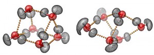
The scientists have provided an illustration of two water hexamer structures,

Three-dimensional representations of the prism (left) and cage (right) structures of the water hexamer, the smallest drop of water. The mesh contours represent the actual quantum-mechanical densities of the oxygen (red) and hydrogen (white) atoms. The small yellow spheres represent the hydrogen bonds between the six water molecules. Characterizing the hydrogen-bond topology of the water hexamer at the molecular level is key to understanding the unique and often surprising properties of liquid water, our life matrix. Images courtesy of Volodymyr Babin and Francesco Paesani, UC San Diego.

Here’s the full citation for the research paper if you want to follow up on it or you can read more in either the news item or news release,

The Water Hexamer: Cage, Prism, or Both. Full Dimensional Quantum Simulations Say Both; Yimin Wang, Volodymyr Babin, Joel M. Bowman, and Francesco Paesani; J. Am. Chem. Soc., 2012, 134 (27), pp 11116–11119 DOI: 10.1021/ja304528m

The article is behind a paywall.