Tag Archives: University of Gothenburg

Growing electrodes in your brain?

This isn’t for everybody. From a February 23, 2023 news item on Nanowerk, Note: A link has been removed,

The boundaries between biology and technology are becoming blurred. Researchers at Linköping, Lund, and Gothenburg universities in Sweden have successfully grown electrodes in living tissue using the body’s molecules as triggers. The result, published in the journal Science (“Metabolite-induced in vivo fabrication of substrate-free organic bioelectronics”), paves the way for the formation of fully integrated electronic circuits in living organisms.
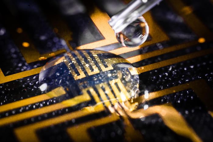
Caption: The injectable gel being tested on a microfabricated circuit. Credit: Thor Balkhed

I have two news releases for this research. First, the February 23, 2023 American Association for the Advancement of Science (AAAS) news release on EurekAlert,

Researchers have developed a way to make bioelectronics directly inside living tissues, an approach they tested by making electrodes in the brain, heart, and fin tissue of living zebrafish, as well as in isolated mammalian muscle tissues. According to the authors, the new method paves the way for in vivo fabrication of fully integrated electronic circuits within the nervous system and other living tissue. “Safety and stability analyses over long periods will be essential to determining whether such technology is useful for chronic implantations,” writes Sahika Inal in a related Perspective. “However, the strategy … suggests that any living tissue can turn into electronic matter and brings the field closer to generating seamless biotic-abiotic interfaces with a potentially long lifetime and minimum harm to tissues.” Implantable electronic devices that can interface with soft biological neural tissues offer a valuable approach to studying the complex electrical signaling of the nervous system and enable the therapeutic modulation of neural circuitry to prevent or treat various diseases and disorders. However, conventional bioelectronic implants often require the use of rigid electronic substrates that are incompatible with delicate living tissues and can provoke injury and inflammation that can affect a device’s electrical properties and long-term performance. Overcoming the incompatibility between static, solid-state electronic materials and dynamic, soft biological tissues has proven challenging. Here, Xenofon Strakosas and colleagues present a method to fabricate polymer-based, substrate-free electronic conducting materials directly inside a tissue. Strakosas et al. developed a complex molecular precursor cocktail that, when injected into a tissue, uses endogenous metabolites (glucose and lactate) to induce polymerization of organic precursors to form conducting polymer gels. To demonstrate the approach, the authors “grew” gel electrodes in the brain, heart, and fin tissue of living zebrafish, with no signs of tissue damage, and in isolated mammalian muscle tissues, including beef, pork and chicken. In medicinal leeches, they showed how the conducting gel could interface nervous tissue with electrodes on a tiny flexible probe.

The second is the February 23, 2023 Linköping University press release on EurekAlert, which originated the news item, and it provides further insight,

“For several decades, we have tried to create electronics that mimic biology. Now we let biology create the electronics for us,” says Professor Magnus Berggren at the Laboratory for Organic Electronics, LOE, at Linköping University.

Linking electronics to biological tissue is important to understand complex biological functions, combat diseases in the brain, and develop future interfaces between man and machine. However, conventional bioelectronics, developed in parallel with the semiconductor industry, have a fixed and static design that is difficult, if not impossible, to combine with living biological signal systems.

To bridge this gap between biology and technology, researchers have developed a method for creating soft, substrate-free, electronically conductive materials in living tissue. By injecting a gel containing enzymes as the “assembly molecules”, the researchers were able to grow electrodes in the tissue of zebrafish and medicinal leeches.

“Contact with the body’s substances changes the structure of the gel and makes it electrically conductive, which it isn’t before injection. Depending on the tissue, we can also adjust the composition of the gel to get the electrical process going,” says Xenofon Strakosas, researcher at LOE and Lund University and one of the study’s main authors.

The body’s endogenous molecules are enough to trigger the formation of electrodes. There is no need for genetic modification or external signals, such as light or electrical energy, which has been necessary in previous experiments. The Swedish researchers are the first in the world to succeed in this.

Their study paves the way for a new paradigm in bioelectronics. Where it previously took implanted physical objects to start electronic processes in the body, injection of a viscous gel will be enough in the future.

In their study, the researchers further show that the method can target the electronically conducting material to specific biological substructures and thereby create suitable interfaces for nerve stimulation. In the long term, the fabrication of fully integrated electronic circuits in living organisms may be possible.

In experiments conducted at Lund University, the team successfully achieved electrode formation in the brain, heart, and tail fins of zebrafish and around the nervous tissue of medicinal leeches. The animals were not harmed by the injected gel and were otherwise not affected by the electrode formation. One of the many challenges in these trials was to take the animals’ immune system into account.

“By making smart changes to the chemistry, we were able to develop electrodes that were accepted by the brain tissue and immune system. The zebrafish is an excellent model for the study of organic electrodes in brains,” says Professor Roger Olsson at the Medical Faculty at Lund University, who also has a chemistry laboratory at the University of Gothenburg.

It was Professor Roger Olsson who took the initiative for the study, after he read about the electronic rose developed by researchers at Linköping University in 2015. One research problem, and an important difference between plants and animals, was the difference in cell structure. Whereas plants have rigid cell walls which allow for the formation of electrodes, animal cells are more like a soft mass. Creating a gel with enough structure and the right combination of substances to form electrodes in such surroundings was a challenge that took many years to solve.

“Our results open up for completely new ways of thinking about biology and electronics. We still have a range of problems to solve, but this study is a good starting point for future research,” says Hanne Biesmans, PhD student at LOE and one of the main authors.

Here’s a link to and a citation for the paper,

Metabolite-induced in vivo fabrication of substrate-free organic bioelectronics by Xenofon Strakosas, Hanne Biesmans, Tobias Abrahamsson, Karin Hellman, Malin Silverå Ejneby, Mary J. Donahue, Peter Ekström, Fredrik Ek, Marios Savvakis, Martin Hjort, David Bliman, Mathieu Linares, Caroline Lindholm, Eleni Stavrinidou, Jennifer Y. Gerasimov, Daniel T. Simon, Roger Olsson, and Magnus Berggren. Science 23 Feb 2023 Vol 379, Issue 6634 pp. 795-802 DOI: 10.1126/science.adc9998

This paper is behind a paywall.

Memristive spintronic neurons

A December 6, 2021 news item on Nanowerk on memristive spintronic neurons (Note: A link has been removed),

Researchers at Tohoku University and the University of Gothenburg have established a new spintronic technology for brain-inspired computing.

Their achievement was published in the journal Nature Materials (“Memristive control of mutual SHNO synchronization for neuromorphic computing”).

Sophisticated cognitive tasks, such as image and speech recognition, have seen recent breakthroughs thanks to deep learning. Even so, the human brain still executes these tasks without exerting much energy and with greater efficiency than any computer. The development of energy-efficient artificial neurons capable of emulating brain-inspired processes has therefore been a major research goal for decades.

A November 29, 2021 Tohoku University press release (also on EurekAlert but published November 30, 2021), which originated the news release, provides more technical detail,

Researchers demonstrated the first integration of a cognitive computing nano-element – the memristor – into another – a spintronic oscillator. Arrays of these memristor-controlled oscillators combine the non-volatile local storage of the memristor function with the microwave frequency computation of the nano-oscillator networks and can closely imitate the non-linear oscillatory neural networks of the human brain.

Resistance of the memristor changed with the voltage hysteresis applied to the top Ti/Cu electrode. Upon voltage application to the electrode, an electric field was applied at the high-resistance state, compared to electric current flows for the low-resistance state. The effects of electric field and current on the oscillator differed from each other, offering various controls of oscillation and synchronization properties.

Professor Johan Åkerman of the University of Gothenburg and leader of the study expressed his hopes for the future and the significance of the finding. “We are particularly interested in emerging quantum-inspired computing schemes, such as Ising Machines. The results also highlight the productive collaboration that we have established in neuromorphic spintronics between the University of Gothenburg and Tohoku University, something that is also part of the Sweden-Japan collaborative network MIRAI 2.0.”

“So far, artificial neurons and synapses have been developed separately in many fields; this work marks an important milestone: two functional elements have been combined into one,” said professor Shunsuke Fukami, who led the project on the Tohoku University side. Dr. Mohammad Zahedinejad of the University of Gothenburg and first author of the study adds, “Using the memristor-controlled spintronic oscillator arrays, we could tune the synaptic interactions between adjacent neurons and program them into mutually different and partially synchronized states.”

To put into practice their discovery, the researchers examined the operation of a test device comprising one oscillator and one memristor. The constricted region of W/CoFeB stack served as an oscillator, i.e., the neuron, whereas the MgO/AlOx/SiNx stack acted as a memristor, i.e., the synapse.

Resistance of the memristor changed with the voltage hysteresis applied to the top Ti/Cu electrode. Upon voltage application to the electrode, an electric field was applied at the high-resistance state, compared to electric current flows for the low-resistance state. The effects of electric field and current on the oscillator differed from each other, offering various controls of oscillation and synchronization properties.

Professor Johan Åkerman of the University of Gothenburg and leader of the study expressed his hopes for the future and the significance of the finding. “We are particularly interested in emerging quantum-inspired computing schemes, such as Ising Machines. The results also highlight the productive collaboration that we have established in neuromorphic spintronics between the University of Gothenburg and Tohoku University, something that is also part of the Sweden-Japan collaborative network MIRAI 2.0.” [sic]

Here’s a link to and a citation for the paper,

Memristive control of mutual spin Hall nano-oscillator synchronization for neuromorphic computing by Mohammad Zahedinejad, Himanshu Fulara, Roman Khymyn, Afshin Houshang, Mykola Dvornik, Shunsuke Fukami, Shun Kanai, Hideo Ohno & Johan Åkerman. Nature Materials (2021) DOI: https://doi.org/10.1038/s41563-021-01153-6 Published 29 November 2021

This paper is behind a paywall.

Living with a mind-controlled prosthetic

This could be described as the second half of an October 10, 2014 post (Mind-controlled prostheses ready for real world activities). Five and a half years later, Sweden’s Chalmers University of Technology has announced mind-controlled prosthetics in daily use that feature the sense of touch. From an April 30, 2020 Chalmers University of Technology press release (also on EurekAlert but published April 29, 2020) by Johanna Wilde,

For the first time, people with arm amputations can experience sensations of touch in a mind-controlled arm prosthesis that they use in everyday life. A study in the New England Journal of Medicine reports on three Swedish patients who have lived, for several years, with this new technology – one of the world’s most integrated interfaces between human and machine.

See the film: “The most natural robotic prosthesis in the world” [Should you not have Swedish language skills, you can click on the subtitle option in the video’s settings field]

The advance is unique: the patients have used a mind-controlled prosthesis in their everyday life for up to seven years. For the last few years, they have also lived with a new function – sensations of touch in the prosthetic hand. This is a new concept for artificial limbs, which are called neuromusculoskeletal prostheses – as they are connected to the user’s nerves, muscles, and skeleton.

The research was led by Max Ortiz Catalan, Associate Professor at Chalmers University of Technology, in collaboration with Sahlgrenska University Hospital, University of Gothenburg, and Integrum AB, all in Gothenburg, Sweden. Researchers at Medical University of Vienna in Austria and the Massachusetts Institute of Technology in the USA were also involved.

“Our study shows that a prosthetic hand, attached to the bone and controlled by electrodes implanted in nerves and muscles, can operate much more precisely than conventional prosthetic hands. We further improved the use of the prosthesis by integrating tactile sensory feedback that the patients use to mediate how hard to grab or squeeze an object. Over time, the ability of the patients to discern smaller changes in the intensity of sensations has improved,” says Max Ortiz Catalan.

“The most important contribution of this study was to demonstrate that this new type of prosthesis is a clinically viable replacement for a lost arm. No matter how sophisticated a neural interface becomes, it can only deliver real benefit to patients if the connection between the patient and the prosthesis is safe and reliable in the long term. Our results are the product of many years of work, and now we can finally present the first bionic arm prosthesis that can be reliably controlled using implanted electrodes, while also conveying sensations to the user in everyday life”, continues Max Ortiz Catalan.

Since receiving their prostheses, the patients have used them daily in all their professional and personal activities.

The new concept of a neuromusculoskeletal prosthesis is unique in that it delivers several different features which have not been presented together in any other prosthetic technology in the world:

[1] It has a direct connection to a person’s nerves, muscles, and skeleton.

[2] It is mind-controlled and delivers sensations that are perceived by the user as arising from the missing hand.

[3] It is self-contained; all electronics needed are contained within the prosthesis, so patients do not need to carry additional equipment or batteries.

[4] It is safe and stable in the long term; the technology has been used without interruption by patients during their everyday activities, without supervision from the researchers, and it is not restricted to confined or controlled environments.

The newest part of the technology, the sensation of touch, is possible through stimulation of the nerves that used to be connected to the biological hand before the amputation. Force sensors located in the thumb of the prosthesis measure contact and pressure applied to an object while grasping. This information is transmitted to the patients’ nerves leading to their brains. Patients can thus feel when they are touching an object, its characteristics, and how hard they are pressing it, which is crucial for imitating a biological hand.

“Currently, the sensors are not the obstacle for restoring sensation,” says Max Ortiz Catalan. “The challenge is creating neural interfaces that can seamlessly transmit large amounts of artificially collected information to the nervous system, in a way that the user can experience sensations naturally and effortlessly.”
The implantation of this new technology took place at Sahlgrenska University Hospital, led by Professor Rickard Brånemark and Doctor Paolo Sassu. Over a million people worldwide suffer from limb loss, and the end goal for the research team, in collaboration with Integrum AB, is to develop a widely available product suitable for as many of these people as possible.

“Right now, patients in Sweden are participating in the clinical validation of this new prosthetic technology for arm amputation,” says Max Ortiz Catalan. “We expect this system to become available outside Sweden within a couple of years, and we are also making considerable progress with a similar technology for leg prostheses, which we plan to implant in a first patient later this year.”

More about: How the technology works:

The implant system for the arm prosthesis is called e-OPRA and is based on the OPRA implant system created by Integrum AB. The implant system anchors the prosthesis to the skeleton in the stump of the amputated limb, through a process called osseointegration (osseo = bone). Electrodes are implanted in muscles and nerves inside the amputation stump, and the e-OPRA system sends signals in both directions between the prosthesis and the brain, just like in a biological arm.

The prosthesis is mind-controlled, via the electrical muscle and nerve signals sent through the arm stump and captured by the electrodes. The signals are passed into the implant, which goes through the skin and connects to the prosthesis. The signals are then interpreted by an embedded control system developed by the researchers. The control system is small enough to fit inside the prosthesis and it processes the signals using sophisticated artificial intelligence algorithms, resulting in control signals for the prosthetic hand’s movements.

The touch sensations arise from force sensors in the prosthetic thumb. The signals from the sensors are converted by the control system in the prosthesis into electrical signals which are sent to stimulate a nerve in the arm stump. The nerve leads to the brain, which then perceives the pressure levels against the hand.

The neuromusculoskeletal implant can connect to any commercially available arm prosthesis, allowing them to operate more effectively.

More about: How the artificial sensation is experienced:

People who lose an arm or leg often experience phantom sensations, as if the missing body part remains although not physically present. When the force sensors in the prosthetic thumb react, the patients in the study feel that the sensation comes from their phantom hand. Precisely where on the phantom hand varies between patients, depending on which nerves in the stump receive the signals. The lowest level of pressure can be compared to touching the skin with the tip of a pencil. As the pressure increases, the feeling becomes stronger and increasingly ‘electric’.

I have read elsewhere that one of the most difficult aspects of dealing with a prosthetic is the loss of touch. This has to be exciting news for a lot of people. Here’s a link to and a citation for the paper,

Self-Contained Neuromusculoskeletal Arm Prostheses by Max Ortiz-Catalan, Enzo Mastinu, Paolo Sassu, Oskar Aszmann, and Rickard Brånemark. N Engl J Med 2020; 382:1732-1738 DOI: 10.1056/NEJMoa1917537 Published: April 30, 2020

This paper is behind a paywall.