Tag Archives: bones

Making better concrete by looking to nature for inspiration

Researchers from the Masssachusetts Institute of Technology (MIT) are working on a new formula for concrete based on bones, shells, and other such natural materials. From a May 25, 2016 news item on Nanowerk (Note: A link has been removed),

Researchers at MIT are seeking to redesign concrete — the most widely used human-made material in the world — by following nature’s blueprints.

In a paper published online in the journal Construction and Building Materials (“Roadmap across the mesoscale for durable and sustainable cement paste – A bioinspired approach”), the team contrasts cement paste — concrete’s binding ingredient — with the structure and properties of natural materials such as bones, shells, and deep-sea sponges. As the researchers observed, these biological materials are exceptionally strong and durable, thanks in part to their precise assembly of structures at multiple length scales, from the molecular to the macro, or visible, level.

A May 26, 2016 MIT news release (also on EurekAlert), which originated the news item, provides more detail,

From their observations, the team, led by Oral Buyukozturk, a professor in MIT’s Department of Civil and Environmental Engineering (CEE), proposed a new bioinspired, “bottom-up” approach for designing cement paste.

“These materials are assembled in a fascinating fashion, with simple constituents arranging in complex geometric configurations that are beautiful to observe,” Buyukozturk says. “We want to see what kinds of micromechanisms exist within them that provide such superior properties, and how we can adopt a similar building-block-based approach for concrete.”

Ultimately, the team hopes to identify materials in nature that may be used as sustainable and longer-lasting alternatives to Portland cement, which requires a huge amount of energy to manufacture.

“If we can replace cement, partially or totally, with some other materials that may be readily and amply available in nature, we can meet our objectives for sustainability,” Buyukozturk says.

“The merger of theory, computation, new synthesis, and characterization methods have enabled a paradigm shift that will likely change the way we produce this ubiquitous material, forever,” Buehler says. “It could lead to more durable roads, bridges, structures, reduce the carbon and energy footprint, and even enable us to sequester carbon dioxide as the material is made. Implementing nanotechnology in concrete is one powerful example [of how] to scale up the power of nanoscience to solve grand engineering challenges.”

From molecules to bridges

Today’s concrete is a random assemblage of crushed rocks and stones, bound together by a cement paste. Concrete’s strength and durability depends partly on its internal structure and configuration of pores. For example, the more porous the material, the more vulnerable it is to cracking. However, there are no techniques available to precisely control concrete’s internal structure and overall properties.

“It’s mostly guesswork,” Buyukozturk says. “We want to change the culture and start controlling the material at the mesoscale.”

As Buyukozturk describes it, the “mesoscale” represents the connection between microscale structures and macroscale properties. For instance, how does cement’s microscopic arrangement affect the overall strength and durability of a tall building or a long bridge? Understanding this connection would help engineers identify features at various length scales that would improve concrete’s overall performance.

“We’re dealing with molecules on the one hand, and building a structure that’s on the order of kilometers in length on the other,” Buyukozturk says. “How do we connect the information we develop at the very small scale, to the information at the large scale? This is the riddle.”

Building from the bottom, up

To start to understand this connection, he and his colleagues looked to biological materials such as bone, deep sea sponges, and nacre (an inner shell layer of mollusks), which have all been studied extensively for their mechanical and microscopic properties. They looked through the scientific literature for information on each biomaterial, and compared their structures and behavior, at the nano-, micro-, and macroscales, with that of cement paste.

They looked for connections between a material’s structure and its mechanical properties. For instance, the researchers found that a deep sea sponge’s onion-like structure of silica layers provides a mechanism for preventing cracks. Nacre has a “brick-and-mortar” arrangement of minerals that generates a strong bond between the mineral layers, making the material extremely tough.

“In this context, there is a wide range of multiscale characterization and computational modeling techniques that are well established for studying the complexities of biological and biomimetic materials, which can be easily translated into the cement community,” says Masic.

Applying the information they learned from investigating biological materials, as well as knowledge they gathered on existing cement paste design tools, the team developed a general, bioinspired framework, or methodology, for engineers to design cement, “from the bottom up.”

The framework is essentially a set of guidelines that engineers can follow, in order to determine how certain additives or ingredients of interest will impact cement’s overall strength and durability. For instance, in a related line of research, Buyukozturk is looking into volcanic ash [emphasis mine] as a cement additive or substitute. To see whether volcanic ash would improve cement paste’s properties, engineers, following the group’s framework, would first use existing experimental techniques, such as nuclear magnetic resonance, scanning electron microscopy, and X-ray diffraction to characterize volcanic ash’s solid and pore configurations over time.

Researchers could then plug these measurements into models that simulate concrete’s long-term evolution, to identify mesoscale relationships between, say, the properties of volcanic ash and the material’s contribution to the strength and durability of an ash-containing concrete bridge. These simulations can then be validated with conventional compression and nanoindentation experiments, to test actual samples of volcanic ash-based concrete.

Ultimately, the researchers hope the framework will help engineers identify ingredients that are structured and evolve in a way, similar to biomaterials, that may improve concrete’s performance and longevity.

“Hopefully this will lead us to some sort of recipe for more sustainable concrete,” Buyukozturk says. “Typically, buildings and bridges are given a certain design life. Can we extend that design life maybe twice or three times? That’s what we aim for. Our framework puts it all on paper, in a very concrete way, for engineers to use.”

This is not the only team looking at new methods for producing the material, my Dec. 24, 2012 posting features a number of ‘concrete’ research projects.

Also, I highlighted the reference to ‘volcanic ash’ as it reminded me of Roman concrete which has lasted for over 2000 years and includes volcanic sand and volcanic rock.  You can read more about it in a Dec. 18, 2014 article by Mark Miller for Ancient Origins where he describes the wonders of the material and what was then a recent discovery of the Romans’ recipe.

I have two links and citations, first, the MIT paper, then the paper on Roman concrete.

Roadmap across the mesoscale for durable and sustainable cement paste – A bioinspired approach by Steven D. Palkovic, Dieter B. Brommer, Kunal Kupwade-Patil, Admir Masic, Markus J. Buehler, Oral Büyüköztürk.Construction and Building Materials Volume 115, 15 July 2016, Pages 13–31.  doi:10.1016/j.conbuildmat.2016.04.020

Mechanical resilience and cementitious processes in Imperial Roman architectural mortar by Marie D. Jackson, Eric N. Landis, Philip F. Brune, Massimo Vitti, Heng Chen, Qinfei Li, Martin Kunz, Hans-Rudolf Wenk, Paulo J. M. Monteiro, and Anthony R. Ingraffea. Proceedings of the National Academy of Sciences  vol. 111 no. 52 18484–18489, doi: 10.1073/pnas.1417456111

The first paper is behind a paywall but the second one appears to be open access.
Nanoparticles in baby formula

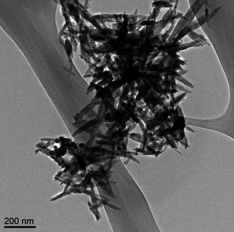
Needle-like particles of hydroxyapatite found in infant formula by ASU researchers. Westerhoff and Schoepf/ASU, CC BY-ND

Needle-like particles of hydroxyapatite found in infant formula by ASU [Arizona State University] researchers. Westerhoff and Schoepf/ASU, CC BY-ND

Nanowerk is featuring an essay about hydroxyapatite nanoparticles in baby formula written by Dr. Andrew Maynard in a May 17, 2016 news item (Note: A link has been removed),

There’s a lot of stuff you’d expect to find in baby formula: proteins, carbs, vitamins, essential minerals. But parents probably wouldn’t anticipate finding extremely small, needle-like particles. Yet this is exactly what a team of scientists here at Arizona State University [ASU] recently discovered.

The research, commissioned and published by Friends of the Earth (FoE) – an environmental advocacy group – analyzed six commonly available off-the-shelf baby formulas (liquid and powder) and found nanometer-scale needle-like particles in three of them. The particles were made of hydroxyapatite – a poorly soluble calcium-rich mineral. Manufacturers use it to regulate acidity in some foods, and it’s also available as a dietary supplement.

Andrew’s May 17, 2016 essay first appeared on The Conversation website,

Looking at these particles at super-high magnification, it’s hard not to feel a little anxious about feeding them to a baby. They appear sharp and dangerous – not the sort of thing that has any place around infants. …

… questions like “should infants be ingesting them?” make a lot of sense. However, as is so often the case, the answers are not quite so straightforward.

Andrew begins by explaining about calcium and hydroxyapatite (from The Conversation),

Calcium is an essential part of a growing infant’s diet, and is a legally required component in formula. But not necessarily in the form of hydroxyapatite nanoparticles.

Hydroxyapatite is a tough, durable mineral. It’s naturally made in our bodies as an essential part of bones and teeth – it’s what makes them so strong. So it’s tempting to assume the substance is safe to eat. But just because our bones and teeth are made of the mineral doesn’t automatically make it safe to ingest outright.

The issue here is what the hydroxyapatite in formula might do before it’s digested, dissolved and reconstituted inside babies’ bodies. The size and shape of the particles ingested has a lot to do with how they behave within a living system.

He then discusses size and shape, which are important at the nanoscale,

Size and shape can make a difference between safe and unsafe when it comes to particles in our food. Small particles aren’t necessarily bad. But they can potentially get to parts of our body that larger ones can’t reach. Think through the gut wall, into the bloodstream, and into organs and cells. Ingested nanoscale particles may be able to interfere with cells – even beneficial gut microbes – in ways that larger particles don’t.

These possibilities don’t necessarily make nanoparticles harmful. Our bodies are pretty well adapted to handling naturally occurring nanoscale particles – you probably ate some last time you had burnt toast (carbon nanoparticles), or poorly washed vegetables (clay nanoparticles from the soil). And of course, how much of a material we’re exposed to is at least as important as how potentially hazardous it is.

Yet there’s a lot we still don’t know about the safety of intentionally engineered nanoparticles in food. Toxicologists have started paying close attention to such particles, just in case their tiny size makes them more harmful than otherwise expected.

Currently, hydroxyapatite is considered safe at the macroscale by the US Food and Drug Administration (FDA). However, the agency has indicated that nanoscale versions of safe materials such as hydroxyapatite may not be safe food additives. From Andrew’s May 17, 2016 essay,

Hydroxyapatite is a tough, durable mineral. It’s naturally made in our bodies as an essential part of bones and teeth – it’s what makes them so strong. So it’s tempting to assume the substance is safe to eat. But just because our bones and teeth are made of the mineral doesn’t automatically make it safe to ingest outright.

The issue here is what the hydroxyapatite in formula might do before it’s digested, dissolved and reconstituted inside babies’ bodies. The size and shape of the particles ingested has a lot to do with how they behave within a living system. Size and shape can make a difference between safe and unsafe when it comes to particles in our food. Small particles aren’t necessarily bad. But they can potentially get to parts of our body that larger ones can’t reach. Think through the gut wall, into the bloodstream, and into organs and cells. Ingested nanoscale particles may be able to interfere with cells – even beneficial gut microbes – in ways that larger particles don’t.These possibilities don’t necessarily make nanoparticles harmful. Our bodies are pretty well adapted to handling naturally occurring nanoscale particles – you probably ate some last time you had burnt toast (carbon nanoparticles), or poorly washed vegetables (clay nanoparticles from the soil). And of course, how much of a material we’re exposed to is at least as important as how potentially hazardous it is.Yet there’s a lot we still don’t know about the safety of intentionally engineered nanoparticles in food. Toxicologists have started paying close attention to such particles, just in case their tiny size makes them more harmful than otherwise expected.

Putting particle size to one side for a moment, hydroxyapatite is classified by the US Food and Drug Administration (FDA) as “Generally Regarded As Safe.” That means it considers the material safe for use in food products – at least in a non-nano form. However, the agency has raised concerns that nanoscale versions of food ingredients may not be as safe as their larger counterparts.Some manufacturers may be interested in the potential benefits of “nanosizing” – such as increasing the uptake of vitamins and minerals, or altering the physical, textural and sensory properties of foods. But because decreasing particle size may also affect product safety, the FDA indicates that intentionally nanosizing already regulated food ingredients could require regulatory reevaluation.In other words, even though non-nanoscale hydroxyapatite is “Generally Regarded As Safe,” according to the FDA, the safety of any nanoscale form of the substance would need to be reevaluated before being added to food products.Despite this size-safety relationship, the FDA confirmed to me that the agency is unaware of any food substance intentionally engineered at the nanoscale that has enough generally available safety data to determine it should be “Generally Regarded As Safe.”Casting further uncertainty on the use of nanoscale hydroxyapatite in food, a 2015 report from the European Scientific Committee on Consumer Safety (SCCS) suggests there may be some cause for concern when it comes to this particular nanomaterial.Prompted by the use of nanoscale hydroxyapatite in dental products to strengthen teeth (which they consider “cosmetic products”), the SCCS reviewed published research on the material’s potential to cause harm. Their conclusion?

The available information indicates that nano-hydroxyapatite in needle-shaped form is of concern in relation to potential toxicity. Therefore, needle-shaped nano-hydroxyapatite should not be used in cosmetic products.

This recommendation was based on a handful of studies, none of which involved exposing people to the substance. Researchers injected hydroxyapatite needles directly into the bloodstream of rats. Others exposed cells outside the body to the material and observed the effects. In each case, there were tantalizing hints that the small particles interfered in some way with normal biological functions. But the results were insufficient to indicate whether the effects were meaningful in people.

As Andrew also notes in his essay, none of the studies examined by the SCCS OEuropean Scientific Committee on Consumer Safety) looked at what happens to nano-hydroxyapatite once it enters your gut and that is what the researchers at Arizona State University were considering (from the May 17, 2016 essay),

The good news is that, according to preliminary studies from ASU researchers, hydroxyapatite needles don’t last long in the digestive system.

This research is still being reviewed for publication. But early indications are that as soon as the needle-like nanoparticles hit the highly acidic fluid in the stomach, they begin to dissolve. So fast in fact, that by the time they leave the stomach – an exceedingly hostile environment – they are no longer the nanoparticles they started out as.

These findings make sense since we know hydroxyapatite dissolves in acids, and small particles typically dissolve faster than larger ones. So maybe nanoscale hydroxyapatite needles in food are safer than they sound.

This doesn’t mean that the nano-needles are completely off the hook, as some of them may get past the stomach intact and reach more vulnerable parts of the gut. But the findings do suggest these ultra-small needle-like particles could be an effective source of dietary calcium – possibly more so than larger or less needle-like particles that may not dissolve as quickly.

Intriguingly, recent research has indicated that calcium phosphate nanoparticles form naturally in our stomachs and go on to be an important part of our immune system. It’s possible that rapidly dissolving hydroxyapatite nano-needles are actually a boon, providing raw material for these natural and essential nanoparticles.

While it’s comforting to know that preliminary research suggests that the hydroxyapatite nanoparticles are likely safe for use in food products, Andrew points out that more needs to be done to insure safety (from the May 17, 2016 essay),

And yet, even if these needle-like hydroxyapatite nanoparticles in infant formula are ultimately a good thing, the FoE report raises a number of unresolved questions. Did the manufacturers knowingly add the nanoparticles to their products? How are they and the FDA ensuring the products’ safety? Do consumers have a right to know when they’re feeding their babies nanoparticles?

Whether the manufacturers knowingly added these particles to their formula is not clear. At this point, it’s not even clear why they might have been added, as hydroxyapatite does not appear to be a substantial source of calcium in most formula. …

And regardless of the benefits and risks of nanoparticles in infant formula, parents have a right to know what’s in the products they’re feeding their children. In Europe, food ingredients must be legally labeled if they are nanoscale. In the U.S., there is no such requirement, leaving American parents to feel somewhat left in the dark by producers, the FDA and policy makers.

As far as I’m aware, the Canadian situation is much the same as the US. If the material is considered safe at the macroscale, there is no requirement to indicate that a nanoscale version of the material is in the product.

I encourage you to read Andrew’s essay in its entirety. As for the FoE report (Nanoparticles in baby formula: Tiny new ingredients are a big concern), that is here.

Bone goo

Most of us think of our bones as being solid matter and in the light of some new research at the University of Cambridge, it seems that we (including scientists and doctors) have not entirely understood the true nature of bone matter. From a March 24, 2014 University of Cambridge news release (also on EurekAlert),

Latest research shows that the chemical citrate – a by-product of natural cell metabolism – is mixed with water to create a viscous fluid that is trapped between the nano-scale crystals that form our bones.

This fluid allows enough movement, or ‘slip’, between these crystals so that bones are flexible, and don’t shatter under pressure. It is the inbuilt shock absorber in bone that, until now, was unknown.

If citrate leaks out, the crystals – made of calcium phosphate – fuse together into bigger and bigger clumps that become inflexible, increasingly brittle and more likely to shatter. This could be the root cause of osteoporosis.

The team from Cambridge’s Department of Chemistry used a combination of NMR spectroscopy, X-ray diffraction, imaging and high-level molecular modelling to reveal the citrate layers in bone.

They say that this is the start of what needs to be an entire shift in focus for studying the cause of brittle bone diseases like osteoporosis, and bone pathologies in general. The study is published today [March 24, 2014] in the journal Proceedings of the National Academy of Sciences.

“Bone mineral was thought to be closely related to this substance called hydroxyapatite. But what we’ve shown is that a large part of bone mineral – possibly as much as half of it in fact – is made up of this goo, where citrate is binding like a gel between mineral crystals,” said Dr Melinda Duer, who led the study.

“This nano-scopic layering of citrate fluid and mineral crystals in bone means that the crystals stay in flat, plate-like shapes that have the facility to slide with respect to each other. Without citrate, all crystals in bone mineral would collapse together, become one big crystal and shatter.

“It’s this layered structure that’s been missing from our knowledge, and we can now see that without it you’re stuffed.”

I appreciate the lively quotes (“… without it you’re stuffed”) and the description which follows,

Duer compares it to two panes of glass with water in the middle, which stick together but are able to slide: “it’s the same thing in these flat bone crystals. But you’ve got to have something that keeps the water there, stops it from drying out and stops the plates from either flying apart or sticking fast together. We now know that thing is citrate.”

Citrate is a ‘spidery’ molecule with four arms, all of which can bond easily to calcium – which bone is packed with, explains Duer. This means that citrate can hold the mineral crystals together at the same time as preventing them from fusing, while trapping the water that allows for the slippery movement which provides bone flexibility. “Without citrate, water would just flow straight through these gaps,” she said.

The body actually delivers bone calcium wrapped in citrate, to prevent it fusing with phosphate and forming large solid – and brittle – mineral crystals in the wrong places. Bone tissue has a protein mesh with holes where the calcium is deposited. In healthy tissue, the holes are very small, so that when the calcium is deposited, the citrate that comes with it can’t escape and is trapped between crystals – creating the flexible layers of fluid and bone plates.

As people age or suffer repeated bone trauma, the protein mesh isn’t repaired so well by the cells that try to replace damaged tissue, but often end up chewing away tissue faster than it can be re-deposited. This causes progressively larger holes in the protein mesh, citrate fluid escapes and crystals fuse together.

What happens then is pure chemistry, says Duer, with little biological control.

The body instigates a form of biological control through the tiny holes in the protein mesh that trap the citrate fluid, along with other molecules that normally control the deposit of mineral. These small spaces force the molecules to be involved with the forming mineral, controlling the process. But if you haven’t got the confined space the chemical reactions spiral out of control.

“In the bigger holes in damaged tissue, pure chemistry takes over. Pretty much the moment calcium and phosphate touch, they form a solid. You end up with these expanding clumps of brittle crystal, with water and citrate relegated to the outside of them,” she said.

“In terms of chemistry, that solid clump of mineral is the most stable structure. Biomechanically, however, it’s hopeless – as soon as you stand on it, it shatters. If we want to cure osteoporosis, we need to figure out how to stop the bigger holes forming in the protein matrix.”

The study is the first in a series of findings, with other studies from the team’s work on bone chemistry expected to come out later in the year.

Here’s a link to and citation for the paper,

Citrate bridges between mineral platelets in bone by Erika Davies, Karin H. Müller, Wai Ching Wong, Chris J. Pickard, David G. Reid, Jeremy N. Skepper, and Melinda J. Duer. PNAS doi: 10.1073/pnas.1315080111

This paper is made available through the PNAS open access option.

Tissue regeneration by injection

I’ve got two items: one from the University of Nottingham (UK) where they’re working on tissue regeneration for bones, muscles, and the heart.The second item is from Simon Fraser University (Vancouver, Canada)where the focus is on regenerating bones.

Here’s more about the work at the University of Nottingham from the [July 3, 2012] news item on Nanowerk,

The University of Nottingham has begun the search for a new class of injectable materials that will stimulate stem cells to regenerate damaged tissue in degenerative and age related disorders of the bone, muscle and heart.

The work, which is currently at the experimental stage, could lead to treatments for diseases that currently have no cure. The aim is to produce radical new treatments that will reduce the need for invasive surgery, optimise recovery and reduce the risk of undesirable scar tissue.

The research, which brings together expertise in The University of Nottingham’s Malaysia Campus (UNMC) and UK campus, is part of the Rational Bioactive Materials Design for Tissue Generation project (Biodesign). This €11m EU funded research project which involves 21 research teams from across Europe is made up of leading experts in degenerative disease and regenerative medicine.

The original July 3, 2012 news release from the University of Nottingham includes a video which offers some additional insight (sadly ,it cannot be embedded here) and more information (Note: I have removed a link),

Kevin Shakesheff, Professor of Advanced Drug Delivery and Tissue Engineering and Head of the School of Pharmacy, said: “This research heralds a step-change in approaches to tissue regeneration. Current biomaterials are poorly suited to the needs of tissue engineering and regenerative medicine. The aim of Biodesign is to develop new materials and medicines that will stimulate tissue regeneration rather than wait for the body to start the process itself. The aim is to fabricate advanced biomaterials that match the basic structure of each tissue so the cells can take over the recovery process themselves.”

The Canadian project at Simon Fraser University features a singular focus on bone regeneration, from the July 19, 2012 news release on EurekAlert,

A Simon Fraser University researcher is leading a team of scientists working to create new drugs to stimulate bone regeneration – research that will be furthered by a $2.5 million grant from the Canadian Institutes of Health Research (CIHR).

Lead researcher Robert Young heads a team of internationally recognized experts in bone disease and drug development. The researchers are focusing on developing small molecule compounds and nano-medicines that stimulate bone regeneration, and hope to identify new therapeutic approaches by improving understanding of bone renewal biology.

Their objective is to develop new therapeutic agents that promote bone repair, regeneration and renewal, and prove their efficiency in reproducing or improving bone strength.

The research involves studying the “natural controls” that guide the development of cells in the bones toward either bone forming or bone resorbing cells, setting the stage for the next generation of bone regenerative therapies.

The grant is one of three announced today by the federal government targeting bone health research and totalling $7 million. The others focus on wrist fractures management and identifying bone loss in gum disease.

The funding is through the CIHR’s Institute of Musculoskeletal Health and Arthritis and addresses priorities identified at a 2009 national Bone Health Consensus Conference.

I’ve decided to focus on tissues today so there will be something about tissue engineering and jellyfish (artificial) shortly.

Speeding up bone growth with a tobacco virus

Steven Powell in a June 22, 2012 article for the University of South Carolina news office describes progress that Qian Wang, a chemistry professor, and his colleagues at the University of South Carolina have made toward cutting down the time it takes to heal a bone. From the June 22, 2012 article (Note: I have removed a link),

Wang, Andrew Lee and co-workers just reported in Molecular Pharmaceutics that surfaces coated with bionanoparticles could greatly accelerate the early phases of bone growth. Their coatings, based in part on genetically modified Tobacco mosaic virus, reduced the amount of time it took to convert stem cells into bone nodules – from two weeks to just two days.

Here’s a description of the healing process,

The human body continuously generates and circulates cells that are undifferentiated; that is, they can be converted into the components of a range of tissues, such as skin or muscle or bone, depending on what the body needs.

The conversion of these cells – called stem cells – is set into motion by external cues. In bone healing, the body senses the break at the cellular level and begins converting stem cells into new bone cells at the location of the break, bonding the fracture back into a single unit.

There are reasons for wanting to speed the process,

The process is very slow, which is helpful in allowing a fracture to be properly set, but after that point the wait is at least an inconvenience, and in some cases highly detrimental.

“With a broken femur, a leg, you can be really incapacitated for a long time,” said Wang. “In cases like that, they sometimes inject a protein-based drug, BMP-2, which is very effective in speeding up the healing process. Unfortunately, it’s very expensive and can also have some side effects.”

Wang and his colleagues stumbled across a new approach to speeding up the healing process (Note: I have removed a link),

In a search for alternatives four years ago, Wang and colleagues uncovered some unexpected accelerants of bone growth: plant viruses. They originally meant for these viruses, which are harmless to humans, to work as controls. They coated glass surfaces with uniform coverings of the Turnip yellow mosaic virus and Tobacco mosaic virus, originally intending to use them as starting points for examining other potential variations.

But they were surprised to find that the coatings alone could reduce the amount of time to grow bone nodules from stem cells. Since then, Wang and co-workers have refined their approach to better define just what it is that accelerates bone growth.

This is a description of their latest refinements and what they imagine to be possible at some time in the future,

In the most recent effort spearheaded by Lee, they built up a layer-by-layer assembly underneath the virus coating to ensure stability. They also genetically modified the viral protein to enhance the interaction between the coating and the stem cells and help drive them toward bone growth.

Their efforts were rewarded with bone nodules that formed just two days after the addition of stem cells, compared to two weeks with a standard glass surface. They’re also carefully following the cellular signs involved with success. BMP-2 is involved, but as an intrinsic cellular product rather than an added drug.

“BMP-2 is bone morphogenetic protein 2. It can be added as a protein-based drug, but it’s a natural protein produced in the cell,” said Wang. “We see upregulation of the BMP-2 within 8 hours with the new scaffold.” They also find osteocalcin expression and calcium sequestration, two processes associated with bone formation, to be much more pronounced with their new coatings.

“What we’ve seen could prove very useful, particularly when it comes to external implants in bones,” said Wang. “With those, you have to add a foreign material, and knowing that a coating might increase the bone growth process is clearly beneficial.”

“But more importantly, we feel we’re making progress in a more general sense in bone engineering. We’re really showing the direct correlation between nanotopography and cellular response. If our results can be further developed, in the future you could use titanium to replace the bone, and you might be able to use different kinds of nanoscale patterning on the titanium surface to create all kinds of different cellular responses.” [emphasis mine]

I had not expected to leap from bone tissue engineering to creating titanium bones  the sort of thing that I imagine much interests the military.  As for “different cellular responses,” my imagination fails. What is being suggested? Thanks to the June 25,2012 news item on Nanowerk for alerting me to this work.

My mother is a cyborg

About 20 or 25 years ago there was a robot/cyborg/ etc. show at the local art gallery. The curators of the show noted that people with hip and/or knee replacements, pacemakers, deep brain stimulators, etc. were cyborgs. It was along time ago and I wasn’t sure I remembered rightly so I checked and found this in a Wikipedia essay,

A cyborg, short for “cybernetic organism”, is a being with both biological and artificial (e.g. electronic, mechanical or robotic) parts. The term was coined in 1960 when Manfred Clynes and Nathan S. Kline used it in an article about the advantages of self-regulating human-machine systems in outer space.D. S. Halacy’s Cyborg: Evolution of the Superman in 1965 featured an introduction which spoke of a “new frontier” that was “not merely space, but more profoundly the relationship between ‘inner space’ to ‘outer space’ – a bridge…between mind and matter.”

My mother became a cyborg five years ago when she had a hip replacement. I don’t believe that I will ever share that information with her; she simply wouldn’t want to know.

Since her operation, I’ve become somewhat interested in hip replacements. From the April 19, 2012 news item by Anne Trafton on Nanowerk about research at MIT (Massachusetts Institute of Technology),

Every year, more than a million Americans receive an artificial hip or knee prosthesis. Such implants are designed to last many years, but in about 17 percent of patients who receive a total joint replacement, the implant eventually loosens and has to be replaced early, which can cause dangerous complications for elderly patients.

To help minimize these burdensome operations, a team of MIT chemical engineers has developed a new coating for implants that could help them better adhere to the patient’s bone, preventing premature failure.

The coating, which induces the body’s own cells to produce bone that fixes the implant in place, could also be used to help heal fractures and to improve dental implants, according to Hammond and lead author Nisarg Shah, a graduate student in Hammond’s [Paula Hammond, senior author] lab.

Here’s what can happen to an artificial hip, (from the April 19, 2012 news release on the MIT website),

Artificial hips consist of a metal ball on a stem, connecting the pelvis and femur. The ball rotates within a plastic cup attached to the inside of the hip socket. Similarly, artificial knees consist of plates and a stem that enable movement of the femur and tibia. To secure the implant, surgeons use bone cement, a polymer that resembles glass when hardened. In some cases, this cement ends up cracking and the implant detaches from the bone, causing chronic pain and loss of mobility for the patient.

“Typically, in such a case, the implant is removed and replaced, which causes tremendous secondary tissue loss in the patient that wouldn’t have happened if the implant hadn’t failed,” Shah says. “Our idea is to prevent failure by coating these implants with materials that can induce native bone that is generated within the body. That bone grows into the implant and helps fix it in place.”

The new coating consists of a very thin film, ranging from 100 nanometers to one micron, composed of layers of materials that help promote rapid bone growth. One of the materials, hydroxyapatite, is a natural component of bone, made of calcium and phosphate. This material attracts mesenchymal stem cells from the bone marrow and provides an interface for the formation of new bone. The other layer releases a growth factor that stimulates mesenchymal stem cells to transform into bone-producing cells called osteoblasts.

The Hammond lab has kindly made an image of  the hydroxyapatite nanoparticles,

Hydroxyapatite nanoparticles are incorporated into multilayer coatings for faster bone tissue growth. Image courtesy of the Hammond Lab

I hope that this improved method for hip implants will be in hospitals in foreseeable future.

ETA April 20, 2012: You can check out Dexter Johnson’s April 19, 2012 posting on Nanoclast (on the Institute of Electrical and Electronics Engineers [IEEE] website).

Scientists use Lego toys to grow bones

Dr. Michelle Oyen, team leader and lecturer in the engineering department [Cambridge University, UK], added: “Research is a funny thing because you might think that we order everything up from scientific catalogues – but actually a lot of the things we use around the lab are household items, things that we picked up at the local home goods store – so our Lego robots just fit in with that mind-set.”

That was from the March 28, 2012 news item (Growing bones with Lego) on physorg.com. Oyen’s group at Cambridge University uses the robots to grow synthetic bones as they discuss in this video (from the Cambridge University webpage hosting the March 27, 2012 news release about Lego robots in the lab [it was part of a Google Science Fair promotion],

Here’s a bit more about the robots and about the team’s bone project (from the Cambridge University news release),

 “To make the bone-like substance you take a sample, then you dip it into one beaker of calcium and protein, then rinse it in some water and dip in into another beaker of phosphate and protein – you have to do it over and over and over again to build up the compound, [as seen in the video]” says Daniel Strange, one of the PhD students working on the research.

After a bit of investigation the researchers decided to build cranes from a Lego Mindstorms robotics kit, which contains microprocessors, motors, and sensors that can be programmed to perform basic tasks on repeat. The sample is tied to string at the end of the crane which then dips it in the different solutions.

The team quickly discovered that the miniature machines made from the famous plastic blocks vastly reduced the human time cost of creating the bone samples: “the great thing about the robots is once you tell them what to do they can do it very precisely over and over again – so a day later I can come back and see a fully made sample,” says Strange.

Bone defects can result from trauma, infection and the removal of tumours, and beyond a certain size of trauma bone is unable to regenerate itself. Current treatments include bone grafts, which can be risky and greatly increase recovery time.

The team at Cambridge are working on hydroxyapatite–gelatin composites to create synthetic bone, and the work is generating considerable interest due to the low energy costs and improved similarity to the tissues they are intended to replace.

Oyen and Strange have published a paper (behind a paywall), Biomimetic bone-like composites fabricated through an automated alternate soaking process, about their biomimetic work and attempts to create scaffolding (synthetic bone) in the journal Acta Biomaterialia. Here’s the abstract,

Hydroxyapatite–gelatin composites have been proposed as suitable scaffolds for bone and dentin tissue regeneration. There is considerable interest in producing these scaffolds using biomimetic methods due to their low energy costs and potential to create composites similar to the tissues they are intended to replace. Here an existing process used to coat a surface with hydroxyapatite under near physiological conditions, the alternate soaking process, is modified and automated using an inexpensive “off the shelf” robotics kit. The process is initially used to precipitate calcium phosphate coatings. Then, in contrast to previous utilizations of the alternate soaking process, gelatin was added directly to the solutions in order to co-precipitate hydroxyapatite–gelatin composites. Samples were investigated by Fourier transform infrared spectroscopy, scanning electron microscopy, energy dispersive X-ray spectroscopy and nanoindentation. Calcium phosphate coatings formed by the alternate soaking process exhibited different calcium to phosphate ratios, with correspondingly distinct structural morphologies. The coatings demonstrated an interconnected structure with measurable mechanical properties, even though they were 95% porous. In contrast, hydroxyapatite–gelatin composite coatings over 2 mm thick could be formed with little visible porosity. The hydroxyapatite–gelatin composites demonstrate a composition and mechanical properties similar to those of cortical bone.


Dem bones, dem bones, dem dry bones

Making sounds with bones—but not as you might imagine.

Image from slideshow of Transjuicer exhibit in Science Gallery, Dublin, 2011 and John Curtin Gallery, Perth 2010

Christopher Mims in his Dec. 27, 2011 (?) article for Fast Company explains what artist Boo Chapple is doing with her Transjuicer installation of speakers made from bone tissue,

Turned on its head, bone’s response to physical stress can be used to produce music—or at least musical tones. That’s what artist Boo Chapple discovered during the course of a year-long collaboration at the University of Western Australia’s SymbioticA lab, the only research facility in the world devoted to providing access to wet labs to artists and artistically minded researchers.

When Chapple began this project, she knew that extensive scientific literature suggested bone had what are known as piezoelectric properties. Basically, when a piezoelectric material is bent, compressed, or otherwise physically stressed, it generates an electric charge. Conversely, applying an electric charge to a piezoelectric material can change its shape. This has made piezoelectrics the backbone of countless environmental sensors and tiny actuators.

Poring through this literature, Chapple realized that applying a current to bone at just the right frequency should make it vibrate like the diaphragm in an audio speaker. And because bone retains its piezoelectric properties even when it’s no longer living, it should be fairly straightforward to transform any old bone into the world’s most outre audio component.

Because Chapple is an artist and not a technologist, her goal wasn’t to pursue this technique until it yielded a new product. Rather, the point was to accomplish what all good art can: “making strange” otherwise familiar objects.

I first heard about the SymbioticA lab when they showed their Fish & Chips project (the report I’ve linked to is undated) at the 2001 Ars Electronic annual event in Linz, Austria. I never did get to see the performance (fish neurons grown on silicon chips and hooked up to software and musical instruments) but their work remains a source of great interest to me. (I last mentioned SymbioticA in my July 5, 2011 posting where they were scheduled for the same session that I was, at the 2011 ISEA conference in Istanbul.)

Here’s a bit more about the SymbioticA lab at the University of Western Australia (from their home page),

SymbioticA is a research facility dedicated to artistic inquiry into knowledge and technology in the life sciences.

Our research embodies:

  • identifying and developing new materials and subjects for artistic manipulation
  • researching strategies and implications of presenting living-art in different contexts
  • developing technologies and protocols as artistic tool kits.

Having access to scientific laboratories and tools, SymbioticA is in a unique position to offer these resources for artistic research. Therefore, SymbioticA encourages and favours research projects that involve hands on development of technical skills and the use of scientific tools.

The research undertaken at SymbioticA is speculative in nature. SymbioticA strives to support non-utilitarian, curiosity based and philosophically motivated research.

Boo Chapple, a resident at the SymbioticA Lab, had this to say about her installation, Transjuicer, and science when it was at Dublin’s Science Gallery (excerpted from the Visceral Interview),

Do you think that work like yours helps to open up science to public discussion and debate; and does this interest you?

I’m not sure that Transjuicer is so much about science as it is about belief, the economy of human-animal relations, and the politics of material transformation. These are all things that are inherent to the practice of science but perhaps not what one might think of when one thinks of public debate around particular scientific discoveries, or technologies.

While I am interested in the philosophical parameters of these debates, I do not see my art practice as an instrument of communication in this respect, nor is Transjuicer engaged with any hot topics of the moment, or designed in such a way as to reveal the technical processes that were employed in making the bone audio speakers.

The work being done at the SymbioticA lab is provocative in the best sense, i.e., meant to provoke thought and discussion.

Printing bones

Apparently all you need is an inkjet printer and some researchers from Washington State University (WSU) at Pullman to create new bone. From the Nov. 29, 2011 news item (written by Eric Sorenson of WSU) on Nanowerk,

Washington State University researchers have used a 3D printer to create a bone-like material and structure that can be used in orthopedic procedures, dental work and to deliver medicine for treating osteoporosis. Paired with actual bone, it acts as a scaffold for new bone to grow on and ultimately dissolves with no apparent ill effects. [emphasis mine]

The authors report on successful in vitro tests in the journal Dental Materials (“Effects of silica and zinc oxide doping on mechanical and biological properties of 3D printed tricalcium phosphate tissue engineering scaffolds” [behind a paywall]) and say they’re already seeing promising results with in vivo tests on rats and rabbits. It’s possible that doctors will be able to custom order replacement bone tissue in a few years, said Susmita Bose, co-author and professor in WSU’s School of Mechanical and Materials Engineering.

The printer works by having an inkjet spray a plastic binder over a bed of powder in layers of 20 microns, about half the width of a human hair. Following a computer’s directions, it creates a channeled cylinder the size of a pencil eraser.

After just a week in a medium with immature human bone cells, the scaffold was supporting a network of new bone cells.

Here’s a video of Dr. Bose discussing the inkjet printer that produces bone-like material,

The Nov. 30, 2011 news item about the bone scaffolding work on BBC News adds more detail,

Prof Bose’s team have spent four years developing the bone-like substance.

Their breakthrough came when they discovered a way to double the strength of the main ceramic powder – calcium phosphate – by adding silica and zinc oxide.

To create the scaffold shapes they customised a printer which had originally been designed to make three-dimensional metal objects.

It sprayed a plastic binder over the loose powder in layers half as thick as the width of a human hair.

The process was repeated layer by layer until completed, at which point the scaffold was dried, cleaned and then baked for two hours at 1250C (2282F).

Earlier this year I highlighted a story about a trachea transplant where they used scaffolding to grow trachea cells in much the same way the WSU team is using a scaffolding to grow bone cells. Here are the posts about the trachea transplant and scaffolding from the first to the last,

Body parts nano style

Making nanotechnology-enabled body parts

More on synthetic windpipe; Swedes and Italians talk about nanoscience and medicine