Monthly Archives: August 2016

Café Scientifique (Vancouver, Canada) August 30, 2016 talk: Titans of the Ice Age—Rise of the Megafauna

For the second time in a row, Vancouver’s (Canada) Café Scientifique is at Yagger’s Downtown (433 W. Pender), which is hosting the upcoming August 2016 Café Scientifique talk. From the August 24, 2016 notice received via email,

Our next café will happen on Tuesday August 30th, 7:30pm in the back room at Yagger’s Downtown (433 W Pender). Our speaker for the evening will be Dr. Greg Bole, from the Department of Zoology at UBC. The title of his talk is:

Titans of the Ice Age—Rise of the Megafauna

The talk will introduce people to some of the biggest members of the Pleistocene megafauna and discuss their evolutionary radiation, including why they were so big, as well as their extinction and possible de-extinction!

This holds the distinction of being the most succinct description of a Café Scientifique talk that I’ve seen.

You can find out a tiny bit more about Greg Bole here and more about Yagger’s Downtown here.

Sonifying a swimmer’s performance to improve technique by listening)

I imagine since the 2016 Olympic Games are over that athletes and their coaches will soon start training for the 2020 Games. Researchers at Bielefeld University (Germany) have developed a new technique for helping swimmers improve their technique (Note: The following video is German language with English language subtitles),

An Aug. 4, 2016 Bielefeld University press release (also on EurekAlert), tells more,

Since 1896, swimming has been an event in the Olympic games. Back then it was the swimmer’s physical condition that was decisive in securing a win, but today it is mostly technique that determines who takes home the title of world champion. Researchers at Bielefeld University have developed a system that professional swimmers can use to optimize their swimming technique. The system expands the athlete’s perception and feel for the water by enabling them to hear, in real time, how the pressure of the water flows created by the swimmer changes with their movements. This gives the swimmer an advantage over his competitors because he can refine the execution of his technique. This “Swimming Sonification” system was developed at the Cluster of Excellence Cognitive Interaction Technology (CITEC) of Bielefeld University. In a video, Bielefeld University’s own “research_tv” reports on the new system.

“Swimmers see the movements of their hands. They also feel how the water glides over their hands, and they sense how quickly they are moving forwards. However, the majority of swimmers are not very aware of one significant factor: how the pressure exerted by the flow of the water on their bodies changes,” says Dr. Thomas Hermann of the Cluster of Excellence Cognitive Interaction Technology (CITEC). The sound researcher is working on converting data into sounds that can be used to benefit the listener. This is called sonification, a process in which measured data values are systematically turned into audible sounds and noises. “In this project, we are using the pressure from water flows as the data source,” says Hermann, who heads CITEC research group Ambient Intelligence. “We convert into sound how the pressure of water flows changes while swimming – in real time. We play the sounds to the swimmer over headphones so that they can then adjust their movements based on what they hear,” explains Hermann.

For this research project on swimming sonification, Dr. Hermann is working together with Dr. Bodo Ungerechts of the Faculty of Psychology and Sports Science. As a biomechanist, Dr. Ungerechts deals with how human beings control their movements, particularly with swimming. “If a swimmer registers how the flow pressure changes by hearing, he can better judge, for instance, how he can produce more thrust at similar energy costs. This give the swimmer a more encompassing perception for his movements in the water,” says Dr. Ungerechts. The researcher even tested the system out for himself. “I was surprised at just how well the sonification and the effects of the water flow, which I felt myself, corresponded with one another,” he says. The system is intuitive and easy to use. “You immediately starts playing with the sounds to hear, for example, what tonal effect spreading your fingers apart or changing the position of your hand has,” says Ungerechts. The new system should open up new training possibilities for athletes. “By using this system, swimmers develop a harmony – a kind of melody. If a swimmer very quickly masters a lap, they can use the recording of the melody to mentally re-imagine and retrace the successful execution of this lap. This mental training can also help athletes perform successfully in competitions.” To this, Thomas Hermann adds “the ear is great at perceiving rhythm and changes in rhythm. In this way, swimmers can find their own rhythm and use this to orient themselves in the water.”

This system includes two gloves with thin tube ends that serve as pressure sensors and are fixed between the fingers. The swimmer wears these gloves during practice. The tubes are linked to a measuring instrument, which is currently connected to the swimmer via a line while he or she is swimming. The measuring device transmits data about water flow pressure to a laptop. A custom-made software then sonifies the data, meaning that it turns the information into sound. “During repeated hand actions, for instance, the system can make rising and sinking flow pressure audible as increasing or decreasing tonal pitches,” says Thomas Hermann. Other settings that sonify features such as symmetry or steadiness can also be activated as needed.

The sounds are transmitted to the swimmer in real time over headphones. When the swimmer modifies a movement, he hears live how this also changes the sound. With the sonification of aquatic flow pressure, the swimmer can now practice the front crawl in way that, for instance, both hands displace the water masses with the same water flow form – to do this, the swimmer just has make sure that he generates the same sound pattern with each hand. Because the coach also hears the sounds over speakers, he can base the instructions he gives to the swimmer not only on the movements he observes, but also on the sounds generated by the swimmer and their rhythm (e.g. “Move your hands so that the tonal pitch increases faster”).

For this sonification project, Thomas Hermann and Bodo Ungerechts are working with Daniel Cesarini, Ph.D., a researcher from the Department of Information Engineering at the University of Pisa in Italy. Dr. Cesarini developed the measuring device that analyzes the aquatic flow pressure data.

In a practical workshop held in September 2015, professional swimmers tested the system out and confirmed that it indeed helped them to optimize their swimming technique. Of the 10 swimmers who participated, three of them qualify for international competitions, and one of the female swimmers is competing this year at the Paralympics in Rio de Janeiro, Brazil. The workshop was funded by the Cluster of Excellence Cognitive Interaction Technology (CITEC). In addition to this, swim teams at the PSV Eindhoven (Philips Sports Union Eindhoven) in the Netherlands tested the new system out for two months, using it as part of their technique training sessions. The PSV swim club competes in the top swimming league in the Netherlands.

“It is advantageous for swimmers to receive immediate feedback on their swimming form,” says Thomas Hermann. “People learn more quickly when they get direct feedback because they can immediately test how the feedback – in this case, the sound – changes when they try out something new.”

The researchers want to continue developing their current prototype. “We are planning to develop a wearable system that can be used independently by the user, without the help of others,” says Thomas Hermann. In addition to this, the new sonification method is planned to be incorporated into long-term training programs in cooperation with swim clubs.

My first post about sonification was this February 7, 2014 post titled, Data sonification: listening to your data instead of visualizing it.

As for this swimmer’s version of data sonification, you can find out more about the project here and/or here.

Canada’s Nanorobotics Laboratory unveils its ‘medical interventional infrastructure’

Located at the Polytechnique Montréal (Canada), the Nanorobotics Laboratory has built a one-of-a-kind ‘medical interventional infrastructure’, the result of a $4.6M investment from various levels of government and from private enterprise.

Before getting to the news release, here’s a video featuring Prof. Sylvain Martel who discusses his work by referencing the movie, Fantastic Voyage. There are subtitles for those whose French fails them,

From an Aug. 24, 2016 Polytechnique Montréal news release (also on EurekAlert),

Fifty years to the day after the film Fantastic Voyage was first shown in theatres, the Polytechnique Montréal Nanorobotics Laboratory is unveiling a unique medical interventional infrastructure devoted to the fight against cancer. The outcome of 15 years of research conducted by Professor Sylvain Martel and his team, it enables microscopic nanorobotic agents to be guided through the vascular systems of living bodies, delivering drugs to targeted areas.

An action-packed 100,000-kilometre journey in the human body

Fantastic Voyage recounted the adventure of a team of researchers shrunk to microscopic size who, aboard a miniature submarine, travelled into a patient’s body to conduct a medical operation in a surgically inoperable area. This science fiction classic has now been eclipsed by procedures and protocols developed by Professor Martel’s multidisciplinary team comprising engineers, scientists and experts from several medical specialties working together on these projects that herald the future of medicine.

“Our work represents a new vision of cancer treatments, with our goal being to develop the most effective transportation systems for the delivery of therapeutic agents right to tumour cells, to areas unreachable by conventional treatments,” says Professor Martel, holder of the Canada Research Chair in Medical Nanorobotics and Director of the Polytechnique Montréal Nanorobotics Laboratory.

Conveying nanorobotic agents into the bloodstream to reach the targeted area right up to the tiniest capillaries without getting lost in this network stretching about 100,000 kilometres—two-and-a-half times the Earth’s circumference—is a scenario that has been turned into reality. This is an adventure-filled journey for these microscopic vehicles that must confront the powerful onslaught of arterial blood flow, the mazes of the vascular network and the narrowness of the capillaries—just like the film’s heroes!

“Doctors” invisible to the naked eye

To conduct this fantastic voyage, Professor Martel’s team is developing various procedures, often playing a pioneering role. These include navigating carriers just a fraction of the thickness of a hair through the arteries using a clinical magnetic resonance imaging (MRI) platform, the first in the world to achieve this in a living organism, in 2006. This exploit was followed in 2011 by the guidance of drug-loaded micro-transporters into the liver of a rabbit.

Limits to the miniaturization of artificial nanorobots prevent them from penetrating the smallest blood vessels, however. For this, Professor Martel plans to have them play the role of Trojan horses, enclosing an “army” of special bacteria loaded with drugs that they will release at the edges of these small vessels.

Able to follow paths smaller than a red blood cell, these self-propelled bacteria move at high speed (200 microns per second, or 200 times their size per second). Once they are inside a tumour, they are able to naturally detect hypoxic (oxygen-starved) zones, which are the most active zones and the hardest to treat by conventional means, including radiotherapy, and then deliver the drug.

Professor Martel’s team has succeeded in using this procedure to administer therapeutic agents in colorectal tumours in mice, guiding them through a magnetic field. This has just been the subject of an article in the renowned journal Nature Nanotechnology, titled Magneto-gerotactic Bacteria Deliver Drug-containing Nanoliposomes to Tumour Hypoxic Regions. “This advanced procedure, which provides optimal targeting of a tumour while preserving surrounding healthy organs and tissue, unlike current chemotherapy or radiotherapy, heralds a new era in cancer treatment,” says Dr. Gerald Batist, Director of the McGill Centre for Translational Research in Cancer, based at the Jewish General Hospital, which is collaborating on the project.

Professor Martel’s projects also focus on the inaccessibility of certain parts of the body, such as the brain, to transporting agents. In 2015, his team also stood out by successfully opening a rat’s blood-brain barrier, temporarily and without damage, providing access to targeted areas of the brain. This feat was achieved through a slight rise in temperature caused by exposing nanoparticles to a radiofrequency field.

“At present, 98% of drug molecules cross the blood-brain barrier only with great difficulty,” notes Dr. Anne-Sophie Carret, a specialist in hematology-oncology at Montréal’s Centre hospitalier universitaire Sainte-Justine and one of the doctors collaborating on the project. “This means surgery is often the only way to treat some patients who have serious brain diseases. But certain tumours are inoperable because of their location. Radiation therapy, for its part, is not without medium- and long-term risk for the brain. This work therefore offers real hope to patients suffering from a brain tumour.”

Here’s who invested, how much they invested, and what the Nanorobotics Laboratory got for its money,

This new investment in the Nanorobotics Laboratory represents $4.6 million in infrastructure, with contributions of $1.85 million each from the Canada Foundation for Innovation (CFI), and the Government of Québec. Companies including Siemens Canada and Mécanik have also made strategic contributions to the project. This laboratory now combines platforms to help develop medical protocols for transferring the procedures developed by Professor Martel to a
clinical setting.

The laboratory contains the following equipment:

  • a clinical MRI platform to navigate microscopic carriers directly into specific areas in the vascular system and for 3D visualization of these carriers in the body;
  • a specially-developed platform that generates the required magnetic field sequences to guide special bacteria loaded with therapeutic agents into tumours;
  • a robotic station (consisting of a robotized bed) for moving a patient from one platform to another;
  • a hyperthermia platform for temporary opening of the blood-brain barrier;
  • a mobile X-ray system;
  • a facility to increase the production of these cancer-fighting bacteria.

Sylvain Martel’s most recent work with nanorobotic agents (as cited in the news release) was featured here in an Aug. 16, 2016 post.

The science behind a hidden portrait by Edgar Degas

Rebecca Morelle’s Aug. 4, 2016 article for BBC (British Broadcasting Corporation) News online describes an intriguing piece of research into artists and how they work,

A hidden portrait by the French Impressionist painter Edgar Degas has been revealed by scientists.

Researchers in Australia used powerful X-rays to bring to light the painting of a young woman concealed beneath a work called Portrait of a Woman.

The researchers believe the subject is Emma Dobigny, who appeared in other Degas paintings.

Dr Daryl Howard, a co-author of the study, told BBC News: “I think what is really exciting is that we have now been able to add one more Degas artwork for the world to see.”

Edgar Degas, French, 1834–1917, Portrait of a Woman (Portrait de Femme), c. 1876–80, oil on canvas, 46.3 × 38.2 cm, National Gallery of Victoria, Melbourne, Felton Bequest, 1937. (a) Visible light image. The boxed region highlights the XRF scan area. (b) X-radiograph. The obscured portrait is rotated 180 degrees relative to the upper portrait. The face and ear of the obscured sitter are the primary source of contrast. (c) Reflected infrared image (detail). A partial outline of the obscured sitter’s face is indicated with a dotted line. The extensive use of highly infrared-absorbing black paint in the final composition provides a limited view of the underlying figure. Courtesy: National Gallery of Victoria, Australia

Edgar Degas, French, 1834–1917, Portrait of a Woman (Portrait de Femme), c. 1876–80, oil on canvas, 46.3 × 38.2 cm, National Gallery of Victoria, Melbourne, Felton Bequest, 1937. (a) Visible light image. The boxed region highlights the XRF scan area. (b) X-radiograph. The obscured portrait is rotated 180 degrees relative to the upper portrait. The face and ear of the obscured sitter are the primary source of contrast. (c) Reflected infrared image (detail). A partial outline of the obscured sitter’s face is indicated with a dotted line. The extensive use of highly infrared-absorbing black paint in the final composition provides a limited view of the underlying figure. Courtesy: National Gallery of Victoria, Australia

Morelle describes how the second portrait deteriorated such that a previous painting on the canvas was becoming perceptible and how scientists were able to ‘peel’ back the original to see what lay beneath,

It had long been known that Degas’ portrait of a woman wearing a black bonnet and dress, which he painted in the late 1870s, covered an earlier painting.

A ghostly impression of the composition appears as a dark stain on the sitter’s face, and over the years has become more prominent as the oil paint thinned.

Conventional X-rays revealed the outline of another image was lurking beneath, but without scraping away the outer painting, the researchers required a much more powerful technique to show any detail.

For that, they used the Australian Synchrotron, a huge accelerator that generates more powerful X-rays, to peer beneath the top layers of paint.

They were able to detect the metallic elements in the pigments that Degas had used in his underlying artwork.

Dr Howard, from the Australian Synchrotron, said: “Each element has its own unique signature, and so that gets collected.

“And what we do is analyse that data and build up these ‘elemental maps’. And that allows us to image all the different pigments used in the painting.”

Through this they were able to see in colour and in remarkable detail Degas’ hidden work: a portrait of a woman with auburn hair.

False colour reconstruction of Degas’ hidden portrait (detail). The image was created from the X-ray fluorescence microscopy elemental maps. (Edgar Degas, French, 1834–1917, Portrait of a Woman (Portrait de femme) c. 1876–80, oil on canvas, 46.3 × 38.2 cm, National Gallery of Victoria, Melbourne, Felton Bequest, 1937).

False colour reconstruction of Degas’ hidden portrait (detail). The image was created from the X-ray fluorescence microscopy elemental maps. (Edgar Degas, French, 1834–1917, Portrait of a Woman (Portrait de femme) c. 1876–80, oil on canvas, 46.3 × 38.2 cm, National Gallery of Victoria, Melbourne, Felton Bequest, 1937).

Apparently, Degas had a tendency, in his early paintings, to give his models pixie-like (longish and pointed) ears. Unusually, he has incorporated some of the features of the first painting into the second painting.

Getting back to the science, the technique used to ‘uncover’ the first painting is nondestructive (many techniques used in conservation are destructive as scrapings are required) and more powerful than previous x-ray techniques used to uncover artists’ secrets.

Here’s a link to and a citation for the paper,

A Hidden Portrait by Edgar Degas by David Thurrowgood, David Paterson, Martin D. de Jonge, Robin Kirkham, Saul Thurrowgood, & Daryl L. Howard. Scientific Reports 6, Article number: 29594 (2016) doi:10.1038/srep29594 Published online: 04 August 2016

This paper is open access but for anyone who doesn’t have the time to read it, here’s a bit from the paper’s Discussion section (Note: Links have been removed),

We are not aware of any other current analytical technique that could have achieved such an imaging outcome for this painting. The data generated by this study has provided a better understanding of the artist’s technique. The 60 μm [micrometre] spatial resolution allows us to observe with confidence that a majority of the hidden sitter’s face has been achieved as one action. However the disproportionate and blurred form of the ears is indicative of several attempts to achieve the final proportions and features. Degas is reported as having painted “pixie” like ears at about this period46. By examining single elemental maps of the painting it is possible to observe such a “pixie” like ear shape (e.g., Mn and Fe, Fig. 3) which appears to have been reworked to a more conventional form (e.g., Co and Hg, Fig. 3). Careful study of the data reveals numerous intricacies of painting technique and brush stroke direction of the underpainting. It reveals stylistic information and elemental composition information that is unlikely to be reproducible by persons attempting to copy a work, and the technique has strong potential for application in authentication studies4,5.

Consideration has been given to the properties of synchrotron radiation, and the research group used visible and chemical observation to look for radiation-induced change in preliminary experiments. Pigment binder matrices were studied by Fourier Transform Infrared (FTIR) spectroscopy before and after extended X-ray exposure at the XFM beamline, and spectroscopic changes were not detected. No evidence for any chemical or physical change was observed for radiation doses 10,000 times that reported for this study, which is in accord with recent findings by other research groups using intense radiation sources47,48.

This study has successfully demonstrated a virtual reconstruction of a hidden portrait by Edgar Degas and has delivered a better understanding of his work and artistic practices. The authors propose that the unfolding technological developments for examining artwork using synchrotron radiation-based techniques will significantly impact the ways cultural heritage is studied for authentication, preservation and scholarly purposes. We anticipate that the high quality outcome presented here and the propagation of the rapid-scanning XRF detector technology used will further stimulate growing interest in the better understanding of our cultural assets. Parallel work using portable XRF systems7 is demonstrating that a version of the technique is becoming viable (at substantially reduced spatial resolution and increased data collection time) outside of a synchrotron facility, raising a strong likelihood that precedents being set at synchrotron facilities will directly influence emerging field-based technologies. Until recently XRF large area scanning facilities were built in-house, and this had limited the technique’s availability. With the introduction of commercial large scanning area instruments on the market49, the technique has the potential to expand rapidly.

And here’s just a bit from the paper’s Methods section (Note: Links have been removed),

The scanning XRF mapping of the painting Portrait of a Woman was performed at the X-ray fluorescence microscopy (XFM) beamline of the Australian Synchrotron31. The X-ray fluorescence was acquired with the Maia 384A detector array, which integrates the sample stage motion with continuous fly scanning, leading to zero data readout overhead50,51. An incident excitation beam energy of 12.6 keV was used to circumvent intense fluorescence from the Pb L absorption edges, which would originate primarily from the painting’s Pb-based ground layer and thereby limit detection sensitivity to other elements in the pictorial paint layers. The low-energy sensitivity of the detector is limited to approximately 4 keV, thus Pb-M fluorescence (~2.3 keV) was not detectable for example. The energy resolution of the detector is 375 eV at Mn Kα.

The artwork was fitted to a custom manufactured cradle for scanning. The painting was placed approximately 13 mm from Maia detector rather than the optimal distance of 10 mm, since the painting was not perfectly flat. The painting is shown mounted at the XFM beamline in Supplementary Material Fig. S1. A 426 × 267 mm2 area was raster-scanned at 16.4 mm s−1, providing a dwell time of approximately 3.7 ms per 60 × 60 μm2 pixel and yielded a 31.6 megapixel data set in 33 h. Given the 10 × 10 μm2 incident beam size used, the average time an area of the painting was in the beam was 0.6 ms. The average incident flux on the painting was 1.5 × 109 photons s−1.

For art historians, conservationists, scientists, and people like me (the curious), this is pretty exciting stuff.

I recommend reading Morelle’s piece for anyone who finds the science a little hard going as she does an excellent job of describing the science and the art.

New form of light could lead to circuits that run on photons instead of electrons

If circuits are running on photons instead of electrons, does that mean there will be no more electricity and electronics?  Apparently, the answer is not exactly. First, an Aug. 5, 2016 news item on ScienceDaily makes the announcement about photons and circuits,

New research suggests that it is possible to create a new form of light by binding light to a single electron, combining the properties of both.

According to the scientists behind the study, from Imperial College London, the coupled light and electron would have properties that could lead to circuits that work with packages of light — photons — instead of electrons.

It would also allow researchers to study quantum physical phenomena, which govern particles smaller than atoms, on a visible scale.

An Aug. 5, 2016 Imperial College of London (ICL) press release, which originated the news item, describes the research further (Note: A link has been removed),

In normal materials, light interacts with a whole host of electrons present on the surface and within the material. But by using theoretical physics to model the behaviour of light and a recently-discovered class of materials known as topological insulators, Imperial researchers have found that it could interact with just one electron on the surface.

This would create a coupling that merges some of the properties of the light and the electron. Normally, light travels in a straight line, but when bound to the electron it would instead follow its path, tracing the surface of the material.

Improved electronics

In the study, published today in Nature Communications, Dr Vincenzo Giannini and colleagues modelled this interaction around a nanoparticle – a small sphere below 0.00000001 metres in diameter – made of a topological insulator.

Their models showed that as well as the light taking the property of the electron and circulating the particle, the electron would also take on some of the properties of the light. [emphasis mine]

Normally, as electrons are travelling along materials, such as electrical circuits, they will stop when faced with a defect. However, Dr Giannini’s team discovered that even if there were imperfections in the surface of the nanoparticle, the electron would still be able to travel onwards with the aid of the light.

If this could be adapted into photonic circuits, they would be more robust and less vulnerable to disruption and physical imperfections.

Quantum experiments

Dr Giannini said: “The results of this research will have a huge impact on the way we conceive light. Topological insulators were only discovered in the last decade, but are already providing us with new phenomena to study and new ways to explore important concepts in physics.”

Dr Giannini added that it should be possible to observe the phenomena he has modelled in experiments using current technology, and the team is working with experimental physicists to make this a reality.

He believes that the process that leads to the creation of this new form of light could be scaled up so that the phenomena could observed much more easily.

Currently, quantum phenomena can only be seen when looking at very small objects or objects that have been super-cooled, but this could allow scientists to study these kinds of behaviour at room temperature.

An electron that takes on the properties of light? I find that fascinating.

Artistic image of light trapped on the surface of a nanoparticle topological insulator. Credit: Vincenzo Giannini

Artistic image of light trapped on the surface of a nanoparticle topological insulator. Credit: Vincenzo Giannini

For those who’d like more information, here’s a link to and a citation for the paper,

Single-electron induced surface plasmons on a topological nanoparticle by G. Siroki, D.K.K. Lee, P. D. Haynes,V. Giannini. Nature Communications 7, Article number: 12375  doi:10.1038/ncomms12375 Published 05 August 2016

This paper is open access.

‘Neural dust’ could lead to introduction of electroceuticals

In case anyone is wondering, the woman who’s manipulating a prosthetic arm so she can eat or a drink of coffee probably has a bulky implant/docking station in her head. Right now that bulky implant is the latest and greatest innovation for tetraplegics (aka, quadriplegics) as it frees, to some extent, people who’ve had no independent movement of any kind. By virtue of the juxtaposition of the footage of the woman with the ‘neural dust’ footage, they seem to be suggesting that neural dust might some day accomplish the same type of connection. At this point, hopes for the ‘neural dust’ are more modest.

An Aug. 3, 2016 news item on ScienceDaily announces the ‘neural dust’,

University of California, Berkeley engineers have built the first dust-sized, wireless sensors that can be implanted in the body, bringing closer the day when a Fitbit-like device could monitor internal nerves, muscles or organs in real time.

Because these batteryless sensors could also be used to stimulate nerves and muscles, the technology also opens the door to “electroceuticals” to treat disorders such as epilepsy or to stimulate the immune system or tamp down inflammation.

An Aug. 3, 2016 University of California at Berkeley news release (also on EurekAlert) by Robert Sanders, which originated the news item, explains further and describes the researchers’ hope that one day the neural dust could be used to control implants and prosthetics,

The so-called neural dust, which the team implanted in the muscles and peripheral nerves of rats, is unique in that ultrasound is used both to power and read out the measurements. Ultrasound technology is already well-developed for hospital use, and ultrasound vibrations can penetrate nearly anywhere in the body, unlike radio waves, the researchers say.

“I think the long-term prospects for neural dust are not only within nerves and the brain, but much broader,“ said Michel Maharbiz, an associate professor of electrical engineering and computer sciences and one of the study’s two main authors. “Having access to in-body telemetry has never been possible because there has been no way to put something supertiny superdeep. But now I can take a speck of nothing and park it next to a nerve or organ, your GI tract or a muscle, and read out the data.“

Maharbiz, neuroscientist Jose Carmena, a professor of electrical engineering and computer sciences and a member of the Helen Wills Neuroscience Institute, and their colleagues will report their findings in the August 3 [2016] issue of the journal Neuron.

The sensors, which the researchers have already shrunk to a 1 millimeter cube – about the size of a large grain of sand – contain a piezoelectric crystal that converts ultrasound vibrations from outside the body into electricity to power a tiny, on-board transistor that is in contact with a nerve or muscle fiber. A voltage spike in the fiber alters the circuit and the vibration of the crystal, which changes the echo detected by the ultrasound receiver, typically the same device that generates the vibrations. The slight change, called backscatter, allows them to determine the voltage.

Motes sprinkled thoughout the body

In their experiment, the UC Berkeley team powered up the passive sensors every 100 microseconds with six 540-nanosecond ultrasound pulses, which gave them a continual, real-time readout. They coated the first-generation motes – 3 millimeters long, 1 millimeter high and 4/5 millimeter thick – with surgical-grade epoxy, but they are currently building motes from biocompatible thin films which would potentially last in the body without degradation for a decade or more.

While the experiments so far have involved the peripheral nervous system and muscles, the neural dust motes could work equally well in the central nervous system and brain to control prosthetics, the researchers say. Today’s implantable electrodes degrade within 1 to 2 years, and all connect to wires that pass through holes in the skull. Wireless sensors – dozens to a hundred – could be sealed in, avoiding infection and unwanted movement of the electrodes.

“The original goal of the neural dust project was to imagine the next generation of brain-machine interfaces, and to make it a viable clinical technology,” said neuroscience graduate student Ryan Neely. “If a paraplegic wants to control a computer or a robotic arm, you would just implant this electrode in the brain and it would last essentially a lifetime.”

In a paper published online in 2013, the researchers estimated that they could shrink the sensors down to a cube 50 microns on a side – about 2 thousandths of an inch, or half the width of a human hair. At that size, the motes could nestle up to just a few nerve axons and continually record their electrical activity.

“The beauty is that now, the sensors are small enough to have a good application in the peripheral nervous system, for bladder control or appetite suppression, for example,“ Carmena said. “The technology is not really there yet to get to the 50-micron target size, which we would need for the brain and central nervous system. Once it’s clinically proven, however, neural dust will just replace wire electrodes. This time, once you close up the brain, you’re done.“

The team is working now to miniaturize the device further, find more biocompatible materials and improve the surface transceiver that sends and receives the ultrasounds, ideally using beam-steering technology to focus the sounds waves on individual motes. They are now building little backpacks for rats to hold the ultrasound transceiver that will record data from implanted motes.

They’re also working to expand the motes’ ability to detect non-electrical signals, such as oxygen or hormone levels.

“The vision is to implant these neural dust motes anywhere in the body, and have a patch over the implanted site send ultrasonic waves to wake up and receive necessary information from the motes for the desired therapy you want,” said Dongjin Seo, a graduate student in electrical engineering and computer sciences. “Eventually you would use multiple implants and one patch that would ping each implant individually, or all simultaneously.”

Ultrasound vs radio

Maharbiz and Carmena conceived of the idea of neural dust about five years ago, but attempts to power an implantable device and read out the data using radio waves were disappointing. Radio attenuates very quickly with distance in tissue, so communicating with devices deep in the body would be difficult without using potentially damaging high-intensity radiation.

Marharbiz hit on the idea of ultrasound, and in 2013 published a paper with Carmena, Seo and their colleagues describing how such a system might work. “Our first study demonstrated that the fundamental physics of ultrasound allowed for very, very small implants that could record and communicate neural data,” said Maharbiz. He and his students have now created that system.

“Ultrasound is much more efficient when you are targeting devices that are on the millimeter scale or smaller and that are embedded deep in the body,” Seo said. “You can get a lot of power into it and a lot more efficient transfer of energy and communication when using ultrasound as opposed to electromagnetic waves, which has been the go-to method for wirelessly transmitting power to miniature implants”

“Now that you have a reliable, minimally invasive neural pickup in your body, the technology could become the driver for a whole gamut of applications, things that today don’t even exist,“ Carmena said.

Here’s a link to and a citation for the team’s latest paper,

Wireless Recording in the Peripheral Nervous System with Ultrasonic Neural Dust by Dongjin Seo, Ryan M. Neely, Konlin Shen, Utkarsh Singhal, Elad Alon, Jan M. Rabaey, Jose M. Carmena. and Michel M. Maharbiz. Neuron Volume 91, Issue 3, p529–539, 3 August 2016 DOI: http://dx.doi.org/10.1016/j.neuron.2016.06.034

This paper appears to be open access.

Being solid and liquid over a range of 1000 degrees Fahrenheit means it’s perpetual ice

Duke University researchers along with their international collaborators have made an extraordinary observation. From an Aug. 3, 2016 news item on ScienceDaily,

Imagine pouring a glass of ice water and having the ice cubes remain unchanged hours later, even under a broiler’s heat or in the very back corner of the freezer.

That’s fundamentally the surprising discovery recently made by an international group of researchers led by an electrical engineering professor at Duke University in a paper published online in Nature Matter on July 25, 2016. But instead of a refreshing mixture of H2O in a pint glass, the researchers were working with the chemical element gallium on a nanoscopic scale.

This image shows a single gallium nanoparticle sitting on top of a sapphire base. The black sphere in the center reveals the presence of solid gallium within the liquid drop exterior. The sapphire base is important, as it is rigid with a relatively high surface energy. As the nanoparticle and sapphire try to minimize their total energy, this combination of properties drives the formation and coexistence of the two phases. Courtesy: Duke University

This image shows a single gallium nanoparticle sitting on top of a sapphire base. The black sphere in the center reveals the presence of solid gallium within the liquid drop exterior. The sapphire base is important, as it is rigid with a relatively high surface energy. As the nanoparticle and sapphire try to minimize their total energy, this combination of properties drives the formation and coexistence of the two phases. Courtesy: Duke University

An Aug. 3, 2016 Duke University news release (also on EurekAlert), which originated the news item, explains more about gallium and about this new state,

Gallium is a soft, silvery bluish metal at room temperature. Raise the heat to 86 degrees Fahrenheit, however, and it melts. Drop the temperature to subzero levels, and it becomes hard and brittle. But when gallium nanoparticles sit on top of a sapphire surface, they form a solid core surrounded by a liquid outer layer. The discovery marks the first time that this stable phase coexistence phenomenon at the nanoscale has ever been directly observed.

“This odd combination of a liquid and solid state existing together has been predicted theoretically and observed indirectly in other materials in narrow bands of specific temperatures,” said April Brown, the John Cocke Professor of Electrical and Computer Engineering at Duke. “But this finding was very unexpected, especially because of its stability over such a large temperature range.”

The temperature range Brown is referring to covers more than 1,000 degrees Fahrenheit, all the way from -135 to 980 degrees.

“At a fundamental level, this finding reveals the need to reconsider all our presumptions about solid–liquid equilibrium,” wrote Andrés Aguado, professor of theoretical, atomic and optical physics at the University of Valladolid in Spain, in a News and Views piece appearing in the same edition of Nature Matter. “At a more applied level, the results hold much promise for future nanotechnology applications.”

Gallium is an important element in electronics and is used in microwave circuits, high-speed switching circuits and infrared circuits. The discovery of this novel part-solid, part-liquid nanoparticle phase could be useful in ultraviolet sensors, molecular sensing devices and enhanced photodetectors.

Brown hopes this work is just the tip of the iceberg, as she is planning on creating a facility at Duke to investigate what other nanoparticles might have similar unexpected phase qualities.

The research was conducted in conjunction with researchers at the Institute of Nanotechnology-CNR-Italy, the University of Western Australia, the University of Melbourne and Johannes Kepler University Linz.

This is an atomic view of liquid and solid gallium coexisting in a single nanoparticle taken by a transmission electron microscope. The circular shape on the left-hand side shows gallium atoms in an organized, crystalline, solid structure, while the atoms on the right are in liquid form, showing no organized structure at all. Courtesy: Duke University

This is an atomic view of liquid and solid gallium coexisting in a single nanoparticle taken by a transmission electron microscope. The circular shape on the left-hand side shows gallium atoms in an organized, crystalline, solid structure, while the atoms on the right are in liquid form, showing no organized structure at all. Courtesy: Duke University

Here’s a link to and a citation for the paper,

Thermally stable coexistence of liquid and solid phases in gallium nanoparticles by Maria Losurdo, Alexandra Suvorova, Sergey Rubanov, Kurt Hingerl, & April S. Brown.  Nature Materials (2016) doi:10.1038/nmat4705 Published online 25 July 2016

This paper is behind a paywall.

Vitamin-driven lithium-ion battery from the University of Toronto

It seems vitamins aren’t just good for health, they’re also good for batteries. My Aug. 2, 2016 post on vitamins and batteries focused on work from Harvard, this time the work is from the University of Toronto (Canada). From an Aug. 3, 2016 news item on ScienceDaily,

A team of University of Toronto chemists has created a battery that stores energy in a biologically derived unit, paving the way for cheaper consumer electronics that are easier on the environment.

The battery is similar to many commercially-available high-energy lithium-ion batteries with one important difference. It uses flavin from vitamin B2 as the cathode: the part that stores the electricity that is released when connected to a device.

“We’ve been looking to nature for a while to find complex molecules for use in a number of consumer electronics applications,” says Dwight Seferos, an associate professor in U of T’s Department of Chemistry and Canada Research Chair in Polymer Nanotechnology.

“When you take something made by nature that is already complex, you end up spending less time making new material,” says Seferos.

An Aug. 2, 2016 University of Toronto news release (also on EurekAlert) by Peter McMahon, which originated the news item, explains further,

To understand the discovery, it’s important to know that modern batteries contain three basic parts:

  • a positive terminal – the metal part that touches devices to power them – connected to a cathode inside the battery casing
  • a negative terminal connected to an anode inside the battery casing
  • an electrolyte solution, in which ions can travel between the cathode and anode electrodes

When a battery is connected to a phone, iPod, camera or other device that requires power, electrons flow from the anode – the negatively charged electrode of the device supplying current – out to the device, then into the cathode and ions migrate through the electrolyte solution to balance the charge. When connected to a charger, this process happens in reverse.

The reaction in the anode creates electrons and the reaction in the cathode absorbs them when discharging. The net product is electricity. The battery will continue to produce electricity until one or both of the electrodes run out of the substance necessary for the reactions to occur.

Organic chemistry is kind of like Lego

While bio-derived battery parts have been created previously, this is the first one that uses bio-derived polymers – long-chain molecules – for one of the electrodes, essentially allowing battery energy to be stored in a vitamin-created plastic, instead of costlier, harder to process, and more environmentally-harmful metals such as cobalt.

“Getting the right material evolved over time and definitely took some test reactions,” says paper co-author and doctoral student Tyler Schon. “In a lot of ways, it looked like this could have failed. It definitely took a lot of perseverance.”

Schon, Seferos and colleagues happened upon the material while testing a variety of long-chain polymers – specifically pendant group polymers: the molecules attached to a ‘backbone’ chain of a long molecule.

“Organic chemistry is kind of like Lego,” he says. “You put things together in a certain order, but some things that look like they’ll fit together on paper don’t in reality. We tried a few approaches and the fifth one worked,” says Seferos.

Building a better power pack

The team created the material from vitamin B2 that originates in genetically-modified fungi using a semi-synthetic process to prepare the polymer by linking two flavin units to a long-chain molecule backbone.

This allows for a green battery with high capacity and high voltage – something increasingly important as the ‘Internet of Things’ continues to link us together more and more through our battery-powered portable devices.

“It’s a pretty safe, natural compound,” Seferos adds. “If you wanted to, you could actually eat the source material it comes from.”

B2’s ability to be reduced and oxidized makes its well-suited for a lithium ion battery.

“B2 can accept up to two electrons at a time,” says Seferos. “This makes it easy to take multiple charges and have a high capacity compared to a lot of other available molecules.”

A step to greener electronics

“It’s been a lot of trial-and-error,” says Schon. “Now we’re looking to design new variants that can be recharged again and again.”

While the current prototype is on the scale of a hearing aid battery, the team hopes their breakthrough could lay the groundwork for powerful, thin, flexible, and even transparent metal-free batteries that could support the next wave of consumer electronics.

Here’s a link to and a citation for the paper,

Bio-Derived Polymers for Sustainable Lithium-Ion Batteries by Tyler B. Schon, Andrew J. Tilley, Colin R. Bridges, Mark B. Miltenburg, and Dwight S. Seferos. Advanced Functional Materials DOI: 10.1002/adfm.201602114 Version of Record online: 14 JUL 2016

© 2016 WILEY-VCH Verlag GmbH & Co. KGaA, Weinheim

This paper is behind a paywall.

Georgina Lohan, Bharti Kher, and Pablo Picasso: the beauty and the beastliness of art (in Vancouver)

Georgina Lohan

Vancouver (Canada) artist Georgina Lohan’s latest show was a departure of sorts. Better known for her tableware and jewelry, her art exhibit showcased ceramic sculptures ranging in height from 16 inches to over seven feet and incorporating concepts from biology, species evolution, mythology, philosophy, sociology, and archaeology to convey imagery associated with the primordial world.

Perhaps one of the most striking elements of Lohan’s work is its beauty. This is not a quality one often sees in contemporary art. If she were fish, Lohan could be seen as swimming against the tide.

Origins II 62" x 24" Porcelain, steel 2016

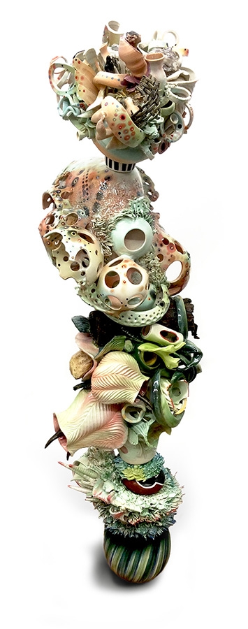
Origins II 62″ x 24″ Porcelain, steel 2016 Courtesy: Georgina Lohan

Within a context that encompasses beauty and the primordial ooze, she is representing many of the disturbing themes seen in contemporary art: fragmentation, loss, destruction, and, indirectly, war.

The artist deliberately exploits the structural fragility of her pieces (four of them had to be anchored to the walls of the gallery).  From Lohan’s own writings about the show,

The repetitive nature of loss and destruction when working with a fragile medium has consolidated my tactic of collage porcelain debris as well as a consideration of the fragment as signifier for a larger totality.

The heat of the kiln is equivalent to an acceleration of time. Gravity becomes a critical force at these high temperatures and strategies of support become more and more necessary the larger and heavier the pieces become. Glazes liquefy, boil and bubble before smoothing out, colour change, the work expands and shrinks, moving and changing it molecular structure, growing crystals and other phenomena. The results can unpredictable and there is a high level of risk, but there are also those alchemical moments when base metals have turned to gold.

Sadly, the show ended Aug. 11, 2016 but Lohan has plans for future shows. You can find out more at her website.

Bharti Kher

The Vancouver Art Gallery (VAG) is showcasing UK-born, New Delhi-based artist Bharti Kher in North America’s first 20 year retrospective of her work, titled ‘Matter’, from July 9, 2016 to Oct. 10, 2016.

I saw the show on a Tuesday (Aug. 16, 2016) which features entry by donation from 5 pm. Depending on how you feel about crowds, you may want to get there early for the lineup. (The Picasso show which is also happening is quite the attraction, more about Picasso: The Artist and His Muses later in this post.

There is a lot to this show so I’m concentrating on  elements of special interest to me: the goddess sculptures, the ‘fabric pieces’, and one of the bindi pieces.

The sculptures of the women incorporating animal pelts, fragile teacups, and/or antlers fascinated me. I was particularly intrigued by ‘And all the while the benevolent slept’ (2008).

Bharti Kher's And all the while the benevolent slept, 2008 Guillaume Ziccarelli

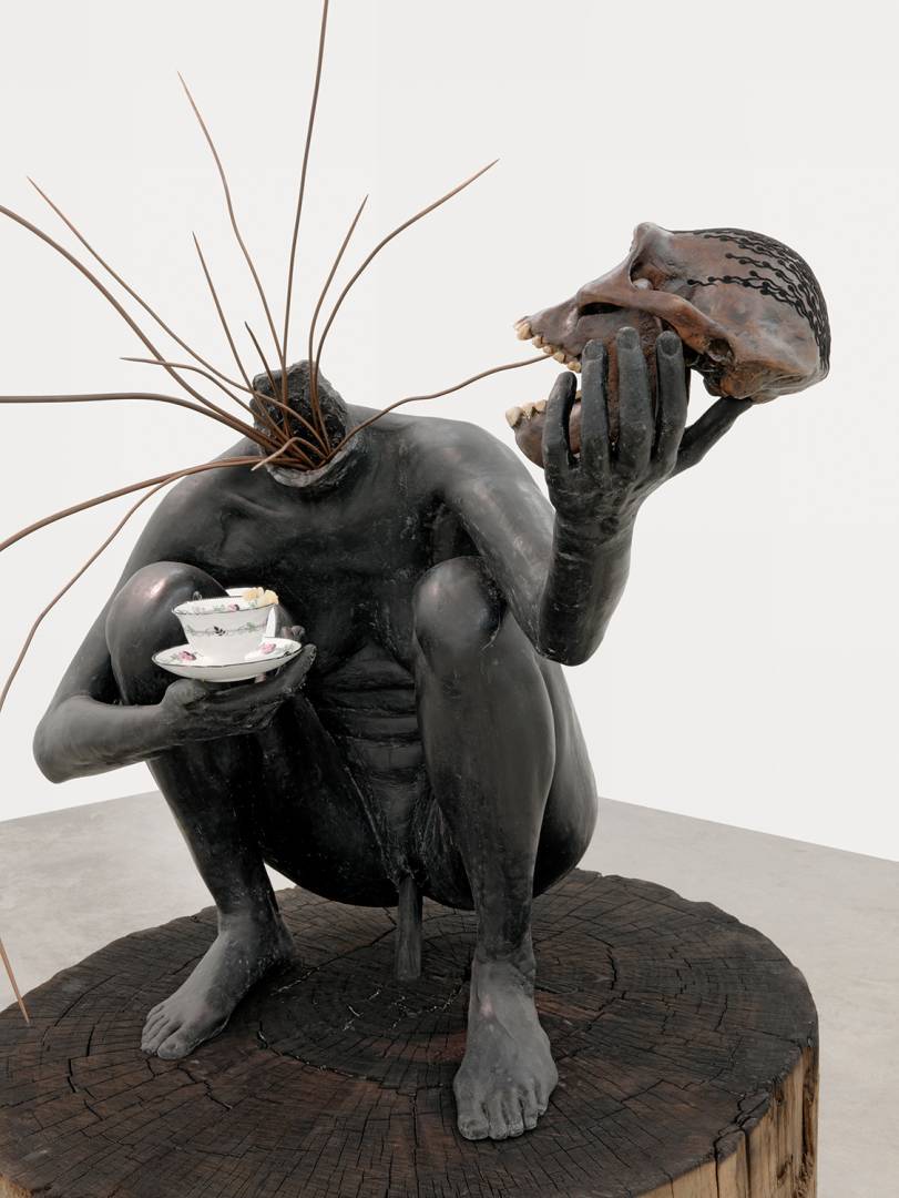
Bharti Kher’s And all the while the benevolent slept, 2008. Credit: Guillaume Ziccarelli

Here’s what Kher is doing with this goddess according to a June 28, 2016 VAG news release,

Through her use of a particular body type or character, Kher’s sculptures make reference to iconic figures from mythology and history. And all the while the benevolent slept (2008) references Chinnamasta, an Indian goddess Kali who, in traditional iconography, holds her own detached head in her hand, blood gushing from her neck, while she stands on top of a copulating couple. Through her self-sacrifice she awakens the awareness of spiritual energy while at the same time incarnating sexual energy

Kher’s ‘Chinnamasta’ stands on a tree stump and has branches growing out of her neck rather than pouring blood. For someone from a province where forestry is a major industry, this piece lends itself to a political/ecological reading, as well as, as a reading of the feminine which is so much a part of Kher’s work. The skull does not seem wholly human.

The artist does not explain the piece beyond noting its origins in traditional Indian iconography. Here’s more about Chinnamasta from its Wikipedia entry (Note: Links have been removed),

Chhinnamasta (Sanskrit: छिन्नमस्ता, Chinnamastā, “She whose head is severed”), often spelled Chinnamasta, and also called Chhinnamastika and Prachanda Chandika, is one of the Mahavidyas, ten Tantric goddesses and a ferocious aspect of Devi, the Hindu Divine Mother. Chhinnamasta can be easily identified by her unusual iconography. The nude self-decapitated goddess, usually standing or seated on a copulating couple, holds her own severed head in one hand, a scimitar in another. Three jets of blood spurt out of her bleeding neck and are drunk by her severed head and two attendants.

Chhinnamasta is a goddess of contradictions. She symbolises both aspects of Devi: a life-giver and a life-taker. She is considered both a symbol of sexual self-control and an embodiment of sexual energy, depending upon interpretation. She represents death, temporality, and destruction as well as life, immortality, and recreation. The goddess conveys spiritual self-realization and the awakening of the kundalini – spiritual energy. The legends of Chhinnamasta emphasise her self-sacrifice – sometimes coupled with a maternal element – sexual dominance, and self-destructive fury.

In reading more about Chinnamasta, the piece grows in intrigue.

Moving on to the ‘fabric pieces, there’s this from the June 28, 2016 VAG news release,

Bharti Kher’s furniture and sari sculptures speaks to socially constructed ideals of femininity and domesticity. Any utilitarian function has been rendered useless, and instead these pieces of furniture become proxies for a body. The sari-draped chairs in Absence (2011) introduces the possibility of domestic narratives filled with mothers, daughters, wives and lovers, whose bodiless garments preserve a former presence. In The day they met (2011), vibrant and richly patterned saris are decisively placed on a staircase, effectively embalming the ritual act of sari unwrapping.

Bharti Kerr, Absence, 2011, sari, resin, wooden chair. Private Collection Courtesy of the Artist and Galerie Peerotin, Photo Guillaume Ziccarelli

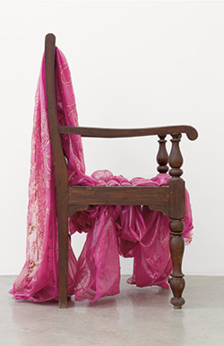
Bharti Kher, Absence, 2011, sari, resin, wooden chair. Private Collection Courtesy of the Artist and Galerie Peerotin, Photo Guillaume Ziccarelli

The saris appear on various pieces of furniture and sometimes appear as twisted, long rolls that could be said to resemble snakes. The fabrics are beautiful and they call to mind Lohan’s work and also ‘women’s work’.

Now for the bindis. For anyone not familiar with bindis, there’s this from its Wikipedia entry (Note: Links have been removed),

A bindi (Hindi: बिंदी, from Sanskrit bindu, meaning “point, drop, dot or small particle”) is a red dot worn on the center of the forehead, commonly by Hindu and Jain women. The word Bindu dates back to the hymn of creation known as Nasadiya Sukta in Rig Veda.[1] Bindu is considered the point at which creation begins and may become unity. It is also described as “the sacred symbol of the cosmos in its unmanifested state”.[2][3] Bindi is a bright dot of red colour applied in the center of the forehead close to the eyebrow worn in Indian Subcontinent (particularly amongst Hindus in India, Pakistan, Bangladesh, Nepal, and Sri Lanka)[2] and Southeast Asia among Bali and Javanese Hindus. Bindi in Hinduism, Buddhism, and Jainism is associated with Ajna Chakra and Bindu[4] is known as the third eye chakra. Bindu is the point or dot around which the mandala is created, representing the universe.[3][5] Bindi has historical and cultural presence in the region of Greater India.[6][7]

The first piece you see in the Matter show is Virus VII (2016). It is comprised of bindis, blues ones rather than the traditional red, painstakingly overlapped in a spiral that extends several feet in height and width and affixed to the wall. The piece is accompanied by a wooden box with a plaque and containing sheets of blue bindis,

Matter exhibition at Vancouver Art Gallery, July 9 - Oct. 10, 2016 Bharti Kher, Virus VII, 2016, Photo: Megan Hill-Carol Vancouver Art Gallery

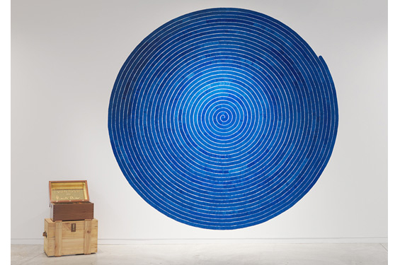
Matter exhibition at Vancouver Art Gallery, July 9 – Oct. 10, 2016 Bharti Kher, Virus VII, 2016, Photo: Megan Hill-Carol Vancouver Art Gallery

It is a stunning piece that almost seems to vibrate and is a fitting and sensual entry to the show.

For an alternative experience of the Kher show, there’s Robin Laurence’s July 6, 2016 preview titled: Bharti Kher’s hybrid vision merges humans with animals to address politics, sociology, and love for the Georgia Straight. Unexpectedly (for me), the first piece she sees is the heart,

The first artwork visitors will see when they enter Bharti Kher’s thoughtful and provocative exhibition at the Vancouver Art Gallery is a life-size sculpture of the heart of a blue sperm whale. The largest creature that now exists on our planet, the blue whale possesses a heart that is also the biggest in the world—the size, the artist says, of a small car. Kher’s realistic, cast-resin depiction of the organ’s two massive chambers, enormous aorta, and branching blood vessels is a work of weird grandeur.

To some, it might suggest an environmental message, a monument to a creature slaughtered by the hundreds of thousands in the 19th century and threatened in our own age by pollution and rising ocean temperatures. The artist, however, says the work is about the nature of love, and its title, An Absence of Assignable Cause, evokes the irrationality of that most vaunted and lamented emotion.

“More things have been written about love and all the ways around it,” she says. “I thought it would be interesting to talk about it using an animal as a metaphor.”

Picasso: The Artist and His Muses

Never having been a big fan of Pablo Picasso’s, I wouldn’t have made a special effort to see the VAG’s Picasso: The Artist and His Muses exhibition (June 11 – Oct. 2, 2016) but since I was already on premise for the Kher exhibit, it seemed to foolish to pass up the opportunity.

The show focuses on six women, his relationship with them, and how his art was affected by those relationships.

His most widely known images of women are those with the distorted features and extra or missing eyes and ears such as this,
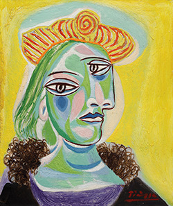
Pablo Picasso Bust of a Woman (Dora Maar), 1938 oil on canvas Hirshhorn Museum and Sculpture Garden, Gift of Joseph H. Hirshhorn, 1966 © Picasso Estate/SODRAC (2016) Photo: Cathy Carver

Pablo Picasso
Bust of a Woman (Dora Maar), 1938
oil on canvas
Hirshhorn Museum and Sculpture Garden, Gift of Joseph H. Hirshhorn, 1966
© Picasso Estate/SODRAC (2016)
Photo: Cathy Carver

These images have always left me cold. Seeing them in real life didn’t make that big a difference although I hadn’t fully appreciated their vibrancy having previously seen reproductions only. I did say I’m not a fan and that is especially true of the images of women most often seen. The surprise in this show, are the naturalistic studies where one can appreciate his extraordinary technique even if one is inclined to shun his distorted women.

I mention this show only because its subject, women, has been the direct and indirect focus of this commentary. For an even more jaundiced view of this show, you can read Robin Laurence’s June 10, 2016 preview of the VAG exhibition,

Muse is such a curiously antiquated term. Divine woman breathing inspiration into the mind of the creative male? Really? Still, Picasso: The Artist and His Muses has a more visitor-friendly sound to it than “Picasso and the Women He Fucked and Painted”. Not that visitor-friendly titles are a necessity where Pablo Picasso exhibitions are concerned.

The mere name of the man—easily the most famous artist of the 20th century, whose personal myth is built as much on his prodigious womanizing as on his protean art-making—guarantees attendance. Irrespective of what’s on view. Irrespective, too, of the challenges his work might pose to contemporary critics.

Organized with Art Centre Basel in Switzerland, the Vancouver Art Gallery’s big-draw summer show includes some 60 paintings, drawings, sculptures, and prints ranging across the years 1905 to 1971. Borrowed from an international array of public and private collections, it is the most ambitious exhibition of Picasso works ever shown in Western Canada.

I recommend reading both of Laurence’s pieces before going to the exhibit.

Final words

It seems when it comes to contemporary art, beauty is transgressive. The distortions with which Picasso experimented seem to have taken root and, like bamboo, taken over. So, an artist risks being shunned if his/her works are intrinsically beautiful (Lohan). Alternatively, an artist can include it by stealth (Kher) so viewers do not experience it as the primary impression.

All of these artists’ exhibitions have in one fashion or another focused on women. Lohan’s material of choice, porcelain, referenced women’s work indirectly and resonated in a fascinating way with Kher’s teacup bearing goddess. While Lohan and Kher are interested in women’s experiences (dressing/undressing and ornamentation (Kher), women’s roles in society (Lohan), meanwhile, Picasso seems to have considered women as raw material for his work.

Protecting soldiers from biological and chemical agents with a ‘second skin’ made of carbon nanotubes

There are lots of ‘second skins’ which promise to protect against various chemical and biological agents, the big plus for this ‘skin’ from the US Lawrence Livermore National Laboratory is breathability. From an Aug. 3, 2016 news item on Nanowerk (Note: A link has been removed),

This material is the first key component of futuristic smart uniforms that also will respond to and protect from environmental chemical hazards. The research appears in the July 27 [2016] edition of the journal, , Advanced Materials (“Carbon Nanotubes: Ultrabreathable and Protective Membranes with Sub-5 nm Carbon Nanotube Pores”).

An Aug. 3, 2016 Lawrence Livermore National Laboratory (LLNL) news release (also on EurekAlert), which originated the news item, explains further (Note: Links have been removed),

High breathability is a critical requirement for protective clothing to prevent heat-stress and exhaustion when military personnel are engaged in missions in contaminated environments. Current protective military uniforms are based on heavyweight full-barrier protection or permeable adsorptive protective garments that cannot meet the critical demand of simultaneous high comfort and protection, and provide a passive rather than active response to an environmental threat.

The LLNL team fabricated flexible polymeric membranes with aligned carbon nanotube (CNT) channels as moisture conductive pores. The size of these pores (less than 5 nanometers, nm) is 5,000 times smaller than the width of a human hair [if 1 nm is 1/100,000 or 1/60,000 of a human hair {the two most commonly used measurements} then wouldn’t 5 nm be between 1/20,000 or1/15,000 of a human hair?] .

“We demonstrated that these membranes provide rates of water vapor transport that surpass those of commercial breathable fabrics like GoreTex, even though the CNT pores are only a few nanometers wide,” said Ngoc Bui, the lead author of the paper.

To provide high breathability, the new composite material takes advantage of the unique transport properties of carbon nanotube pores. By quantifying the membrane permeability to water vapor, the team found for the first time that, when a concentration gradient is used as a driving force, CNT nanochannels can sustain gas-transport rates exceeding that of a well-known diffusion theory by more than one order of magnitude.

These membranes also provide protection from biological agents due to their very small pore size — less than 5 nanometers (nm) wide. Biological threats like bacteria or viruses are much larger and typically more than 10-nm in size. Performed tests demonstrated that the CNT membranes repelled Dengue virus from aqueous solutions during filtration tests. This confirms that LLNL-developed CNT membranes provide effective protection from biological threats by size exclusion rather than by merely preventing wetting.

Furthermore, the results show that CNT pores combine high breathability and bio-protection in a single functional material.

However, chemical agents are much smaller in size and require the membrane pores to be able to react to block the threat. To encode the membrane with a smart and dynamic response to small chemical hazards, LLNL scientists and collaborators are surface modifying these prototype carbon nanotube membranes with chemical-threat-responsive functional groups. These functional groups will sense and block the threat like gatekeepers on the pore entrance. A second response scheme also is in development — similar to how living skin peels off when challenged with dangerous external factors. The fabric will exfoliate upon reaction with the chemical agent.

“The material will be like a smart second skin that responds to the environment,” said Kuang Jen Wu, leader of LLNL’s Biosecurity & Biosciences Group. “In this way, the fabric will be able to block chemical agents such as sulfur mustard (blister agent), GD and VX nerve agents, toxins such as staphylococcal enterotoxin and biological spores such as anthrax.”

Current work is directed toward designing this multifunctional material to undergo a rapid transition from the breathable state to the protective state.

“These responsive membranes are expected to be particularly effective in mitigating a physiological burden because a less breathable but protective state can be actuated locally and only when needed,” said Francesco Fornasiero, LLNL’s principal investigator of the project.

The new uniforms could be deployed in the field in less than 10 years.

“The goal of this science and technology program is to develop a focused, innovative technological solution for future chemical biological defense protective clothing,” said Tracee Whitfield, the DTRA [US Defense Threat Reduction Agency] science and technology manager for the Dynamic Multifunctional Material for a Second Skin Program. “Swatch-level evaluations will occur in early 2018 to demonstrate the concept of ‘second skin,’ a major milestone that is a key step in the maturation of this technology.”

The researchers have prepared a video describing their work,

Here’s a link to and a citation for the paper,

Ultrabreathable and Protective Membranes with Sub-5 nm Carbon Nanotube Pores by Ngoc Bui, Eric R. Meshot, Sangil Kim, José Peña, Phillip W. Gibson, Kuang Jen Wu, and Francesco Fornasiero. Advanced Materials Volume 28, Issue 28, pages 5871–5877, July 27, 2016 DOI: 10.1002/adma.201600740 Version of Record online: 9 MAY 2016

© 2016 WILEY-VCH Verlag GmbH & Co. KGaA, Weinheim

This paper is behind a paywall.