Tag Archives: brain

Nanoparticle-based radiogenetics to control brain cells

While the title for this post sounds like an opening for a zombie-themed story, this Oct. 8, 2014 news item on Nanowerk actually concerns brain research at Rockefeller University (US), Note: A link has been removed,

A proposal to develop a new way to remotely control brain cells from Sarah Stanley, a Research Associate in Rockefeller University’s Laboratory of Molecular Genetics, headed by Jeffrey M. Friedman, is among the first to receive funding from the BRAIN initiative. The project will make use of a technique called radiogenetics that combines the use of radio waves or magnetic fields with nanoparticles to turn neurons on or off.

An Oct. 7, 2014 Rockefeller University news release, which originated the news item, further describes the BRAIN initiative and the research (Note: Links have been removed),

The NIH [National Institutes of Health]  is one of four federal agencies involved in the BRAIN (Brain Research through Advancing Innovative Neurotechnologies) initiative. Following in the ambitious footsteps of the Human Genome Project, the BRAIN initiative seeks to create a dynamic map of the brain in action, a goal that requires the development of new technologies. The BRAIN initiative working group, which outlined the broad scope of the ambitious project, was co-chaired by Rockefeller’s Cori Bargmann, head of the Laboratory of Neural Circuits and Behavior.

Stanley’s grant, for $1.26 million over three years, is one of 58 projects to get BRAIN grants, the NIH announced. The NIH’s plan for its part of this national project, which has been pitched as “America’s next moonshot,” calls for $4.5 billion in federal funds over 12 years.

The technology Stanley is developing would enable researchers to manipulate the activity of neurons, as well as other cell types, in freely moving animals in order to better understand what these cells do. Other techniques for controlling selected groups of neurons exist, but her new nanoparticle-based technique has a unique combination of features that may enable new types of experimentation. For instance, it would allow researchers to rapidly activate or silence neurons within a small area of the brain or dispersed across a larger region, including those in difficult-to-access locations. Stanley also plans to explore the potential this method has for use treating patients.

“Francis Collins, director of the NIH, has discussed the need for studying the circuitry of the brain, which is formed by interconnected neurons. Our remote-control technology may provide a tool with which researchers can ask new questions about the roles of complex circuits in regulating behavior,” Stanley says.

Here’s an image that Rockefeller University has used to illustrate the concept of radio-controlled brain cells,


BRAIN control: The new technology uses radio waves to activate or silence cells remotely. The bright spots above represent cells with increased calcium after treatment with radio waves, a change that would allow neurons to fire. [downloaded from: http://newswire.rockefeller.edu/2014/10/07/rockefeller-neurobiology-lab-is-awarded-first-round-brain-initiative-grant/]

BRAIN control: The new technology uses radio waves to activate or silence cells remotely. The bright spots above represent cells with increased calcium after treatment with radio waves, a change that would allow neurons to fire. [downloaded from: http://newswire.rockefeller.edu/2014/10/07/rockefeller-neurobiology-lab-is-awarded-first-round-brain-initiative-grant/]

You can find out more about the US BRAIN initiative here.

DARPA (US Defense Advanced Research Projects Agency) awards funds for implantable neural interface

I’m not a huge fan of neural implantable devices (at least not the ones that facilitate phone calls directly to and from the brain as per my April 30, 2010 posting; scroll down about 40% of the way) but they are important from a therapeutic perspective. On that  note, the Lawrence Livermore National Laboratory (LLNL) has received an award of $5.6M from the US Defense Advanced Research Projects Agency (DARPA) to advance their work on neural implantable interfaces. From a June 13, 2014 news item on Azonano,

Lawrence Livermore National Laboratory recently received $5.6 million from the Department of Defense’s Defense Advanced Research Projects Agency (DARPA) to develop an implantable neural interface with the ability to record and stimulate neurons within the brain for treating neuropsychiatric disorders.

The technology will help doctors to better understand and treat post-traumatic stress disorder (PTSD), traumatic brain injury (TBI), chronic pain and other conditions.

Several years ago, researchers at Lawrence Livermore in conjunction with Second Sight Medical Products developed the world’s first neural interface (an artificial retina) that was successfully implanted into blind patients to help partially restore their vision. The new neural device is based on similar technology used to create the artificial retina.

An LLNL June 11, 2014 news release, which originated the news item, provides some fascinating insight into the interrelations between various US programs focused on the brain and neural implants,

“DARPA is an organization that advances technology by leaps and bounds,” said LLNL’s project leader Satinderpall Pannu, director of the Lab’s Center for Micro- and Nanotechnology and Center for Bioengineering, a facility dedicated to fabricating biocompatible neural interfaces. “This DARPA program will allow us to develop a revolutionary device to help patients suffering from neuropsychiatric disorders and other neural conditions.”

The project is part of DARPA’s SUBNETS (Systems-Based Neurotechnology for Emerging Therapies) program. The agency is launching new programs to support President Obama’s BRAIN (Brain Research through Advancing Innovative Neurotechnologies) Initiative, a new research effort aimed to revolutionize our understanding of the human mind and uncover ways to treat, prevent and cure brain disorders.

LLNL and Medtronic are collaborating with UCSF, UC Berkeley, Cornell University, New York University, PositScience Inc. and Cortera Neurotechnologies on the DARPA SUBNETS project. Some collaborators will be developing the electronic components of the device, while others will be validating and characterizing it.

As part of its collaboration with LLNL, Medtronic will consult on the development of new technologies and provide its investigational Activa PC+S deep brain stimulation (DBS) system, which is the first to enable the sensing and recording of brain signals while simultaneously providing targeted DBS. This system has recently been made available to leading researchers for early-stage research and could lead to a better understanding of how various devastating neurological conditions develop and progress. The knowledge gained as part of this collaboration could lead to the next generation of advanced systems for treating neural disease.

As for what LLNL will contribute (from the news release),

The LLNL Neural Technology group will develop an implantable neural device with hundreds of electrodes by leveraging their thin-film neural interface technology, a more than tenfold increase over current Deep Brain Stimulation (DBS) devices. The electrodes will be integrated with electronics using advanced LLNL integration and 3D packaging technologies. The goal is to seal the electronic components in miniaturized, self-contained, wireless neural hardware. The microelectrodes that are the heart of this device are embedded in a biocompatible, flexible polymer.

Surgically implanted into the brain, the neural device is designed to help researchers understand the underlying dynamics of neuropsychiatric disorders and re-train neural networks to unlearn these disorders and restore proper function. This will enable the device to be eventually removed from the patient instead of being dependent on it.

This image from LLNL illustrates their next generation neural implant,

This rendering shows the next generation neural device capable of recording and stimulating the human central nervous system being developed at Lawrence Livermore National Laboratory. The implantable neural interface will record from and stimulate neurons within the brain for treating neuropsychiatric disorders.

This rendering shows the next generation neural device capable of recording and stimulating the human central nervous system being developed at Lawrence Livermore National Laboratory. The implantable neural interface will record from and stimulate neurons within the brain for treating neuropsychiatric disorders.

i expect there will be many more ‘brain’ projects to come with the advent of the US BRAIN initiative (funds of $100M in 2014 and $200M in 2015) and the European Union’s Human Brain Project (1B Euros to be spent on research over a 10 year period).

Wireless nano for remotely activating neurons

Every once in a while, there’s a piece of research that disconcerts me and this would be one of those pieces. From a May 22, 2014 news item on Nanowerk,

Yang Xiang, PhD, assistant professor of neurobiology at University of Massachusetts Medical School, has received a three-year, $900,000 grant from the Human Frontiers Science Program to lead an international team of scientists, including Gang Han, PhD, assistant professor of biochemistry & molecular pharmacology, in the development and implementation of a new optogenetic platform that can remotely activate neurons inside a free-moving organism.

Using a new class of nanoparticles developed by Dr. Han, Dr. Xiang and colleagues propose to selectively turn on non-image forming photoreceptors (NIFP) inside mice and Drosophila unencumbered by the fiber optic wires used in currently available optogenetic technologies. By wirelessly stimulating these photoreceptors, which are able to sense light even though they don’t generate vision, scientists can better understand their role in regulating physiological functions such as circadian rhythm, sleep and melatonin secretion. The hope is that this new technology can also be used to study the links between other types of neurons, physiology and behavior.

A May 22, 2014 University of Massachusetts Medical School news release by Jim Fessenden, which originated the news item, describes optogenetics and some of its challenges,

“Current optogenetic technologies are limited in their application because they require using ‘wired’ fiber optic implants to deliver blue light to activate neuron activities,” said Xiang. “This is a major technological problem that has become an obstacle to understanding the physiological role NIFP play in animal behavior. If we’re able to overcome this hurdle by using the nanoparticles developed by Dr. Han, it would open the door to more informed investigations of not only NIFP but a wide range of neurons and their effect on behavior.”

In use for only about a decade, optogenetic technology combines techniques from optics and genetics, allowing scientists to precisely control activities of individual neurons using light. By genetically inserting light-activated biological molecules such as channelrhodopsins, a family of proteins found in algae, into neurons, scientists can instantaneously turn them on using beams of blue light with millisecond precision.

A limiting factor to the wider application of this technology, however, is that blue wavelengths are unable to penetrate skin, bone and other tissues deep enough to activate the neurons inside free-moving animals. To overcome this obstacle, current techniques require the insertion of fiber optic wires close enough to the neurons so the light that activates them can be delivered. This technique restricts animal movement and makes it difficult to observe behavioral responses in natural conditions. This fiber optic approach further limits scientists’ ability to study behavior over longer periods of time as the effectiveness of light delivery is relatively short due to scarring.

The news release describes the new technique proposed by Xiang and his associates,

Han has developed an “upconversion nanoparticle” (UCNP) that has the potential to solve the limitations of wired optogenetic techniques. These nanoparticles are capable of absorbing infrared light that can’t be seen and converting it into visible blue light. In contrast to blue light, infrared light is capable of penetrating skin and tissue to a depth of several centimeters. Xiang and Han believe these nanoparticles, tuned to emit blue light, can be inserted into the brain and used as a substitute for traditional fiber optics to wirelessly activate neurons in animals.

The hope is that the nanoparticles will absorb infrared light that passes through the tissue, and convert it to blue light inside the animal. This blue light would then activate the NFIPs. If successful, Xiang and colleagues will be able to observe any changes in animal behavior brought about by activating these non-image forming photoreceptors.

“The nanoparticles act as a kind of relay station,” said Han. “They convert the low-energy red light into a high-energy blue light that can activate the neurons. This technique completely alleviates the need to use intrusive fiber optic wires. It vastly simplifies the technology and expands the potential uses for optogenetics.”

Xiang said, “In many ways, this is the perfect bridge between a technological advancement and an important biological question. With these nanoparticles it’s possible for us to begin answering fundamental neurobiological questions about NIFPs.

“More broadly, it would open up the possibility of using other model organisms, such as Drosophila, that can’t be used with the current wired optogenetic technologies, to investigate and answer important questions about how neural activities regulate behavior.”

Illogical as it is, the idea that neurons could be wirelessly and remotely activated by someone other the owner of those neurons disturbs me even though I know drugs are commonly used to do much the same thing in humans.

In any event, the news release provides this final paragraph about the funding,

HFSP [Human Frontiers Science Program] awards are given to highly innovative teams that demonstrate that they have developed and can test a paradigm-shifting idea that holds promise for the development of new approaches to problems in the life sciences with potential to advance the field of research significantly.

I looked up the HFSP online and found this on the About Us page on the HFSP website,

The Human Frontier Science Program is a program of funding for frontier research in the life sciences. It is implemented by the International Human Frontier Science Program Organization (HFSPO) with its office in Strasbourg.

The members of the HFSPO, the so-called Management Supporting Parties (MSPs) are the contributing countries and the European Union, which contributes on behalf of the non-G7 EU members.

The current MSPs are Australia, Canada, France, Germany, India, Italy, Japan, Republic of Korea, Norway, New Zealand, Switzerland the United Kingdom, the United States of America and the European Union. [emphasis mine]

I was not expecting to find Canada on that list.

Controversial theory of consciousness confirmed (maybe)

There’s a very interesting event taking place today (Jan. 16, 2014) in Amsterdam, Netherlands titled: NEW PROOF OF REVOLUTIONARY THEORY OF CONSCIOUSNESS (programme).,which is one of a month’s worth of events themed around the brain (The Brainstorming Sessions).  The speakers at this event have recently published a paper and a Jan. 16, 2014 news item on ScienceDaily gives some insight into why theirbrainstorming session has the word revolutionary in the title,

A review and update of a controversial 20-year-old theory of consciousness published in Physics of Life Reviews claims that consciousness derives from deeper level, finer scale activities inside brain neurons. The recent discovery of quantum vibrations in “microtubules” inside brain neurons corroborates this theory, according to review authors Stuart Hameroff and Sir Roger Penrose. They suggest that EEG rhythms (brain waves) also derive from deeper level microtubule vibrations, and that from a practical standpoint, treating brain microtubule vibrations could benefit a host of mental, neurological, and cognitive conditions.

A Jan. 16, 2014 Elsevier press release,which originated the news item, provides more details about the theory,

The theory, called “orchestrated objective reduction” (‘Orch OR’), was first put forward in the mid-1990s by eminent mathematical physicist Sir Roger Penrose, FRS, Mathematical Institute and Wadham College, University of Oxford, and prominent anesthesiologist Stuart Hameroff, MD, Anesthesiology, Psychology and Center for Consciousness Studies, The University of Arizona, Tucson. They suggested that quantum vibrational computations in microtubules were “orchestrated” (“Orch”) by synaptic inputs and memory stored in microtubules, and terminated by Penrose “objective reduction” (‘OR’), hence “Orch OR.” Microtubules are major components of the cell structural skeleton.

Orch OR was harshly criticized from its inception, as the brain was considered too “warm, wet, and noisy” for seemingly delicate quantum processes. However, evidence has now shown warm quantum coherence in plant photosynthesis, bird brain navigation, our sense of smell, and brain microtubules. The recent discovery of warm temperature quantum vibrations in microtubules inside brain neurons by the research group led by Anirban Bandyopadhyay, PhD, at the National Institute of Material Sciences in Tsukuba, Japan (and now at MIT), corroborates the pair’s theory and suggests that EEG rhythms also derive from deeper level microtubule vibrations. In addition, work from the laboratory of Roderick G. Eckenhoff, MD, at the University of Pennsylvania, suggests that anesthesia, which selectively erases consciousness while sparing non-conscious brain activities, acts via microtubules in brain neurons.

“The origin of consciousness reflects our place in the universe, the nature of our existence. Did consciousness evolve from complex computations among brain neurons, as most scientists assert? Or has consciousness, in some sense, been here all along, as spiritual approaches maintain?” ask Hameroff and Penrose in the current review. “This opens a potential Pandora’s Box, but our theory accommodates both these views, suggesting consciousness derives from quantum vibrations in microtubules, protein polymers inside brain neurons, which both govern neuronal and synaptic function, and connect brain processes to self-organizing processes in the fine scale, ‘proto-conscious’ quantum structure of reality.”

After 20 years of skeptical criticism, “the evidence now clearly supports Orch OR,” continue Hameroff and Penrose. “Our new paper updates the evidence, clarifies Orch OR quantum bits, or “qubits,” as helical pathways in microtubule lattices, rebuts critics, and reviews 20 testable predictions of Orch OR published in 1998 – of these, six are confirmed and none refuted.”

An important new facet of the theory is introduced. Microtubule quantum vibrations (e.g. in megahertz) appear to interfere and produce much slower EEG “beat frequencies.” Despite a century of clinical use, the underlying origins of EEG rhythms have remained a mystery. Clinical trials of brief brain stimulation aimed at microtubule resonances with megahertz mechanical vibrations using transcranial ultrasound have shown reported improvements in mood, and may prove useful against Alzheimer’s disease and brain injury in the future.

Lead author Stuart Hameroff concludes, “Orch OR is the most rigorous, comprehensive and successfully-tested theory of consciousness ever put forth. From a practical standpoint, treating brain microtubule vibrations could benefit a host of mental, neurological, and cognitive conditions.

The review is accompanied by eight commentaries from outside authorities, including an Australian group of Orch OR arch-skeptics. To all, Hameroff and Penrose respond robustly.

The press release ends with this information about the event in Amsterdam,

Penrose, Hameroff and Bandyopadhyay will explore their theories during a session on “Microtubules and the Big Consciousness Debate” at the Brainstorm Sessions, a public three-day event at the Brakke Grond in Amsterdam, the Netherlands, January 16-18, 2014. They will engage skeptics in a debate on the nature of consciousness, and Bandyopadhyay and his team will couple microtubule vibrations from active neurons to play Indian musical instruments. “Consciousness depends on anharmonic vibrations of microtubules inside neurons, similar to certain kinds of Indian music, but unlike Western music which is harmonic,” Hameroff explains.

I wasn’t able to locate information about the three-day event in the press release but I did find this about the month-long series, The Brainstorm Sessions (Dutch language first, scroll down for English language version),

Europe and the USA are looking to completely unravel the secrets of our brains within the next ten years. Europe has designated 2014 as The Year of the Brain. We have decided to dedicate a month to the grey matter. A month in which guest curator Frank Theys – filmmaker, philosopher and visual artist – i.c.w. Damiaan Denys (neuroscientist, philosopher and professor of psychiatry at the AMC-UvA, the Amsterdam Medical Centre of the University of Amsterdam) will bring together elements he considers interesting from an artistic and philosophical viewpoint related to this theme.

Featuring an exhibition at the intersection between artistic and scientific experiments; the first ever performance by ‘stand-up scientist’ Damiaan Denys, Head of Psychiatry at the AMC hospital; a ‘neuro-concert’ by nanoscientist Anirban Bandyopadyay and a film programme in the Kriterion cinema in cooperation with Patricia Pisters, author of The Neuro-Image.

PROGRAMME FOR AN INTERNATIONAL AUDIENCE
Fri 13 Dec – Sun 19 Jan: Exhibition Neurons Firing
Thur 09 Jan / 20h30: Sonic Soirée #22 a musical pillaging of the brain
Mon 13 Jan / 20h30: Lecture: Film and the Brain in Digital Era, by Patricia Pisters
Thu 16 Jan / 20h30: Lecture: Microtubules & the Big Consciousness Debate, by Roger Penrose & Anirban Bandyopadhyay
Fr 17 Jan / 20h30: Scientific demonstration Sapta Rishi (The Seven Stars)
Sa 18 Jan / 20h30: Scientific concert: Ajeya Chhandam – The Invincible Rhythm

I’m not sure what your chances are for attending the events on Jan. 17 or Jan. 18 but I wish you good luck! For those of us who weren’t able to attend the Jan.16, 2014 event featuring Penrose amd Hameroff, there are recently published papers.

First, the researchers offer a review of their theory along with some refinements,

Consciousness in the universe: A review of the ‘Orch OR’ theory by Stuart Hameroff and Roger Penrose. Physics of Life Reviews Available online 20 August 2013, Phys Life Rev. 2013 Aug 20. pii: S1571-0645(13)00118-8. doi: 10.1016/j.plrev.2013.08.002.

This paper is open access as of Jan. 16, 2014.

The next two papers have similar titles and were published at about the same time,

Reply to criticism of the ‘Orch OR qubit’ – ‘Orchestrated objective reduction’ is scientifically justified by Stuart Hameroff and Roger Penrose. Physics of Life Reviews Available online 12 December 2013. Phys Life Rev. 2013 Dec 12. pii: S1571-0645(13)00191-7. doi: 10.1016/j.plrev.2013.11.014.

Reply to seven commentaries on “Consciousness in the universe: Review of the ‘Orch OR’ theory by Stuart Hameroff and Roger Penrose. Physics of Life Reviews Available online 12 December 2013 Phys Life Rev. 2013 Dec 12. pii: S1571-0645(13)00190-5. doi: 10.1016/j.plrev.2013.11.013.

These papers are behind a paywall.

Two bits about the brain: fiction affects your brain and the US’s BRAIN Initiative is soliciting grant submissions

As a writer I love to believe my words have a lasting impact and while this research is focused on fiction, something I write more rarely than nonfiction, hope springs eternal that one day nonfiction too will be proved as having an impact (in a good way) on the brain. From a Jan. 3, 2014 news release on EurekAlert (or you can read the Dec. 17, 2013 Emory University news release by Carol Clark),

Many people can recall reading at least one cherished story that they say changed their life. Now researchers at Emory University have detected what may be biological traces related to this feeling: Actual changes in the brain that linger, at least for a few days, after reading a novel.

“Stories shape our lives and in some cases help define a person,” says neuroscientist Gregory Berns, lead author of the study and the director of Emory’s Center for Neuropolicy. “We want to understand how stories get into your brain, and what they do to it.”

His co-authors included Kristina Blaine and Brandon Pye from the Center for Neuropolicy, and Michael Prietula from Emory’s Goizueta Business School.

Neurobiological research using functional magnetic resonance imaging (fMRI) has begun to identify brain networks associated with reading stories. Most previous studies have focused on the cognitive processes involved in short stories, while subjects are actually reading them while they are in the fMRI scanner.

All of the study subjects read the same novel, “Pompeii,” a 2003 thriller by Robert Harris that is based on the real-life eruption of Mount Vesuvius in ancient Italy.

“The story follows a protagonist, who is outside the city of Pompeii and notices steam and strange things happening around the volcano,” Berns says. “He tries to get back to Pompeii in time to save the woman he loves. Meanwhile, the volcano continues to bubble and nobody in the city recognizes the signs.”

The researchers chose the book due to its page-turning plot. “It depicts true events in a fictional and dramatic way,” Berns says. “It was important to us that the book had a strong narrative line.”

For the first five days, the participants came in each morning for a base-line fMRI scan of their brains in a resting state. Then they were fed nine sections of the novel, about 30 pages each, over a nine-day period. They were asked to read the assigned section in the evening, and come in the following morning. After taking a quiz to ensure they had finished the assigned reading, the participants underwent an fMRI scan of their brain in a non-reading, resting state. After completing all nine sections of the novel, the participants returned for five more mornings to undergo additional scans in a resting state.

The results showed heightened connectivity in the left temporal cortex, an area of the brain associated with receptivity for language, on the mornings following the reading assignments. “Even though the participants were not actually reading the novel while they were in the scanner, they retained this heightened connectivity,” Berns says. “We call that a ‘shadow activity,’ almost like a muscle memory.”

Heightened connectivity was also seen in the central sulcus of the brain, the primary sensory motor region of the brain. Neurons of this region have been associated with making representations of sensation for the body, a phenomenon known as grounded cognition. Just thinking about running, for instance, can activate the neurons associated with the physical act of running.

“The neural changes that we found associated with physical sensation and movement systems suggest that reading a novel can transport you into the body of the protagonist,” Berns says. “We already knew that good stories can put you in someone else’s shoes in a figurative sense. Now we’re seeing that something may also be happening biologically.”

The neural changes were not just immediate reactions, Berns says, since they persisted the morning after the readings, and for the five days after the participants completed the novel.

“It remains an open question how long these neural changes might last,” Berns says. “But the fact that we’re detecting them over a few days for a randomly assigned novel suggests that your favorite novels could certainly have a bigger and longer-lasting effect on the biology of your brain.”

Here’s a link to and a citation for the paper,

Short- and Long-Term Effects of a Novel on Connectivity in the Brain by Gregory S. Berns, Kristina Blaine, Michael J. Prietula, and Brandon E. Pye. Brain Connectivity. 2013, 3(6): 590-600. doi:10.1089/brain.2013.0166.

This is an open access paper where you’ll notice the participants cover a narrow range of ages (from the Materials and Methods section of the paper,

A total of 21 participants were studied. Two were excluded from the fMRI analyses: one for insufficient attendance, and the other for image abnormalities. Before the experiment, participants were screened for the presence of medical and psychiatric diagnoses, and none were taking medications. There were 12 female and 9 male participants between the ages of 19 and 27 (mean 21.5). Emory University’s Institutional Review Board approved all procedures, and all participants gave written informed consent.

It’s always good to remember that university research often draws from student populations and the question one might want to ask is whether or not those results will remain the same, more or less, throughout someone’s life span.In any event, I find this research intriguing and hope they follow this up.

Currently known as the BRAIN (Brain Research through Advancing Innovative Neurotechnologies), I first wrote about the project under its old name BAM (Brain Activity Map) in two postings, first in a March 4, 2013 posting featuring brain-to-brain communication and other brain-related tidbits, then again, in an April 2, 2013 posting featuring an announcement about its federal funding. Today (Jan. 6, 2014), I stumbled across some BRAIN funding opportunities for researchers, from the BRAIN Initiative funding opportunities webpage,

NIH released six funding opportunity announcements in support of the President’s BRAIN Initiative. Collectively, these opportunities focus on building a new arsenal of tools and technologies for helping scientists unlock the mysteries of the brain. NIH [US National Institutes of Health] plans to invest $40 million in Fiscal Year 2014 through these opportunities, contingent upon the submission of a sufficient number of scientifically meritorious applications.

The opportunities currently available are as follows:

  • Transformative Approaches for Cell-Type Classification in the Brain (U01) (RFA-MH-14-215) – aims to pilot classification strategies to generate a systematic inventory/cell census of cell types in the brain, integrating molecular identity of cell types with connectivity, morphology, and location. These pilot projects and methodologies should be designed to demonstrate their utility and scalability to ultimately complete a comprehensive cell census of the human brain.
    Contact Email: BRAIN-info-NIMH@mail.nih.gov
    Application Receipt: March 13, 2014
  • Development and Validation of Novel Tools to Analyze Cell-Specific and Circuit-Specific Processes in the Brain (U01) (RFA-MH-14-216) – aims to develop and validate novel tools that possess a high degree of cell-type and/or circuit-level specificity to facilitate the detailed analysis of complex circuits and provide insights into cellular interactions that underlie brain function. A particular emphasis is the development of new genetic and non-genetic tools for delivering genes, proteins and chemicals to cells of interest; new approaches are also expected to target specific cell types and or circuits in the nervous system with greater precision and sensitivity than currently established methods.
    Contact Email: BRAIN-info-NIMH@mail.nih.gov
    Application Receipt: March 13, 2014
  • New Technologies and Novel Approaches for Large-Scale Recording and Modulation in the Nervous System (U01) (RFA-NS-14-007) – focuses on development and proof-of-concept testing of new technologies and novel approaches for large scale recording and manipulation of neural activity, with cellular resolution, at multiple spatial and/or temporal scales, in any region and throughout the entire depth of the brain. The proposed research may be high risk, but if successful could profoundly change the course of neuroscience research.
    Contact Email: NINDS-Brain-Initiative@nih.gov
    Application Receipt: March 24, 2014
  • Optimization of Transformative Technologies for Large Scale Recording and Modulation in the Nervous System (U01) (RFA-NS-14-008) – aims to optimize existing and emerging technologies and approaches that have the potential to address major challenges associated with recording and manipulating neural activity. This FOA is intended for the iterative refinement of emergent technologies and approaches that have already demonstrated their transformative potential through initial proof-of-concept testing, and are appropriate for accelerated engineering development with an end-goal of broad dissemination and incorporation into regular neuroscience research.
    Contact Email: NINDS-Brain-Initiative@nih.gov
    Application Receipt: March 24, 2014
  • Integrated Approaches to Understanding Circuit Function in the Nervous System (U01) (RFA-NS-14-009) – focuses onexploratory studies that use new and emerging methods for large scale recording and manipulation to elucidate the contributions of dynamic circuit activity to a specific behavioral or neural system. Applications should propose teams of investigators that seek to cross boundaries of interdisciplinary collaboration, for integrated development of experimental, analytic and theoretical capabilities in preparation for a future competition for large-scale awards.
    Contact Email: NINDS-Brain-Initiative@nih.gov
    Application Receipt: March 24, 2014
  • Planning for Next Generation Human Brain Imaging (R24) (RFA-MH-14-217) – aims to create teams of imaging scientist together with other experts from a range of disciplines such as engineering, material sciences, nanotechnology and computer science, to plan for a new generation of non-invasive imaging techniques that would be used to understand human brain function. Incremental improvements to existing technologies will not be funded under this announcement.
    Contact Email: sgrant@nida.nih.gov
    Application Receipt: March 13, 2014

For the interested, in the near future there will be some informational conference calls regarding these opportunities,

Informational Conference Calls for Prospective Applicants

NIH will be hosting a series of informational conference calls to address technical questions regarding applications to each of the RFAs released under the BRAIN Initiative.   Information on dates and contacts for each of the conference calls is as follows:

January 10, 2014, 2:00-3:00 PM EST
RFA-MH-14-215, Transformative Approaches for Cell-Type Classification in the Brain

For call-in information, contact Andrea Beckel-Mitchener at BRAIN-info-NIMH@mail.nih.gov.

January 13, 2014, 2:00-3:00 PM EST
RFA-MH-14-216, Development and Validation of Novel Tools to Analyze Cell-Specific and Circuit-Specific Processes in the Brain

For call-in information, contact Michelle Freund at BRAIN-info-NIMH@mail.nih.gov.

January 15, 2014, 1:00-2:00 PM EST
RFA-MH-14-217, Planning for Next Generation Human Brain Imaging

For call-in information, contact Greg Farber at BRAIN-info-NIMH@mail.nih.gov.

February 4, 2014, 1:00-2:30 PM EST
RFA-NS-14-007, New Technologies and Novel Approaches for Large-Scale Recording and Modulation in the Nervous System
RFA-NS-14-008, Optimization of Transformative Technologies for Large Scale Recording and Modulation in the Nervous System
RFA-NS-14-009, Integrated Approaches to Understanding Circuit Function in the Nervous System

For call-in information, contact Karen David at NINDS-Brain-Initiative@nih.gov.
In addition to accessing the information provided in the upcoming conference calls, applicants are strongly encouraged to consult with the Scientific/Research Contacts listed in each of the RFAs to discuss the alignment of their proposed work with the goals of the RFA to which they intend to apply.

Good luck!

It’s kind of fascinating to see this much emphasis on brains what with the BRAIN Initiative in the US and the Human Brain Project in Europe (my Jan. 28, 2013 posting announcing the European Union’s winning Future and Emerging Technologies (FET) research projects, The prizes (1B Euros to be paid out over 10 years to each winner) had been won by the Human Brain FET project and the Graphene FET project, respectively

AAAS 2013 meeting in Boston,US and Canadian research excellence

The 2013 annual meeting for the American Association for the Advancement of Science (AAAS) will be held in Boston, Massachusetts from Feb. 14 – 18, 2013 with a much better theme this year, The Beauty and Benefits of Science, than last year’s, Flattening the World. (It didn’t take much to improve the theme, eh?)

Plenary speakers range from AAAS’s president, William N. Press to Nathan Myhrvold, a venture capitalist to astrophysicist, Robert Kirshner to Cynthia Kenyon, a molecular biologist to Sherry Turkle. From the AAAS webpage describing Turkle’s 2013 plenary lecture,

Sherry Turkle

Abby Rockefeller Mauzé Professor of the Social Studies of Science and Technology in the Program in Science, Technology, and Society, MIT

The Robotic Moment: What Do We Forget When We Talk to Machines?

Dr. Turkle is founder and director of the MIT Initiative on Technology and Self. She received a joint doctorate in sociology and personality psychology from Harvard University and is a licensed clinical psychologist. Her research focuses on the psychology of human relationships with technology, especially in the realm of how people relate to computational objects. She is an expert on mobile technology, social networking, and sociable robotics and a regular media commentator on the social and psychological effects of technology. Her most recent book is Alone Together: Why We Expect More from Technology and Less from Each Other.

Given my experience last year in the 2012 meeting media room, I’m surprised to see a social media session is planned, from the session webpage,

Engaging with Social Media
Communicating Science
Thursday, February 14, 2013: 3:00 PM-4:30 PM
Ballroom A (Hynes Convention Center)

In a constantly changing online landscape, what is the best way for scientists and engineers to engage the public through social media? This session will discuss how people are accessing science information via blogs and social networks and the importance of researchers getting involved directly. [emphasis mine]  Speakers will address the ways that researchers can create meaningful interactions with the public through social media.

Organizer: Cornelia Dean, The New York Times
Co-Organizer: Dennis Meredith, Science Communication Consultant
Moderator: Carl Zimmer, Independent Science Journalist

Speakers:
XXXX Scicurious, Neurotic Physiology
Science Blogging for Fun and Profit
Christie Wilcox, University of Hawaii
Science in a Digital Age
Dominique Brossard, University of Wisconsin
Science and the Public in New Information Environments

I’d love to see how the theme of ‘researcher engaging directly’ gets developed. In theory, I have no problems with the concept. Unfortunately, those words are sometimes code for this perspective, ‘only experts (scientists/accredited journalists) should discuss or write about science’. A couple of quick comments, my Jan. 13, 2012 posting featured an interview with Carl Zimmer, this session’s moderator, about his science tattoo book and Dominique Brossard, one of the speakers, was last mentioned here in my Jan. 24, 2013 posting titled, Tweet your nano, in the context of a research study on social media and nanotechnology.

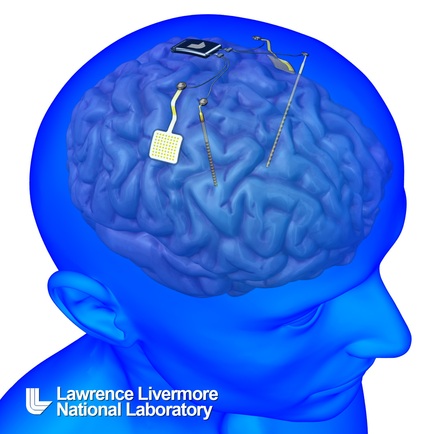
In keeping with the times (as per my Jan. 28, 2013 posting about the colossal research prizes for the Graphene and Human Brain Project initiatives), the 2012 AAAS annual meeting features a Brain Function and Plasticity thread or subtheme. There’s this session amongst others,

The Connectome: From the Synapse to Brain Networks in Health and Disease
Brain Function and Plasticity
Saturday, February 16, 2013: 8:30 AM-11:30 AM
Room 304 (Hynes Convention Center)

A series of innovative studies are being done to map the brain from the molecular to the systems level both structurally and functionally. At the synaptic level, how neurotransmitters, their receptors, and signaling pathways influence neural function and plasticity is becoming much better understood. Integrating neuronal function at the level of single neurons and groups of neurons into larger circuits at the anatomical level in the mammalian brain, while a daunting task, is being studied by advanced imaging techniques requiring vast amounts of information storage and processing. To integrate local circuit function with whole brain function, understanding the structure and processing of brain networks is critical. A major project to accomplish this task, the Human Connectome Project, is in the process of integrating the structure and function of brain networks using the most advanced imaging and analysis techniques in 1,200 people, including twins and their nontwin siblings. This step will allow for major new insights into not only brain structure and function, but also their genetic underpinnings. Comparing this information in both the normal brain and in different brain disorders such as neurodegenerative diseases is providing novel insights into how understanding brain function from the molecular to the systems level will provide insights into normal brain function and disease pathogenesis as well as provide new treatment strategies.

Organizer:

David Holtzman, Washington University

Speakers:

Mark F. Bear, Massachusetts Institute of Technology
Molecules and Mechanisms Involved in Synaptic Plasticity in Health and Disease
Jeff Lichtman, Harvard University
Connectomics: Developing a Wiring Diagram for the Mammalian Brain
Steve Petersen, Washington University
The Human Connectome Project
Marcus E. Raichle, Washington University
The Brain’s Dark Energy and the Default Mode Network
Nicole Calakos, Duke University
Synaptic Plasticity in the Basal Ganglia in Health and Disease
William W. Seeley, University of California
Brain Networks: Linking Structure and Function in Neurodegenerative Diseases

Then, there’s this session featuring graphene,

What’s Hot in Cold
Sunday, February 17, 2013: 8:30 AM-11:30 AM
Room 308 (Hynes Convention Center)

The study of ultracold atoms and molecules is now the frontier of low-temperature science, reaching temperatures of a few hundred picokelvin above absolute zero. This field was made possible by a technique that did not exist 30 years ago: laser cooling of atoms. It is hardly obvious that the laser, which produces the most intense light on Earth and is routinely used in industrial applications for cutting and welding medal, would also provide the most powerful coolant. Such are the surprises of science, where a breakthrough in one area transforms others in unexpected ways. Since 1997, eight Nobel Laureates in physics have been recognized for contributions to ultracold atomic and molecular science, which has become one of the most vibrant fields in physics, cutting across traditional disciplinary boundaries, e.g., atomic, molecular, and optical; condensed matter; statistical physics; and nuclear and particle physics. This field builds on two accomplishments that it was the first to achieve: first, the production of quantum degenerate matter using a wide range of elements and, second, exquisite control of quantum degenerate matter at the atomic level. These have led to record low temperatures, ultraprecise atomic clocks, and new forms of quantum matter that generalize ideas from magnetism superconductivity and graphene physics.

Organizer:

Charles W. Clark, Joint Quantum Institute

Speakers:

Markus Greiner, Harvard University
Quantum Simulation: A Microscopic View of Quantum Matter
Ana Maria Rey, University of Colorado
Atomic Clocks: From Precise Timekeepers to Quantum Simulators
Daniel Greif, ETH Zurich
Exploring Dirac Points with Ultracold Fermions in a Tunable Honeycomb Lattice
Gretchen Campbell, Joint Quantum Institute
Superflow in Bose-Einstein Condensate Rings: Tunable Weak Links in Atom Circuits
Benjamin Lev, Stanford University
New Physics in Strongly Magnetic Ultracold Gases

Amongst all these other sessions, there’s a session about Canadian science,

Introduction to Canadian Research Excellence: Evidence & Examples
Friday, February 15, 2013: 11:00 AM-12:00 PM
Room 205 (Hynes Convention Center)

The Canada Pavilion in the Exhibit Hall gives a taste of what lies north of Boston and the 49th parallel. Join us at this workshop to learn about opportunities in Canada for research and study. Canada recently completed a comprehensive analysis of its domestic science and technology strengths. The final report of the expert panel of the Council of Canadian Academies will be presented, including the use of global benchmarks and insights on international collaborations. Two of the drivers for Canadian excellence will be introduced: large-scale science facilities in key fields and a system of targeted fellowships and research chairs that recruit globally.

Coordinator:

Tim Meyer, TRIUMF

Presenters:

Tim Meyer, TRIUMF,
Chad Gaffield, Social Sciences and Humanities Research Council of Canada
Eliot Phillipson, University of Toronto

“Introduced,” really? Large scale science facilities are not new in Canada or anywhere else for that matter and the programmes of targeted fellowships have been around long enough and successful enough that it is being copied.

First, there was the Canada Research Chair programme, which was instituted in 2000. From the About Us page (Note: A link has been removed),

The Canada Research Chairs program stands at the centre of a national strategy to make Canada one of the world’s top countries in research and development. [emphasis mine]

In 2000, the Government of Canada created a permanent program to establish 2000 research professorships—Canada Research Chairs—in eligible degree-granting institutions across the country.

The Canada Research Chairs program invests $300 million per year to attract and retain some of the world’s most accomplished and promising minds.

This was programme was followed up with the Canada Excellence Research Chairs Program in 2008, from the Background page (Note: A link has been removed),

Launched in 2008, the Canada Excellence Research Chairs (CERC) Program supports Canadian universities in their efforts to build on Canada’s growing reputation as a global leader in research and innovation. The program awards world-renowned researchers and their teams up to $10 million over seven years to establish ambitious research programs at Canadian universities. These awards are among the most prestigious and generous available globally.

In May 2010, the first group of Canada Excellence Research Chairs was announced. Selected through a rigorous, multilevel peer review process, these chairholders are helping Canada build a critical mass of expertise in the four priority research areas of the federal government’s science and technology strategy …

Here’s an excerpt from my Feb. 21, 2012 posting,

Canadians have been throwing money at scientists for some years now (my May 20, 2010 posting about the Canada Excellence Research Chairs programme). We’ve attempted to recruit from around the world with our ‘research chairs’ and our ‘excellence research chairs’ and our Network Centres of Excellence (NCE) all serving as enticements.

The European Research Council (ERC) has announced that they will be trying to beat us at our own game at the AAAS 2012 annual meeting in Vancouver (this new ERC programme was launched in Boston, Massachusetts in January 2012).

The Canadian report these folks will be discussing was released in Sept. 2012 and was  featured here in a two-part commentary,

The State of Science and Technology in Canada, 2012 report—examined (part 1: the executive summary)

The State of Science and Technology in Canada, 2012 report—examined (part 2: the rest of the report)

My Sept. 27, 2012 posting features my response to the report’s launch on that day.

As for the AAAS 2013 annual meeting, there’s a lot, lot more of it and it’s worth checking out, if for no other reason than to anticipate the types of science stories you will be seeing in the coming months.

Can you deflate your spike-studded balloon?

Researchers at North Carolina State University have developed a means for embedding carbon nanofiber spikes (or needles)  into an elastic-like membrane to create a studded balloon that could potentially be used for drug delivery according to a Jan. 15, 2013 news item on ScienceDailyOnline,

The research community is interested in finding new ways to deliver precise doses of drugs to specific targets, such as regions of the brain. One idea is to create balloons embedded with nanoscale spikes that are coated with the relevant drug. Theoretically, the deflated balloon could be inserted into the target area and then inflated, allowing the spikes on the balloon’s surface to pierce the surrounding cell walls and deliver the drug. The balloon could then be deflated and withdrawn.

But to test this concept, researchers first needed to develop an elastic material that is embedded with these aligned, nanoscale needles. That’s where the NC State [North Carolina State University] research team came in.

“We have now developed a way of embedding carbon nanofibers in an elastic silicone membrane and ensuring that the nanofibers are both perpendicular to the membrane’s surface and sturdy enough to impale cells,” says Dr. Anatoli Melechko, an associate professor of materials science and engineering at NC State and co-author of a paper on the work.

For some reason this description brought to mind medieval weapons of war such as this  flail (the ball

Flail-Klassischer-Flegel (Deutsch: Ein mit einem Lederriemen verzierter klassischer Flegel mit kugelförmigem Kopf und Kette als Faustriemen) Credit: Tim Avatar Bartel [downloaded from: http://en.wikipedia.org/wiki/File:Klassischer-Flegel.jpg]

Flail-Klassischer-Flegel (Deutsch: Ein mit einem Lederriemen verzierter klassischer Flegel mit kugelförmigem Kopf und Kette als Faustriemen) Credit: Tim Avatar Bartel [downloaded from: http://en.wikipedia.org/wiki/File:Klassischer-Flegel.jpg]

not the stick. There’s much more about the flail and its use as a weapon in this Wikipedia essay.

As for this nanoscaled balloon studded with carbon nanofibers, the Jan. 15, 2013 North Carolina State University news release, which originated the news item, goes on to describe the technique,

The researchers first “grew” the nanofibers on an aluminum bed, or substrate. They then added a drop of liquid silicone polymer. The polymer, nanofibers and substrate were then spun, so that centrifugal force spread the liquid polymer in a thin layer between the nanofibers – allowing the nanofibers to stick out above the surface. The polymer was then “cured,” turning the liquid polymer into a solid, elastic membrane. Researchers then dissolved the aluminum substrate, leaving the membrane embedded with the carbon nanofibers “needles.”

“This technique is relatively easy and inexpensive,” says Melechko, “so we are hoping this development will facilitate new research on targeted drug-delivery methods.”

The paper, “Transfer of Vertically Aligned Carbon Nanofibers to Polydimethylsiloxane (PDMS) while Maintaining their Alignment and Impalefection Functionality,” is published online in the journal ACS Applied Materials & Interfaces. Lead authors on the paper are Ryan Pearce, a Ph.D. student at NC State, and Justin Railsback, a former NC State student now pursuing a Ph.D. at Northwestern University. Co-authors are Melechko; Dr. Joseph Tracy, an assistant professor of materials science and engineering at NC State; Bryan Anderson and Mehmet Sarac, Ph.D. students at NC State; and Timothy McKnight of Oak Ridge National Laboratory.

It’s very interesting but I wonder how they plan to deflate the balloon and what will happen to the carbon nanofiber needles and balloon membrane after their usage?

Using carbon nanotubes to treat neural injuries?

It’s more usual to hear about toxicology when discussing carbon nanotubes (CNTs) and health but recent work from Duke University Medical Center suggests that CNTs could be used in therapeutic treatments for neural injuries. From the Dec. 10,2012 news item on ScienceDaily,

A nanomaterial engineered by researchers at Duke can help regulate chloride levels in nerve cells that contribute to chronic pain, epilepsy, and traumatic brain injury.

The findings, published online Dec. 10, 2012, in the journal Small, were demonstrated in individual nerve cells as well as in the brains of mice and rats, and may have future applications in intracranial or spinal devices to help treat neural injuries.

The Dec. 10, 2012 news release from Duke Medicine News and Communications discusses carbon nanotubes and the applications they are usually associated with,

Carbon nanotubes are a nanomaterial with unique features, including mechanical strength and electrical conductivity. These characteristics, along with their tiny size, make them appealing to researchers in technology and medicine alike.

In a world of shrinking computers and smartphones, carbon nanotubes have been tapped as a solution for improving microchips. They outpace silicon microchips in size and performance, meeting a demand for smaller, faster devices. For people with nerve injury and certain neurological disorders, devices coated with or entirely made of carbon nanotubes could offer a new avenue for improving treatment options.

“Carbon nanotubes hold great promise for an array of applications, and we are only beginning to see their enormous potential,” said lead author Wolfgang Liedtke, M.D., PhD, associate professor of medicine and neurobiology at Duke. “Their exceptional mechanical and electrical properties make them ideal for developing devices that interface with nervous tissues. However, the precise mechanisms behind carbon nanotubes and their effect on neurons remain elusive.”

One of the Duke researchers actually developed a new kind of carbon nanotube for this research (from the Duke news release),

Not all carbon nanotubes are the same. Jie Liu, PhD, George Barth Geller Professor of Chemistry at Duke University and senior author of the study, developed specific carbon nanotubes that are extraordinarily pure. Termed few-walled carbon nanotubes, they have superior properties to their commercially-available counterparts.

Duke researchers initially set out to gauge if carbon nanotubes had toxic or adverse effects on living tissue. Studying neurons cultured from rodents, representing a “cerebral cortex in a dish,” they found the opposite. Exposing the cells to carbon nanotubes appeared to have a nourishing effect on the neurons, making them bigger and stronger.

“Previous studies have looked at the behavior of carbon nanotubes on neurons. However, the impurity in the nanotubes significantly affected the results. After we developed pure few-walled carbon nanotubes in our lab, we discovered that nanotubes actually accelerated the growth of the neuronal cells significantly,” said Liu.

Here’s what happens in some cases of neural injury and the impact that few-walled carbon nanotubes might have on future therapeutics (from the Duke news release),

Neural circuits can be corrupted by elevated chloride within neurons. A number of diseases involve such neural circuit damage, including chronic pain, epilepsy, and traumatic brain injury.

Low levels of chloride within neurons are maintained by a chloride transporter protein called KCC2, which functions by churning chloride ions out of the cell. In mature neurons, there is no back-up for this function.

The immature neurons cultured in Liedtke’s laboratory had high levels of chloride, but as the cells matured, their chloride levels dropped as KCC2 increased. When the neurons were exposed to carbon nanotubes, the cells matured much faster, and the chloride levels dropped more quickly. Researchers learned that younger cells exposed to carbon nanotubes produced more KCC2 protein.

“Carbon nanotubes enhanced the regulation of chloride in neurons to normal levels. These changes are of enormous significance to the cell,” Liedtke said.

The increase in KCC2 protein was also connected to a rise in calcium in the neurons. The increased calcium levels activated a protein found in the brain called CaMKII which signals a neuron to make more KCC2.

Similar results were observed in the brains of mice, as the carbon nanotubes prompted an increase in activity of the KCC2 gene, suggesting that the few-walled carbon nanotubes influence gene regulation of KCC2.

These findings may lead to the development of a new generation of neural engineering devices using carbon nanotubes. Existing devices that modulate the function of nerve cells use electrical systems that date back several decades.

“We hope that carbon nanotubes will work as well in injured nerves as they did in our study of developing neurons,” Liedtke continued. “The use of carbon nanotubes is just in its infancy, and we are excited to be part of a developing field with so much potential.”

Naturally (sarcasm alert), the researchers have done this (from the Duke news release),

Liedtke and Liu have filed a preliminary patent application for the few-walled carbon nanotubes used in this research. [emphasis mine]

How how many new therapies will be developed (or even researched) if the materials needed for the research are patented?

For anyone who’s interested in the paper, here’s a citation and link (from the ScienceDaily news item),

Wolfgang Liedtke, Michele Yeo, Hongbo Zhang, Yiding Wang, Michelle Gignac, Sara Miller, Ken Berglund and Jie Liu. Highly Conductive Carbon Nanotube Matrix Accelerates Developmental Chloride Extrusion in Central Nervous System Neurons by Increased Expression of Chloride Transporter KCC2. Small, 10 DEC 2012 DOI: 10.1002/smll.201201994

This paper is behind a paywall.

It should be mentioned that ScienceDaily offers a choice of citation formats, APA or MLA. This citation is in APA format.

Researcher infects self with computer virus

It’s called body hacking—the practice of adding a magnetic chip or computer chip to your body—and a UK researcher recently became the first person to deliberately infect a computer chip he’d previously inserted into his body. From the news item on Nanowerk,

Dr Mark Gasson, from the School of Systems Engineering [University of Reading], contaminated a computer chip which had been inserted into his hand as part of research into human enhancement and the potential risks of implantable devices.

These results could have huge implications for implantable computing technologies used medically to improve health, such as heart pacemakers and cochlear implants, and as new applications are found to enhance healthy humans.

Dr. Gasson goes a little further than pacemakers and the like in his speculations,

“I believe it is necessary to acknowledge that our next evolutionary step may well mean that we all become part machine as we look to enhance ourselves. Indeed we may find that there are significant social pressures to have implantable technologies, either because it becomes as much of a social norm as say mobile phones, or because we’ll be disadvantaged if we do not. However we must be mindful of the new threats this step brings.” [emphases mine]

An interesting contrast to last week’s discussion of synthetic biology (on the occasion of Craig Venter’s May.20.10 announcement) where the focus is on creating new life forms, this more closely resembles the biotech discussion with its emphasis on genetic modifications and transgenic organisms although in this case, it’s not two biological organisms which are being grafted together but a biological organism and a machine.

I first came across body hacking last year in Tracy Picha’s article in Flare magazine’s August 2009 issue (blog entry here and here in my series on human enhancement and robots) but was focused on related questions.

This time after doing a little research about body hacking specifically, I found the queen of body hackers, Quinn Norton who is an early adopter (she hacked herself in 2005), a journalist, and a public speaker on the phenomenon. There’s a 2007 article by Cory Doctorow in Boing,  Boing (here) which leads you to a slide show put together by Norton, there’s a YouTube clip (here) of a talk Norton gave at the 23rd  (2007?) Chaos Communication Congress (Wikipedia entry about this hacker’s conference). If you’re squeamish (I am), you may not want to view Norton’s slide show or talk as she mentions there’s blood. From the 23rd Chaos Communication Congress webpage about Norton’s presentation,

What happens when we leave behind cosmetics and start to modify our bodies and minds to enhance who we are and what we can do? In this talk, journalist Quinn Norton explores how technology and flesh are coming together.

She’ll explain what’s possible and what people are doing, inside the established medical system and in the growing grey and black markets of body hacking. She’ll touch on her own experiences and talk about what’s coming next- and the ethical questions we will soon face as people choose to become something post human.

In September of 2005 journalist Quinn Norton began to explore the world of functional body modification with an implanted rare earth magnet that gave her a sense for Electro-Magnetic fields- until it began to go wrong. Since then she’s research the edges of what’s currently possible and what’s likely to become possible in the near term. Technology that was the traditional purview of the medical establishment is migrating into the hands of body hackers, and the medical establishment itself is finding ways to enhance humans, not just cure disease, and faces a new dilemma about whether and who should be enhanced. All of these advancements come with health dangers and unanticipated possibilities, as well as an ethical debate about what it means to be human. This talk will touch on the latest medical advances in neurological understanding and interface as well as physical enhancements in sports and prosthetics. But more time will be given to how the body hackers and renegades of the world are likely to go forward with or without societal permission. Quinn will touch on sensory extension, home surgery, medical tourism, nervous system interfaces, and controlling parts of our bodies and minds once thought to be nature’s fate for us.

How society is likely to react to enhancement technologies or enhanced humans? Early adopters face dangers including pain, disfigurement, and death- how will that shape progress? Technology and flesh are going to come together, but will they come together in you? Bring your own stories of modification, and you own ideas about what constitutes post human- and whether that’s a good or bad thing.

I don’t know if a practice that was transgressive in 2005 has become ‘normalized’ in 2010 such that an academic, Dr. Mark Gasson, can choose to study a hacked body (his own) as part of his research but it seems to have been rapidly adopted. Even Vancouver (which I consider to be a bit of a backwater) had body hackers by January 2006 as Gillian Shaw of the Vancouver Sun notes in her article,

Amal Graafstra and his girlfriend Jennifer Tomblin never have to worry about forgetting the keys to her Vancouver home or locking themselves out of Graafstra’s Volkswagen GT.

They can simply walk up to the door and, with a wave of a hand, the lock will open. Ditto for the computer. No more struggling to remember complicated passwords and no more lost keys.

As Graafstra puts it, he could be buck naked and still be carrying the virtual keys to unlock his home.

“I did it for the very real function of replacing keys. …

Think of the tiny ampoule that your vet implants under the skin of your dog or cat for identification if the animal is lost. All it takes is a special reader flashed over the skin and Fido can be on his way home.

Graafstra did much the same, only the three-by-13 millimetre chip was put under the skin of his left hand by a surgeon. A second one, measuring two-by-12 millimetres, is in his right hand.

Using his computer skills, Graafstra was able to modify the locks on his car and his house so they would be activated by a built-in reader.

There is a picture that goes with the story if you want to see what Graafstra’s ‘chipped’ hand looks like.

Dr. Mark Gasson’s chip, like Graafstra’s, gives building access but also includes mobile phone access and allows Gasson to be tracked and profiled. As for what happened when Gasson’s chip was infected—two things,

Once infected, the chip corrupted the main system used to communicate with it. Should other devices have been connected to the system, the virus would have been passed on.

[and]

While it is exciting to be the first person to become infected by a computer virus in this way, I found it a surprisingly violating experience because the implant is so intimately connected to me but the situation is potentially out of my control. [emphasis mine]

If you want to know more about the experience, Gasson will be presenting at the IEEE [Institute of Electrical and Electronics Engineers] International Symposium on Technology and Society in Australia next month (June 2010).

ETA May 28, 2010: Amal Graafstra will be at the IEEE meeting (aka ISTAS 2010) to offer his thoughts about it all. I’m not sure if he’s presenting or if this will be done on a more informal basis. If you want a preview, you can read this posting on the Amal Graafstra blog.

On a related note, I have previously posted on the idea of implanting devices in the brain:

Stephen Fry, Cambridge University, and nanotechnology (read the part about the video and Mark Welland’s speculations about a telephone in your brain)

Nano devices in your brain (a device that could melt into your brain)

Nanotechnology and biocompatibility; carbon nanotubes in agriculture; venture capital for nanotechnology

One of the big nanotechnology toxicity issues centers around the question of its biocompatibility i.e. what effect do the particles have on cells in human bodies, plants, and other biological organisms? Right now, the results are mixed. Two studies have recently been published which suggest that there are neutral or even positive responses to nanoparticles.

Researchers at Lund University (Sweden) have conducted tests of nanowires, which they are hoping could be used as electrodes in the future, showing that microglial cells break down the nanowires and almost completely clean them away over a period of weeks. You can read more about the work here on Nanowerk. I would expect they’ll need to do more studies confirming these results as well more tests establishing what happens to the nanowire debris over longer periods of time and what problems, if any, emerge when electrodes are introduced in succession (i.e. how many times can you implant nanowires and have them ‘mostly’ cleaned away?).

The other biocompatibility story centers on food stuffs. Apparently carbon nanotubes can have a positive effect on crops. According to researchers in Arkansas*, Mariya Khodakovskaya, Alexandru Biris, and their colleagues, the treated seeds (tomato) sprouted twice as fast and grew more than twice as much as their untreated neighbours. The news item is here on Nanowerk and there is a more in-depth article about agriculture and nanotechnology here in Nanowerk Spotlight. (Note: I have checked and both of the papers have been published although I believe they’re both behind paywalls.)

It seems be to a Nanowerk day as I’m featuring the site again for this item. They have made a guide to finding venture capital for startup nanotechnology companies available on their site. From the item,

To help potential nanotechnology start-up founders with shaping their plans, Nanowerk, the leading nanotechnology information service, and Nanostart, the world’s leading nanotechnology venture capital company, have teamed up to provide this useful guide which particularly addresses the funding aspects of nanotechnology start-ups, along with answers to some of the most commonly asked questions.

You can read more here.

*’Arkansaa’ corrected to ‘Arkansas’ on Dec. 7, 2017.