Tag Archives: Xuanhe Zhao

Soft, inflatable, and potentially low-cost neuroprosthetic hand?

An August 16, 2021 news item on ScienceDaily describes a new type of neuroprosthetic,

For the more than 5 million people in the world who have undergone an upper-limb amputation, prosthetics have come a long way. Beyond traditional mannequin-like appendages, there is a growing number of commercial neuroprosthetics — highly articulated bionic limbs, engineered to sense a user’s residual muscle signals and robotically mimic their intended motions.

But this high-tech dexterity comes at a price. Neuroprosthetics can cost tens of thousands of dollars and are built around metal skeletons, with electrical motors that can be heavy and rigid.

Now engineers at MIT [Massachusetts Institute of Technology] and Shanghai Jiao Tong University have designed a soft, lightweight, and potentially low-cost neuroprosthetic hand. Amputees who tested the artificial limb performed daily activities, such as zipping a suitcase, pouring a carton of juice, and petting a cat, just as well as — and in some cases better than — those with more rigid neuroprosthetics.

Here’s a video demonstration,

An August 16, 2021 MIT news news release (also on EurekAlert), which originated the news item, provides more detail,

The researchers found the prosthetic, designed with a system for tactile feedback, restored some primitive sensation in a volunteer’s residual limb. The new design is also surprisingly durable, quickly recovering after being struck with a hammer or run over with a car.

The smart hand is soft and elastic, and weighs about half a pound. Its components total around $500 — a fraction of the weight and material cost associated with more rigid smart limbs.

“This is not a product yet, but the performance is already similar or superior to existing neuroprosthetics, which we’re excited about,” says Xuanhe Zhao, professor of mechanical engineering and of civil and environmental engineering at MIT. “There’s huge potential to make this soft prosthetic very low cost, for low-income families who have suffered from amputation.”

Zhao and his colleagues have published their work today [August 16, 2021] in Nature Biomedical Engineering. Co-authors include MIT postdoc Shaoting Lin, along with Guoying Gu, Xiangyang Zhu, and collaborators at Shanghai Jiao Tong University in China.

Big Hero hand

The team’s pliable new design bears an uncanny resemblance to a certain inflatable robot in the animated film “Big Hero 6.” Like the squishy android, the team’s artificial hand is made from soft, stretchy material — in this case, the commercial elastomer EcoFlex. The prosthetic comprises five balloon-like fingers, each embedded with segments of fiber, similar to articulated bones in actual fingers. The bendy digits are connected to a 3-D-printed “palm,” shaped like a human hand.

Rather than controlling each finger using mounted electrical motors, as most neuroprosthetics do, the researchers used a simple pneumatic system to precisely inflate fingers and bend them in specific positions. This system, including a small pump and valves, can be worn at the waist, significantly reducing the prosthetic’s weight.

Lin developed a computer model to relate a finger’s desired position to the corresponding pressure a pump would have to apply to achieve that position. Using this model, the team developed a controller that directs the pneumatic system to inflate the fingers, in positions that mimic five common grasps, including pinching two and three fingers together, making a balled-up fist, and cupping the palm.

The pneumatic system receives signals from EMG sensors — electromyography sensors that measure electrical signals generated by motor neurons to control muscles. The sensors are fitted at the prosthetic’s opening, where it attaches to a user’s limb. In this arrangement, the sensors can pick up signals from a residual limb, such as when an amputee imagines making a fist.

The team then used an existing algorithm that “decodes” muscle signals and relates them to common grasp types. They used this algorithm to program the controller for their pneumatic system. When an amputee imagines, for instance, holding a wine glass, the sensors pick up the residual muscle signals, which the controller then translates into corresponding pressures. The pump then applies those pressures to inflate each finger and produce the amputee’s intended grasp.

Going a step further in their design, the researchers looked to enable tactile feedback — a feature that is not incorporated in most commercial neuroprosthetics. To do this, they stitched to each fingertip a pressure sensor, which when touched or squeezed produces an electrical signal proportional to the sensed pressure. Each sensor is wired to a specific location on an amputee’s residual limb, so the user can “feel” when the prosthetic’s thumb is pressed, for example, versus the forefinger.

Good grip

To test the inflatable hand, the researchers enlisted two volunteers, each with upper-limb amputations. Once outfitted with the neuroprosthetic, the volunteers learned to use it by repeatedly contracting the muscles in their arm while imagining making five common grasps.

After completing this 15-minute training, the volunteers were asked to perform a number of standardized tests to demonstrate manual strength and dexterity. These tasks included stacking checkers, turning pages, writing with a pen, lifting heavy balls, and picking up fragile objects like strawberries and bread. They repeated the same tests using a more rigid, commercially available bionic hand and found that the inflatable prosthetic was as good, or even better, at most tasks, compared to its rigid counterpart.

One volunteer was also able to intuitively use the soft prosthetic in daily activities, for instance to eat food like crackers, cake, and apples, and to handle objects and tools, such as laptops, bottles, hammers, and pliers. This volunteer could also safely manipulate the squishy prosthetic, for instance to shake someone’s hand, touch a flower, and pet a cat.

In a particularly exciting exercise, the researchers blindfolded the volunteer and found he could discern which prosthetic finger they poked and brushed. He was also able to “feel” bottles of different sizes that were placed in the prosthetic hand, and lifted them in response. The team sees these experiments as a promising sign that amputees can regain a form of sensation and real-time control with the inflatable hand.

The team has filed a patent on the design, through MIT, and is working to improve its sensing and range of motion.

“We now have four grasp types. There can be more,” Zhao says. “This design can be improved, with better decoding technology, higher-density myoelectric arrays, and a more compact pump that could be worn on the wrist. We also want to customize the design for mass production, so we can translate soft robotic technology to benefit society.”

Here’s a link to and a citation for the paper,

A soft neuroprosthetic hand providing simultaneous myoelectric control and tactile feedback by Guoying Gu, Ningbin Zhang, Haipeng Xu, Shaoting Lin, Yang Yu, Guohong Chai, Lisen Ge, Houle Yang, Qiwen Shao, Xinjun Sheng, Xiangyang Zhu, Xuanhe Zhao. Nature Biomedical Engineering (2021) DOI: https://doi.org/10.1038/s41551-021-00767-0 Published: 16 August 2021

This paper is behind a paywall.
A lobster’s stretch and strength in a hydrogel

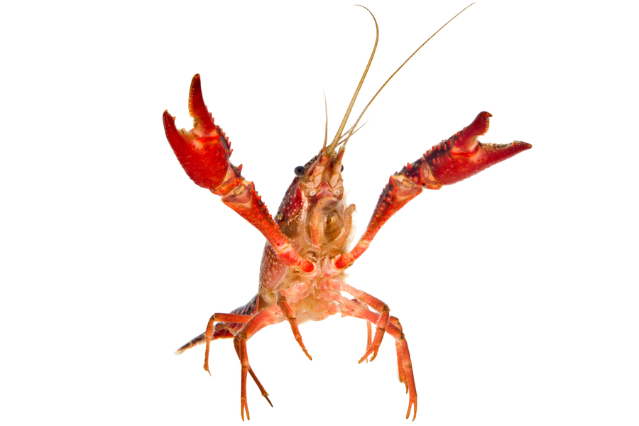
An MIT team has fabricated a hydrogel-based material that mimics the structure of the lobster’s underbelly, the toughest known hydrogel found in nature. Credits: Courtesy of the researchers

I love this lobster. In most photos, they’re food. This shows off the lobster as a living entity while showcasing its underbelly, which is what this story is all about. From an April 23, 2021 news item on phys.org (Note: A link has been removed),

A lobster’s underbelly is lined with a thin, translucent membrane that is both stretchy and surprisingly tough. This marine under-armor, as MIT [Massachusetts Institute of Technology] engineers reported in 2019, is made from the toughest known hydrogel in nature, which also happens to be highly flexible. This combination of strength and stretch helps shield a lobster as it scrabbles across the seafloor, while also allowing it to flex back and forth to swim.

Now a separate MIT team has fabricated a hydrogel-based material that mimics the structure of the lobster’s underbelly. The researchers ran the material through a battery of stretch and impact tests, and showed that, similar to the lobster underbelly, the synthetic material is remarkably “fatigue-resistant,” able to withstand repeated stretches and strains without tearing.

If the fabrication process could be significantly scaled up, materials made from nanofibrous hydrogels could be used to make stretchy and strong replacement tissues such as artificial tendons and ligaments.

The team’s results are published in the journal Matter. The paper’s MIT co-authors include postdocs Jiahua Ni and Shaoting Lin; graduate students Xinyue Liu and Yuchen Sun; professor of aeronautics and astronautics Raul Radovitzky; professor of chemistry Keith Nelson; mechanical engineering professor Xuanhe Zhao; and former research scientist David Veysset Ph.D. ’16, now at Stanford University; along with Zhao Qin, assistant professor at Syracuse University, and Alex Hsieh of the Army Research Laboratory.

An April 23, 2021 MIT news release (also on EurekAlert) by Jennifer Chu, which originated the news item, offers an overview of the groundwork for this latest research along with technical detail about the latest work,

Nature’s twist

In 2019, Lin and other members of Zhao’s group developed a new kind of fatigue-resistant material made from hydrogel — a gelatin-like class of materials made primarily of water and cross-linked polymers. They fabricated the material from ultrathin fibers of hydrogel, which aligned like many strands of gathered straw when the material was repeatedly stretched. This workout also happened to increase the hydrogel’s fatigue resistance.

“At that moment, we had a feeling nanofibers in hydrogels were important, and hoped to manipulate the fibril structures so that we could optimize fatigue resistance,” says Lin.

In their new study, the researchers combined a number of techniques to create stronger hydrogel nanofibers. The process starts with electrospinning, a fiber production technique that uses electric charges to draw ultrathin threads out of polymer solutions. The team used high-voltage charges to spin nanofibers from a polymer solution, to form a flat film of nanofibers, each measuring about 800 nanometers — a fraction of the diameter of a human hair.

They placed the film in a high-humidity chamber to weld the individual fibers into a sturdy, interconnected network, and then set the film in an incubator to crystallize the individual nanofibers at high temperatures, further strengthening the material.

They tested the film’s fatigue-resistance by placing it in a machine that stretched it repeatedly over tens of thousands of cycles. They also made notches in some films and observed how the cracks propagated as the films were stretched repeatedly. From these tests, they calculated that the nanofibrous films were 50 times more fatigue-resistant than the conventional nanofibrous hydrogels.

Around this time, they read with interest a study by Ming Guo, associate professor of mechanical engineering at MIT, who characterized the mechanical properties of a lobster’s underbelly. This protective membrane is made from thin sheets of chitin, a natural, fibrous material that is similar in makeup to the group’s hydrogel nanofibers.

Guo found that a cross-section of the lobster membrane revealed sheets of chitin stacked at 36-degree angles, similar to twisted plywood, or a spiral staircase. This rotating, layered configuration, known as a bouligand structure, enhanced the membrane’s properties of stretch and strength.

“We learned that this bouligand structure in the lobster underbelly has high mechanical performance, which motivated us to see if we could reproduce such structures in synthetic materials,” Lin says.

Angled architecture

Ni, Lin, and members of Zhao’s group teamed up with Nelson’s lab and Radovitzky’s group in MIT’s Institute for Soldier Nanotechnologies, and Qin’s lab at Syracuse University, to see if they could reproduce the lobster’s bouligand membrane structure using their synthetic, fatigue-resistant films.

“We prepared aligned nanofibers by electrospinning to mimic the chinic fibers existed in the lobster underbelly,” Ni says.

After electrospinning nanofibrous films, the researchers stacked each of five films in successive, 36-degree angles to form a single bouligand structure, which they then welded and crystallized to fortify the material. The final product measured 9 square centimeters and about 30 to 40 microns thick — about the size of a small piece of Scotch tape.

Stretch tests showed that the lobster-inspired material performed similarly to its natural counterpart, able to stretch repeatedly while resisting tears and cracks — a fatigue-resistance Lin attributes to the structure’s angled architecture.

“Intuitively, once a crack in the material propagates through one layer, it’s impeded by adjacent layers, where fibers are aligned at different angles,” Lin explains.

The team also subjected the material to microballistic impact tests with an experiment designed by Nelson’s group. They imaged the material as they shot it with microparticles at high velocity, and measured the particles’ speed before and after tearing through the material. The difference in velocity gave them a direct measurement of the material’s impact resistance, or the amount of energy it can absorb, which turned out to be a surprisingly tough 40 kilojoules per kilogram. This number is measured in the hydrated state.

“That means that a 5-millimeter steel ball launched at 200 meters per second would be arrested by 13 millimeters of the material,” Veysset says. “It is not as resistant as Kevlar, which would require 1 millimeter, but the material beats Kevlar in many other categories.”

It’s no surprise that the new material isn’t as tough as commercial antiballistic materials. It is, however, significantly sturdier than most other nanofibrous hydrogels such as gelatin and synthetic polymers like PVA. The material is also much stretchier than Kevlar. This combination of stretch and strength suggests that, if their fabrication can be sped up, and more films stacked in bouligand structures, nanofibrous hydrogels may serve as flexible and tough artificial tissues.

“For a hydrogel material to be a load-bearing artificial tissue, both strength and deformability are required,” Lin says. “Our material design could achieve these two properties.”

If you have the time and the interest, do check out the April 23, 2021 MIT news release, which features a couple of informative GIFs.

Here’s a link to and a citation for the paper,

Strong fatigue-resistant nanofibrous hydrogels inspired by lobster underbelly by Jiahua Ni, Shaoting Lin, Zhao Qin, David Veysset, Xinyue Liu, Yuchen Sun, Alex J. Hsieh, Raul Radovitzky, Keith A. Nelson, Xuanhe Zhao. Matter, 2021; DOI: 10.1016/j.matt.2021.03.023 Published April 23, 2021

This paper is behind a paywall.
New capacitor for better wearable electronics?

Supercapacitors are a predictable source of scientific interest and excitement. The latest entry in the ‘supercapacitor stakes’ is from a Russian/Finnish/US team according to a June 11, 2020 Skoltech (Skolkovo Institute of Science and Technology) press release (also on EurekAlert),

Researchers from Skoltech [Russia], Aalto University [Finland] and Massachusetts Institute of Technology [MIT; US] have designed a high-performance, low-cost, environmentally friendly, and stretchable supercapacitor that can potentially be used in wearable electronics. The paper was published in the Journal of Energy Storage.

Supercapacitors, with their high power density, fast charge-discharge rates, long cycle life, and cost-effectiveness, are a promising power source for everything from mobile and wearable electronics to electric vehicles. However, combining high energy density, safety, and eco-friendliness in one supercapacitor suitable for small devices has been rather challenging.

“Usually, organic solvents are used to increase the energy density. These are hazardous, not environmentally friendly, and they reduce the power density compared to aqueous electrolytes with higher conductivity,” says Professor Tanja Kallio from Aalto University, a co-author of the paper.

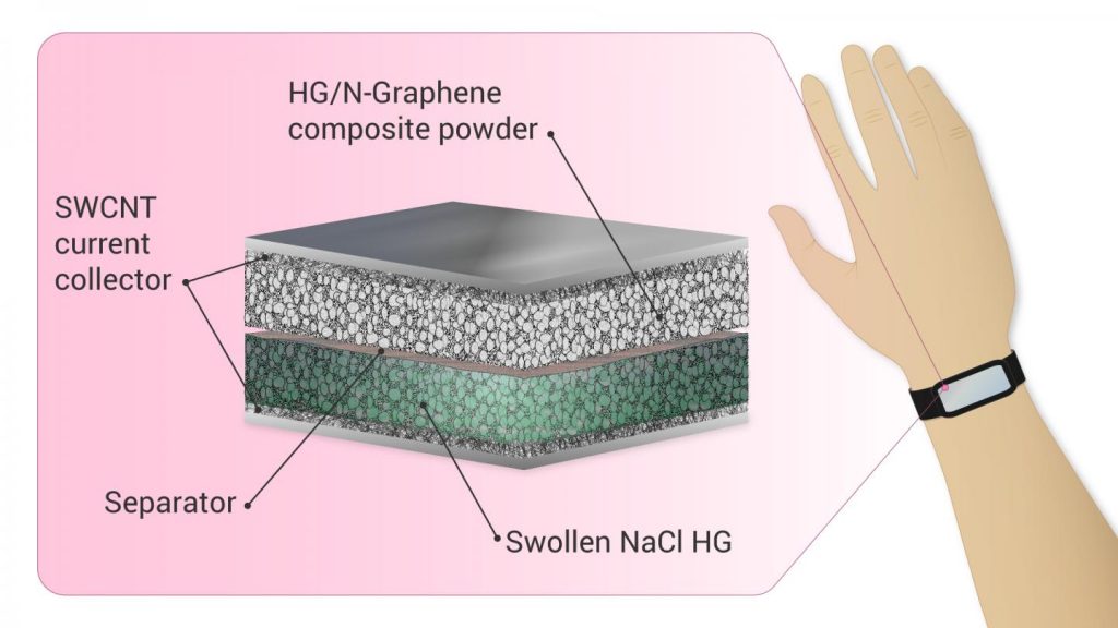
The researchers proposed a new design for a “green” and simple-to-fabricate supercapacitor. It consists of a solid-state material based on nitrogen-doped graphene flake electrodes distributed in the NaCl-containing hydrogel electrolyte. This structure is sandwiched between two single-walled carbon nanotube film current collectors, which provides stretchability. Hydrogel in the supercapacitor design enables compact packing and high energy density and allows them to use the environmentally friendly electrolyte.

The scientists managed to improve the volumetric capacitive performance, high energy density and power density for the prototype over analogous supercapacitors described in previous research. “We fabricated a prototype with unchanged performance under the 50% strain after a thousand stretching cycles. To ensure lower cost and better environmental performance, we used a NaCl-based electrolyte. Still the fabrication cost can be lowered down by implementation of 3D printing or other advanced fabrication techniques,” concluded Skoltech professor Albert Nasibulin.

Here’s a link to and a citation for the paper,

Superior environmentally friendly stretchable supercapacitor based on nitrogen-doped graphene/hydrogel and single-walled carbon nanotubes by Evgeniia Gilshtein, Cristina Flox, Farhan S.M. Ali, Bahareh Mehrabimatin, Fedor S.Fedorov, Shaoting Lin, Xuanhe Zhao, Albert G. Nasibulin, Tanja Kallio. Journal of Energy Storage Volume 30, August 2020, 101505 DOI: https://doi.org/10.1016/j.est.2020.101505

This paper is behind a paywall.

I’m trying to remember if I’ve ever before seen a material that combines graphene and single-walled carbon nanotubes (SWCNTs). Anyway, here’s an image the researchers are using illustrate their work,

Caption: This is an outline of the new supercapacitor. Credit: Pavel Odinev / Skoltech

A 3D printed ‘living’ tattoo

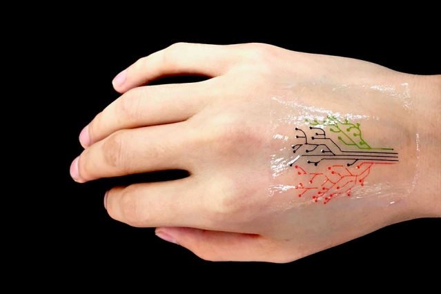
MIT engineers have devised a 3-D printing technique that uses a new kind of ink made from genetically programmed living cells. Courtesy of the researchers [and MIT]

If that image isn’t enough, there’s also a video abstract (I don’t think I’ve seen one of these before) for the paper,

For those who’d still like to read the text, here’s more from a December 5, 2017 MIT (Massachusetts Institute of Technology) news release (also on EurekAlert),

MIT engineers have devised a 3-D printing technique that uses a new kind of ink made from genetically programmed living cells.

The cells are engineered to light up in response to a variety of stimuli. When mixed with a slurry of hydrogel and nutrients, the cells can be printed, layer by layer, to form three-dimensional, interactive structures and devices.

The team has then demonstrated its technique by printing a “living tattoo” — a thin, transparent patch patterned with live bacteria cells in the shape of a tree. Each branch of the tree is lined with cells sensitive to a different chemical or molecular compound. When the patch is adhered to skin that has been exposed to the same compounds, corresponding regions of the tree light up in response.

The researchers, led by Xuanhe Zhao, the Noyce Career Development Professor in MIT’s Department of Mechanical Engineering, and Timothy Lu, associate professor of biological engineering and of electrical engineering and computer science, say that their technique can be used to fabricate “active” materials for wearable sensors and interactive displays. Such materials can be patterned with live cells engineered to sense environmental chemicals and pollutants as well as changes in pH and temperature.

What’s more, the team developed a model to predict the interactions between cells within a given 3-D-printed structure, under a variety of conditions. The team says researchers can use the model as a guide in designing responsive living materials.

Zhao, Lu, and their colleagues have published their results today [December 5, 2017] in the journal Advanced Materials. The paper’s co-authors are graduate students Xinyue Liu, Hyunwoo Yuk, Shaoting Lin, German Alberto Parada, Tzu-Chieh Tang, Eléonore Tham, and postdoc Cesar de la Fuente-Nunez.

A hardy alternative

In recent years, scientists have explored a variety of responsive materials as the basis for 3D-printed inks. For instance, scientists have used inks made from temperature-sensitive polymers to print heat-responsive shape-shifting objects. Others have printed photoactivated structures from polymers that shrink and stretch in response to light.

Zhao’s team, working with bioengineers in Lu’s lab, realized that live cells might also serve as responsive materials for 3D-printed inks, particularly as they can be genetically engineered to respond to a variety of stimuli. The researchers are not the first to consider 3-D printing genetically engineered cells; others have attempted to do so using live mammalian cells, but with little success.

“It turns out those cells were dying during the printing process, because mammalian cells are basically lipid bilayer balloons,” Yuk says. “They are too weak, and they easily rupture.”

Instead, the team identified a hardier cell type in bacteria. Bacterial cells have tough cell walls that are able to survive relatively harsh conditions, such as the forces applied to ink as it is pushed through a printer’s nozzle. Furthermore, bacteria, unlike mammalian cells, are compatible with most hydrogels — gel-like materials that are made from a mix of mostly water and a bit of polymer. The group found that hydrogels can provide an aqueous environment that can support living bacteria.

The researchers carried out a screening test to identify the type of hydrogel that would best host bacterial cells. After an extensive search, a hydrogel with pluronic acid was found to be the most compatible material. The hydrogel also exhibited an ideal consistency for 3-D printing.

“This hydrogel has ideal flow characteristics for printing through a nozzle,” Zhao says. “It’s like squeezing out toothpaste. You need [the ink] to flow out of a nozzle like toothpaste, and it can maintain its shape after it’s printed.”

From tattoos to living computers

Lu provided the team with bacterial cells engineered to light up in response to a variety of chemical stimuli. The researchers then came up with a recipe for their 3-D ink, using a combination of bacteria, hydrogel, and nutrients to sustain the cells and maintain their functionality.

“We found this new ink formula works very well and can print at a high resolution of about 30 micrometers per feature,” Zhao says. “That means each line we print contains only a few cells. We can also print relatively large-scale structures, measuring several centimeters.”

They printed the ink using a custom 3-D printer that they built using standard elements combined with fixtures they machined themselves. To demonstrate the technique, the team printed a pattern of hydrogel with cells in the shape of a tree on an elastomer layer. After printing, they solidified, or cured, the patch by exposing it to ultraviolet radiation. They then adhere the transparent elastomer layer with the living patterns on it, to skin.

To test the patch, the researchers smeared several chemical compounds onto the back of a test subject’s hand, then pressed the hydrogel patch over the exposed skin. Over several hours, branches of the patch’s tree lit up when bacteria sensed their corresponding chemical stimuli.

The researchers also engineered bacteria to communicate with each other; for instance they programmed some cells to light up only when they receive a certain signal from another cell. To test this type of communication in a 3-D structure, they printed a thin sheet of hydrogel filaments with “input,” or signal-producing bacteria and chemicals, overlaid with another layer of filaments of an “output,” or signal-receiving bacteria. They found the output filaments lit up only when they overlapped and received input signals from corresponding bacteria .

Yuk says in the future, researchers may use the team’s technique to print “living computers” — structures with multiple types of cells that communicate with each other, passing signals back and forth, much like transistors on a microchip.

“This is very future work, but we expect to be able to print living computational platforms that could be wearable,” Yuk says.

For more near-term applications, the researchers are aiming to fabricate customized sensors, in the form of flexible patches and stickers that could be engineered to detect a variety of chemical and molecular compounds. They also envision their technique may be used to manufacture drug capsules and surgical implants, containing cells engineered produce compounds such as glucose, to be released therapeutically over time.

“We can use bacterial cells like workers in a 3-D factory,” Liu says. “They can be engineered to produce drugs within a 3-D scaffold, and applications should not be confined to epidermal devices. As long as the fabrication method and approach are viable, applications such as implants and ingestibles should be possible.”

Here’s a link to and a citation for the paper,

3D Printing of Living Responsive Materials and Devices by Xinyue Liu, Hyunwoo Yuk, Shaoting Lin, German Alberto Parada, Tzu-Chieh Tang, Eléonore Tham, Cesar de la Fuente-Nunez, Timothy K. Lu, and Xuanhe Zhao. Advanced Materials DOI: 10.1002/adma.201704821 Version of Record online: 5 DEC 2017

© 2017 WILEY-VCH Verlag GmbH & Co. KGaA, Weinheim

This paper is behind a paywall.

Semi-living gloves as sensors

Researchers at the Massachusetts Institute of Technology (MIT) are calling it a new ‘living material’ according to a Feb. 16, 2017 news item on Nanowerk,

Engineers and biologists at MIT have teamed up to design a new “living material” — a tough, stretchy, biocompatible sheet of hydrogel injected with live cells that are genetically programmed to light up in the presence of certain chemicals.

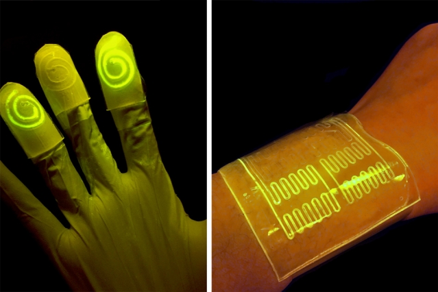
Researchers have found that the hydrogel’s mostly watery environment helps keep nutrients and programmed bacteria alive and active. When the bacteria reacts to a certain chemical, the bacteria are programmed to light up, as seen on the left. Courtesy of the researchers

A Feb. 15, 2017 MIT news release, which originated the news item, provides more information about this work,

In a paper published this week in the Proceedings of the National Academy of Sciences, the researchers demonstrate the new material’s potential for sensing chemicals, both in the environment and in the human body.

The team fabricated various wearable sensors from the cell-infused hydrogel, including a rubber glove with fingertips that glow after touching a chemically contaminated surface, and bandages that light up when pressed against chemicals on a person’s skin.

Xuanhe Zhao, the Robert N. Noyce Career Development associate professor of mechanical engineering at MIT, says the group’s living material design may be adapted to sense other chemicals and contaminants, for uses ranging from crime scene investigation and forensic science, to pollution monitoring and medical diagnostics.

“With this design, people can put different types of bacteria in these devices to indicate toxins in the environment, or disease on the skin,” says Timothy Lu, associate professor of biological engineering and of electrical engineering and computer science. “We’re demonstrating the potential for living materials and devices.”

The paper’s co-authors are graduate students Xinyue Liu, Tzu-Chieh Tang, Eleonore Tham, Hyunwoo Yuk, and Shaoting Lin.

Infusing life in materials

Lu and his colleagues in MIT’s Synthetic Biology Group specialize in creating biological circuits, genetically reprogramming the biological parts in living cells such as E. coli to work together in sequence, much like logic steps in an electrical circuit. In this way, scientists can reengineer living cells to carry out specific functions, including the ability to sense and signal the presence of viruses and toxins.

However, many of these newly programmed cells have only been demonstrated in situ, within Petri dishes, where scientists can carefully control the nutrient levels necessary to keep the cells alive and active — an environment that has proven extremely difficult to replicate in synthetic materials.

“The challenge to making living materials is how to maintain those living cells, to make them viable and functional in the device,” Lu says. “They require humidity, nutrients, and some require oxygen. The second challenge is how to prevent them from escaping from the material.”

To get around these roadblocks, others have used freeze-dried chemical extracts from genetically engineered cells, incorporating them into paper to create low-cost, virus-detecting diagnostic strips. But extracts, Lu says, are not the same as living cells, which can maintain their functionality over a longer period of time and may have higher sensitivity for detecting pathogens.

Other groups have seeded heart muscle cells onto thin rubber films to make soft, “living” actuators, or robots. When bent repeatedly, however, these films can crack, allowing the live cells to leak out.

A lively host

Zhao’s group in MIT’s Soft Active Materials Laboratory has developed a material that may be ideal for hosting living cells. For the past few years, his team has come up with various formulations of hydrogel — a tough, highly stretchable, biocompatible material made from a mix of polymer and water. Their latest designs have contained up to 95 percent water, providing an environment which Zhao and Lu recognized might be suitable for sustaining living cells. The material also resists cracking even when repeatedly stretched and pulled — a property that could help contain cells within the material.

The two groups teamed up to integrate Lu’s genetically programmed bacterial cells into Zhao’s sheets of hydrogel material. They first fabricated layers of hydrogel and patterned narrow channels within the layers using 3-D printing and micromolding techniques. They fused the hydrogel to a layer of elastomer, or rubber, that is porous enough to let in oxygen. They then injected E. coli cells into the hydrogel’s channels. The cells were programmed to fluoresce, or light up, when in contact with certain chemicals that pass through the hydrogel, in this case a natural compound known as DAPG (2,4-diacetylphloroglucinol).

The researchers then soaked the hydrogel/elastomer material in a bath of nutrients which infused throughout the hydrogel and helped to keep the bacterial cells alive and active for several days.

To demonstrate the material’s potential uses, the researchers first fabricated a sheet of the material with four separate, narrow channels, each containing a type of bacteria engineered to glow green in response to a different chemical compound. They found each channel reliably lit up when exposed to its respective chemical.

Next, the team fashioned the material into a bandage, or “living patch,” patterned with channels containing bacteria sensitive to rhamnose, a naturally occurring sugar. The researchers swabbed a volunteer’s wrist with a cotton ball soaked in rhamnose, then applied the hydrogel patch, which instantly lit up in response to the chemical.

Finally, the researchers fabricated a hydrogel/elastomer glove whose fingertips contained swirl-like channels, each of which they filled with different chemical-sensing bacterial cells. Each fingertip glowed in response to picking up a cotton ball soaked with a respective compound.

The group has also developed a theoretical model to help guide others in designing similar living materials and devices.

“The model helps us to design living devices more efficiently,” Zhao says. “It tells you things like the thickness of the hydrogel layer you should use, the distance between channels, how to pattern the channels, and how much bacteria to use.”

Ultimately, Zhao envisions products made from living materials, such as gloves and rubber soles lined with chemical-sensing hydrogel, or bandages, patches, and even clothing that may detect signs of infection or disease.

Here’s a link to and a citation for the paper,

Stretchable living materials and devices with hydrogel–elastomer hybrids hosting programmed cells by Xinyue Liu, Tzu-Chieh Tang, Eléonore Tham, Hyunwoo Yuk, Shaoting Lin, Timothy K. Lu, and Xuanhe Zhao. PNAS February 15, 2017 doi: 10.1073/pnas.1618307114 Published online before print February 15, 2017

This paper appears to be open access.

Stretchy optical materials for implants that could pulse light

An Oct. 17, 2016 Massachusetts Institute of Technology (MIT) news release (also on EurekAlert) by Emily Chu describes research that could lead to long-lasting implants offering preventive health strategies,

Researchers from MIT and Harvard Medical School have developed a biocompatible and highly stretchable optical fiber made from hydrogel — an elastic, rubbery material composed mostly of water. The fiber, which is as bendable as a rope of licorice, may one day be implanted in the body to deliver therapeutic pulses of light or light up at the first sign of disease. [emphasis mine]

The researchers say the fiber may serve as a long-lasting implant that would bend and twist with the body without breaking down. The team has published its results online in the journal Advanced Materials.

Using light to activate cells, and particularly neurons in the brain, is a highly active field known as optogenetics, in which researchers deliver short pulses of light to targeted tissues using needle-like fibers, through which they shine light from an LED source.

“But the brain is like a bowl of Jell-O, whereas these fibers are like glass — very rigid, which can possibly damage brain tissues,” says Xuanhe Zhao, the Robert N. Noyce Career Development Associate Professor in MIT’s Department of Mechanical Engineering. “If these fibers could match the flexibility and softness of the brain, they could provide long-term more effective stimulation and therapy.”

Getting to the core of it

Zhao’s group at MIT, including graduate students Xinyue Liu and Hyunwoo Yuk, specializes in tuning the mechanical properties of hydrogels. The researchers have devised multiple recipes for making tough yet pliable hydrogels out of various biopolymers. The team has also come up with ways to bond hydrogels with various surfaces such as metallic sensors and LEDs, to create stretchable electronics.

The researchers only thought to explore hydrogel’s use in optical fibers after conversations with the bio-optics group at Harvard Medical School, led by Associate Professor Seok-Hyun (Andy) Yun. Yun’s group had previously fabricated an optical fiber from hydrogel material that successfully transmitted light through the fiber. However, the material broke apart when bent or slightly stretched. Zhao’s hydrogels, in contrast, could stretch and bend like taffy. The two groups joined efforts and looked for ways to incorporate Zhao’s hydrogel into Yun’s optical fiber design.

Yun’s design consists of a core material encased in an outer cladding. To transmit the maximum amount of light through the core of the fiber, the core and the cladding should be made of materials with very different refractive indices, or degrees to which they can bend light.

“If these two things are too similar, whatever light source flows through the fiber will just fade away,” Yuk explains. “In optical fibers, people want to have a much higher refractive index in the core, versus cladding, so that when light goes through the core, it bounces off the interface of the cladding and stays within the core.”

Happily, they found that Zhao’s hydrogel material was highly transparent and possessed a refractive index that was ideal as a core material. But when they tried to coat the hydrogel with a cladding polymer solution, the two materials tended to peel apart when the fiber was stretched or bent.

To bond the two materials together, the researchers added conjugation chemicals to the cladding solution, which, when coated over the hydrogel core, generated chemical links between the outer surfaces of both materials.

“It clicks together the carboxyl groups in the cladding, and the amine groups in the core material, like molecular-level glue,” Yuk says.

Sensing strain

The researchers tested the optical fibers’ ability to propagate light by shining a laser through fibers of various lengths. Each fiber transmitted light without significant attenuation, or fading. They also found that fibers could be stretched over seven times their original length without breaking.

Now that they had developed a highly flexible and robust optical fiber, made from a hydrogel material that was also biocompatible, the researchers began to play with the fiber’s optical properties, to see if they could design a fiber that could sense when and where it was being stretched.

They first loaded a fiber with red, green, and blue organic dyes, placed at specific spots along the fiber’s length. Next, they shone a laser through the fiber and stretched, for instance, the red region. They measured the spectrum of light that made it all the way through the fiber, and noted the intensity of the red light. They reasoned that this intensity relates directly to the amount of light absorbed by the red dye, as a result of that region being stretched.

In other words, by measuring the amount of light at the far end of the fiber, the researchers can quantitatively determine where and by how much a fiber was stretched.

“When you stretch a certain portion of the fiber, the dimensions of that part of the fiber changes, along with the amount of light that region absorbs and scatters, so in this way, the fiber can serve as a sensor of strain,” Liu explains.

“This is like a multistrain sensor through a single fiber,” Yuk adds. “So it can be an implantable or wearable strain gauge.”

The researchers imagine that such stretchable, strain-sensing optical fibers could be implanted or fitted along the length of a patient’s arm or leg, to monitor for signs of improving mobility.

Zhao envisions the fibers may also serve as sensors, lighting up in response to signs of disease.

“We may be able to use optical fibers for long-term diagnostics, to optically monitor tumors or inflammation,” he says. “The applications can be impactful.”

Here’s a link to and a citation for the paper,

Highly Stretchable, Strain Sensing Hydrogel Optical Fibers by Jingjing Guo, Xinyue Liu, Nan Jiang, Ali K. Yetisen, Hyunwoo Yuk, Changxi Yang, Ali Khademhosseini, Xuanhe Zhao, and Seok-Hyun Yun. Advanced Materials DOI: 10.1002/adma.201603160 Version of Record online: 7 OCT 2016

© 2016 WILEY-VCH Verlag GmbH & Co. KGaA, Weinheim

This paper is behind a paywall.

Next supercapacitor: crumpled graphene?

An Oct. 3, 2014 news item on ScienceDaily features the use of graphene as a possible supercapacitor,

When someone crumples a sheet of paper, that usually means it’s about to be thrown away. But researchers have now found that crumpling a piece of graphene “paper” — a material formed by bonding together layers of the two-dimensional form of carbon — can actually yield new properties that could be useful for creating extremely stretchable supercapacitors to store energy for flexible electronic devices.

The finding is reported in the journal Scientific Reports by MIT’s {Massachusetts Institute of Technology] Xuanhe Zhao, an assistant professor of mechanical engineering and civil and environmental engineering, and four other authors. The new, flexible superconductors should be easy and inexpensive to fabricate, the team says.

An Oct. 3, 2014 MIT news release by David Chandler (also on EurekAlert), which originated the news item, explains the technology at more length,

“Many people are exploring graphene paper: It’s a good candidate for making supercapacitors, because of its large surface area per mass,” Zhao says. Now, he says, the development of flexible electronic devices, such as wearable or implantable biomedical sensors or monitoring devices, will require flexible power-storage systems.

Like batteries, supercapacitors can store electrical energy, but they primarily do so electrostatically, rather than chemically — meaning they can deliver their energy faster than batteries can. Now Zhao and his team have demonstrated that by crumpling a sheet of graphene paper into a chaotic mass of folds, they can make a supercapacitor that can easily be bent, folded, or stretched to as much as 800 percent of its original size. The team has made a simple supercapacitor using this method as a proof of principle.

The material can be crumpled and flattened up to 1,000 times, the team has demonstrated, without a significant loss of performance. “The graphene paper is pretty robust,” Zhao says, “and we can achieve very large deformations over multiple cycles.” Graphene, a structure of pure carbon just one atom thick with its carbon atoms arranged in a hexagonal array, is one of the strongest materials known.

To make the crumpled graphene paper, a sheet of the material was placed in a mechanical device that first compressed it in one direction, creating a series of parallel folds or pleats, and then in the other direction, leading to a chaotic, rumpled surface. When stretched, the material’s folds simply smooth themselves out.

Forming a capacitor requires two conductive layers — in this case, two sheets of crumpled graphene paper — with an insulating layer in between, which in this demonstration was made from a hydrogel material. Like the crumpled graphene, the hydrogel is highly deformable and stretchable, so the three layers remain in contact even while being flexed and pulled.

Though this initial demonstration was specifically to make a supercapacitor, the same crumpling technique could be applied to other uses, Zhao says. For example, the crumpled graphene material might be used as one electrode in a flexible battery, or could be used to make a stretchable sensor for specific chemical or biological molecules.

Here is a link to and a citation for the paper,

Stretchable and High-Performance Supercapacitors with Crumpled Graphene Papers by Jianfeng Zang, Changyong Cao, Yaying Feng, Jie Liu, & Xuanhe Zhao. Scientific Reports 4, Article number: 6492 doi:10.1038/srep06492 Published 01 October 2014

This is an open access article.

ETA Oct. 8, 2014: Dexter Johnson of the Nanoclast blog on the IEEE (Institute of Electrical and Electronics Engineers) website has an Oct. 7, 2014 post where he comments about the ‘flexibility’ aspect of this work.

No more boat scraping with new coating from Duke University

There’s a lot of interest in finding ways to discourage bacteria from growing on various surfaces, for example, Sharklet, which is based on nanostructures on sharkskin, is a product being developed for hospitals (my Feb. 10, 2011 posting) and there are polymers that ‘uninvite’ bacteria at the University of Nottingham (my Aug. 13, 2012 posting).

A Jan. 31, 2013 news item on Nanowerk highlights the latest work being done at Duke University,

Duke University engineers have developed a material that can be applied like paint to the hull of a ship and will literally be able to dislodge bacteria, keeping it from accumulating on the ship’s surface. This buildup on ships increases drag and reduces the energy efficiency of the vessel, as well as blocking or clogging undersea sensors.

The team’s research was published online,

Bioinspired Surfaces with Dynamic Topography for Active Control of Biofouling by Phanindhar Shivapooja, Qiming Wang, Beatriz Orihuela, Daniel Rittschof, Gabriel P. López1, Xuanhe Zhao. Advanced Materials, Article first published online: 6 JAN 2013, DOI: 10.1002/adma.201203374

Copyright © 2013 WILEY-VCH Verlag GmbH & Co. KGaA, Weinheim

The article is behind a paywall but the abstract is freely available,

Dynamic change of surface area and topology of elastomers is used as a general, environmentally friendly approach for effectively detaching micro- and macro-fouling organisms adhered on the elastomer surfaces. Deformation of elastomer surfaces under electrical or pneumatic actuation can debond various biofilms and barnacles. The bio-inspired dynamic surfaces can be fabricated over large areas through simple and practical processes. This new mechanism is complementary with existing materials and methods for biofouling control.

Duke University’s Jan. 31, 2013 news release by Richard Merritt, which originated the news item, provides more detail from the researchers,

“We have developed a material that ‘wrinkles,’ or changes it surface in response to a stimulus, such as stretching or pressure or electricity,” said Duke engineer Xuanhe Zhao, assistant professor in Duke’s Pratt School of Engineering. “This deformation can effectively detach biofilms and other organisms that have accumulated on the surface.”

Zhao has already demonstrated the ability of electric current to deform, or change, the surface of polymers.

The researchers tested their approach in the laboratory with simulated seawater, as well as on barnacles. These experiments were conducted in collaboration with Daniel Rittsch of the Duke University Marine Lab in Beaufort, N.C.

Keeping bacteria from attaching to ship hulls or other submerged objects can prevent a larger cascade of events that can reduce performance or efficiency. Once they have taken up residence on a surface, bacteria often attract larger organisms, such as seaweed and larva of other marine organisms, such as worms, bivalves, barnacles or mussels.

There are other ways to introduce efficiencies in marine transp0rtation as per my June 27, 2012 posting about Zyvex Marine and its new composites which will make for lighter vessels.

Coatings that shake off bacteria and biological photocopying

The American Vacuum Society (AVS) is holding its 58th International Symposium and Exhibition from Oct. 30 – Nov. 4, 2011 in Nashville, Tennessee. Presentations are not focused on vacuuming (hoovering) floors but rather on something called vacuum science and they span from a presentation on bacteria and coatings to another on photocopying DNA to more.

From the Oct. 31, 2011 news item on Nanowerk,

“Sea water is a very aggressive biological system,” says Gabriel Lopez, whose lab at Duke University studies the interface of marine bacterial films with submerged surfaces. While the teeming abundance of ocean life makes coral reefs and tide pools attractive tourist destinations, for ships whose hulls become covered with slime, all this life can, quite literally, be a big drag. On just one class of U.S. Navy destroyer, biological build-up is estimated to cost more than $50 million a year, mostly in extra fuel, according to a 2010 study performed by researchers from the U.S. Naval Academy and Naval Surface Warfare Center in Maryland. Marine biofouling can also disrupt the operation of ocean sensors, heat-exchangers that suck in water to cool mechanical systems, and other underwater equipment.

I think rather than describing sea water as ‘aggressive’  which suggests intent, I’d use ‘active’ as Lopez does later in another context (excerpted from the news item),

Lopez and his group focus on a class of materials called stimuli-responsive surfaces. As the name implies, the materials will alter their physical or chemical properties in response to a stimulus, such as a temperature change. The coatings being tested in Lopez’s lab wrinkle on the micro- or nano-scale, shaking off slimy colonies of marine bacteria in a manner similar to how a horse might twitch its skin to shoo away flies. The researchers also consider how a stimulus might alter the chemical properties of a surface in a way that could decrease a marine organism’s ability to stick.

At the AVS Symposium, held Oct. 30 – Nov. 4 in Nashville, Tenn., Lopez will present results from experiments on two different types of stimuli-responsive surfaces: one that changes its texture in response to temperature and the other in response to an applied voltage. The voltage-responsive surfaces are being developed in collaboration with the laboratory of Xuanhe Zhao, also a Duke researcher, who found that insulating cables can fail if they deform under voltages. “Surprisingly, the same failure mechanism can be made useful in deforming surfaces of coatings and detaching biofouling,” Zhao said.

“The idea of an active surface is inspired by nature,” adds Lopez, who remembers being intrigued by the question of how a sea anemone’s waving tentacles are able to clean themselves. [emphasis mine] Other biological surfaces, such as shark skin, have already been copied by engineers seeking to learn from nature’s own successful anti-fouling systems.

(I did profile some biomimicry work being done with shark skin in my comments on part 4 of the Making Stuff programmes broadcast as part of the Nova series on PBS (US Public Broadcasting Stations) in my Feb. 10, 2011 posting.)

This next presentation is in the area of synthetic biology. From the Oct. 31, 2011 news item (DNA origami from inkjet synthesis produced strands) on Nanowerk,

In the emerging field of synthetic biology, engineers use biological building blocks, such as snippets of DNA, to construct novel technologies. One of the key challenges in the field is finding a way to quickly and economically synthesize the desired DNA strands. Now scientists from Duke University have fabricated a reusable DNA chip that may help address this problem by acting as a template from which multiple batches of DNA building blocks can be photocopied. The researchers have used the device to create strands of DNA which they then folded into unique nanoscale structures.

“We found that we had an “immortal” DNA chip in our hands,” says Ishtiaq Saaem, a biomedical engineering researcher at Duke and member of the team. [emphasis mine] “Essentially, we were able to do the biological copying process to release material off the chip tens of times. [emphasis mine] The process seems to work even using a chip that we made, used, stored in -20C for a while, and brought out and used again.”

After releasing the DNA from the chip, the team “cooked” it together with a piece of long viral DNA. “In the cooking process, the viral DNA is stapled into a desired shape by the smaller chip-derived DNA,” explains Saaem. One of the team’s first examples of DNA origami was a rectangle shape with a triangle attached on one side, which the researchers dubbed a “nano-house.” The structure could be used to spatially orient organic and inorganic materials, serve as a scaffold for drug delivery, or act as a nanoscale ruler, Saaem says.

I’m not very comfortable with the notion of an “immortal DNA chip” but then I have many reservations about synthetic biology. Still, I think it’s important to pay attention and consider the possibility that my fears about synthetic biology might make as much sense as the fears many had about electricity in the 19th century.