Tag Archives: human-on-a-chip

Human-on-a-chip predicts in vivo results based on in vitro model … for the first time

If successful the hope is that ‘human-on-a-chip’ will replace most, if not all, animal testing. This July 3, 2019 Hesperos news release (also on EurekAlert) suggests scientists are making serious gains in the drive to replace animal testing (Note: For anyone having difficulty with the terms, pharmacokinetics and pharmacodynamics, there are definitions towards the end of this posting, which may prove helpful),

Hesperos Inc., pioneers* of the “human-on-a-chip” in vitro system has announced the use of its innovative multi-organ model to successfully measure the concentration and metabolism of two known cardiotoxic small molecules over time, to accurately describe the drug behavior and toxic effects in vivo. The findings further support the potential of body-on-a-chip systems to transform the drug discovery process.

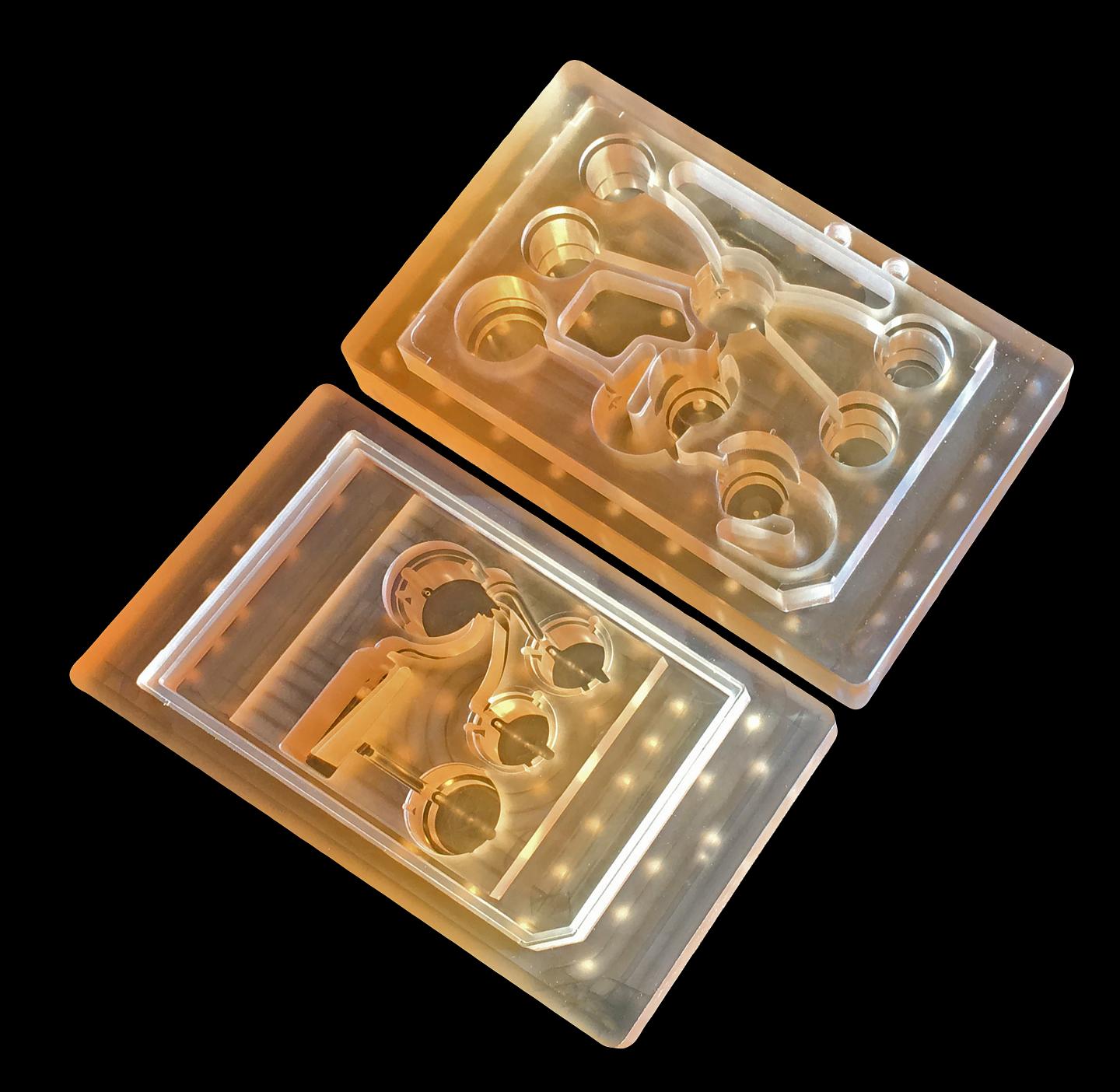
In a study published in Nature Scientific Reports, in collaboration with AstraZeneca, Hesperos described how they used a pumpless heart model and a heart:liver system to evaluate the temporal pharmacokinetic/pharmacodynamic (PKPD) relationship for terfenadine, an antihistamine that was banned due to toxic cardiac effects, as well as determine its mechanism of toxicity.

The study found there was a time-dependent, drug-induced response in the heart model. Further experiments were conducted, adding a metabolically competent liver module to the Hesperos Human-on-a-Chip® system to observe what happened when terfenadine was converted to fexofenadine. By doing so, the researchers were able to determine the driver of the pharmacodynamic (PD) effect and develop a mathematical model to predict the effect of terfenadine in preclinical species. This is the first time an in vitro human-on-a-chip system has been shown to predict in vivo outcomes, which could be used to predict clinical trial outcomes in the future.

“The ability to examine PKPD relationships in vitro would enable us to understand compound behavior prior to in vivo testing, offering significant cost and time savings,” said Dr. Shuler, President and CEO, Hesperos, Inc and Professor Emeritus, Cornell University. “We are excited about the potential of this technology to help us ensure that potential new drug candidates have a higher probability of success during the clinical trial process.”

Understanding the inter-relationship between pharmacokinetics (PK), the drug’s time course for absorption, distribution, metabolism and excretion, and PD, the biological effect of a drug, is crucial in drug discovery and development. Scientists have learned that the maximum drug effect is not always driven by the peak drug concentration. In some cases, time is a critical factor influencing drug effect, but often this concentration-effect-time relationship only comes to light during the advanced stages of the preclinical program. In addition, often the data cannot be reliably extrapolated to humans.

“It is costly and time consuming to discover that potential drug candidates may have poor therapeutic qualities preventing their onward progression,” said James Hickman, Chief Scientist at Hesperos and Professor at the University of Central Florida. “Being able to define this during early drug discovery will be a valuable contribution to the optimization of potential new drug candidates.”

As demonstrated with the terfenadine experiment, the PKPD modelling approach was critical for understanding both the flux of compound between compartments as well as the resulting PD response in the context of dynamic exposure profiles of both parent and metabolite, as indicated by Dr. Shuler.

In order to test the viability of their system in a real-world drug discovery setting, the Hesperos team collaborated with scientists at AstraZeneca, to test one of their failed small molecules, known to have a CV [cardiovscular?] risk.

One of the main measurements used to assess the electrical properties of the heart is the QT interval, which approximates the time taken from when the cardiac ventricles start to contract to when they finish relaxing. Prolongation of the QT interval on the electrocardiogram can lead to a fatal arrhythmia known as Torsade de Pointes. Consequently, it is a mandatory requirement prior to first-in-human administration of potential new drug candidates that their ability to inhibit the hERG channel (a biomarker for QT prolongation) is investigated.

In the case of the AstraZeneca molecule, the molecule was assessed for hERG inhibition early on, and it was concluded to have a low potential to cause in vivo QT prolongation up to 100 μM. In later pre-clinical testing, the QT interval increased by 22% at a concentration of just 3 μM. Subsequent investigations found that a major metabolite was responsible. Hesperos was able to detect a clear PD effect at concentrations above 3 μM and worked to determine the mechanism of toxicity of the molecule.

The ability of these systems to assess cardiac function non-invasively in the presence of both parent molecule and metabolite over time, using multiplexed and repeat drug dosing regimes, provides an opportunity to run long-term studies for chronic administration of drugs to study their potential toxic effects.

Hesperos, Inc. is the first company spun out from the Tissue Chip Program at NCATS (National Center for Advancing Translational Sciences), which was established in 2011 to address the long timelines, steep costs and high failure rates associated with the drug development process. Hesperos currently is funded through NCATS’ Small Business Innovation Research program to undertake these studies and make tissue chips technology available as a service based company.

“The application of tissue chip technology in drug testing can lead to advances in predicting the potential effects of candidate medicines in people,” said Danilo Tagle, Ph.D., associate director for special initiatives at NCATS.

###

About Hesperos
Hesperos, Inc. is a leader in efforts to characterize an individual’s biology with human-on-a-chip microfluidic systems. Founders Michael L. Shuler and James J. Hickman have been at the forefront of every major scientific discovery in this realm, from individual organ-on-a-chip constructs to fully functional, interconnected multi-organ systems. With a mission to revolutionize toxicology testing as well as efficacy evaluation for drug discovery, the company has created pumpless platforms with serum-free cellular mediums that allow multi-organ system communication and integrated computational PKPD modeling of live physiological responses utilizing functional readouts from neurons, cardiac, muscle, barrier tissues and neuromuscular junctions as well as responses from liver, pancreas and barrier tissues. Created from human stem cells, the fully human systems are the first in vitro solutions that accurately utilize in vitro systems to predict in vivo functions without the use of animal models, as featured in Science. More information is available at http://www.
hesperosinc.com

Years ago I went to a congress focused on alternatives to animal testing (August 22, 2014 posting) and saw a video of heart cells in a petri dish (in vitro) beating in a heartlike rhythm. It was something like this,

ipscira
Published on Oct 17, 2010 https://www.youtube.com/watch?v=BqzW9Jq-OVA

I found it amazing as did the scientist who drew my attention to it. After, it’s just a collection of heart cells. How do they start beating and keep time with each other?

Getting back to the latest research, here’s a link and a citation for the paper,

On the potential of in vitro organ-chip models to define temporal pharmacokinetic-pharmacodynamic relationships by Christopher W. McAleer, Amy Pointon, Christopher J. Long, Rocky L. Brighton, Benjamin D. Wilkin, L. Richard Bridges, Narasimham Narasimhan Sriram, Kristin Fabre, Robin McDougall, Victorine P. Muse, Jerome T. Mettetal, Abhishek Srivastava, Dominic Williams, Mark T. Schnepper, Jeff L. Roles, Michael L. Shuler, James J. Hickman & Lorna Ewart. Scientific Reports volume 9, Article number: 9619 (2019) DOI: https://doi.org/10.1038/s41598-019-45656-4 Published: 03 July 2019

This paper is open access.

I happened to look at the paper and found good definitions of pharmacokinetics and pharmacodynamics. I know it’s not for everyone but if you’ve ever been curious about the difference (from the Introduction of On the potential of in vitro organ-chip models to define temporal pharmacokinetic-pharmacodynamic relationships),

Integrative pharmacology is a discipline that builds an understanding of the inter-relationship between pharmacokinetics (PK), the drug’s time course for absorption, distribution, metabolism and excretion and pharmacodynamics (PD), the biological effect of a drug. In drug discovery, this multi-variate approach guides medicinal chemists to modify structural properties of a drug molecule to improve its chance of becoming a medicine in a process known as “lead optimization”.

*More than one person and more than one company and more than one country claims pioneer status where ‘human-on-a-chip’ is concerned.

Body-on-a-chip (10 organs)

Also known as human-on-a-chip, the 10-organ body-on-a-chip was being discussed at the 9th World Congress on Alternatives to Animal Testing in the Life Sciences in 2014 in Prague, Czech Republic (see this July 1, 2015 posting for more). At the time, scientists were predicting success at achieving their goal of 10 organs on-a-chip in 2017 (the best at the time was four organs). Only a few months past that deadline, scientists from the Massachusetts Institute of Technology (MIT) seem to have announced a ’10 organ chip’ in a March 14, 2018 news item on ScienceDaily,

MIT engineers have developed new technology that could be used to evaluate new drugs and detect possible side effects before the drugs are tested in humans. Using a microfluidic platform that connects engineered tissues from up to 10 organs, the researchers can accurately replicate human organ interactions for weeks at a time, allowing them to measure the effects of drugs on different parts of the body.

Such a system could reveal, for example, whether a drug that is intended to treat one organ will have adverse effects on another.

A March 14, 2018 MIT news release (also on EurekAlert), which originated the news item, expands on the theme,

“Some of these effects are really hard to predict from animal models because the situations that lead to them are idiosyncratic,” says Linda Griffith, the School of Engineering Professor of Teaching Innovation, a professor of biological engineering and mechanical engineering, and one of the senior authors of the study. “With our chip, you can distribute a drug and then look for the effects on other tissues, and measure the exposure and how it is metabolized.”

These chips could also be used to evaluate antibody drugs and other immunotherapies, which are difficult to test thoroughly in animals because they are designed to interact with the human immune system.

David Trumper, an MIT professor of mechanical engineering, and Murat Cirit, a research scientist in the Department of Biological Engineering, are also senior authors of the paper, which appears in the journal Scientific Reports. The paper’s lead authors are former MIT postdocs Collin Edington and Wen Li Kelly Chen.

Modeling organs

When developing a new drug, researchers identify drug targets based on what they know about the biology of the disease, and then create compounds that affect those targets. Preclinical testing in animals can offer information about a drug’s safety and effectiveness before human testing begins, but those tests may not reveal potential side effects, Griffith says. Furthermore, drugs that work in animals often fail in human trials.

“Animals do not represent people in all the facets that you need to develop drugs and understand disease,” Griffith says. “That is becoming more and more apparent as we look across all kinds of drugs.”

Complications can also arise due to variability among individual patients, including their genetic background, environmental influences, lifestyles, and other drugs they may be taking. “A lot of the time you don’t see problems with a drug, particularly something that might be widely prescribed, until it goes on the market,” Griffith says.

As part of a project spearheaded by the Defense Advanced Research Projects Agency (DARPA), Griffith and her colleagues decided to pursue a technology that they call a “physiome on a chip,” which they believe could offer a way to model potential drug effects more accurately and rapidly. To achieve this, the researchers needed new equipment — a platform that would allow tissues to grow and interact with each other — as well as engineered tissue that would accurately mimic the functions of human organs.

Before this project was launched, no one had succeeded in connecting more than a few different tissue types on a platform. Furthermore, most researchers working on this kind of chip were working with closed microfluidic systems, which allow fluid to flow in and out but do not offer an easy way to manipulate what is happening inside the chip. These systems also require external pumps.

The MIT team decided to create an open system, which essentially removes the lid and makes it easier to manipulate the system and remove samples for analysis. Their system, adapted from technology they previously developed and commercialized through U.K.-based CN BioInnovations, also incorporates several on-board pumps that can control the flow of liquid between the “organs,” replicating the circulation of blood, immune cells, and proteins through the human body. The pumps also allow larger engineered tissues, for example tumors within an organ, to be evaluated.

Complex interactions

The researchers created several versions of their chip, linking up to 10 organ types: liver, lung, gut, endometrium, brain, heart, pancreas, kidney, skin, and skeletal muscle. Each “organ” consists of clusters of 1 million to 2 million cells. These tissues don’t replicate the entire organ, but they do perform many of its important functions. Significantly, most of the tissues come directly from patient samples rather than from cell lines that have been developed for lab use. These so-called “primary cells” are more difficult to work with but offer a more representative model of organ function, Griffith says.

Using this system, the researchers showed that they could deliver a drug to the gastrointestinal tissue, mimicking oral ingestion of a drug, and then observe as the drug was transported to other tissues and metabolized. They could measure where the drugs went, the effects of the drugs on different tissues, and how the drugs were broken down. In a related publication, the researchers modeled how drugs can cause unexpected stress on the liver by making the gastrointestinal tract “leaky,” allowing bacteria to enter the bloodstream and produce inflammation in the liver.

Kevin Healy, a professor of bioengineering and materials science and engineering at the University of California at Berkeley, says that this kind of system holds great potential for accurate prediction of complex adverse drug reactions.

“While microphysiological systems (MPS) featuring single organs can be of great use for both pharmaceutical testing and basic organ-level studies, the huge potential of MPS technology is revealed by connecting multiple organ chips in an integrated system for in vitro pharmacology. This study beautifully illustrates that multi-MPS “physiome-on-a-chip” approaches, which combine the genetic background of human cells with physiologically relevant tissue-to-media volumes, allow accurate prediction of drug pharmacokinetics and drug absorption, distribution, metabolism, and excretion,” says Healy, who was not involved in the research.

Griffith believes that the most immediate applications for this technology involve modeling two to four organs. Her lab is now developing a model system for Parkinson’s disease that includes brain, liver, and gastrointestinal tissue, which she plans to use to investigate the hypothesis that bacteria found in the gut can influence the development of Parkinson’s disease.

Other applications include modeling tumors that metastasize to other parts of the body, she says.

“An advantage of our platform is that we can scale it up or down and accommodate a lot of different configurations,” Griffith says. “I think the field is going to go through a transition where we start to get more information out of a three-organ or four-organ system, and it will start to become cost-competitive because the information you’re getting is so much more valuable.”

The research was funded by the U.S. Army Research Office and DARPA.

Caption: MIT engineers have developed new technology that could be used to evaluate new drugs and detect possible side effects before the drugs are tested in humans. Using a microfluidic platform that connects engineered tissues from up to 10 organs, the researchers can accurately replicate human organ interactions for weeks at a time, allowing them to measure the effects of drugs on different parts of the body. Credit: Felice Frankel

Here’s a link to and a citation for the paper,

Interconnected Microphysiological Systems for Quantitative Biology and Pharmacology Studies by Collin D. Edington, Wen Li Kelly Chen, Emily Geishecker, Timothy Kassis, Luis R. Soenksen, Brij M. Bhushan, Duncan Freake, Jared Kirschner, Christian Maass, Nikolaos Tsamandouras, Jorge Valdez, Christi D. Cook, Tom Parent, Stephen Snyder, Jiajie Yu, Emily Suter, Michael Shockley, Jason Velazquez, Jeremy J. Velazquez, Linda Stockdale, Julia P. Papps, Iris Lee, Nicholas Vann, Mario Gamboa, Matthew E. LaBarge, Zhe Zhong, Xin Wang, Laurie A. Boyer, Douglas A. Lauffenburger, Rebecca L. Carrier, Catherine Communal, Steven R. Tannenbaum, Cynthia L. Stokes, David J. Hughes, Gaurav Rohatgi, David L. Trumper, Murat Cirit, Linda G. Griffith. Scientific Reports, 2018; 8 (1) DOI: 10.1038/s41598-018-22749-0 Published online:

This paper which describes testing for four-, seven-, and ten-organs-on-a-chip, is open access. From the paper’s Discussion,

In summary, we have demonstrated a generalizable approach to linking MPSs [microphysiological systems] within a fluidic platform to create a physiome-on-a-chip approach capable of generating complex molecular distribution profiles for advanced drug discovery applications. This adaptable, reusable system has unique and complementary advantages to existing microfluidic and PDMS-based approaches, especially for applications involving high logD substances (drugs and hormones), those requiring precise and flexible control over inter-MPS flow partitioning and drug distribution, and those requiring long-term (weeks) culture with reliable fluidic and sampling operation. We anticipate this platform can be applied to a wide range of problems in disease modeling and pre-clinical drug development, especially for tractable lower-order (2–4) interactions.

Congratulations to the researchers!

3D brain-on-a-chip from the University of Twente

Dutch researchers have developed a 3D brain-on-a-chip according to a June 23, 2016 news item on Nanowerk,

To study brain cell’s operation and test the effect of medication on individual cells, the conventional Petri dish with flat electrodes is not sufficient. For truly realistic studies, cells have to flourish within three-dimensional surroundings.

Bart Schurink, researcher at University of Twente’s MESA+ Institute for Nanotechnology, has developed a sieve with 900 openings, each of which has the shape of an inverted pyramid. On top of this array of pyramids, a micro-reactor takes care of cell growth. Schurink defends his PhD thesis June 23 [2016].

A June 23, 2016 University of Twente press release, which originated the news item, provides more detail,

A brain-on-a-chip demands more than a series of electrodes in 2D, on which brain cells can be cultured. To mimic the brain in a realistic way, you need facilities for fluid flow, and the cells need some freedom for themselves even when they are kept at predefined spaces. Schurink therefore developed a micro sieve structure with hundreds of openings on a 2 by 2 mm surface. Each of these holes has the shape of  an inverted pyramid. Each pyramid, in turn, is equipped with an electrode, for measuring electrical signals or sending stimuli to the network. At the same time, liquids can flow through tiny holes, needed to capture the cells and for sending nutrients or medication to a single cell.

NEURONAL NETWORK

After neurons have been placed inside all the pyramids, they will start to form a network. This is not just a 2D network between the holes: by placing a micro reactor on top of the sieve, a neuron network can develop in the vertical direction as well. Growth and electrical activity can be monitored subsequently: each individual cell can be identified by the pyramid it is in. Manufacturing this system, demands a lot of both the production facilities at UT’s NanoLab and of creative solutions the designers come up with. For example, finding the proper way of guaranteeing  the same dimensions for every hole, is quite challenging.

Schurink’s new µSEA (micro sieve electrode array) has been tested with living cells, from the brains of laboratory rats. Both the positioning of the cells and neuronal network growth have been tested. The result of this PhD research is a fully new research platform for performing research on the brain, diseases and effects of medication.

Schurink (1982) has conducted his research within the group Meso Scale Chemical Systems, of Prof Han Gardeniers. The group is part of the MESA+ Institute for Nanotechnology of the University of Twente. Schurink’s thesis is titled ‘Microfabrication and microfluidics for 3D brain-on-chip’ …

I have written about one other piece about a ‘3D’ organ-on-a-chip project in China (my Jan. 29, 2016 posting).

University of Toronto (Canada) researchers and lab-grown heart and liver tissue (person-on-a-chip)

Usually called ‘human-on-a-chip’, a team at the University of Toronto have developed a two-organ ‘person on a chip’ according to a March 7, 2016 news item on phys.org (Note: Links have been removed),

Researchers at U of T [University of Toronto] Engineering have developed a new way of growing realistic human tissues outside the body. Their “person-on-a-chip” technology, called AngioChip, is a powerful platform for discovering and testing new drugs, and could eventually be used to repair or replace damaged organs.

Professor Milica Radisic (IBBME, ChemE), graduate student Boyang Zhang and the rest of the team are among those research groups around the world racing to find ways to grow human tissues in the lab, under conditions that mimic a real person’s body. They have developed unique methods for manufacturing small, intricate scaffolds for individual cells to grow on. These artificial environments produce cells and tissues that resemble the real thing more closely than those grown lying flat in a petri dish.

The team’s recent creations have included BiowireTM—an innovative method of growing heart cells around a silk suture—as well as a scaffold for heart cells that snaps together like sheets of Velcro. But AngioChip takes tissue engineering to a whole new level. “It’s a fully three-dimensional structure complete with internal blood vessels,” says Radisic. “It behaves just like vasculature, and around it there is a lattice for other cells to attach and grow.” …

A March 7, 2016 University of Toronto news release (also on EurekAlert), which originated the news item, provides more detail about the AngioChip,

Zhang built the scaffold out of POMaC, a polymer that is both biodegradable and biocompatible. The scaffold is built out of a series of thin layers, stamped with a pattern of channels that are each about 50 to 100 micrometres wide. The layers, which resemble the computer microchips, are then stacked into a 3D structure of synthetic blood vessels. As each layer is added, UV light is used to cross-link the polymer and bond it to the layer below.

When the structure is finished, it is bathed in a liquid containing living cells. The cells quickly attach to the inside and outside of the channels and begin growing just as they would in the human body.

“Previously, people could only do this using devices that squish the cells between sheets of silicone and glass,” says Radisic. “You needed several pumps and vacuum lines to run just one chip. Our system runs in a normal cell culture dish, and there are no pumps; we use pressure heads to perfuse media through the vasculature. The wells are open, so you can easily access the tissue.”

Using the platform, the team has built model versions of both heart and liver tissues that function like the real thing. “Our liver actually produced urea and metabolized drugs,” says Radisic. They can connect the blood vessels of the two artificial organs, thereby modelling not just the organs themselves, but the interactions between them. They’ve even injected white blood cells into the vessels and watched as they squeezed through gaps in the vessel wall to reach the tissue on the other side, just as they do in the human body.

The news release also mentions potential markets and the work that needs to be accomplished before AngioChip is available for purchase,

AngioChip has great potential in the field of pharmaceutical testing. Current drug-testing methods, such as animal testing and controlled clinical trials, are costly and fraught with ethical concerns. Testing on lab-grown human tissues would provide a realistic model at a fraction of the cost, but this area of research is still in its infancy. “In the last few years, it has become possible to order cultures of human cells for testing, but they’re grown on a plate, a two-dimensional environment,” says Radisic. “They don’t capture all the functional hallmarks of a real heart muscle, for example.”

A more realistic platform like AngioChip could enable drug companies to detect dangerous side effects and interactions between organ compartments long before their products reach the market, saving countless lives. It could also be used to understand and validate the effectiveness of current drugs and even to screen libraries of chemical compounds to discover new drugs. Through TARA Biosystems Inc., a spin-off company co-founded by Radisic, the team is already working on commercializing the technology.

In future, Radisic envisions her lab-grown tissues being implanted into the body to repair organs damaged by disease. Because the cells used to seed the platform can come from anyone, the new tissues could be genetically identical to the intended host, reducing the risk of organ rejection. Even in its current form, the team has shown that the AngioChip can be implanted into a living animal, its artificial blood vessels connected to a real circulatory system. The polymer scaffolding itself simply biodegrades after several months.

The team still has much work to do. Each AngioChip is currently made by hand; if the platform is to be used industrially, the team will need to develop high-throughput manufacturing methods to create many copies at once. Still, the potential is obvious. “It really is multifunctional, and solves many problems in the tissue engineering space,” says Radisic. “It’s truly next-generation.”

Here’s a link to and a citation for the paper,

Biodegradable scaffold with built-in vasculature for organ-on-a-chip engineering and direct surgical anastomosis by Boyang Zhang, Miles Montgomery, M. Dean Chamberlain, Shinichiro Ogawa, Anastasia Korolj, Aric Pahnke, Laura A. Wells, Stéphane Massé, Jihye Kim, Lewis Reis, Abdul Momen, Sara S. Nunes, Aaron R. Wheeler, Kumaraswamy Nanthakumar, Gordon Keller, Michael V. Sefton, & Milica Radisic. Nature Materials (2016) doi:10.1038/nmat4570 Published online 07 March 2016

This paper is behind a paywall.

The researchers have made two images illustrating their work available. There’s this still image,

These tiny polymer scaffolds contain channels that are about 100 micrometres wide, about the same diameter as a human hair. When seeded with cells, the channels act as artificial blood vessels. By mimicking tissues in the human heart and other organs, these scaffolds provide a new way to test drugs for potentially dangerous side effects. (Image: Tyler Irving/Boyang Zhang/Kevin Soobrian)

These tiny polymer scaffolds contain channels that are about 100 micrometres wide, about the same diameter as a human hair. When seeded with cells, the channels act as artificial blood vessels. By mimicking tissues in the human heart and other organs, these scaffolds provide a new way to test drugs for potentially dangerous side effects. (Image: Tyler Irving/Boyang Zhang/Kevin Soobrian)

Perhaps more intriguing is this one,

UofT_AngioChipMoving

When seeded with heart cells, the flexible polymer scaffold contracts with a regular rhythm, just like real heart tissue. (Image: Boyang Zhang)

I have mentioned ‘human-on-a-chip’ projects many times here and as the news release writer notes, there is an international race. My July 1, 2015 posting (cross-posted from the June 30, 2015 posting [Testing times: the future of animal alternatives] on the International Innovation blog [a CORDIS-listed project dissemination partner for FP7 and H2020 projects]) notes a couple of those projects,

Organ-on-a-chip projects use stem cells to create human tissues that replicate the functions of human organs. Discussions about human-on-a-chip activities – a phrase used to describe 10 interlinked organ chips – were a highlight of the 9th World Congress on Alternatives to Animal Testing held in Prague, Czech Republic, last year. One project highlighted at the event was a joint US National Institutes of Health (NIH), US Food and Drug Administration (FDA) and US Defense Advanced Research Projects Agency (DARPA) project led by Dan Tagle that claimed it would develop functioning human-on-a-chip by 2017. However, he and his team were surprisingly close-mouthed and provided few details making it difficult to assess how close they are to achieving their goal.

By contrast, Uwe Marx – Leader of the ‘Multi-Organ-Chip’ programme in the Institute of Biotechnology at the Technical University of Berlin and Scientific Founder of TissUse, a human-on-a-chip start-up company – claims to have sold two-organ chips. He also claims to have successfully developed a four-organ chip and that he is on his way to building a human-on-a-chip. Though these chips remain to be seen, if they are, they will integrate microfluidics, cultured cells and materials patterned at the nanoscale to mimic various organs, and will allow chemical testing in an environment that somewhat mirrors a human.

As for where the University of Toronto efforts fit into the race, I don’t know for sure. It’s the first time I’ve come across a reference to liver tissue producing urea but I believe there’s at least one other team in China which has achieved a three-dimensional, more lifelike aspect for liver tissue in my Jan. 29, 2016 posting ‘Constructing a liver’.

A pragmatic approach to alternatives to animal testing

Retitled and cross-posted from the June 30, 2015 posting (Testing times: the future of animal alternatives) on the International Innovation blog (a CORDIS-listed project dissemination partner for FP7 and H2020 projects).

Maryse de la Giroday explains how emerging innovations can provide much-needed alternatives to animal testing. She also shares highlights of the 9th World Congress on Alternatives to Animal Testing.

‘Guinea pigging’ is the practice of testing drugs that have passed in vitro and in vivo tests on healthy humans in a Phase I clinical trial. In fact, healthy humans can make quite a bit of money as guinea pigs. The practice is sufficiently well-entrenched that there is a magazine, Guinea Pig Zero, devoted to professionals. While most participants anticipate some unpleasant side effects, guinea pigging can sometimes be a dangerous ‘profession’.

HARMFUL TO HEALTH

One infamous incident highlighting the dangers of guinea pigging occurred in 2006 at Northwick Park Hospital outside London. Volunteers were offered £2,000 to participate in a Phase I clinical trial to test a prospective treatment – a monoclonal antibody designed for rheumatoid arthritis and multiple sclerosis. The drug, called TGN1412, caused catastrophic systemic organ failure in participants. All six individuals receiving the drug required hospital treatment. One participant reportedly underwent amputation of fingers and toes. Another reacted with symptoms comparable to John Merrick, the Elephant Man.

The root of the disaster lay in subtle immune system differences between humans and cynomolgus monkeys – the model animal tested prior to the clinical trial. The drug was designed for the CD28 receptor on T cells. The monkeys’ receptors closely resemble those found in humans. However, unlike these monkeys, humans have other immune cells that carry CD28. The trial participants received a starting dosage that was 0.2 per cent of what the monkeys received in their final tests, but failure to take these additional receptors into account meant a dosage that was supposed to occupy 10 per cent of the available CD28 receptors instead occupied 90 per cent. After the event, a Russian inventor purchased the commercial rights to the drug and renamed it TAB08. It has been further developed by Russian company, TheraMAB, and TAB08 is reportedly in Phase II clinical trials.

HUMAN-ON-A-CHIP AND ORGANOID PROJECTS

While animal testing has been a powerful and useful tool for determining safe usage for pharmaceuticals and other types of chemicals, it is also a cruel and imperfect practice. Moreover, it typically only predicts 30-60 per cent of human responses to new drugs. Nanotechnology and other emerging innovations present possibilities for reducing, and in some cases eliminating, the use of animal models.

People for the Ethical Treatment of Animals (PETA), still better known for its publicity stunts, maintains a webpage outlining a number of alternatives including in silico testing (computer modelling), and, perhaps most interestingly, human-on-a-chip and organoid (tissue engineering) projects.

Organ-on-a-chip projects use stem cells to create human tissues that replicate the functions of human organs. Discussions about human-on-a-chip activities – a phrase used to describe 10 interlinked organ chips – were a highlight of the 9th World Congress on Alternatives to Animal Testing held in Prague, Czech Republic, last year. One project highlighted at the event was a joint US National Institutes of Health (NIH), US Food and Drug Administration (FDA) and US Defense Advanced Research Projects Agency (DARPA) project led by Dan Tagle that claimed it would develop functioning human-on-a-chip by 2017. However, he and his team were surprisingly close-mouthed and provided few details making it difficult to assess how close they are to achieving their goal.

By contrast, Uwe Marx – Leader of the ‘Multi-Organ-Chip’ programme in the Institute of Biotechnology at the Technical University of Berlin and Scientific Founder of TissUse, a human-on-a-chip start-up company – claims to have sold two-organ chips. He also claims to have successfully developed a four-organ chip and that he is on his way to building a human-on-a-chip. Though these chips remain to be seen, if they are, they will integrate microfluidics, cultured cells and materials patterned at the nanoscale to mimic various organs, and will allow chemical testing in an environment that somewhat mirrors a human.

Another interesting alternative for animal testing is organoids – a feature in regenerative medicine that can function as test sites. Engineers based at Cornell University recently published a paper on their functional, synthetic immune organ. Inspired by the lymph node, the organoid is comprised of gelatin-based biomaterials, which are reinforced with silicate nanoparticles (to keep the tissue from melting when reaching body temperature) and seeded with cells allowing it to mimic the anatomical microenvironment of a lymphatic node. It behaves like its inspiration converting B cells to germinal centres which activate, mature and mutate antibody genes when the body is under attack. The engineers claim to be able to control the immune response and to outperform 2D cultures with their 3D organoid. If the results are reproducible, the organoid could be used to develop new therapeutics.

Maryse de la Giroday is a science communications consultant and writer.

Full disclosure: Maryse de la Giroday received transportation and accommodation for the 9th World Congress on Alternatives to Animal Testing from SEURAT-1, a European Union project, making scientific inquiries to facilitate the transition to animal testing alternatives, where possible.

ETA July 1, 2015: I would like to acknowledge more sources for the information in this article,

Sources:

The guinea pigging term, the ‘professional aspect, the Northwick Park story, and the Guinea Pig Zero magazine can be found in Carl Elliot’s excellent 2006 story titled ‘Guinea-Pigging’ for New Yorker magazine.

http://www.newyorker.com/magazine/2008/01/07/guinea-pigging

Information about the drug used in the Northwick Park Hospital disaster, the sale of the rights to a Russian inventor, and the June 2015 date for the current Phase II clinical trials were found in this Wikipedia essay titled, TGN 1412.

http://en.wikipedia.org/wiki/TGN1412

Additional information about the renamed drug, TAB08 and its Phase II clinical trials was found on (a) a US government website for information on clinical trials, (b) in a Dec. 2014 (?) TheraMAB  advertisement in a Nature group magazine and a Jan. 2014 press release,

https://www.clinicaltrials.gov/ct2/show/NCT01990157?term=TAB08_RA01&rank=1

http://www.theramab.ru/TheraMAB_NAture.pdf

http://theramab.ru/en/news/phase_II

An April 2015 article (Experimental drug that injured UK volunteers resumes in human trials) by Owen Dyer for the British Medical Journal also mentioned the 2015 TheraMab Phase II clinical trials and provided information about the information about Macaque (cynomolgus) monkey tests.

http://www.bmj.com.proxy.lib.sfu.ca/content/350/bmj.h1831

BMJ 2015; 350 doi: http://dx.doi.org.proxy.lib.sfu.ca/10.1136/bmj.h1831 (Published 02 April 2015) Cite this as: BMJ 2015;350:h1831

A 2009 study by Christopher Horvath and Mark Milton somewhat contradicts the Dyer article’s contention that a species Macaque monkey was used as an animal model. (As the Dyer article is more recent and the Horvath/Milton analysis is more complex covering TGN 1412 in the context of other MAB drugs and their precursor tests along with specific TGN 1412 tests, I opted for the simple description.)

The TeGenero Incident [another name for the Northwick Park Accident] and the Duff Report Conclusions: A Series of Unfortunate Events or an Avoidable Event? by Christopher J. Horvath and Mark N. Milton. Published online before print February 24, 2009, doi: 10.1177/0192623309332986 Toxicol Pathol April 2009 vol. 37 no. 3 372-383

http://tpx.sagepub.com/content/37/3/372.full

Philippa Roxbuy’s May 24, 2013 BBC news online article provided confirmation and an additional detail or two about the Northwick Park Hospital accident. It notes that other models, in addition to animal models, are being developed.

http://www.bbc.com/news/health-22556736

Anne Ju’s excellent June 10,2015 news release about the Cornell University organoid (synthetic immune organ) project was very helpful.

http://www.news.cornell.edu/stories/2015/06/engineers-synthetic-immune-organ-produces-antibodies

There will also be a magazine article in International Innovation, which will differ somewhat from the blog posting, due to editorial style and other requirements.

ETA July 22, 2015: I now have a link to the magazine article.

Two-organ tests (body-on-a-chip) show liver damage possible from nanoparticles

This is the first time I’ve seen testing of two organs for possible adverse effects from nanoparticles. In this case, the researchers were especially interested in the liver. From an Aug. 12, 2014 news item on Azonano,

Nanoparticles in food, sunscreen and other everyday products have many benefits. But Cornell [University] biomedical scientists are finding that at certain doses, the particles might cause human organ damage.

A recently published study in Lab on a Chip by the Royal Society of Chemistry and led by senior research associate Mandy Esch shows that nanoparticles injure liver cells when they are in microfluidic devices designed to mimic organs of the human body. The injury was worse when tested in two-organ systems, as opposed to single organs – potentially raising concerns for humans and animals.

Anne Ju’s Aug. 11, 2014 article for Cornell University’s Chronicle describes the motivation for this work and the research itself in more detail,

“We are looking at the effects of what are considered to be harmless nanoparticles in humans,” Esch said. “These particles are not necessarily lethal, but … are there other consequences? We’re looking at the non-lethal consequences.”

She used 50-nanometer carboxylated polystyrene nanoparticles, found in some animal food sources and considered model inert particles. Shuler’s lab specializes in “body-on-a-chip” microfluidics, which are engineered chips with carved compartments that contain cell cultures to represent the chemistry of individual organs.

In Esch’s experiment, she made a human intestinal compartment, a liver compartment and a compartment to represent surrounding tissues in the body. She then observed the effects of fluorescently labeled nanoparticles as they traveled through the system.

Esch found that both single nanoparticles as well as small clusters crossed the gastrointestinal barrier and reached liver cells, and the liver cells released an enzyme called aspartate transaminase, known to be released during cell death or damage.

It’s unclear exactly what damage is occurring or why, but the results indicate that the nanoparticles must be undergoing changes as they cross the gastrointestinal barrier, and that these alterations may change their toxic potential, Esch said. Long-term consequences for organs in proximity could be a concern, she said.

“The motivation behind this study was twofold: one, to show that multi-organ, in vitro systems give us more information when testing for the interaction of a substance with the human body, and two … to look at nanoparticles because they have a huge potential for medicine, yet adverse effects have not been studied in detail yet,” Esch said.

Mary Macleod’s July 3, 2014 article for Chemistry World features a diagram of the two-organ system and more technical details about the research,

Schematic of the two-organ system [downloaded from http://www.rsc.org/chemistryworld/2014/07/nanoparticle-liver-gastrointestinal-tract-microfluidic-chip]

Schematic of the two-organ system [downloaded from http://www.rsc.org/chemistryworld/2014/07/nanoparticle-liver-gastrointestinal-tract-microfluidic-chip]

HepG2/C3A cells were used to represent the liver, with the intestinal cell co-culture consisting of enterocytes (Caco-2) and mucin-producing (HT29-MTX) cells. Carboxylated polystyrene nanoparticles were fluorescently labelled so their movement between the chambers could be tracked. Levels of aspartate transaminase, a cytosolic enzyme released into the culture medium upon cell death, were measured to give an indication of liver damage.

The study saw that single nanoparticles and smaller nanoparticle aggregates were able to cross the GI barrier and reach the liver cells. The increased zeta potentials of these nanoparticles suggest that crossing the barrier may raise their toxic potential. However, larger nanoparticles, which interact with cell membranes and aggregate into clusters, were stopped much more effectively by the GI tract barrier.

The gastrointestinal tract is an important barrier preventing ingested substances crossing into systemic circulation. Initial results indicate that soluble mediators released upon low-level injury to liver cells may enhance the initial injury by damaging the cells which form the GI tract. These adverse effects were not seen in conventional single-organ tests.

Here’s a link to and a citation for the paper,

Body-on-a-chip simulation with gastrointestinal tract and liver tissues suggests that ingested nanoparticles have the potential to cause liver injury by Mandy B. Esch, Gretchen J. Mahler, Tracy Stokol, and Michael L. Shuler. Lab Chip, 2014,14, 3081-3092 DOI: 10.1039/C4LC00371C First published online 27 Jun 2014

This paper is open access until Aug. 12, 2014.

While this research is deeply concerning, it should be noted the researchers are being very careful in their conclusions as per Ju’s article, “It’s unclear exactly what damage is occurring or why, but the results indicate that the nanoparticles must be undergoing changes as they cross the gastrointestinal barrier, and that these alterations may change their toxic potential … Long-term consequences for organs in proximity could be a concern … .”