Tag Archives: point-of-care diagnostics

Point-of-care diagnostics made easier to read with silver nanocubes

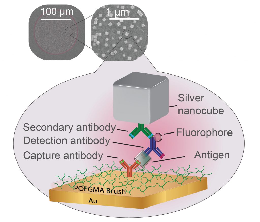
Researchers have shown that plasmonics can enhance the fluorescent markers used to signal positive samples in certain types of tests for diseases. A polymer brush coating keeps unwanted biomolecules away while a capture antibody (red) catches biomarkers of disease (clear). A detection antibody (blue) then latches on to the biomarker and emits light from an attached fluorophore (sphere). All of this is sandwiched by a thin layer of gold and a silver nanocube that is attached by a third antibody (green), creating conditions for the fluorophore to emit brighter light. Courtesy: Duke University

A May 12, 2020 news item on Nanowerk announces new work from scientists at Duke University on making point-of-care diagnostics easier to use by making the readouts brighter,

Engineers at Duke University [North Carolina, US] have shown that nanosized silver cubes can make diagnostic tests that rely on fluorescence easier to read by making them more than 150 times brighter. Combined with an emerging point-of-care diagnostic platform already shown capable of detecting small traces of viruses and other biomarkers, the approach could allow such tests to become much cheaper and more widespread.

A May 12, 2020 Duke University news release (also on EurekAlert), which originated the news item, provides more detail about the work,

Plasmonics is a scientific field that traps energy in a feedback loop called a plasmon onto the surface of silver nanocubes. When fluorescent molecules are sandwiched between one of these nanocubes and a metal surface, the interaction between their electromagnetic fields causes the molecules to emit light much more vigorously. Maiken Mikkelsen, the James N. and Elizabeth H. Barton Associate Professor of Electrical and Computer Engineering at Duke, has been working with her laboratory at Duke to create new types of hyperspectral cameras and superfast optical signals using plasmonics for nearly a decade.

At the same time, researchers in the laboratory of Ashutosh Chilkoti, the Alan L. Kaganov Distinguished Professor of Biomedical Engineering, have been working on a self-contained, point-of-care diagnostic test that can pick out trace amounts of specific biomarkers from biomedical fluids such as blood. But because the tests rely on fluorescent markers to indicate the presence of the biomarkers, seeing the faint light of a barely positive test requires expensive and bulky equipment.

“Our research has already shown that plasmonics can enhance the brightness of fluorescent molecules tens of thousands of times over,” said Mikkelsen. “Using it to enhance diagnostic assays that are limited by their fluorescence was clearly a very exciting idea.”

“There are not a lot of examples of people using plasmon-enhanced fluorescence for point-of-care diagnostics, and the few that exist have not been yet implemented into clinical practice,” added Daria Semeniak, a graduate student in Chilkoti’s laboratory. “It’s taken us a couple of years, but we think we’ve developed a system that can work.”

In the new paper, researchers from the Chilkoti lab build their super-sensitive diagnostic platform called the D4 Assay onto a thin film of gold, the preferred yin to the plasmonic silver nanocube’s yang. The platform starts with a thin layer of polymer brush coating, which stops anything from sticking to the gold surface that the researchers don’t want to stick there. The researchers then use an ink-jet printer to attach two groups of molecules tailored to latch on to the biomarker that the test is trying to detect. One set is attached permanently to the gold surface and catches one part of the biomarker. The other is washed off of the surface once the test begins, attaches itself to another piece of the biomarker, and flashes light to indicate it’s found its target.

After several minutes pass to allow the reactions to occur, the rest of the sample is washed away, leaving behind only the molecules that have managed to find their biomarker matches, floating like fluorescent beacons tethered to a golden floor.

“The real significance of the assay is the polymer brush coating,” said Chilkoti. “The polymer brush allows us to store all of the tools we need on the chip while maintaining a simple design.”

While the D4 Assay is very good at grabbing small traces of specific biomarkers, if there are only trace amounts, the fluorescent beacons can be difficult to see. The challenge for Mikkelsen and her colleagues was to place their plasmonic silver nanocubes above the beacons in such a way that they supercharged the beacons’ fluorescence.

But as is usually the case, this was easier said than done.

“The distance between the silver nanocubes and the gold film dictates how much brighter the fluorescent molecule becomes,” said Daniela Cruz, a graduate student working in Mikkelsen’s laboratory. “Our challenge was to make the polymer brush coating thick enough to capture the biomarkers–and only the biomarkers of interest–but thin enough to still enhance the diagnostic lights.”

The researchers attempted two approaches to solve this Goldilocks riddle. They first added an electrostatic layer that binds to the detector molecules that carry the fluorescent proteins, creating a sort of “second floor” that the silver nanocubes could sit on top of. They also tried functionalizing the silver nanocubes so that they would stick directly to individual detector molecules on a one-on-one basis.

While both approaches succeeded in boosting the amount of light coming from the beacons, the former showed the best improvement, increasing its fluorescence by more than 150 times. However, this method also requires an extra step of creating a “second floor,” which adds another hurdle to engineering a way to make this work on a commercial point-of-care diagnostic rather than only in a laboratory. And while the fluorescence didn’t improve as much in the second approach, the test’s accuracy did.

“Building microfluidic lab-on-a-chip devices through either approach would take time and resources, but they’re both doable in theory,” said Cassio Fontes, a graduate student in the Chilkoti laboratory. “That’s what the D4 Assay is moving toward.”

And the project is moving forward. Earlier in the year, the researchers used preliminary results from this research to secure a five-year, $3.4 million R01 research award from the National Heart, Lung, and Blood Institute. The collaborators will be working to optimize these fluorescence enhancements while integrating wells, microfluidic channels and other low-cost solutions into a single-step diagnostic device that can run through all of these steps automatically and be read by a common smartphone camera in a low-cost device.

“One of the big challenges in point-of-care tests is the ability to read out results, which usually requires very expensive detectors,” said Mikkelsen. “That’s a major roadblock to having disposable tests to allow patients to monitor chronic diseases at home or for use in low-resource settings. We see this technology not only as a way to get around that bottleneck, but also as a way to enhance the accuracy and threshold of these diagnostic devices.”

Here’s a link to and a citation for the paper,

Ultrabright Fluorescence Readout of an Ink-Jet Printed Immunoassay Using Plasmonic Nanogap Cavities by Daniela F. Cruz, Cassio M. Fontes, Daria Semeniak, Jiani Huang, Angus Hucknall, Ashutosh Chilkoti, Maiken H. Mikkelsen. Nano Lett. 2020, XXXX, XXX, XXX-XXX DOI: https://doi.org/10.1021/acs.nanolett.0c01051 Publication Date:May 6, 2020 Copyright © 2020 American Chemical Society

This paper is behind a paywall.

Acoustofluidics and lab-on-a-chip for asthma and tuberculosis diagnostics

This is my first exposure to acoustofluidics (although it’s been around for a few years) and it concerns lab-on-a-chip diagnostics for asthma and tuberculosis. From an Aug. 3, 2015 news item on Azonano,

A device to mix liquids utilizing ultrasonics is the first and most difficult component in a miniaturized system for low-cost analysis of sputum from patients with pulmonary diseases such as tuberculosis and asthma.

The device, developed by engineers at Penn State in collaboration with researchers at the National Heart, Lung, and Blood Institute (NHLBI), part of the National Institutes of Health, and the Washington University School of Medicine, will benefit patients in the U.S., where 12 percent of the population, or around 19 million people, have asthma, and in undeveloped regions where TB is still a widespread and often deadly contagion.

“To develop more accurate diagnosis and treatment approaches for patients with pulmonary diseases, we have to analyze sample cells directly from the lungs rather than by drawing blood,” said Tony Jun Huang, professor of engineering science and mechanics at Penn State and the inventor, with his group, of this and other acoustofluidic devices based on ultrasonic waves. “For instance, different drugs are used to treat different types of asthma patients. If you know what a person’s immunophenotype is, you can provide personalized medicine for their particular disease.

A July 29, 2015 Pennsylvania State University news release, which originated the news item, describes the disadvantages of the current sputum analyses techniques and explains how this new technique in an improvement,

There are several issues with the current standard method for sputum analysis. The first is that human specimens can be contagious, and sputum analysis requires handling of specimens in several discrete machines. With a lab on a chip device, all biospecimens are safely contained in a single disposable component.

Another issue is the sample size required for analysis in the current system, which is often larger than a person can easily produce. The acoustofluidic sputum liquefier created by Huang’s group requires 100 times less sample while still providing accuracy equivalent to the standard system.

A further issue is that current systems are difficult to use and require trained operators. With the lab on a chip system, a nurse can operate the device with a touch of a few buttons and get a read out, or the patient could even operate the device at home. In addition, the disposable portion of the device should cost less than a dollar to manufacture.

Po-Hsun Huang, a graduate student in the Huang group and the first author on the recent paper describing the device in the Royal Society of Chemistry journal Lab on a Chip, said “This will offer quick analysis of samples without having to send them out to a centralized lab. While I have been working on the liquefaction component of the device, my lab mates are working on the flow cytometry analysis component, which should be ready soon. This is the first on-chip sputum liquefier anyone has developed.”

Stewart J. Levine, a Senior Investigator and Chief of the Laboratory of Asthma and Lung Inflammation in the Division of Intramural Research at NHLBI, said “This on-chip sputum liquefier is a significant advance regarding our goal of developing a point-of-care diagnostic device that will determine the type of inflammation present in the lungs of asthmatics. This will allow health care providers to individualize asthma treatments for each patient and advance the goal of bringing precision medicine into clinical practice.”

Here’s a link to and a citation for the paper,

An acoustofluidic sputum liquefier by Po-Hsun Huang, Liqiang Ren, Nitesh Nama, Sixing Li, Peng Li, Xianglan Yao, Rosemarie A. Cuento, Cheng-Hsin Wei, Yuchao Chen, Yuliang Xie, Ahmad Ahsan Nawaz, Yael G. Alevy, Michael J. Holtzman, J. Philip McCoy, Stewart J. Levine, and  Tony Jun Huang. Lab Chip, 2015,15, 3125-3131 DOI: 10.1039/C5LC00539F

First published online 17 Jun 2015

This is an open access paper but you do need to register for a free (British) Royal Society of Chemistry publishing personal account.

2014 Sanofi BioGENEius Challenge Canada (SBCC) national winners announced

Last week on May 23, 2014, the Sanofi BioGENEius Challenge Canada (SBCC) National winners were announced in Ottawa. (A Feb. 20, 2013 posting recounts the organization’s history and accomplishments on its 20th anniversary). Here’s more about the 2014 national winners from a May 23, 2014 Sanofi BioGENEius Challenge Canada news release,

A novel method of HIV detection for newborns under the age of 18 months and for adults before three months post-transmission earned a grade 10, British Columbia student top national honours today [May 23, 2014] in the 2014 “Sanofi BioGENEius Challenge Canada” (SBCC).

Nicole Ticea, 15, from York House School in Burnaby, BC was awarded the top prize of $5,000 by a panel of eminent Canadian scientists assembled at the Ottawa headquarters of the National Research Council of Canada (NRC).

Her impressive research project, mentored at Simon Fraser University by associate professor, Dr. Mark Brockman, is the first test capable of analyzing HIV viral nucleic acids in a point-of-care, low-resource setting.Nicole’s research, was deemed an incredibly innovative solution to a global challenge according to the judges led by Dr. Julie Ducharme, General Manager, Human Health Therapeutics, NRC.

See a full project description below and online here: http://sanofibiogeneiuschallenge.ca/2014/05/23/

Ten brilliant young scientists from nine Canadian regions, all just 15 to 18 years old, took part in the national finals. They had placed first at earlier regional SBCC competitions, conducted between March 27 and May 22, 2014.

High school and CEGEP students from Victoria to Saskatoon to St. John’s, focused on biotechnology fields of discovery and study, submitted more than 200 proposals. Working closely with mentors, these students conducted research in diverse areas such as telomeres, diabetes, stress management, Alzheimer’s, autism and pulp production. Since its inauguration in 1994, more than 4,700 young Canadians have competed in SBCC, with the majority of competitors going on to pursue careers in science and biotechnology.

1st place winner, Nicole Ticea will compete for Canada on June 22-25 at the International BioGENEius Challenge, conducted at the annual BIO conference in San Diego, CA.

2nd place, $4,000 – Ontario: Varsha Jayasankar, 17, grade 12, Sir Winston Churchill Secondary School, St. Catherines won with research into how an extract created from mango ginger can be used to inhibit the growth of multiple antibiotic-resistant bacteria. Project description: http://sanofibiogeneiuschallenge.ca/2014/05/23/

3rd place, $3,000 – Ontario: Anoop Manjunath, 17, grade 11, University of Toronto Schools, Toronto investigated image processing techniques for the analysis of ultrasound stimulated bubble interactions with fibrin clots.Project description: http://sanofibiogeneiuschallenge.ca/2014/05/23/

There were a couple of other projects (one for its ‘nano’ focus and the other for its ‘wheat’ focus), which caught my attention, from the SBCC 2014 National Competitor Project Descriptions page by Anne Ramsay,

Amit Scheer, Grade 10

Colonel By Secondary School, Ottawa, ON

“Development of a Novel Quantum Dot-Aptamer Bioconjugate Targeted Cancer Therapy for Precision Nanomedicine Applications”

A novel nanoparticle for targeted cancer therapeutics is described. This research was effectuated to create a theranostic bioconjugate with an optimal effective therapeutic index, achieved by biomarker-specific targeting. Estimates show that over 14 million new cases of cancer are diagnosed annually worldwide. Aptamer-quantum dot (APT-QD) bioconjugates were synthesized by conjugating cadmium-telluride quantum dots (QDs, semiconductor nanoparticles) to aptamers (nucleic-acid based ligands), by amide crosslinking. Aptamers targeted mucin-1 (MUC1), a glycosylated surface protein overexpressed on many cancers, including MCF7 breast cancer cells, and only minimally expressed in MCF-10A non-cancerous cells. The bioconjugate and unmodified QD treatments (the control) were tested for cellular uptake and cytotoxicity in MCF7 (cancerous) and MCF-10A (comparison) cell cultures. MTT assays, which quantify cellular viability by assessing mitochondrial activity, were used for dose-response analysis at several treatment concentrations. APT-QDs caused a statistically significant decrease in viability specifically in MUC1-overexpressing cultures, suggesting cell-specific internalization by receptor-mediated endocytosis. Apoptosis and necrosis were quantified using immunofluorescence assays; bioconjugate-treated cells were early apoptotic after 4 hours, proving effective initiation of programmed cell death. Finally, confocal microscopy was used for aptamer-dependent nanoparticle internalization analysis, demonstrating that APT-QDs accumulate outside of nuclei. A fluorochrome-modified DNA complement to the aptamer was synthesized for co-localization of aptamers and QDs, proving effective endosomal escape for both components. The bioconjugate has applications in combination and theranostic treatments for cancer, and in precision medicine to diversify targeting based on patient-specific panomics analyses. The researcher created a novel bioconjugate nanoparticle and has proven numerous viable applications in cancer therapeutics.

Wenyu Ruan, Grade 9, & Amy Yu Ruiyun Wang, Grade 10

Walter Murray Collegiate Institute, Saskatoon, SK

“Identification of Leaf Rust Resistance in Wheat”

Leaf rust is the most common disease in wheat, a crop which contributes $11B annually to Canada’s economy. The most effective strategy to control leaf rust has been to grow resistant varieties. There are two general types of resistance genes found in wheat: Race-specific genes confer a high-level of resistance to specific strains of leaf rust but can be easily overcome by genetic mutation in pathogen populations, while slow rusting (APR) resistance provides partial resistance to a broad spectrum of races, but is typically effective only at the adult stage of plant growth. A three-phase experiment was conducted on a doubled-haploid population derived from the cross RL4452/AC Domain to determine if the resistance of a recently discovered gene (Lr2BS) worked with other resistance genes to synergistically enhance resistance to leaf rust. Linkage and quantitative trait loci (QTL) mapping were performed by combining our new genotypic data with a previously generated genetic map for this population, then adding rust disease data from our experiment to identify genomic regions associated with leaf rust resistance. In addition, a fluorescent microscope was used to examine host-pathogen interaction on a cellular level. These experiments showed that lines carrying Lr2BS alone, and in combination with other APR genes were susceptible at the seedling stage, which suggests that Lr2BS is an adult plant gene. It appears that the synergistic effect of some multiple gene combinations, including Lr2BS, enhances leaf rust resistance. Furthermore, QTL mapping identified an uncharacterized resistance gene (LrUsw4B) that conferred resistance at the seedling stage.

I am sorry to see they are not sending all three national finalists to the international competition as they did in 2012. As I noted in my July 16, 2012 posting the international standings did not reflect the national standings,

As the 2012 winner of the Sanofi BioGENEius Challenge Canada competition, Tam was invited to compete in this year’s international Sanofi BioGENEisu Challenge held in Boston, Massachusetts on June 19, 2012. [Janelle] Tam received an honourable mention for her work while Rui Song of Saskatoon placed third internationally.

Presumably the costs are too high to continue the practice.

Getting back to 2014, congratulations to all the competitors and the winners! And, good luck to Nicole Ticea at the International BioGENEius Challenge which will be conducted at the annual BIO conference, June 22-25  2014, in San Diego, CA!

Namdiatream; a European multimodal diagnostics project

I’ve written about lab-on-a-chip projects, point-of-care diagnostics, and other such initiatives on several occasions, most recently in a Mar. 1, 2013 posting about a technique where powder is used to make the diagnostic device more portable. This time it was a Europe-wide project described in a Mar. 4, 2013 news item on Nanowerk,which caught my attention (Note: A link has been removed),

The plan of the EU-funded consortium Nanotechnological toolkits for multi-modal disease diagnostics and treatment monitoring (Namdiatream) is not to cure cancer, per se, but to boost the sensitivity of diagnostics and the ability to monitor progress during treatment. They focused on three types – breast, prostate and lung cancer.

… The prototype devices being developed during the four-year project will detect common cancer cells much earlier and, with timely treatment, improve the chances of recovery.

According to the project leader, Professor Yuri Volkov of Trinity College Dublin’s School of Medicine, the portable nanodevices are based on innovative lab-on-a-chip, -bead and -wire technologies applicable in different settings – clinical, research, or point of care (i.e. hospitals). These lab-on-x technologies exploit the photo-luminescent (‘glow-in-the-dark’ light emitting), plasmonic (‘light-on-a-wire’), magnetic and unique optical properties of nanomaterials.

Volkov offers some insight into how the project started and its current state of evolution (from the news item),

This is ground-breaking work made possible thanks to advanced technology but also to EU funding for cross-border investigations. Teams across Europe were doing related but fragmented research, suggests Prof. Volkov. This risked leaving a team dangling if their approach failed or lacked funding.

“So we integrated our research and identified joint strengths to help one another develop the best technological approaches in case something didn’t work in one, or synergies were identified, thereby increasing the chances of wider success.”

At its half-way stage, notes Prof. Volkov, Namdiatream underwent a natural evolution when it became clear that by merging and refocusing work in some areas – i.e. in fluorescent nanomaterial technology and magnetic nanowire barcodes – it would speed up industrial implementation efforts.

“Now, work on the preclinical prototype devices is well under way,” he confirms. But one of the many remaining challenges is to calibrate their sensitivity, so that they do not give false readings, for instance.

The Namdiatream (Nanotechnological Toolkits for Multi-Modal Disease Diagnostics and Treatment Monitoring) home page offers more detail about the project,

Namdiatream is a truly interdisciplinary and Pan-european consortium that builds around 7 High-Tech SMEs [small to medium enterprises], 2 Multinational industries and 13 academic institutions. NAMDIATREAM will develop nanotechnology-based toolkit to enable early detection and imaging of molecular biomarkers of the most common cancer types and of cancer metastases, as well as permitting the identification of cells indicative of early-stage disease onset. The project is built on the innovative technology concepts of super-sensitive “lab-on-a-bead”, “lab-on-a-chip” and “lab-on-a-wire” nano-devices.

Interestingly, this too was on the home page,

The ETP Nanomedicine documents point out that nanotechnology has yet to deliver practical solutions for the patients and clinicians in their struggle against common, socially and economically important diseases such as cancer. Therefore NAMDIATREAM results will firstly aim to deliver to the diagnostic and medical imaging device companies involved in the consortium, and the clinical and academic partners. This could further provide the basis for cancer therapeutics as it will be possible to accurately assess the kinetics of cancer cell destruction during the course of appropriate therapy.

Home pregnancy tests inspire simple diagnostics containing gold nanoparticles

PhD student Kyryl Zagorovsky and Professor Warren Chan of the University of Toronto’s Institute of Biomaterials and Biomedical Engineering (IBBME) have created a rapid diagnostic biosensor according to a Feb. 28, 2013 news item on phys.org,

A diagnostic “cocktail” containing a single drop of blood, a dribble of water, and a dose of DNA powder with gold particles could mean rapid diagnosis and treatment of the world’s leading diseases in the near future. …

The recent winner of the NSERC E.W.R. Steacie Memorial Fellowship, Professor Chan and his lab study nanoparticles: in particular, the use of gold particles in sizes so small that they are measured in the nanoscale. Chan and his group are working on custom-designing nanoparticles to target and illuminate cancer cells and tumours, with the potential of one day being able to deliver drugs to cancer cells.

But it’s a study recently published in Angewandte Chemie that’s raising some interesting questions about the future of this relatively new frontier of science.

Zagorovsky’s rapid diagnostic biosensor will allow technicians to test for multiple diseases at one time with one small sample, and with high accuracy and sensitivity. The biosensor relies upon gold particles in much the same vein as your average pregnancy test. With a pregnancy test, gold particles turn the test window red because the particles are linked with an antigen that detects a certain hormone in the urine of a pregnant woman.

(Until now, I’d never thought about how a pregnancy test actually works and always assumed it was similar to a litmus paper test of acid.) The University of Toronto’s Feb. 28, 2013 news release, which originated the news item, describes the technology in more detail,

Currently, scientists can target a particular disease by linking gold particles with DNA strands. When a sample containing the disease gene (e.g., Malaria) is present, it clumps the gold particles, turning the sample blue.

Rather than clumping the particles together, Zagorovsky immerses the gold particles in a DNA-based enzyme solution (DNA-zyme) that, when the disease gene is introduced, ‘snip’ the DNA from the gold particles, turning the sample red.

“It’s like a pair of scissors,” said Zagorovsky. “The target gene activates the scissors that cut the DNA links holding gold particles together.”

The advantage is that far less of the gene needs to be present for the solution to show noticeable colour changes, amplifying detection. A single DNA-zyme can clip up to 600 ‘links’ between the target genes.

Just a single drop from a biological sample such as saliva or blood can potentially be tested in parallel, so that multiple diseases can be tested in one sitting.

But the team has also demonstrated that [it] can transform the testing solution into a powder, making it light and far easier to ship than solutions, which degrade over time. Powder can be stored for years at a time, and offers hope that the technology can be developed into efficient, cheap, over-the-counter tests for diseases such as HIV and malaria for developing countries, where access to portable diagnostics is a necessity. [emphases mine]

I think the fact that the testing solution can be made into powder is exciting news. Medical technologies can be wonderful but if they require special handling and training (e.g., a standard vaccine is in a solution which needs to be refrigerated [that’s expensive in some parts of the world] and someone who is specially trained has to administer the injection) then they’re confined to the few who have access and can afford it.

Here’s a citation and a link to the researchers’ paper,

A Plasmonic DNAzyme Strategy for Point-of-Care Genetic Detection of Infectious Pathogens by Kyryl Zagorovsky, and Dr. Warren C. W. Chan. Angewandte Chemie International Edition DOI: 10.1002/anie.201208715 Article first published online: 10 FEB 2013

Copyright © 2013 WILEY-VCH Verlag GmbH & Co. KGaA, Weinheim

This article is behind a paywall.

ETA Mar. 1, 2013 10:42 am PST: I made a quick change to the title. Hopefully this one makes more sense than the first one did.

A breath-based and handheld diagnostic device

Researcher Perena Gouma and her team at Stony Brook University (New York, US) are hoping that eventually their device will be available over the counter so anyone will be able to perform a preliminary diagnostic test as casually as you take a breath. From the May 7, 2012 news item on Nanowerk,

You blow into a small valve attached to a box that is about half the size of your typical shoebox and weighs less than one pound. Once you blow into it, the lights on top of the box will give you an instant readout. A green light means you pass (and your bad breath is not indicative of an underlying disease; perhaps it’s just a result of the raw onions you ingested recently); however, a red light means you might need to take a trip to the doctor’s office to check if something more serious is an issue.

Here’s a bit more about the device and the researchers’ hopes in a video from the US National Science Foundation (NSF) featuring the NSF’s Miles O’Brien as the reporter,

O’Brien in his May 7, 2012 article for the NSF’s Science Nation online magazine describes the technology,

With support from the National Science Foundation (NSF), Professor Perena Gouma and her team at Stony Brook University in New York developed a sensor chip that you might say is the “brain” of the breathalyzer. It’s coated with tiny nanowires that look like microscopic spaghetti and are able to detect minute amounts of chemical compounds in the breath. “These nanowires enable the sensor to detect just a few molecules of the disease marker gas in a ‘sea’ of billions of molecules of other compounds that the breath consists of,” Gouma explains. This is what nanotechnology is all about.

The manufacturing process that creates the single crystal nanowires is called “electrospinning.” It starts with a liquid compound being shot from a syringe into an electrical field. The electric field crystallizes the inserted liquid into a tiny thread or “wire” that collects onto an aluminum backing. Gouma says enough nanowire can be produced in one syringe to stretch from her lab in Stony Brook, N.Y. to the moon and still be a single grain (monocrystal).

“There can be different types of nanowires, each with a tailored arrangement of metal and oxygen atoms along their configuration, so as to capture a particular compound,” explains Gouma. “For example, some nanowires might be able to capture ammonia molecules, while others capture just acetone and others just the nitric oxide. Each of these biomarkers signal a specific disease or metabolic malfunction so a distinct diagnostic breathalyzer can be designed.”

Gouma also says the nanowires can be rigged to detect infectious viruses and microbes like Salmonella, E. coli or even anthrax. “There will be so many other applications we haven’t envisioned. It’s very exciting; it’s a whole new world,” she says.

I think most (if not all) of the handheld diagnostic projects I’ve covered have been fluids-based, i.e., they need a sample of saliva, blood, urine, etc. to perform their diagnostic function. I believe this is the first breath-based project I’ve seen.

Alberta’s Domino (point-of-care diagnostic) and Navacim (nano drug delivery) competing for $175,000 prize

It’s interesting that two nanomedicine products are in contention for TEC Edmonton‘s NanoVenture Prize. It’s a new prize category for the business accelerator in this, their 10th anniversary year. From TEC Edmonton’s March 27, 2012 news release,

The NanoVenturePrize finalists are Aquila Diagnostics of Edmonton and Calgary’s Parvus Therapeutics.

Aquila Diagnostics uses the Domino nanotechnology platform developed at the University of Alberta to provide on-site, easy-to-use genetic testing that can quickly test for infectious diseases and pathogens in livestock. The mobile diagnostic platform is portable, low-cost, fast and easy to use.

Parvus Therapeutics’ breakthrough nanomedicines may hold the cure for difficult-to-treat autoimmune diseases like type 1 diabetes, multiple sclerosis and inflammatory bowel disease. Parvus’ new Navacim medicines are nanoparticles coated with immune system proteins that can target specific autoimmune conditions.

The University of Alberta has issued its own April 24, 2012 news release by Bryan Alary about the Domino,

Dubbed the Domino, the technology—developed by a U of A research team—has the potential to revolutionize point-of-care medicine. The innovation has also earned Aquila Diagnostic Systems, the Edmonton-based nano startup that licensed the technology, a shot at $175,000 as a finalist for the TEC NanoVenturePrize award.

“We’re basically replacing millions of dollars of equipment that would be in a conventional, consolidated lab with something that costs pennies to produce and is field portable so you can take it where needed. That’s where this technology shines,” said Jason Acker, an associate professor of laboratory medicine and pathology at the U of A and chief technology officer with Aquila.

The Domino employs polymerase chain reaction technology used to amplify and detect targeted sequences of DNA, but in a miniaturized form that fits on a plastic chip the size of two postage stamps. The chip contains 20 gel posts—each the size of a pinhead—capable of identifying sequences of DNA with a single drop of blood.

Each post performs its own genetic test, meaning you can not only find out whether you have malaria, but also determine the type of malaria and whether your DNA makes you resistant to certain antimalarial drugs. It takes less than an hour to process one chip, making it possible to screen large populations in a short time.

“That’s the real value proposition—being able to do multiple tests at the same time,” Acker said, adding that the Domino has been used in several recently published studies, showing similar accuracy to centralized labs.

Linda Pilarski, an oncology professor at the University of Alberta (mentioned in my Jan. 4, 2012 posting about her diagnostics-on-a-chip work), and her team developed Domino according to the April 25, 2012 news item on Nanowerk,

In 2008, her team received $5 million over five years from Alberta Innovates Health Solutions to perfect and commercialize the technology. As an oncologist, Pilarski is interested in its pharmacogenomic testing capabilities, such as determining whether breast cancer patients are genetically disposed to resist certain drugs.

“With most cancers you want to treat the patient with the most effective therapeutic as possible,” she said. “That’s what this does: it really enables personalized medicine. It will be able to test every patient at the right time, right in their doctor’s office. That’s currently not feasible because it’s too expensive.”

This product is intended for the market but not the one you might expect (from the April 25, 2012 news item on Nanowerk),

Along with its versatility, two key selling points are affordability and portability, with each portable box expected to cost about $5,000 and each chip a few dollars, says Aquila president David Alton. It’s also designed to be easy to use and rugged—important features for the livestock industry, the company’s first target market. [emphasis mine] The Domino will be put through trials within a year at one of the country’s largest feedlots in southern Alberta.

Alton credits Aquila’s relationship with the U of A, not just for the research but for the business relationship with TEC Edmonton that has helped the company license and patent Domino. TEC Edmonton is a joint venture between the U of A and Edmonton Economic Development Corporation with resources and expertise to help startups in the early stages of operations.

“We see a huge potential market for the technology and we’re looking at applying the technology developed here at the U of A to markets first in Alberta and then globally, to address important health issues here and throughout the world.”

Given that the originator is an oncologist I really wasn’t expecting the first market to be livestock industry.

I have had a little less luck getting information about Parvus Therapeutics’ Navacim technology as they’ve not issued a news release about their competition for this prize but I did find some information on their website, from an April 8, 2010 news release about the Navacim technology being featured in a Popular Science article,

Parvus Therapeutics reports that an article entitled “Nanotech Vaccine Successfully Cures Type-1 Diabetes in Mice” has been published at the website of Popular Science. The article, authored by Alessandra Calderin, describes the Parvus Navacim technology and includes remarks from Parvus’ Founder and Chief Scientific Officer, Dr. Pere Santamaria.

The article notes that,

“The technology behind the nanovaccine, following further research, may prove widely applicable to treat other autoimmune diseases, like arthritis and multiple sclerosis, as well.”

You may want to take a look at the news brief by Calderin. Here’s more about the technology, from the Introducing Navacims webpage on the Parvus Therapeutics website,

Our nanotechnology-based therapeutic platform and Navacims, the therapeutic candidates, are the result of two related discoveries: A new class of immune cell, and a new way to treat autoimmunity that these cells provide. Here we provide a very brief summary of how these discoveries came about and what they have led to since.

This summary is also intended as a roadmap to the contents of this technology section of our website, which we will role out over a period of weeks and adapt based on reader feedback and requests. The casual reader may find the background information helpful, while our professional colleagues will probably want to get straight down to the technical details and published papers. We have tried to design the content to cater to all tastes and it can be read in any order, although like all good stories, we highly recommend starting at the beginning.

As with the remainder of our site, we have injected a little colour and a little humour to keep your spirits up if the science appears a little daunting. In all, we have attempted to strike a balance between scientific detail and general accessibility and if you think we have that balance wrong, or you feel something is missing, please let us know — via the form on the Contacts page — and we will try to put it right. We love to hear from you.

The Story So Far

[1] In a series of experiments, only tangentially related to our current activities, we designed p-MHC-coated nanoparticles (NPs) as a way to load iron into effector T-cells and have them ferry the iron to the pancreas so we could visualize pancreatic islet cell inflammation in-vivo, in real-time — this amounts to the use of a Magnetic Resonance Imaging (MRI) contrast agent.

[2] It occurred to us that we might be able to use these p-MHC-NPs to delete the high avidity cytotoxic effector T cells driving disease in the NOD mouse model of type 1 diabetes (T1D).

[3] Too our surprise, therapy did not delete, but rather, very significantly expanded autoregulatory T cell pools.

[4] After careful analysis we were able to conclude that:

pMHC-NPs, now called Navacims, selectively expand a population of low avidity autoregulatory memory T cells that the disease itself generates — this population of cells was previously unknown to science. These cells target and kill antigen presenting cells (APCs), and consequently, interput the process whereby all the cytotoxic effector T cell lineages active in a disease are activated and expanded.

Navacims also directly deplete the high avidity cytotoxic effector T cells cognate to the pMHC carried by the nanoparticle. This removes one lineage of cells that cause damage in disease, but given the many antigens, and consequently the many T cell lineages, the overall therapeutic effect of removing one type is inconsequential compared to the indirect effect of the Navacim on APCs that removes all lineages.

The removal of APCs and the concomitant loss of multiple cytotoxic effector T-cell lineages that drive disease amounted to a cure for T1D in the NOD mouse model.

[5] We believe that Navacims have the potential to become the long sought after ideal treatment for autoimmunity; a therapeutic that restores immunological tolerance — the principal problem in autoimmunity — while depleting autoreactive cells that mediate the damaging effects of disease.

[6] Navacims appear to be safe and very well tolerated in animal experiments that have lasted many months, although we caution that we have yet to complete formal toxicological studies.

[7] Navacims are highly modular and a family of Navacims can be almost identical, differing only in the very short antigenic peptide that gives each one its specificity for a particular disease.

[8] Because they are so similar, we beleive that industry-standard manufacturing processes will need few if any modifications in order to produce a particular Navacim.

[9] We have protected our discoveries with patent applications in the United States, Europe, Canada, and beyond.

[10] Our work has been published in top-ranked peer-reviewed journals and showcased in the best of the popular science publications.

Good luck to both companies in their future endeavours.

ETA April 30,2012: According to the April 27, 2012 article in the Edmonton Journal, Parvus Therapeutics won the $175, 000 prize in TEC Edmonton’s new prize category.,

This year’s awards, the 10th consecutive, added a new category for nanotechnology firms. TEC partnered with Alberta Innovates — Technology Futures for the new award. Calgary’s Parvus Therapeutics, which makes medicine aimed at autoimmune diseases such as Type 1 diabetes and multiple sclerosis, beat out Edmonton’s Aquila Diagnostic Systems for first place. The category’s prizes totalled $175,000 in cash and services.

The French and the Russians work on point-of-care diagnostic devices

RUSNANO (Russian Corporation of Nanotechnologies), a regular visitor to my postings, has just signed a deal with French company, Magnisense SE (why do I keep wanting to call it (en français) Magnificence?), for a three year development deal. From the Feb. 6, 2012 news item on Nanowerk,

RUSNANO and Magnisense today announced that they have finalized details for investment in Magnisense SE, a French developer of the next generation of in vitro bioassays for diagnostic testing in healthcare, veterinary medicine, food safety, and environmental protection. RUSNANO will invest up to €28.5 million over a three-year period in the joint project whose total cost is put at €44.3 million. The remaining co-investment will come from Magnisense’s existing and new shareholders.

The new project will manufacture in Russia an advanced diagnostic system based on Magnisense’s proprietary technology MIAtek®—a magnetic immunoassay in which nanosized magnetic beads are attached to an antibody that selectively binds target molecules, micro-organisms, or other antibodies in test media.

I gather not all of the products that are going to be manufactured in Russia will be point-of-care diagnostic devices but all of them will be based on the Magnisense technology,

Magnisense will establish a Russian subsidiary to produce two product lines:

·  MIAstrip®: point-of-care testing strips that identify a target by detecting known markers for a number of conditions—cardiac arrest, bacterial infections including tetanus, viral infections such as avian flu, and parasitic and fungal infections;

·  MIAflo®: disposable cartridges that detect and quantify such targets as bacterial contaminants in food (e.g., Listeria and Salmonella) or water (e.g., Legionella). Magnisense’s Russian manufacturing facility is expected to open in 2015 with production capacity of 3.5 million test media. The media will be sold domestically and exported, primarily to Europe, the US, and Japan.

In the future, Magnisense intends to increase production in Russia and to focus on the professional segment of the decentralized diagnostic testing market generally known as point-of-care (POC). It will expand into the CIS, Europe, the US, Japan, and China. POC diagnostics—tests performed in medical environments, the workplace, and at home—have become the driving force in the world’s healthcare market. According to Kalorama Information, a market research company specializing in healthcare and related areas, decentralized diagnostics is the fastest growing segment in healthcare. And the professional POC market, already $5.2 billion in 2009, is estimated to be growing at an annual rate of 6 percent with its rate of growth accelerating.

For anyone interested in magnetic immunoassay Magnisense has devoted a webpage to the technology they have developed,

Magnetic immunoassay was developed for the rapid and sensitive detection of proteins, viruses or bacteria in biological or food samples. The system incorporates the use of coated magnetic beads as labels in lateral flow (MIAstrip®) or flow through (MIAflo®) or chip (MIAchip®) formats.

The technology is versatile. It is compatible with multiple assays and uses standard liquid handling robots installed in many clinical labs worldwide.

The robots are designed to deliver reagents via removable tips to carry out ELISA on multi-well plates. As opposed to standard applications, the MIAflo® is realized on the 3D filters inside the tips, and the plate is used to hold the reagents. MIAflo® provides more sensitive and faster results compared to ELISA due to the absence of kinetic limitations and immunofiltration of the antigens on a large immune-active surface. In addition, MIAflo® successfully works even with the whole blood of patients, decreasing the sample preparation time.

I’m not all that interested in pursuing the differences between this technique and ELISA (you can read more about ELISA in this Wikipedia essay) but what does interest me is this worldwide competition to develop point-of-care diagnostics.

Are we creating a Star Trek world? T-rays and tricorders

There’s been quite a flutter online (even the Huffington Post has published a piece) about ‘Star Trek-hand-held medical scanners’ becoming possible due to some recent work in the area of T-rays. From the Jan. 20, 2012 news item on Nanowerk,

Scientists who have developed a new way to create a type of radiation known as Terahertz (THz) or T-rays – the technology behind full-body security scanners – say their new, stronger and more efficient continuous wave T-rays could be used to make better medical scanning gadgets and may one day lead to innovations similar to the “tricorder” scanner used in Star Trek.

In a study published recently in Nature Photonics (“Greatly enhanced continuous-wave terahertz emission by nano-electrodes in a photoconductive photomixer” [behind a paywall]), researchers from the Institute of Materials Research and Engineering (IMRE), a research institute of the Agency for Science, Technology and Research (A*STAR) in Singapore and Imperial College London in the UK have made T-rays into a much stronger directional beam than was previously thought possible and have efficiently produced T-rays at room-temperature conditions. This breakthrough allows future T-ray systems to be smaller, more portable, easier to operate, and much cheaper.

For anyone who’s not familiar with ‘Star Trek world’ and tricorders, here’s a brief description from a Wikipedia essay about tricorders,

In the fictional Star Trek universe, a tricorder is a multifunction handheld device used for sensor scanning, data analysis, and recording data.

David Freeman in his Jan. 21, 2012 article for the Huffington Post about the research puts it this way,

Trekkies, take heart. A scientific breakthrough involving a form of infrared radiation known as terahertz (THz) waves could lead to handheld medical scanners reminiscent of the “tricorder” featured on the original Star Trek television series.

What’s the breakthrough? Using nanotechnology, physicists in London and Singapore found a way to make a beam of the”T-rays”–which are now used in full-body airport security scanners–stronger and more directional.

Here’s how the improved T-ray technology works (from the Jan. 20, 2012 news item on Nanowerk),

In the new technique, the researchers demonstrated that it is possible to produce a strong beam of T-rays by shining light of differing wavelengths on a pair of electrodes – two pointed strips of metal separated by a 100 nanometre gap on top of a semiconductor wafer. The unique tip-to-tip nano-sized gap electrode structure greatly enhances the THz field and acts like a nano-antenna that amplifies the THz wave generated. The waves are produced by an interaction between the electromagnetic waves of the light pulses and a powerful current passing between the semiconductor electrodes from the carriers generated in the underlying semiconductor. The scientists are able to tune the wavelength of the T-rays to create a beam that is useable in the scanning technology.

Lead author Dr Jing Hua Teng, from A*STAR’s IMRE, said: “The secret behind the innovation lies in the new nano-antenna that we had developed and integrated into the semiconductor chip.” ….

Research co-author Stefan Maier, a Visiting Scientist at A*STAR’s IMRE and Professor in the Department of Physics at Imperial College London, said: “T-rays promise to revolutionise medical scanning to make it faster and more convenient, potentially relieving patients from the inconvenience of complicated diagnostic procedures and the stress of waiting for accurate results. Thanks to modern nanotechnology and nanofabrication, we have made a real breakthrough in the generation of T-rays that takes us a step closer to these new scanning devices. …”

It’s another story about handheld (or point-of-care) diagnostic devices and I have posted on this topic previously:

  • Jan. 4, 2012 about work in Alberta;
  •  Dec. 22, 2011 on grants to scientists in the US and Canada working on these devices;
  •  Aug. 4, 2011 about a diagnostic device the size of a credit card;
  •  Mar. 1, 2011 about nanoLAB from Stanford University (my last sentence in that posting “It’s not quite Star Trek yet but we’re getting there.”); and,
  •  Feb. 5, 2011 about the Argento and PROOF initiatives.

I see I had four articles last year and this year (one month old), I already have two articles on these devices. It reflects my own interest, as well as, the amount work being done in this area.


Alberta’s diagnostic tool on a chip (aka point-of-care diagnostics)

2012 seems to be continuing a trend that 2011 enjoyed, the race to develop diagnostics-on-a-chip (aka handheld diagnostics or point-of-care diagnostics). The latest story is from Tannara Yelland for Canadian University Press in a Jan. 3, 2012 article titled, Where nanotechnology and medicine meet; University of Alberta researcher shrinks medical tests, makes them more affordable,

Researchers have made great strides in diagnostic tools for detecting the genetic abnormalities that lead to or signal cancers, but many of these remain solely the province of experimental labs because of practical impediments like the cost of equipment.

Aiming specifically to make clinical medicine easier and less expensive to conduct, Pilarski [Linda Pilarski, a University of Alberta oncology professor and Canada Research Chair in Biomedical Nanotechnology] and her team have created a microfluidic chip about the size of a thumbnail that can test for up to 80 different genetic markers of cancer.

“Most of the things we were doing were much too complicated to do in a clinical lab,” Pilarski said. “Their technology has to be far more regulated than what we’re doing in the lab. It may be feasible [to use current experimental tests] in a big research hospital, but not in Stony Plains [Alberta], in our little health care centre, for example.

“And with tests that are feasible, they’re feasible only because they study many samples at once.”

… They have reversed the normal procedure, studying several samples for one disease, in the hopes of making tests easier to do in more remote locations.

There are about 80 small posts attached to a glass chip, and each post carries out a different test for a different mutation. Unlike the currently used larger equipment, Pilarski says these chips should allow clinicians to perform the tests within an hour, and rather than make patients wait a nerve-wracking few days for their results, they can find out before they leave the lab.

While Pilarski’s work has focused on cancer, the chip she has developed could be used to test for any number of illnesses, which is precisely what medical equipment company Aquila Diagnostics plans to do with Pilarski’s technology.

“Some of the first things to come out might not be for cancer but for infectious diseases,” Pilarski said.

My most recent posting on handheld diagnostic tools, Dec. 22, 2011, noted the Grand Challenges grants (from the Bill & Melinda Gates Foundation and from the Canadian not-for-profit agency called Grand Challenges) awarded to researchers working on the problem of diagnosing infectious diseases in the developing world. From the posting,

The grants announced today are part of the Point-of-Care Diagnostics (POC Dx) Initiative [of the Bill & Melinda Gates Foundation], a research and development program with the goal of creating new diagnostic platforms that enable high-quality, low-cost diagnosis of disease, and also facilitate sustainable markets for diagnostic products, a key challenge in the developing world. This first phase of the POC Dx Initiative is focused on developing new technologies and identifying implementation issues to address the key barriers for clinical diagnostics in the developing world.

Getting back to  Pilarski and the Alberta initiative, the company mentioned in the article, Aquila Diagnostics is based in Edmonton, Alberta and is associated with the University of Alberta. From the company website home page,

Aquila is a medical device company focused on point-of-care diagnosis testing for blood borne infectious diseases and cancer. The Company is developing a portable diagnostic system that delivers rapid, low-cost, multiparameter tests without the need for highly-skilled operators. Aquila’s gel post PCR technology is protected and under licence from the University of Alberta.

I look forward to hearing more about these initiatives as they get closer to market.