Tag Archives: Emilie Ringe

Boron nitride nanotubes

Most of the talk about nanotubes is focused on carbon nanotubes but there are other kinds as a May 21, 2018 Rice University news release (also received via email and on EurekAlert and in a May 21, 2018 news item on ScienceDaily), notes,

Boron nitride nanotubes are primed to become effective building blocks for next-generation composite and polymer materials based on a new discovery at Rice University – and a previous one.

Scientists at known-for-nano Rice have found a way to enhance a unique class of nanotubes using a chemical process pioneered at the university. The Rice lab of chemist Angel Martí took advantage of the Billups-Birch reaction process to enhance boron nitride nanotubes.

The work is described in the American Chemical Society journal ACS Applied Nano Materials.

Boron nitride nanotubes, like their carbon cousins, are rolled sheets of hexagonal arrays. Unlike carbon nanotubes, they’re electrically insulating hybrids made of alternating boron and nitrogen atoms.

Insulating nanotubes that can be functionalized will be a valuable building block for nanoengineering projects, Martí said. “Carbon nanotubes have outstanding properties, but you can only get them in semiconducting or metallic conducting types,” he said. “Boron nitride nanotubes are complementary materials that can fill that gap.”

Until now, these nanotubes have steadfastly resisted functionalization, the “decorating” of structures with chemical additives that allows them to be customized for applications. The very properties that give boron nitride nanotubes strength and stability, especially at high temperatures, also make them hard to modify for their use in the production of advanced materials.

But the Billups-Birch reaction developed by Rice Professor Emeritus of Chemistry Edward Billups, which frees electrons to bind with other atoms, allowed Martí and lead author Carlos de los Reyes to give the electrically inert boron nitride nanotubes a negative charge.

That, in turn, opened them up to functionalization with other small molecules, including aliphatic carbon chains.

“Functionalizing the nanotubes modifies or tunes their properties,” Martí said. “When they’re pristine they are dispersible in water, but once we attach these alkyl chains, they are extremely hydrophobic (water-avoiding). Then, if you put them in very hydrophobic solvents like those with long-chain hydrocarbons, they are more dispersible than their pristine form.

“This allows us to tune the properties of the nanotubes and will make it easier to take the next step toward composites,” he said. “For that, the materials need to be compatible.”

After he discovered the phenomenon, de los Reyes spent months trying to reproduce it reliably. “There was a period where I had to do a reaction every day to achieve reproducibility,” he said. But that turned out to be an advantage, as the process only required about a day from start to finish. “That’s the advantage over other processes to functionalize carbon nanotubes. There are some that are very effective, but they may take a few days.”

The process begins with adding pure ammonia gas to the nanotubes and cooling it to -70 degrees Celsius (-94 degrees Fahrenheit). “When it combines with sodium, lithium or potassium — we use lithium — it creates a sea of electrons,” Martí said. “When the lithium dissolves in the ammonia, it expels the electrons.”

The freed electrons quickly bind with the nanotubes and provide hooks for other molecules. De los Reyes enhanced Billups-Birch when he found that adding the alkyl chains slowly, rather than all at once, improved their ability to bind.

The researchers also discovered the process is reversible. Unlike carbon nanotubes that burn away, boron nitride nanotubes can stand the heat. Placing functionalized boron nitride tubes into a furnace at 600 degrees Celsius (1,112 degrees Fahrenheit) stripped them of the added molecules and returned them to their nearly pristine state.

“We call it defunctionalization,” Martí said. “You can functionalize them for an application and then remove the chemical groups to regain the pristine material. That’s something else the material brings that is a little different.”

The researchers have provided this pretty illustration of boron nitride nanotube,

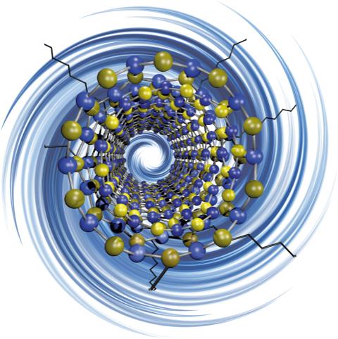
Caption: Rice University researchers have discovered a way to ‘decorate’ electrically insulating boron nitride nanotubes with functional groups, making them more suitable for use with polymers and composite materials. Credit: Martí Research Group/Rice University

Here’s a link to and a citation for the paper,

Chemical Decoration of Boron Nitride Nanotubes Using the Billups-Birch Reaction: Toward Enhanced Thermostable Reinforced Polymer and Ceramic Nanocomposites by Carlos A. de los Reyes, Kendahl L. Walz Mitra, Ashleigh D. Smith, Sadegh Yazdi, Axel Loredo, Frank J. Frankovsky, Emilie Ringe, Matteo Pasquali, and Angel A. Martí. ACS Appl. Nano Mater., Article ASAP DOI: 10.1021/acsanm.8b00633 Publication Date (Web): May 16, 2018

Copyright © 2018 American Chemical Society

This paper is behind a paywall.

Nanorods as multistate switches

This research goes beyond the binary (0 or 1) and to an analog state that resembles quantum states. Fascinating, yes? An Oct. 10, 2016 news item on phys.org tells more,

Rice University scientists have discovered how to subtly change the interior structure of semi-hollow nanorods in a way that alters how they interact with light, and because the changes are reversible, the method could form the basis of a nanoscale switch with enormous potential.

“It’s not 0-1, it’s 1-2-3-4-5-6-7-8-9-10,” said Rice materials scientist Emilie Ringe, lead scientist on the project, which is detailed in the American Chemical Society journal Nano Letters. “You can differentiate between multiple plasmonic states in a single particle. That gives you a kind of analog version of quantum states, but on a larger, more accessible scale.”

Ringe and colleagues used an electron beam to move silver from one location to another inside gold-and-silver nanoparticles, something like a nanoscale Etch A Sketch. The result is a reconfigurable optical switch that may form the basis for a new type of multiple-state computer memory, sensor or catalyst.

An Oct. 10, 2016 Rice University news release, which originated the news item, describes the work in additional detail,

At about 200 nanometers long, 500 of the metal rods placed end-to-end would span the width of a human hair. However, they are large in comparison with modern integrated circuits. Their multistate capabilities make them more like reprogrammable bar codes than simple memory bits, she said.

“No one has been able to reversibly change the shape of a single particle with the level of control we have, so we’re really excited about this,” Ringe said.

Altering a nanoparticle’s internal structure also alters its external plasmonic response. Plasmons are the electrical ripples that propagate across the surface of metallic materials when excited by light, and their oscillations can be easily read with a spectrometer — or even the human eye — as they interact with visible light.

The Rice researchers found they could reconfigure nanoparticle cores with pinpoint precision. That means memories made of nanorods need not be merely on-off, Ringe said, because a particle can be programmed to emit many distinct plasmonic patterns.

The discovery came about when Ringe and her team, which manages Rice’s advanced electron microscopy lab, were asked by her colleague and co-author Denis Boudreau, a professor at Laval University in Quebec, to characterize hollow nanorods made primarily of gold but containing silver.

“Most nanoshells are leaky,” Ringe said. “They have pinholes. But we realized these nanorods were defect-free and contained pockets of water that were trapped inside when the particles were synthesized. We thought: We have something here.”

Ringe and the study’s lead author, Rice research scientist Sadegh Yazdi, quickly realized how they might manipulate the water. “Obviously, it’s difficult to do chemistry there, because you can’t put molecules into a sealed nanoshell. But we could put electrons in,” she said.

Focusing a subnanometer electron beam on the interior cavity split the water and inserted solvated electrons – free electrons that can exist in a solution. “The electrons reacted directly with silver ions in the water, drawing them to the beam to form silver,” Ringe said. The now-silver-poor liquid moved away from the beam, and its silver ions were replenished by a reaction of water-splitting byproducts with the solid silver in other parts of the rod.

“We actually were moving silver in the solution, reconfiguring it,” she said. “Because it’s a closed system, we weren’t losing anything and we weren’t gaining anything. We were just moving it around, and could do so as many times as we wished.”

The researchers were then able to map the plasmon-induced near-field properties without disturbing the internal structure — and that’s when they realized the implications of their discovery.

“We made different shapes inside the nanorods, and because we specialize in plasmonics, we mapped the plasmons and it turned out to have a very nice effect,” Ringe said. “We basically saw different electric-field distributions at different energies for different shapes.” Numerical results provided by collaborators Nicolas Large of the University of Texas at San Antonio and George Schatz of Northwestern University helped explain the origin of the modes and how the presence of a water-filled pocket created a multitude of plasmons, she said.

The next challenge is to test nanoshells of other shapes and sizes, and to see if there are other ways to activate their switching potentials. Ringe suspects electron beams may remain the best and perhaps only way to catalyze reactions inside particles, and she is hopeful.

“Using an electron beam is actually not as technologically irrelevant as you might think,” she said. “Electron beams are very easy to generate. And yes, things need to be in vacuum, but other than that, people have generated electron beams for nearly 100 years. I’m sure 40 years ago people were saying, ‘You’re going to put a laser in a disk reader? That’s crazy!’ But they managed to do it.

“I don’t think it’s unfeasible to miniaturize electron-beam technology. Humans are good at moving electrons and electricity around. We figured that out a long time ago,” Ringe said.

The research should trigger the imaginations of scientists working to create nanoscale machines and processes, she said.

“This is a reconfigurable unit that you can access with light,” she said. “Reading something with light is much faster than reading with electrons, so I think this is going to get attention from people who think about dynamic systems and people who think about how to go beyond current nanotechnology. This really opens up a new field.”

Here’s a link to and a citation for the paper,

Reversible Shape and Plasmon Tuning in Hollow AgAu Nanorods by Sadegh Yazdi, Josée R. Daniel, Nicolas Large, George C. Schatz, Denis Boudreau, and Emilie Ringe. Nano Lett., Article ASAP DOI: 10.1021/acs.nanolett.6b02946 Publication Date (Web): October 5, 2016

Copyright © 2016 American Chemical Society

This paper is behind a paywall.

The researchers have made this video available for the public,

‘Stained glass nanotechnology’ for color displays

From a Dec. 4, 2015 news item on ScienceDaily,

A new method for building “drawbridges” between metal nanoparticles may allow electronics makers to build full-color displays using light-scattering nanoparticles that are similar to the gold materials that medieval artisans used to create red stained-glass.

“Wouldn’t it be interesting if we could create stained-glass windows that changed colors at the flip of a switch?” said Christy Landes, associate professor of chemistry at Rice and the lead researcher on a new study about the drawbridge method that appears this week in the open-access journal Science Advances.

The research by Landes and other experts at Rice University’s Smalley-Curl Institute could allow engineers to use standard electrical switching techniques to construct color displays from pairs of nanoparticles that scatter different colors of light.

For centuries, stained-glass makers have tapped the light-scattering properties of tiny gold nanoparticles to produce glass with rich red tones. Similar types of materials could increasingly find use in modern electronics as manufacturers work to make smaller, faster and more energy-efficient components that operate at optical frequencies.

A Dec. 4, 2015 Rice University news release (also on EurekAlert), which originated the news item, describes the research in more detail,

Though metal nanoparticles scatter bright light, researchers have found it difficult to coax them to produce dramatically different colors, Landes said.

Rice’s new drawbridge method for color switching incorporates metal nanoparticles that absorb light energy and convert it into plasmons, waves of electrons that flow like a fluid across a particle’s surface. Each plasmon scatters and absorbs a characteristic frequency of light, and even minor changes in the wave-like sloshing of a plasmon shift that frequency. The greater the change in plasmonic frequency, the greater the difference between the colors observed.

“Engineers hoping to make a display from optically active nanoparticles need to be able to switch the color,” Landes said. “That type of switching has proven very difficult to achieve with nanoparticles. People have achieved moderate success using various plasmon-coupling schemes in particle assemblies. What we’ve shown though is variation of the coupling mechanism itself, which can be used to produce huge color changes both rapidly and reversibly.”

To demonstrate the method, Landes and study lead author Chad Byers, a graduate student in her lab, anchored pairs of gold nanoparticles to a glass surface covered with indium tin oxide (ITO), the same conductor that’s used in many smartphone screens. By sealing the particles in a chamber filled with a saltwater electrolyte and a silver electrode, Byers and Landes were able form a device with a complete circuit. They then showed they could apply a small voltage to the ITO to electroplate silver onto the surface of the gold particles. In that process, the particles were first coated with a thin layer of silver chloride. By later applying a negative voltage, the researchers caused a conductive silver “drawbridge” to form. Reversing the voltage caused the bridge to withdraw.

“The great thing about these chemical bridges is that we can create and eliminate them simply by applying or reversing a voltage,” Landes said. “This is the first method yet demonstrated to produce dramatic, reversible color changes for devices built from light-activated nanoparticles.”

This research has its roots in previous work (from the news release),

Byers said his research into the plasmonic behavior of gold dimers began about two years ago.

“We were pursuing the idea that we could make significant changes in optical properties of individual particles simply by altering charge density,” he said. “Theory predicts that colors can be changed just by adding or removing electrons, and we wanted to see if we could do that reversibly, simply by turning a voltage on or off.”

The experiments worked. The color shift was observed and reversible, but the change in the color was minute.

“It wasn’t going to get anybody excited about any sort of switchable display applications,” Landes said.

But she and Byers also noticed that their results differed from the theoretical predictions.

Landes said that was because the predictions were based upon using an inert electrode made of a metal like palladium that isn’t subject to oxidation. But silver is not inert. It reacts easily with oxygen in air or water to form a coat of unsightly silver oxide. This oxidizing layer can also form from silver chloride, and Landes said that is what was occurring when the silver counter electrode was used in Byers’ first experiments.

The scientists decided to embrace imperfection (from the news release),

“It was an imperfection that was throwing off our results, but rather than run away from it, we decided to use it to our advantage,” Landes said.

Rice plasmonics pioneer and study co-author Naomi Halas, director of the Smalley-Curl Institute, said the new research shows how plasmonic components could be used to produce electronically switchable color-displays.

“Gold nanoparticles are particularly attractive for display purposes,” said Halas, Rice’s Stanley C. Moore Professor of Electrical and Computer Engineering and professor of chemistry, bioengineering, physics and astronomy, and materials science and nanoengineering. “Depending upon their shape, they can produce a variety of specific colors. They are also extremely stable, and even though gold is expensive, very little is needed to produce an extremely bright color.”

In designing, testing and analyzing the follow-up experiments on dimers, Landes and Byers engaged with a brain trust of Rice plasmonics experts that included Halas, physicist and engineer Peter Nordlander, chemist Stephan Link, materials scientist Emilie Ringe and their students, as well as Paul Mulvaney of the University of Melbourne in Australia.

Together, the team confirmed the composition and spacing of the dimers and showed how metal drawbridges could be used to induce large color shifts based on voltage inputs.

Nordlander and Hui Zhang, the two theorists in the group, examined the device’s “plasmonic coupling,” the interacting dance that plasmons engage in when they are in close contact. For instance, plasmonic dimers are known to act as light-activated capacitors, and prior research has shown that connecting dimers with nanowire bridges brings about a new state of resonance known as a “charge-transfer plasmon,” which has its own distinct optical signature.

“The electrochemical bridging of the interparticle gap enables a fully reversible transition between two plasmonic coupling regimes, one capacitive and the other conductive,” Nordlander said. “The shift between these regimes is evident from the dynamic evolution of the charge transfer plasmon.”

Halas said the method provides plasmonic researchers with a valuable tool for precisely controlling the gaps between dimers and other multiparticle plasmonic configurations.

“In an applied sense, gap control is important for the development of active plasmonic devices like switches and modulators, but it is also an important tool for basic scientists who are conducting curiosity-driven research in the emerging field of quantum plasmonics.”

I’m glad the news release writer included the background work leading to this new research and to hint at the level of collaboration needed to achieve the scientists’ new understanding of color switching.

Here’s a link to and a citation for the paper,

From tunable core-shell nanoparticles to plasmonic drawbridges: Active control of nanoparticle optical properties by Chad P. Byers, Hui Zhang, Dayne F. Swearer, Mustafa Yorulmaz, Benjamin S. Hoener, Da Huang, Anneli Hoggard, Wei-Shun Chang, Paul Mulvaney, Emilie Ringe, Naomi J. Halas, Peter Nordlander, Stephan Link, and Christy F. Landes. Science Advances  04 Dec 2015: Vol. 1, no. 11, e1500988 DOI: 10.1126/sciadv.1500988

In case you missed it in the news release, this is an open access paper.

Combining gold and palladium for catalytic and plasmonic octopods

Hopefully I did not the change meaning when I made the title for this piece more succinct. In any event, this research comes from the always prolific Rice University in Texas, US (from a Nov. 30, 2015 news item on Nanotechnology Now),

Catalysts are substances that speed up chemical reactions and are essential to many industries, including petroleum, food processing and pharmaceuticals. Common catalysts include palladium and platinum, both found in cars’ catalytic converters. Plasmons are waves of electrons that oscillate in particles, usually metallic, when excited by light. Plasmonic metals like gold and silver can be used as sensors in biological applications and for chemical detection, among others.

Plasmonic materials are not the best catalysts, and catalysts are typically very poor for plasmonics. But combining them in the right way shows promise for industrial and scientific applications, said Emilie Ringe, a Rice assistant professor of materials science and nanoengineering and of chemistry who led the study that appears in Scientific Reports.

“Plasmonic particles are magnets for light,” said Ringe, who worked on the project with colleagues in the U.S., the United Kingdom and Germany. “They couple with light and create big electric fields that can drive chemical processes. By combining these electric fields with a catalytic surface, we could further push chemical reactions. That’s why we’re studying how palladium and gold can be incorporated together.”

The researchers created eight-armed specks of gold and coated them with a gold-palladium alloy. The octopods proved to be efficient catalysts and sensors.

A Nov. 30, 2015 Rice University news release (also on EurekAlert), which originated the news item, expands on the theme,

“If you simply mix gold and palladium, you may end up with a bad plasmonic material and a pretty bad catalyst, because palladium does not attract light like gold does,” Ringe said. “But our particles have gold cores with palladium at the tips, so they retain their plasmonic properties and the surfaces are catalytic.”

Just as important, Ringe said, the team established characterization techniques that will allow scientists to tune application-specific alloys that report on their catalytic activity in real time.

The researchers analyzed octopods with a variety of instruments, including Rice’s new Titan Themis microscope, one of the most powerful electron microscopes in the nation. “We confirmed that even though we put palladium on a particle, it’s still capable of doing everything that a similar gold shape would do. That’s really a big deal,” she said.

“If you shine a light on these nanoparticles, it creates strong electric fields. Those fields enhance the catalysis, but they also report on the catalysis and the molecules present at the surface of the particles,” Ringe said.

The researchers used electron energy loss spectroscopy, cathodoluminescence and energy dispersive X-ray spectroscopy to make 3-D maps of the electric fields produced by exciting the plasmons. They found that strong fields were produced at the palladium-rich tips, where plasmons were the least likely to be excited.

Ringe expects further research will produce multifunctional nanoparticles in a variety of shapes that can be greatly refined for applications. Her own Rice lab is working on a metal catalyst to turn inert petroleum derivatives into backbone molecules for novel drugs.

Here’s a link to and a citation for the paper,

Resonances of nanoparticles with poor plasmonic metal tips by Emilie Ringe, Christopher J. DeSantis, Sean M. Collins, Martial Duchamp, Rafal E. Dunin-Borkowski, Sara E. Skrabalak, & Paul A. Midgley.  Scientific Reports 5, Article number: 17431 (2015)  doi:10.1038/srep17431 Published online: 30 November 2015

This is an open access paper,

“This is the best microscope we could ever dream of”—Rice University (US) gets new microscope

I believe it’s Emilie Ringe who’s hosting this video about the new microscope at Rice University (Texas, US) and, as you will be able to tell, she’s thrilled.

A June 29, 2015 news item on Nanotechnology Now explains some of Ringe’s excitement,

Rice University, renowned for nanoscale science, has installed microscopes that will allow researchers to peer deeper than ever into the fabric of the universe.

The Titan Themis scanning/transmission electron microscope, one of the most powerful in the United States, will enable scientists from Rice as well as academic and industrial partners to view and analyze materials smaller than a nanometer — a billionth of a meter — with startling clarity.

The microscope has the ability to take images of materials at angstrom-scale (one-tenth of a nanometer) resolution, about the size of a single hydrogen atom.

Images will be captured with a variety of detectors, including X-ray, optical and multiple electron detectors and a 4K-resolution camera, equivalent to the number of pixels in the most modern high-resolution televisions. The microscope gives researchers the ability to create three-dimensional structural reconstructions and carry out electric field mapping of subnanoscale materials.

“Seeing single atoms is exciting, of course, and it’s beautiful,” said Emilie Ringe, a Rice assistant professor of materials science and nanoengineering and of chemistry. “But scientists saw single atoms in the ’90s, and even before. Now, the real breakthrough is that we can identify the composition of those atoms, and do it easily and reliably.” Ringe’s research group will operate the Titan Themis and a companion microscope that will image larger samples.

A June 29, 2015 Rice University news release, which originated the news item, provides more information about electron microscopes, incident electron beams, and the specifics of the second new piece of equipment being installed,

Electron microscopes use beams of electrons rather than rays of light to illuminate objects of interest. Because the wavelength of electrons is so much smaller than that of photons, the microscopes are able to capture images of much smaller things with greater detail than even the highest-resolution optical microscope.

“The beauty of these newer instruments is their analytical capabilities,” Ringe said. “Before, in order to see single atoms, we had to work a machine for an entire day and get it just right and then take a picture and hold our breath. These days, seeing atoms is routine.

“And now we can probe a particular atom’s chemical composition. Through various techniques, either via scattering intensity, X-rays emission or electron-beam absorption, we can figure out, say, that we’re looking at a palladium atom or a carbon atom. We couldn’t do that before.”

Ringe said when an electron beam ejects a bound electron from a target atom, it creates an empty site. “That can be filled by another electron within the atom, and the energy difference between this electron and the missing electron is emitted as an X-ray,” she said. “That X-ray is like a fingerprint, which we can read. Different types of atoms have different energies.”

She said the incident electron beam loses a bit of energy when it knocks an atom’s electron loose, and that energy loss can also be measured with a spectroscope to identify the atom. The X-ray and electron techniques are independent but complementary. “Typically, you use either/or, and it depends on what element you’re looking at,” Ringe said.

The second instrument, a Helios NanoLab 600 DualBeam microscope, will be used for three-dimensional imaging, analysis of larger samples and preparation of thin slices of samples for the more powerful Titan next door.

Both tools reside in the university’s Brockman Hall for Physics, which opened in 2011 and features sophisticated vibration-dampening capabilities. The microscopes require the best possible isolation from vibration, electric fields and acoustic noise to produce the best images, Ringe said.

“We have wanted a high-end microscopy facility at Rice because so many of us are working on nanomaterials,” said Pulickel Ajayan, a professor and founding chair of Rice’s Department of Materials Science and NanoEngineering. “This has been an issue because in order to be competitive you have to have the best atomic-scale characterization techniques. This will put us in business in terms of imaging and understanding new materials.”

He said the facility will position Rice as one of the most competitive institutions to recruit students and faculty, attract grants and publish groundbreaking results.

“A visual image of something on an atomic level can give you so much more information than a few numbers can,” said Peter Rossky, a theoretical chemist and dean of Rice’s Wiess School of Natural Sciences. Comparing images of the same material taken by an older electron microscope and the Titan Themis was like “the difference between a black-and-white TV and high-definition color,” he said.

Ringe said Rice’s Titan is a fourth-generation model manufactured in the Netherlands. It’s the latest and most powerful model and the first to be installed in the United States.

“Taking a complex image — not just a picture but a spectrum image that has lots of energy information — in the older model would take about 35 minutes,” she said. “By that time, the electron beam has destroyed whatever you were trying to look at.

“With this generation, you have the data you need in about two minutes. You can generate a lot more data more quickly. It’s not just better; it’s enabling.”

Edwin Thomas, the William and Stephanie Sick Dean of Rice’s George R. Brown School of Engineering, expects the new instruments to ignite the already strong research culture at the university. “This is going to influence the kind of people who will be attracted to apply to and then come to Rice,” said Thomas, a materials scientist. “I’m sure there will be people on campus who, once they find out the capabilities, are going to shift their compasses and take advantage of these machines. The whole point is to have an impact on science and society.”

Rice plans to host a two-day workshop in September to introduce the microscopes and their capabilities to the research community at the university and beyond. [emphasis mine] Beginning this summer, Ringe said, the electron microscopy center will be open to Rice students and faculty as well as researchers from other universities and industry.

Ringe looks forward to bringing researchers into the new microscopy lab — and to the research that will emerge.

“I hope everyone’s going to come out with a blockbuster paper with images from these instruments,” she said. “I would like every paper from Rice to have fantastic, crystal-clear, atomic-resolution images and the best possible characterization.”

To sum this up, there are two new pieces of equipment (Titan Themis scanning/transmission electron microscope and Helios NanoLab 600 DualBeam microscope) in Rice University’s 2011 facility, Brockman Hall for Physics. They are very excited about having the most powerful microscope in the US (the Titan) and hope to be holding a two-day workshop on these new microscopes for the research community at Rice and at other institutions.