Tag Archives: US National Institutes of Health

North Carolina universities go beyond organ-on-a-chip

The researchers in the North Carolina universities involved in this project have high hopes according to an Oct. 9, 2015 news item on Nanowerk,

A team of researchers from the University of North Carolina at Chapel Hill and NC State University has received a $5.3 million, five-year Transformative Research (R01) Award from the National Institutes of Health (NIH) to create fully functioning versions of the human gut that fit on a chip the size of a dime.

Such “organs-on-a-chip” have become vital for biomedical research, as researchers seek alternatives to animal models for drug discovery and testing. The new grant will fund a technology that represents a major step forward for the field, overcoming limitations that have mired other efforts.

The technology will use primary cells derived directly from human biopsies, which are known to provide more relevant results than the immortalized cell lines used in current approaches. In addition, the device will sculpt these cells into the sophisticated architecture of the gut, rather than the disorganized ball of cells that are created in other miniature organ systems.

“We are building a device that goes far beyond the organ-on-a-chip,” said Nancy L. Allbritton, MD, PhD, professor and chair of the UNC-NC State joint department of biomedical engineering and one of four principle investigators on the NIH grant. “We call it a ‘simulacrum,’ [emphasis mine] a term used in science fiction to describe a duplicate. The idea is to create something that is indistinguishable from your own gut.”

I’ve come across the term ‘simulacrum’ in relation to philosophy so it’s a bit of a surprise to find it in a news release about an organ-on-a-chip where it seems to have been redefined somewhat. Here’s more from the Simulacrum entry on Wikipedia (Note: Links have been removed),

A simulacrum (plural: simulacra from Latin: simulacrum, which means “likeness, similarity”), is a representation or imitation of a person or thing.[1] The word was first recorded in the English language in the late 16th century, used to describe a representation, such as a statue or a painting, especially of a god. By the late 19th century, it had gathered a secondary association of inferiority: an image without the substance or qualities of the original.[2] Philosopher Fredric Jameson offers photorealism as an example of artistic simulacrum, where a painting is sometimes created by copying a photograph that is itself a copy of the real.[3] Other art forms that play with simulacra include trompe-l’œil,[4] pop art, Italian neorealism, and French New Wave.[3]

Philosophy

The simulacrum has long been of interest to philosophers. In his Sophist, Plato speaks of two kinds of image making. The first is a faithful reproduction, attempted to copy precisely the original. The second is intentionally distorted in order to make the copy appear correct to viewers. He gives the example of Greek statuary, which was crafted larger on the top than on the bottom so that viewers on the ground would see it correctly. If they could view it in scale, they would realize it was malformed. This example from the visual arts serves as a metaphor for the philosophical arts and the tendency of some philosophers to distort truth so that it appears accurate unless viewed from the proper angle.[5] Nietzsche addresses the concept of simulacrum (but does not use the term) in the Twilight of the Idols, suggesting that most philosophers, by ignoring the reliable input of their senses and resorting to the constructs of language and reason, arrive at a distorted copy of reality.[6]

Postmodernist French social theorist Jean Baudrillard argues that a simulacrum is not a copy of the real, but becomes truth in its own right: the hyperreal. Where Plato saw two types of representation—faithful and intentionally distorted (simulacrum)—Baudrillard sees four: (1) basic reflection of reality; (2) perversion of reality; (3) pretence of reality (where there is no model); and (4) simulacrum, which “bears no relation to any reality whatsoever”.[7] In Baudrillard’s concept, like Nietzsche’s, simulacra are perceived as negative, but another modern philosopher who addressed the topic, Gilles Deleuze, takes a different view, seeing simulacra as the avenue by which an accepted ideal or “privileged position” could be “challenged and overturned”.[8] Deleuze defines simulacra as “those systems in which different relates to different by means of difference itself. What is essential is that we find in these systems no prior identity, no internal resemblance”.[9]

Getting back to the proposed research, an Oct. (?), 2015 University of North Carolina news release, which originated the news item, describes the proposed work in more detail,

Allbritton is an expert at microfabrication and microengineering. Also on the team are intestinal stem cell expert Scott T. Magness, associate professor of medicine, biomedical engineering, and cell and molecular physiology in the UNC School of Medicine; microbiome expert Scott Bultman, associate professor of genetics in the UNC School of Medicine; and bioinformatics expert Shawn Gomez, associate professor of biomedical engineering in UNC’s College of Arts and Sciences and NC State.

The impetus for the “organ-on-chip” movement comes largely from the failings of the pharmaceutical industry. For just a single drug to go through the discovery, testing, and approval process can take as many as 15 years and as much as $5 billion dollars. Animal models are expensive to work with and often don’t respond to drugs and diseases the same way humans do. Human cells grown in flat sheets on Petri dishes are also a poor proxy. Three-dimensional “organoids” are an improvement, but these hollow balls are made of a mishmash of cells that doesn’t accurately mimic the structure and function of the real organ.

Basically, the human gut is a 30-foot long hollow tube made up of a continuous single-layer of specialized cells. Regenerative stem cells reside deep inside millions of small pits or “crypts” along the tube, and mature differentiated cells are linked to the pits and live further out toward the surface. The gut also contains trillions of microbes, which are estimated to outnumber human cells by ten to one. These diverse microbial communities – collectively known as the microbiota – process toxins and pharmaceuticals, stimulate immunity, and even release hormones to impact behavior.

To create a dime-sized version of this complex microenvironment, the UNC-NC State team borrowed fabrication technologies from the electronics and microfluidics world. The device is composed of a polymer base containing an array of imprinted or shaped “hydrogels,” a mesh of molecules that can absorb water like a sponge. These hydrogels are specifically engineered to provide the structural support and biochemical cues for growing cells from the gut. Plugged into the device will be various kinds of plumbing that bring in chemicals, fluids, and gases to provide cues that tell the cells how and where to differentiate and grow. For example, the researchers will engineer a steep oxygen gradient into the device that will enable oxygen-loving human cells and anaerobic microbes to coexist in close proximity.

“The underlying concept – to simply grow a piece of human tissue in a dish – doesn’t seem that groundbreaking,” said Magness. “We have been doing that for a long time with cancer cells, but those efforts do not replicate human physiology. Using native stem cells from the small intestine or colon, we can now develop gut tissue layers in a dish that contains stem cells and all the differentiated cells of the gut. That is the thing stem cell biologists and engineers have been shooting for, to make real tissue behave properly in a dish to create better models for drug screening and cell-based therapies. With this work, we made a big leap toward that goal.”

Right now, the team has a working prototype that can physically and chemically guide mouse intestinal stem cells into the appropriate structure and function of the gut. For several years, Magness has been isolating and banking human stem cells from samples from patients undergoing routine colonoscopies at UNC Hospitals.

As part of the grant, he will work with the rest of the team to apply these stem cells to the new device and create “simulacra” that are representative of each patient’s individual gut. The approach will enable researchers to explore in a personalized way how both the human and microbial cells of the gut behave during healthy and diseased states.

“Having a system like this will advance microbiota research tremendously,” said Bultman. “Right now microbiota studies involve taking samples, doing sequencing, and then compiling an inventory of all the microbes in the disease cases and healthy controls. These studies just draw associations, so it is difficult to glean cause and effect. This device will enable us to probe the microbiota, and gain a better understanding of whether changes in these microbial communities are the cause or the consequence of disease.”

I wish them good luck with their work and to end on another interesting note, the concept of organs-on-a-chip won a design award. From a June 22, 2015 article by Oliver Wainwright for the Guardian (Note: Links have been removed),

Meet the Lung-on-a-chip, a simulation of the biological processes inside the human lung, developed by the Wyss Institute for Biologically Inspired Engineering at Harvard University – and now crowned Design of the Year by London’s Design Museum.

Lined with living human cells, the “organs-on-chips” mimic the tissue structures and mechanical motions of human organs, promising to accelerate drug discovery, decrease development costs and potentially usher in a future of personalised medicine.

“This is the epitome of design innovation,” says Paola Antonelli, design curator at New York’s Museum of Modern Art [MOMA], who nominated the project for the award and recently acquired organs-on-chips for MoMA’s permanent collection. “Removing some of the pitfalls of human and animal testing means, theoretically, that drug trials could be conducted faster and their viable results disseminated more quickly.”

Whodathunkit? (Tor those unfamiliar with slang written in this form: Who would have thought it?)
Acoustofluidics and lab-on-a-chip for asthma and tuberculosis diagnostics

This is my first exposure to acoustofluidics (although it’s been around for a few years) and it concerns lab-on-a-chip diagnostics for asthma and tuberculosis. From an Aug. 3, 2015 news item on Azonano,

A device to mix liquids utilizing ultrasonics is the first and most difficult component in a miniaturized system for low-cost analysis of sputum from patients with pulmonary diseases such as tuberculosis and asthma.

The device, developed by engineers at Penn State in collaboration with researchers at the National Heart, Lung, and Blood Institute (NHLBI), part of the National Institutes of Health, and the Washington University School of Medicine, will benefit patients in the U.S., where 12 percent of the population, or around 19 million people, have asthma, and in undeveloped regions where TB is still a widespread and often deadly contagion.

“To develop more accurate diagnosis and treatment approaches for patients with pulmonary diseases, we have to analyze sample cells directly from the lungs rather than by drawing blood,” said Tony Jun Huang, professor of engineering science and mechanics at Penn State and the inventor, with his group, of this and other acoustofluidic devices based on ultrasonic waves. “For instance, different drugs are used to treat different types of asthma patients. If you know what a person’s immunophenotype is, you can provide personalized medicine for their particular disease.

A July 29, 2015 Pennsylvania State University news release, which originated the news item, describes the disadvantages of the current sputum analyses techniques and explains how this new technique in an improvement,

There are several issues with the current standard method for sputum analysis. The first is that human specimens can be contagious, and sputum analysis requires handling of specimens in several discrete machines. With a lab on a chip device, all biospecimens are safely contained in a single disposable component.

Another issue is the sample size required for analysis in the current system, which is often larger than a person can easily produce. The acoustofluidic sputum liquefier created by Huang’s group requires 100 times less sample while still providing accuracy equivalent to the standard system.

A further issue is that current systems are difficult to use and require trained operators. With the lab on a chip system, a nurse can operate the device with a touch of a few buttons and get a read out, or the patient could even operate the device at home. In addition, the disposable portion of the device should cost less than a dollar to manufacture.

Po-Hsun Huang, a graduate student in the Huang group and the first author on the recent paper describing the device in the Royal Society of Chemistry journal Lab on a Chip, said “This will offer quick analysis of samples without having to send them out to a centralized lab. While I have been working on the liquefaction component of the device, my lab mates are working on the flow cytometry analysis component, which should be ready soon. This is the first on-chip sputum liquefier anyone has developed.”

Stewart J. Levine, a Senior Investigator and Chief of the Laboratory of Asthma and Lung Inflammation in the Division of Intramural Research at NHLBI, said “This on-chip sputum liquefier is a significant advance regarding our goal of developing a point-of-care diagnostic device that will determine the type of inflammation present in the lungs of asthmatics. This will allow health care providers to individualize asthma treatments for each patient and advance the goal of bringing precision medicine into clinical practice.”

Here’s a link to and a citation for the paper,

An acoustofluidic sputum liquefier by Po-Hsun Huang, Liqiang Ren, Nitesh Nama, Sixing Li, Peng Li, Xianglan Yao, Rosemarie A. Cuento, Cheng-Hsin Wei, Yuchao Chen, Yuliang Xie, Ahmad Ahsan Nawaz, Yael G. Alevy, Michael J. Holtzman, J. Philip McCoy, Stewart J. Levine, and  Tony Jun Huang. Lab Chip, 2015,15, 3125-3131 DOI: 10.1039/C5LC00539F

First published online 17 Jun 2015

This is an open access paper but you do need to register for a free (British) Royal Society of Chemistry publishing personal account.

A pragmatic approach to alternatives to animal testing

Retitled and cross-posted from the June 30, 2015 posting (Testing times: the future of animal alternatives) on the International Innovation blog (a CORDIS-listed project dissemination partner for FP7 and H2020 projects).

Maryse de la Giroday explains how emerging innovations can provide much-needed alternatives to animal testing. She also shares highlights of the 9th World Congress on Alternatives to Animal Testing.

‘Guinea pigging’ is the practice of testing drugs that have passed in vitro and in vivo tests on healthy humans in a Phase I clinical trial. In fact, healthy humans can make quite a bit of money as guinea pigs. The practice is sufficiently well-entrenched that there is a magazine, Guinea Pig Zero, devoted to professionals. While most participants anticipate some unpleasant side effects, guinea pigging can sometimes be a dangerous ‘profession’.

HARMFUL TO HEALTH

One infamous incident highlighting the dangers of guinea pigging occurred in 2006 at Northwick Park Hospital outside London. Volunteers were offered £2,000 to participate in a Phase I clinical trial to test a prospective treatment – a monoclonal antibody designed for rheumatoid arthritis and multiple sclerosis. The drug, called TGN1412, caused catastrophic systemic organ failure in participants. All six individuals receiving the drug required hospital treatment. One participant reportedly underwent amputation of fingers and toes. Another reacted with symptoms comparable to John Merrick, the Elephant Man.

The root of the disaster lay in subtle immune system differences between humans and cynomolgus monkeys – the model animal tested prior to the clinical trial. The drug was designed for the CD28 receptor on T cells. The monkeys’ receptors closely resemble those found in humans. However, unlike these monkeys, humans have other immune cells that carry CD28. The trial participants received a starting dosage that was 0.2 per cent of what the monkeys received in their final tests, but failure to take these additional receptors into account meant a dosage that was supposed to occupy 10 per cent of the available CD28 receptors instead occupied 90 per cent. After the event, a Russian inventor purchased the commercial rights to the drug and renamed it TAB08. It has been further developed by Russian company, TheraMAB, and TAB08 is reportedly in Phase II clinical trials.

HUMAN-ON-A-CHIP AND ORGANOID PROJECTS

While animal testing has been a powerful and useful tool for determining safe usage for pharmaceuticals and other types of chemicals, it is also a cruel and imperfect practice. Moreover, it typically only predicts 30-60 per cent of human responses to new drugs. Nanotechnology and other emerging innovations present possibilities for reducing, and in some cases eliminating, the use of animal models.

People for the Ethical Treatment of Animals (PETA), still better known for its publicity stunts, maintains a webpage outlining a number of alternatives including in silico testing (computer modelling), and, perhaps most interestingly, human-on-a-chip and organoid (tissue engineering) projects.

Organ-on-a-chip projects use stem cells to create human tissues that replicate the functions of human organs. Discussions about human-on-a-chip activities – a phrase used to describe 10 interlinked organ chips – were a highlight of the 9th World Congress on Alternatives to Animal Testing held in Prague, Czech Republic, last year. One project highlighted at the event was a joint US National Institutes of Health (NIH), US Food and Drug Administration (FDA) and US Defense Advanced Research Projects Agency (DARPA) project led by Dan Tagle that claimed it would develop functioning human-on-a-chip by 2017. However, he and his team were surprisingly close-mouthed and provided few details making it difficult to assess how close they are to achieving their goal.

By contrast, Uwe Marx – Leader of the ‘Multi-Organ-Chip’ programme in the Institute of Biotechnology at the Technical University of Berlin and Scientific Founder of TissUse, a human-on-a-chip start-up company – claims to have sold two-organ chips. He also claims to have successfully developed a four-organ chip and that he is on his way to building a human-on-a-chip. Though these chips remain to be seen, if they are, they will integrate microfluidics, cultured cells and materials patterned at the nanoscale to mimic various organs, and will allow chemical testing in an environment that somewhat mirrors a human.

Another interesting alternative for animal testing is organoids – a feature in regenerative medicine that can function as test sites. Engineers based at Cornell University recently published a paper on their functional, synthetic immune organ. Inspired by the lymph node, the organoid is comprised of gelatin-based biomaterials, which are reinforced with silicate nanoparticles (to keep the tissue from melting when reaching body temperature) and seeded with cells allowing it to mimic the anatomical microenvironment of a lymphatic node. It behaves like its inspiration converting B cells to germinal centres which activate, mature and mutate antibody genes when the body is under attack. The engineers claim to be able to control the immune response and to outperform 2D cultures with their 3D organoid. If the results are reproducible, the organoid could be used to develop new therapeutics.

Maryse de la Giroday is a science communications consultant and writer.

Full disclosure: Maryse de la Giroday received transportation and accommodation for the 9th World Congress on Alternatives to Animal Testing from SEURAT-1, a European Union project, making scientific inquiries to facilitate the transition to animal testing alternatives, where possible.

ETA July 1, 2015: I would like to acknowledge more sources for the information in this article,

Sources:

The guinea pigging term, the ‘professional aspect, the Northwick Park story, and the Guinea Pig Zero magazine can be found in Carl Elliot’s excellent 2006 story titled ‘Guinea-Pigging’ for New Yorker magazine.

http://www.newyorker.com/magazine/2008/01/07/guinea-pigging

Information about the drug used in the Northwick Park Hospital disaster, the sale of the rights to a Russian inventor, and the June 2015 date for the current Phase II clinical trials were found in this Wikipedia essay titled, TGN 1412.

http://en.wikipedia.org/wiki/TGN1412

Additional information about the renamed drug, TAB08 and its Phase II clinical trials was found on (a) a US government website for information on clinical trials, (b) in a Dec. 2014 (?) TheraMAB  advertisement in a Nature group magazine and a Jan. 2014 press release,

https://www.clinicaltrials.gov/ct2/show/NCT01990157?term=TAB08_RA01&rank=1

http://www.theramab.ru/TheraMAB_NAture.pdf

http://theramab.ru/en/news/phase_II

An April 2015 article (Experimental drug that injured UK volunteers resumes in human trials) by Owen Dyer for the British Medical Journal also mentioned the 2015 TheraMab Phase II clinical trials and provided information about the information about Macaque (cynomolgus) monkey tests.

http://www.bmj.com.proxy.lib.sfu.ca/content/350/bmj.h1831

BMJ 2015; 350 doi: http://dx.doi.org.proxy.lib.sfu.ca/10.1136/bmj.h1831 (Published 02 April 2015) Cite this as: BMJ 2015;350:h1831

A 2009 study by Christopher Horvath and Mark Milton somewhat contradicts the Dyer article’s contention that a species Macaque monkey was used as an animal model. (As the Dyer article is more recent and the Horvath/Milton analysis is more complex covering TGN 1412 in the context of other MAB drugs and their precursor tests along with specific TGN 1412 tests, I opted for the simple description.)

The TeGenero Incident [another name for the Northwick Park Accident] and the Duff Report Conclusions: A Series of Unfortunate Events or an Avoidable Event? by Christopher J. Horvath and Mark N. Milton. Published online before print February 24, 2009, doi: 10.1177/0192623309332986 Toxicol Pathol April 2009 vol. 37 no. 3 372-383

http://tpx.sagepub.com/content/37/3/372.full

Philippa Roxbuy’s May 24, 2013 BBC news online article provided confirmation and an additional detail or two about the Northwick Park Hospital accident. It notes that other models, in addition to animal models, are being developed.

http://www.bbc.com/news/health-22556736

Anne Ju’s excellent June 10,2015 news release about the Cornell University organoid (synthetic immune organ) project was very helpful.

http://www.news.cornell.edu/stories/2015/06/engineers-synthetic-immune-organ-produces-antibodies

There will also be a magazine article in International Innovation, which will differ somewhat from the blog posting, due to editorial style and other requirements.

ETA July 22, 2015: I now have a link to the magazine article.

‘Glow in the dark’, paint-on bandage heals

Somewhat unexpectedly (to me), this research about a ‘smart’ paint-on bandage is being published by The Optical Society of America (OSA). Here’s more about the work from an Oct. 1, 2014 news item on Nanowerk,

Inspired by a desire to help wounded soldiers, an international, multidisciplinary team of researchers led by Assistant Professor Conor L. Evans at the Wellman Center for Photomedicine of Massachusetts General Hospital (MGH) and Harvard Medical School (HMS) has created a paint-on, see-through, “smart” bandage that glows to indicate a wound’s tissue oxygenation concentration. Because oxygen plays a critical role in healing, mapping these levels in severe wounds and burns can help to significantly improve the success of surgeries to restore limbs and physical functions.

An Oct. 1, 2014 OSA news release (also on EurekAlert), which originated the news item, describes the interest in oxygenation in more detail,

“Information about tissue oxygenation is clinically relevant but is often inaccessible due to a lack of accurate or noninvasive measurements,” explained lead author Zongxi Li, an HMS research fellow on Evans’ team.

Now, the “smart” bandage developed by the team provides direct, noninvasive measurement of tissue oxygenation by combining three simple, compact and inexpensive components: a bright sensor molecule with a long phosphorescence lifetime and appropriate dynamic range; a bandage material compatible with the sensor molecule that conforms to the skin’s surface to form an airtight seal; and an imaging device capable of capturing the oxygen-dependent signals from the bandage with high signal-to-noise ratio.

This work is part of the team’s long-term program “to develop a Sensing, Monitoring And Release of Therapeutics (SMART) bandage for improved care of patients with acute or chronic wounds,” says Evans …

The news release goes on to briefly explain the technology,

For starters, the bandage’s not-so-secret key ingredient is phosphors—molecules that absorb light and then emit it via a process known as phosphorescence.

Phosphorescence is encountered by many on a daily basis—ranging from glow-in-the-dark dials on watches to t-shirt lettering. “How brightly our phosphorescent molecules emit light depends on how much oxygen is present,” said Li. “As the concentration of oxygen is reduced, the phosphors glow both longer and more brightly.” To make the bandage simple to interpret, the team also incorporated a green oxygen-insensitive reference dye, so that changes in tissue oxygenation are displayed as a green-to-red colormap.

The bandage is applied by “painting” it onto the skin’s surface as a viscous liquid, which dries to a solid thin film within a minute. Once the first layer has dried, a transparent barrier layer is then applied atop it to protect the film and slow the rate of oxygen exchange between the bandage and room air—making the bandage sensitive to the oxygen within tissue.

The final piece involves a camera-based readout device, which performs two functions: it provides a burst of excitation light that triggers the emission of the phosphors inside the bandage, and then it records the phosphors’ emission. “Depending on the camera’s configuration, we can measure either the brightness or color of the emitted light across the bandage or the change in brightness over time,” Li said. “Both of these signals can be used to create an oxygenation map.”  The emitted light from the bandage is bright enough that it can be acquired using a regular camera or smartphone—opening the possibility to a portable, field-ready device.

There are some immediate applications, as well as, plans for research that will yield applications (from the news release),

Immediate applications for the oxygen-sensing bandage include monitoring patients with a risk of developing ischemic (restricted blood supply) conditions, postoperative monitoring of skin grafts or flaps, and burn-depth determination as a guide for surgical debridement—the removal of dead or damaged tissue from the body.

“The need for a reliable, accurate and easy-to-use method of rapid assessment of blood flow to the skin for patients remains a clinical necessity,” said co-author Samuel Lin, an HMS associate professor of surgery at Beth Israel Deaconess Medical Center. “Plastic surgeons continuously monitor the state of blood flow to the skin, so the liquid-bandage oxygenation sensor is an exciting step toward improving patient care within the realm of vascular blood flow examination of the skin.”

What’s the next step for the bandage? “We’re developing brighter sensor molecules to improve the bandage’s oxygen sensing efficiency,” said Emmanuel Roussakis, another research fellow in Evans’ laboratory and co-author, who is leading the sensor development effort.  The team’s laboratory research will also focus on expanding the sensing capability of the bandage to other treatment-related parameters—such as pH, bacterial load, oxidative states and specific disease markers—and incorporating an on-demand drug release capacity.

“In the future, our goal for the bandage is to incorporate therapeutic release capabilities that allow for on-demand drug administration at a desired location,” says Evans. “It allows for the visual assessment of the wound bed, so treatment-related wound parameters are readily accessible without the need for bandage removal—preventing unnecessary wound disruption and reducing the chance for bacterial infection.”

Should you be interested, the researchers are looking for industry partners,

Beyond the lab, the team’s aim is to move this technology from the bench to the bedside, so they are actively searching for industry partners. They acknowledge research support from the Military Medical Photonics Program from the U.S. Department of Defense, and National Institutes of Health.

Here’s a link to and a citation for the paper,

Non-invasive transdermal two-dimensional mapping of cutaneous oxygenation with a rapid-drying liquid bandage by Zongxi Li, Emmanuel Roussakis, Pieter G. L. Koolen, Ahmed M. S. Ibrahim, Kuylhee Kim, Lloyd F. Rose, Jesse Wu, Alexander J. Nichols, Yunjung Baek, Reginald Birngruber, Gabriela Apiou-Sbirlea, Robina Matyal, Thomas Huang, Rodney Chan, Samuel J. Lin, and Conor L. Evans. Biomedical Optics Express, Vol. 5, Issue 11, pp. 3748-3764 (2014) http://dx.doi.org/10.1364/BOE.5.003748

This article is open access.

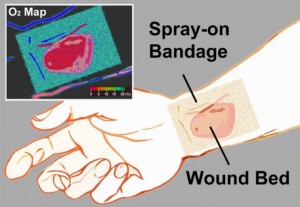
The researcher’s have provided an illustration of the bandage,

Caption: The transparent liquid bandage displays a quantitative, oxygenation-sensitive colormap that can be easily acquired using a simple camera or smartphone. Credit: Li/Wellman Center for Photomedicine.

Caption: The transparent liquid bandage displays a quantitative, oxygenation-sensitive colormap that can be easily acquired using a simple camera or smartphone. Credit: Li/Wellman Center for Photomedicine.

Nanotechnology, tobacco plants, and the Ebola virus

Before presenting information about the current Ebola crisis and issues with vaccines and curatives, here’s a description of the disease from its Wikipedia entry,

Ebola virus disease (EVD) or Ebola hemorrhagic fever (EHF) is a disease of humans and other primates caused by an ebola virus. Symptoms start two days to three weeks after contracting the virus, with a fever, sore throat, muscle pain, and headaches. Typically nausea, vomiting, and diarrhea follow, along with decreased functioning of the liver and kidneys. Around this time, affected people may begin to bleed both within the body and externally. [1]

As for the current crisis in countries situated on the west coast of the African continent, there’s this from an Aug. 14, 2014 news item on ScienceDaily,

The outbreak of Ebola virus disease that has claimed more than 1,000 lives in West Africa this year poses a serious, ongoing threat to that region: the spread to capital cities and Nigeria — Africa’s most populous nation — presents new challenges for healthcare professionals. The situation has garnered significant attention and fear around the world, but proven public health measures and sharpened clinical vigilance will contain the epidemic and thwart a global spread, according to a new commentary by Anthony S. Fauci, M.D., director of the National Institute of Allergy and Infectious Diseases (NIAID), part of the National Institutes of Health.

Dr. Fauci’s Aug. 13, 2014 commentary (open access) in the New England Journal of Medicine provides more detail (Note: A link has been removed),

An outbreak of Ebola virus disease (EVD) has jolted West Africa, claiming more than 1000 lives since the virus emerged in Guinea in early 2014 (see figure) Ebola Virus Cases and Deaths in West Africa (Guinea, Liberia, Nigeria, and Sierra Leone), as of August 11, 2014 (Panel A), and Over Time (Panel B).). The rapidly increasing numbers of cases in the African countries of Guinea, Liberia, and Sierra Leone have had public health authorities on high alert throughout the spring and summer. More recent events including the spread of EVD to Nigeria (Africa’s most populous country) and the recent evacuation to the United States of two American health care workers with EVD have captivated the world’s attention and concern. Health professionals and the general public are struggling to comprehend these unfolding dynamics and to separate misinformation and speculation from truth.

In early 2014, EVD emerged in a remote region of Guinea near its borders with Sierra Leone and Liberia. Since then, the epidemic has grown dramatically, fueled by several factors. First, Guinea, Sierra Leone, and Liberia are resource-poor countries already coping with major health challenges, such as malaria and other endemic diseases, some of which may be confused with EVD. Next, their borders are porous, and movement between countries is constant. Health care infrastructure is inadequate, and health workers and essential supplies including personal protective equipment are scarce. Traditional practices, such as bathing of corpses before burial, have facilitated transmission. The epidemic has spread to cities, which complicates tracing of contacts. Finally, decades of conflict have left the populations distrustful of governing officials and authority figures such as health professionals. Add to these problems a rapidly spreading virus with a high mortality rate, and the scope of the challenge becomes clear.

Although the regional threat of Ebola in West Africa looms large, the chance that the virus will establish a foothold in the United States or another high-resource country remains extremely small. Although global air transit could, and most likely will, allow an infected, asymptomatic person to board a plane and unknowingly carry Ebola virus to a higher-income country, containment should be readily achievable. Hospitals in such countries generally have excellent capacity to isolate persons with suspected cases and to care for them safely should they become ill. Public health authorities have the resources and training necessary to trace and monitor contacts. Protocols exist for the appropriate handling of corpses and disposal of biohazardous materials. In addition, characteristics of the virus itself limit its spread. Numerous studies indicate that direct contact with infected bodily fluids — usually feces, vomit, or blood — is necessary for transmission and that the virus is not transmitted from person to person through the air or by casual contact. Isolation procedures have been clearly outlined by the Centers for Disease Control and Prevention (CDC). A high index of suspicion, proper infection-control practices, and epidemiologic investigations should quickly limit the spread of the virus.

Fauci’s article makes it clear that public concerns are rising in the US and I imagine that’s true of Canada too and many other parts of the world, not to mention the countries currently experiencing the EVD outbreak. In the midst of all this comes a US Food and Drug Administration (FDA) warning as per an Aug. 15, 2014 news item (originated by Reuters reporter Toni Clarke) on Nanowerk,

The U.S. Food and Drug Administration said on Thursday [Aug. 14, 2014] it has become aware of products being sold online that fraudulently claim to prevent or treat Ebola.

The FDA’s warning comes on the heels of comments by Nigeria’s top health official, Onyebuchi Chukwu, who reportedly said earlier Thursday [Aug. 14, 2014] that eight Ebola patients in Lagos, the country’s capital, will receive an experimental treatment containing nano-silver.

Erica Jefferson, a spokeswoman for the FDA, said she could not provide any information about the product referenced by the Nigerians.

The Aug. 14,  2014 FDA warning reads in part,

The U.S. Food and Drug Administration is advising consumers to be aware of products sold online claiming to prevent or treat the Ebola virus. Since the outbreak of the Ebola virus in West Africa, the FDA has seen and received consumer complaints about a variety of products claiming to either prevent the Ebola virus or treat the infection.

There are currently no FDA-approved vaccines or drugs to prevent or treat Ebola. Although there are experimental Ebola vaccines and treatments under development, these investigational products are in the early stages of product development, have not yet been fully tested for safety or effectiveness, and the supply is very limited. There are no approved vaccines, drugs, or investigational products specifically for Ebola available for purchase on the Internet. By law, dietary supplements cannot claim to prevent or cure disease.

As per the FDA’s reference to experimental vaccines, an Aug. 6, 2014 article by Caroline Chen, Mark Niquette, Mark Langreth, and Marie French for Bloomberg describes the ZMapp vaccine/treatment (Note: Links have been removed),

On a small plot of land incongruously tucked amid a Kentucky industrial park sit five weather-beaten greenhouses. At the site, tobacco plants contain one of the most promising hopes for developing an effective treatment for the deadly Ebola virus.

The plants contain designer antibodies developed by San Diego-based Mapp Biopharmaceutical Inc. and are grown in Kentucky by a unit of Reynolds American Inc. Two stricken U.S. health workers received an experimental treatment containing the antibodies in Liberia last week. Since receiving doses of the drug, both patients’ conditions have improved.

Tobacco plant-derived medicines, which are also being developed by a company whose investors include Philip Morris International Inc., are part of a handful of cutting edge plant-based treatments that are in the works for everything from pandemic flu to rabies using plants such as lettuce, carrots and even duckweed. While the technique has existed for years, the treatments have only recently begun to reach the marketplace.

Researchers try to identify the best antibodies in the lab, before testing them on mice, then eventually on monkeys. Mapp’s experimental drug, dubbed ZMapp, has three antibodies, which work together to alert the immune system and neutralize the Ebola virus, she [Erica Ollman Saphire, a molecular biologist at the Scripps Research Institute,] said.

This is where the tobacco comes in: the plants are used as hosts to grow large amounts of the antibodies. Genes for the desired antibodies are fused to genes for a natural tobacco virus, Charles Arntzen, a plant biotechnology expert at Arizona State University, said in an Aug. 4 [2014] telephone interview.

The tobacco plants are then infected with this new artificial virus, and antibodies are grown inside the plant. Eventually, the tobacco is ground up and the antibody is extracted, Arntzen said.

The process of growing antibodies in mammals risks transferring viruses that could infect humans, whereas “plants are so far removed, so if they had some sort of plant virus we wouldn’t get sick because viruses are host-specific,” said Qiang Chen, a plant biologist at Arizona State University in Tempe, Arizona, in a telephone interview.

There is a Canadian (?) company working on a tobacco-based vaccines including one for EVD but as the Bloomberg writers note the project is highly secret,

Another tobacco giant-backed company working on biotech drugs grown in tobacco plants is Medicago Inc. in Quebec City, which is owned by Mitsubishi Tanabe Pharma Corp. and Philip Morris. [emphasis mine]

Medicago is working on testing a vaccine for pandemic influenza and has a production greenhouse facility in North Carolina, said Jean-Luc Martre, senior director for government affairs at Medicago. Medicago is planning a final stage trial of the pandemic flu vaccine for next year, he said in a telephone interview.

The plant method is flexible and capable of making antibodies and vaccines for numerous types of viruses, said Martre. In addition to influenza, the company’s website says it is in early stages of testing products for rabies and rotavirus.

Medicago ‘‘is currently closely working with partners for the production of an Ebola antibody as well as other antibodies that are of interest for bio-defense,” he said in an e-mail. He would not disclose who the partners were. [emphasis mine]

I have checked both the English and French language versions of Medicago’s website and cannot find any information about their work on ebola. (The Bloomberg article provides a good overview of the ebola situation and more. I recommend reading it and/or the Aug. 15, 2014 posting on CTV [Canadian Television Network] which originated from an Associated Press article by Malcolm Ritter).

Moving on to more research and ebola, Dexter Johnson in an Aug. 14, 2014 posting (on his Nanoclast blog on the IEEE [Institute of Electrical and Electronics Engineers] website,) describes some work from Northeastern University (US), Note: Links have been removed,

With the Ebola virus death toll now topping 1000 and even the much publicized experimental treatment ZMapp failing to save the life of a Spanish missionary priest who was treated with it, it is clear that scientists need to explore new ways of fighting the deadly disease. For researchers at Northeastern University in Boston, one possibility may be using nanotechnology.

“It has been very hard to develop a vaccine or treatment for Ebola or similar viruses because they mutate so quickly,” said Thomas Webster, the chair of Northeastern’s chemical engineering department, in a press release. “In nanotechnology we turned our attention to developing nanoparticles that could be attached chemically to the viruses and stop them from spreading.”

Webster, along with many researchers in the nanotechnology community, have been trying to use gold nanoparticles, in combination with near-infrared light, to kill cancer cells with heat. The hope is that the same approach could be used to kill the Ebola virus.

There is also an Aug. 6, 2014 Northeastern University news release by Joe O’Connell describing the technique being used by Webster’s team,

… According to Web­ster, gold nanopar­ti­cles are cur­rently being used to treat cancer. Infrared waves, he explained, heat up the gold nanopar­ti­cles, which, in turn, attack and destroy every­thing from viruses to cancer cells, but not healthy cells.

Rec­og­nizing that a larger sur­face area would lead to a quicker heat-​​up time, Webster’s team cre­ated gold nanos­tars. “The star has a lot more sur­face area, so it can heat up much faster than a sphere can,” Web­ster said. “And that greater sur­face area allows it to attack more viruses once they absorb to the par­ti­cles.” The problem the researchers face, how­ever, is making sure the hot gold nanopar­ti­cles attack the virus or cancer cells rather than the healthy cells.

At this point, there don’t seem to be any curative measures generally available although some are available experimentally in very small quantities.

Researchers propose massive shift in science funding enterprise

The massive science funding shift that researchers are proposing won’t fundamentally change who or what research is funded so much as it will require fewer resources as described in a Jan. 8, 2014 news item on Nanowerk (Note: A link has been removed),

Researchers in the United States have suggested an alternative way to allocate science funding. The method, which is described in EMBO reports (“From funding agencies to scientific agency”), depends on a collective distribution of funding by the scientific community, requires only a fraction of the costs associated with the traditional peer review of grant proposals and, according to the authors, may yield comparable or even better results.

The Jan. 8, 2014 EMBO [European Molecular Biology Organization] news release, which originated the news item, quotes the lead author’s perspective on the current funding systems and describes the proposed solution which is meant for all science funding,

“Peer review of scientific proposals and grants has served science very well for decades. However, there is a strong sense in the scientific community that things could be improved,” said Johan Bollen, professor and lead author of the study from the School of Informatics and Computing at Indiana University. “Our most productive researchers invest an increasing amount of time, energy, and effort into writing and reviewing research proposals, most of which do not get funded. That time could be spent performing the proposed research in the first place.” He added: “Our proposal does not just save time and money but also encourages innovation.”

The new approach is possible due to recent advances in mathematics and  computer technologies. The system involves giving all scientists an annual, unconditional fixed amount of funding to conduct their research. All funded scientists are, however, obliged to donate a fixed percentage of all of the funding that they previously received to other researchers. As a result, the funding circulates through the community, converging on researchers that are expected to make the best use of it. “Our alternative funding system is inspired by the mathematical models used to search the internet for relevant information,” said Bollen. “The decentralized funding model uses the wisdom of the entire scientific community to determine a fair distribution of funding.”

The authors believe that this system can lead to sophisticated behavior at a global level. It would certainly liberate researchers from the time-consuming process of submitting and reviewing project proposals, but could also reduce the uncertainty associated with funding cycles, give researchers much greater flexibility, and allow the community to fund risky but high-reward projects that existing funding systems may overlook.

“You could think of it as a Google-inspired crowd-funding system that encourages all researchers to make autonomous, individual funding decisions towards people, not projects or proposals,” said Bollen. “All you need is a centralized web site where researchers could log-in, enter the names of the scientists they chose to donate to, and specify how much they each should receive.”

The authors emphasize that the system would require oversight to prevent misuse, such as conflicts of interests and collusion. Funding agencies may need to confidentially monitor the flow of funding and may even play a role in directing it. For example they can provide incentives to donate to specific large-scale research challenges that are deemed priorities but which the scientific community can overlook.

“The savings of financial and human resources could be used to identify new targets of funding, to support the translation of scientific results into products and jobs, and to help communicate advances in science and technology,” added Bollen. “This funding system may even have the side-effect of changing publication practices for the better: researchers will want to clearly communicate their vision and research goals to as wide an audience as possible.”

While the research is US-centric, it’s easy to see its applicabllity in many, if not all, jurisdictions around the world.

I have two links and two citations. The first is for the EMBO Reports paper,

From funding agencies to scientific agency; Collective allocation of science funding as an alternative to peer review by  Johan Bollen, David Crandall, Damion Junk, Ying Ding, & Katy Börner. Article first published online: 7 JAN 2014 DOI: 10.1002/embr.201338068

© 2014 The Authors

This paper is behind a paywall.

The second link and citation is for an earlier version of the paper on arXiv.org, which is an open access archive,

Collective allocation of science funding: from funding agencies to scientific agency
by Johan Bollen, David Crandall, Damion Junk, Ying Ding, & Katy Boerner.
(Submitted on 3 Apr 2013)

Here’s the abstract from the April 2013 version of the paper on arXiv.org,

Public agencies like the U.S. National Science Foundation (NSF) and the National Institutes of Health (NIH) award tens of billions of dollars in annual science funding. How can this money be distributed as efficiently as possible to best promote scientific innovation and productivity? The present system relies primarily on peer review of project proposals. In 2010 alone, NSF convened more than 15,000 scientists to review 55,542 proposals. [emphasis mine] Although considered the scientific gold standard, peer review requires significant overhead costs, and may be subject to biases, inconsistencies, and oversights. We investigate a class of funding models in which all participants receive an equal portion of yearly funding, but are then required to anonymously donate a fraction of their funding to peers. The funding thus flows from one participant to the next, each acting as if he or she were a funding agency themselves. Here we show through a simulation conducted over large-scale citation data (37M articles, 770M citations) that such a distributed system for science may yield funding patterns similar to existing NIH and NSF distributions, but may do so at much lower overhead while exhibiting a range of other desirable features. Self-correcting mechanisms in scientific peer evaluation can yield an efficient and fair distribution of funding. The proposed model can be applied in many situations in which top-down or bottom-up allocation of public resources is either impractical or undesirable, e.g. public investments, distribution chains, and shared resource management.

It’s interesting to note the agencies which supported the research (from the news release),

Awards from the National Science Foundation, the Andrew W. Mellon Foundation and the National Institutes of Health supported the work.

It would seem there’s an appetite for change given the National Science Foundation (NSF) and the National Institutes of Health (NIH) are the two largest science funding agencies in the US.

Evolution is the best problem-solver

Dr. Jeffrey Karp of Brigham and Women’s Hospital says, “I truly believe evolution is the best problem-solver,” when discussing his medical biomimcry work in this video,

You can find the video and more in a Mar. 20, 2013 news item on Nanowerk which was originated by a Mar. 6, 2013 article by Alisa Zapp Machalek for the US National Institutes of Health (NIH) National Institute of General Medical Sciences (NGMS) Inside Life Science webpage,

Velcro® was inspired by the grappling hooks of burrs. Supersonic jets have structures that work like the nostrils of peregrine falcons in a speed dive. Full-body swimsuits, now banned from the Olympics, lend athletes a smooth, streamlined shape like fish.

Nature’s designs are also giving researchers funded by the National Institutes of Health ideas for new technologies that could help wounds heal, make injections less painful and provide new materials for a variety of purposes.

… scientists [Jeffrey Karp and Robert Langer] discovered, to their surprise, that a [porcupine] quill’s puncture power comes from its barbed tip. Barbs seem to work like the points on a serrated knife, concentrating pressure onto small areas to aid penetration. Because they require significantly less force to puncture skin, barbed shafts don’t hurt as much when they enter flesh as their smooth-tipped counterparts do.

Zapp Machalek goes on to detail work inspired by gecko feet and spider webs, as well as, porcupine quills.

Brain, brains, brains: a roundup

I’ve decided to do a roundup of the various brain-related projects I’ve been coming across in the last several months. I was inspired by this article (Real-life Jedi: Pushing the limits of mind control) by Katia Moskvitch,

You don’t have to be a Jedi to make things move with your mind.

Granted, we may not be able to lift a spaceship out of a swamp like Yoda does in The Empire Strikes Back, but it is possible to steer a model car, drive a wheelchair and control a robotic exoskeleton with just your thoughts.

We are standing in a testing room at IBM’s Emerging Technologies lab in Winchester, England.

On my head is a strange headset that looks like a black plastic squid. Its 14 tendrils, each capped with a moistened electrode, are supposed to detect specific brain signals.

In front of us is a computer screen, displaying an image of a floating cube.

As I think about pushing it, the cube responds by drifting into the distance.

Moskvitch goes on to discuss a number of projects that translate thought into movement via various pieces of equipment before she mentions a project at Brown University (US) where researchers are implanting computer chips into brains,

Headsets and helmets offer cheap, easy-to-use ways of tapping into the mind. But there are other,

Imagine some kind of a wireless computer device in your head that you’ll use for mind control – what if people hacked into that”

At Brown Institute for Brain Science in the US, scientists are busy inserting chips right into the human brain.

The technology, dubbed BrainGate, sends mental commands directly to a PC.

Subjects still have to be physically “plugged” into a computer via cables coming out of their heads, in a setup reminiscent of the film The Matrix. However, the team is now working on miniaturising the chips and making them wireless.

The researchers are recruiting for human clinical trials, from the BrainGate Clinical Trials webpage,

Clinical Trials – Now Recruiting

The purpose of the first phase of the pilot clinical study of the BrainGate2 Neural Interface System is to obtain preliminary device safety information and to demonstrate the feasibility of people with tetraplegia using the System to control a computer cursor and other assistive devices with their thoughts. Another goal of the study is to determine the participants’ ability to operate communication software, such as e-mail, simply by imagining the movement of their own hand. The study is invasive and requires surgery.

Individuals with limited or no ability to use both hands due to cervical spinal cord injury, brainstem stroke, muscular dystrophy, or amyotrophic lateral sclerosis (ALS) or other motor neuron diseases are being recruited into a clinical study at Massachusetts General Hospital (MGH) and Stanford University Medical Center. Clinical trial participants must live within a three-hour drive of Boston, MA or Palo Alto, CA. Clinical trial sites at other locations may be opened in the future. The study requires a commitment of 13 months.

They have been recruiting since at least November 2011, from the Nov. 14, 2011 news item by Tanya Lewis on MedicalXpress,

Stanford University researchers are enrolling participants in a pioneering study investigating the feasibility of people with paralysis using a technology that interfaces directly with the brain to control computer cursors, robotic arms and other assistive devices.

The pilot clinical trial, known as BrainGate2, is based on technology developed at Brown University and is led by researchers at Massachusetts General Hospital, Brown and the Providence Veterans Affairs Medical Center. The researchers have now invited the Stanford team to establish the only trial site outside of New England.

Under development since 2002, BrainGate is a combination of hardware and software that directly senses electrical signals in the brain that control movement. The device — a baby-aspirin-sized array of electrodes — is implanted in the cerebral cortex (the outer layer of the brain) and records its signals; computer algorithms then translate the signals into digital instructions that may allow people with paralysis to control external devices.

Confusingly, there seemto be two BrainGate organizations. One appears to be a research entity where a number of institutions collaborate and the other is some sort of jointly held company. From the About Us webpage of the BrainGate research entity,

In the late 1990s, the initial translation of fundamental neuroengineering research from “bench to bedside” – that is, to pilot clinical testing – would require a level of financial commitment ($10s of millions) available only from private sources. In 2002, a Brown University spin-off/startup medical device company, Cyberkinetics, Inc. (later, Cyberkinetics Neurotechnology Systems, Inc.) was formed to collect the regulatory permissions and financial resources required to launch pilot clinical trials of a first-generation neural interface system. The company’s efforts and substantial initial capital investment led to the translation of the preclinical research at Brown University to an initial human device, the BrainGate Neural Interface System [Caution: Investigational Device. Limited by Federal Law to Investigational Use]. The BrainGate system uses a brain-implantable sensor to detect neural signals that are then decoded to provide control signals for assistive technologies. In 2004, Cyberkinetics received from the U.S. Food and Drug Administration (FDA) the first of two Investigational Device Exemptions (IDEs) to perform this research. Hospitals in Rhode Island, Massachusetts, and Illinois were established as clinical sites for the pilot clinical trial run by Cyberkinetics. Four trial participants with tetraplegia (decreased ability to use the arms and legs) were enrolled in the study and further helped to develop the BrainGate device. Initial results from these trials have been published or presented, with additional publications in preparation.

While scientific progress towards the creation of this promising technology has been steady and encouraging, Cyberkinetics’ financial sponsorship of the BrainGate research – without which the research could not have been started – began to wane. In 2007, in response to business pressures and changes in the capital markets, Cyberkinetics turned its focus to other medical devices. Although Cyberkinetics’ own funds became unavailable for BrainGate research, the research continued through grants and subcontracts from federal sources. By early 2008 it became clear that Cyberkinetics would eventually need to withdraw completely from directing the pilot clinical trials of the BrainGate device. Also in 2008, Cyberkinetics spun off its device manufacturing to new ownership, BlackRock Microsystems, Inc., which now produces and is further developing research products as well as clinically-validated (510(k)-cleared) implantable neural recording devices.

Beginning in mid 2008, with the agreement of Cyberkinetics, a new, fully academically-based IDE application (for the “BrainGate2 Neural Interface System”) was developed to continue this important research. In May 2009, the FDA provided a new IDE for the BrainGate2 pilot clinical trial. [Caution: Investigational Device. Limited by Federal Law to Investigational Use.] The BrainGate2 pilot clinical trial is directed by faculty in the Department of Neurology at Massachusetts General Hospital, a teaching affiliate of Harvard Medical School; the research is performed in close scientific collaboration with Brown University’s Department of Neuroscience, School of Engineering, and Brown Institute for Brain Sciences, and the Rehabilitation Research and Development Service of the U.S. Department of Veteran’s Affairs at the Providence VA Medical Center. Additionally, in late 2011, Stanford University joined the BrainGate Research Team as a clinical site and is currently enrolling participants in the clinical trial. This interdisciplinary research team includes scientific partners from the Functional Electrical Stimulation Center at Case Western Reserve University and the Cleveland VA Medical Center. As was true of the decades of fundamental, preclinical research that provided the basis for the recent clinical studies, funding for BrainGate research is now entirely from federal and philanthropic sources.

The BrainGate Research Team at Brown University, Massachusetts General Hospital, Stanford University, and Providence VA Medical Center comprises physicians, scientists, and engineers working together to advance understanding of human brain function and to develop neurotechnologies for people with neurologic disease, injury, or limb loss.

I think they’re saying there was a reverse takeover of Cyberkinetics, from the BrainGate company About webpage,

The BrainGate™ Co. is a privately-held firm focused on the advancement of the BrainGate™ Neural Interface System.  The Company owns the Intellectual property of the BrainGate™ system as well as new technology being developed by the BrainGate company.  In addition, the Company also owns  the intellectual property of Cyberkinetics which it purchased in April 2009.

Meanwhile, in Europe there are two projects BrainAble and the Human Brain Project. The BrainAble project is similar to BrainGate in that it is intended for people with injuries but they seem to be concentrating on a helmet or cap for thought transmission (as per Moskovitch’s experience at the beginning of this posting). From the Feb. 28, 2012 news item on Science Daily,

In the 2009 film Surrogates, humans live vicariously through robots while safely remaining in their own homes. That sci-fi future is still a long way off, but recent advances in technology, supported by EU funding, are bringing this technology a step closer to reality in order to give disabled people more autonomy and independence than ever before.

“Our aim is to give people with motor disabilities as much autonomy as technology currently allows and in turn greatly improve their quality of life,” says Felip Miralles at Barcelona Digital Technology Centre, a Spanish ICT research centre.

Mr. Miralles is coordinating the BrainAble* project (http://www.brainable.org/), a three-year initiative supported by EUR 2.3 million in funding from the European Commission to develop and integrate a range of different technologies, services and applications into a commercial system for people with motor disabilities.

Here’s more from the BrainAble home page,

In terms of HCI [human-computer interface], BrainAble improves both direct and indirect interaction between the user and his smart home. Direct control is upgraded by creating tools that allow controlling inner and outer environments using a “hybrid” Brain Computer Interface (BNCI) system able to take into account other sources of information such as measures of boredom, confusion, frustration by means of the so-called physiological and affective sensors.

Furthermore, interaction is enhanced by means of Ambient Intelligence (AmI) focused on creating a proactive and context-aware environments by adding intelligence to the user’s surroundings. AmI’s main purpose is to aid and facilitate the user’s living conditions by creating proactive environments to provide assistance.

Human-Computer Interfaces are complemented by an intelligent Virtual Reality-based user interface with avatars and scenarios that will help the disabled move around freely, and interact with any sort of devices. Even more the VR will provide self-expression assets using music, pictures and text, communicate online and offline with other people, play games to counteract cognitive decline, and get trained in new functionalities and tasks.

Perhaps this video helps,

Another European project, NeuroCare, which I discussed in my March 5, 2012 posting, is focused on creating neural implants to replace damaged and/or destroyed sensory cells in the eye or the ear.

The Human Brain Project is, despite its title, a neuromorphic engineering project (although the researchers do mention some medical applications on the project’s home page)  in common with the work being done at the University of Michigan/HRL Labs mentioned in my April 19, 2012 posting (A step closer to artificial synapses courtesy of memritors) about that project. From the April 11, 2012 news item about the Human Brain Project on Science Daily,

Researchers at the EPFL [Ecole Polytechnique Fédérale de Lausanne] have discovered rules that relate the genes that a neuron switches on and off, to the shape of that neuron, its electrical properties and its location in the brain.

The discovery, using state-of-the-art informatics tools, increases the likelihood that it will be possible to predict much of the fundamental structure and function of the brain without having to measure every aspect of it. That in turn makes the Holy Grail of modelling the brain in silico — the goal of the proposed Human Brain Project — a more realistic, less Herculean, prospect. “It is the door that opens to a world of predictive biology,” says Henry Markram, the senior author on the study, which is published this week in PLoS ONE.

Here’s a bit more about the Human Brain Project (from the home page),

Today, simulating a single neuron requires the full power of a laptop computer. But the brain has billions of neurons and simulating all them simultaneously is a huge challenge. To get round this problem, the project will develop novel techniques of multi-level simulation in which only groups of neurons that are highly active are simulated in detail. But even in this way, simulating the complete human brain will require a computer a thousand times more powerful than the most powerful machine available today. This means that some of the key players in the Human Brain Project will be specialists in supercomputing. Their task: to work with industry to provide the project with the computing power it will need at each stage of its work.

The Human Brain Project will impact many different areas of society. Brain simulation will provide new insights into the basic causes of neurological diseases such as autism, depression, Parkinson’s, and Alzheimer’s. It will give us new ways of testing drugs and understanding the way they work. It will provide a test platform for new drugs that directly target the causes of disease and that have fewer side effects than current treatments. It will allow us to design prosthetic devices to help people with disabilities. The benefits are potentially huge. As world populations grow older, more than a third will be affected by some kind of brain disease. Brain simulation provides us with a powerful new strategy to tackle the problem.

The project also promises to become a source of new Information Technologies. Unlike the computers of today, the brain has the ability to repair itself, to take decisions, to learn, and to think creatively – all while consuming no more energy than an electric light bulb. The Human Brain Project will bring these capabilities to a new generation of neuromorphic computing devices, with circuitry directly derived from the circuitry of the brain. The new devices will help us to build a new generation of genuinely intelligent robots to help us at work and in our daily lives.

The Human Brain Project builds on the work of the Blue Brain Project. Led by Henry Markram of the Ecole Polytechnique Fédérale de Lausanne (EPFL), the Blue Brain Project has already taken an essential first towards simulation of the complete brain. Over the last six years, the project has developed a prototype facility with the tools, know-how and supercomputing technology necessary to build brain models, potentially of any species at any stage in its development. As a proof of concept, the project has successfully built the first ever, detailed model of the neocortical column, one of the brain’s basic building blocks.

The Human Brain Project is a flagship project  in contention for the 1B Euro research prize that I’ve mentioned in the context of the GRAPHENE-CA flagship project (my Feb. 13, 2012 posting gives a better description of these flagship projects while mentioned both GRAPHENE-CA and another brain-computer interface project, PRESENCCIA).

Part of the reason for doing this roundup, is the opportunity to look at a number of these projects in one posting; the effect is more overwhelming than I expected.

For anyone who’s interested in Markram’s paper (open access),

Georges Khazen, Sean L. Hill, Felix Schürmann, Henry Markram. Combinatorial Expression Rules of Ion Channel Genes in Juvenile Rat (Rattus norvegicus) Neocortical Neurons. PLoS ONE, 2012; 7 (4): e34786 DOI: 10.1371/journal.pone.0034786

I do have earlier postings on brains and neuroprostheses, one of the more recent ones is this March 16, 2012 posting. Meanwhile, there are  new announcements from Northwestern University (US) and the US National Institutes of Health (National Institute of Neurological Disorders and Stroke). From the April 18, 2012 news item (originating from the National Institutes of Health) on Science Daily,

An artificial connection between the brain and muscles can restore complex hand movements in monkeys following paralysis, according to a study funded by the National Institutes of Health.

In a report in the journal Nature, researchers describe how they combined two pieces of technology to create a neuroprosthesis — a device that replaces lost or impaired nervous system function. One piece is a multi-electrode array implanted directly into the brain which serves as a brain-computer interface (BCI). The array allows researchers to detect the activity of about 100 brain cells and decipher the signals that generate arm and hand movements. The second piece is a functional electrical stimulation (FES) device that delivers electrical current to the paralyzed muscles, causing them to contract. The brain array activates the FES device directly, bypassing the spinal cord to allow intentional, brain-controlled muscle contractions and restore movement.

From the April 19, 2012 news item (originating from Northwestern University) on Science Daily,

A new Northwestern Medicine brain-machine technology delivers messages from the brain directly to the muscles — bypassing the spinal cord — to enable voluntary and complex movement of a paralyzed hand. The device could eventually be tested on, and perhaps aid, paralyzed patients.

The research was done in monkeys, whose electrical brain and muscle signals were recorded by implanted electrodes when they grasped a ball, lifted it and released it into a small tube. Those recordings allowed the researchers to develop an algorithm or “decoder” that enabled them to process the brain signals and predict the patterns of muscle activity when the monkeys wanted to move the ball.

These experiments were performed by Christian Ethier, a post-doctoral fellow, and Emily Oby, a graduate student in neuroscience, both at the Feinberg School of Medicine. The researchers gave the monkeys a local anesthetic to block nerve activity at the elbow, causing temporary, painless paralysis of the hand. With the help of the special devices in the brain and the arm — together called a neuroprosthesis — the monkeys’ brain signals were used to control tiny electric currents delivered in less than 40 milliseconds to their muscles, causing them to contract, and allowing the monkeys to pick up the ball and complete the task nearly as well as they did before.

“The monkey won’t use his hand perfectly, but there is a process of motor learning that we think is very similar to the process you go through when you learn to use a new computer mouse or a different tennis racquet. Things are different and you learn to adjust to them,” said Miller [Lee E. Miller], also a professor of physiology and of physical medicine and rehabilitation at Feinberg and a Sensory Motor Performance Program lab chief at the Rehabilitation Institute of Chicago.

The National Institutes of Health news item supplies a little history and background for this latest breakthrough while the Northwestern University news item offers more technical details more technical details.

You can find the researchers’ paper with this citation (assuming you can get past the paywall,

C. Ethier, E. R. Oby, M. J. Bauman, L. E. Miller. Restoration of grasp following paralysis through brain-controlled stimulation of muscles. Nature, 2012; DOI: 10.1038/nature10987

I was surprised to find the Health Research Fund of Québec listed as one of the funders but perhaps Christian Ethier has some connection with the province.