Tag Archives: electrophysiology

A bioengineered robot hand with its own nervous system: machine/flesh and a job opening

A November 14, 2017 news item on phys.org announces a grant for a research project which will see engineered robot hands combined with regenerative medicine to imbue neuroprosthetic hands with the sense of touch,

The sense of touch is often taken for granted. For someone without a limb or hand, losing that sense of touch can be devastating. While highly sophisticated prostheses with complex moving fingers and joints are available to mimic almost every hand motion, they remain frustratingly difficult and unnatural for the user. This is largely because they lack the tactile experience that guides every movement. This void in sensation results in limited use or abandonment of these very expensive artificial devices. So why not make a prosthesis that can actually “feel” its environment?

That is exactly what an interdisciplinary team of scientists from Florida Atlantic University and the University of Utah School of Medicine aims to do. They are developing a first-of-its-kind bioengineered robotic hand that will grow and adapt to its environment. This “living” robot will have its own peripheral nervous system directly linking robotic sensors and actuators. FAU’s College of Engineering and Computer Science is leading the multidisciplinary team that has received a four-year, $1.3 million grant from the National Institute of Biomedical Imaging and Bioengineering of the [US] National Institutes of Health for a project titled “Virtual Neuroprosthesis: Restoring Autonomy to People Suffering from Neurotrauma.”

A November14, 2017 Florida Atlantic University (FAU) news release by Gisele Galoustian, which originated the news item, goes into more detail,

With expertise in robotics, bioengineering, behavioral science, nerve regeneration, electrophysiology, microfluidic devices, and orthopedic surgery, the research team is creating a living pathway from the robot’s touch sensation to the user’s brain to help amputees control the robotic hand. A neuroprosthesis platform will enable them to explore how neurons and behavior can work together to regenerate the sensation of touch in an artificial limb.

At the core of this project is a cutting-edge robotic hand and arm developed in the BioRobotics Laboratory in FAU’s College of Engineering and Computer Science. Just like human fingertips, the robotic hand is equipped with numerous sensory receptors that respond to changes in the environment. Controlled by a human, it can sense pressure changes, interpret the information it is receiving and interact with various objects. It adjusts its grip based on an object’s weight or fragility. But the real challenge is figuring out how to send that information back to the brain using living residual neural pathways to replace those that have been damaged or destroyed by trauma.

“When the peripheral nerve is cut or damaged, it uses the rich electrical activity that tactile receptors create to restore itself. We want to examine how the fingertip sensors can help damaged or severed nerves regenerate,” said Erik Engeberg, Ph.D., principal investigator, an associate professor in FAU’s Department of Ocean and Mechanical Engineering, and director of FAU’s BioRobotics Laboratory. “To accomplish this, we are going to directly connect these living nerves in vitro and then electrically stimulate them on a daily basis with sensors from the robotic hand to see how the nerves grow and regenerate while the hand is operated by limb-absent people.”

For the study, the neurons will not be kept in conventional petri dishes. Instead, they will be placed in  biocompatible microfluidic chambers that provide a nurturing environment mimicking the basic function of living cells. Sarah E. Du, Ph.D., co-principal investigator, an assistant professor in FAU’s Department of Ocean and Mechanical Engineering, and an expert in the emerging field of microfluidics, has developed these tiny customized artificial chambers with embedded micro-electrodes. The research team will be able to stimulate the neurons with electrical impulses from the robot’s hand to help regrowth after injury. They will morphologically and electrically measure in real-time how much neural tissue has been restored.

Jianning Wei, Ph.D., co-principal investigator, an associate professor of biomedical science in FAU’s Charles E. Schmidt College of Medicine, and an expert in neural damage and regeneration, will prepare the neurons in vitro, observe them grow and see how they fare and regenerate in the aftermath of injury. This “virtual” method will give the research team multiple opportunities to test and retest the nerves without any harm to subjects.

Using an electroencephalogram (EEG) to detect electrical activity in the brain, Emmanuelle Tognoli, Ph.D., co-principal investigator, associate research professor in FAU’s Center for Complex Systems and Brain Sciences in the Charles E. Schmidt College of Science, and an expert in electrophysiology and neural, behavioral, and cognitive sciences, will examine how the tactile information from the robotic sensors is passed onto the brain to distinguish scenarios with successful or unsuccessful functional restoration of the sense of touch. Her objective: to understand how behavior helps nerve regeneration and how this nerve regeneration helps the behavior.

Once the nerve impulses from the robot’s tactile sensors have gone through the microfluidic chamber, they are sent back to the human user manipulating the robotic hand. This is done with a special device that converts the signals coming from the microfluidic chambers into a controllable pressure at a cuff placed on the remaining portion of the amputated person’s arm. Users will know if they are squeezing the object too hard or if they are losing their grip.

Engeberg also is working with Douglas T. Hutchinson, M.D., co-principal investigator and a professor in the Department of Orthopedics at the University of Utah School of Medicine, who specializes in hand and orthopedic surgery. They are developing a set of tasks and behavioral neural indicators of performance that will ultimately reveal how to promote a healthy sensation of touch in amputees and limb-absent people using robotic devices. The research team also is seeking a post-doctoral researcher with multi-disciplinary experience to work on this breakthrough project.

Here’s more about the job opportunity from the FAU BioRobotics Laboratory job posting, (I checked on January 30, 2018 and it seems applications are still being accepted.)

Post-doctoral Opportunity

Dated Posted: Oct. 13, 2017

The BioRobotics Lab at Florida Atlantic University (FAU) invites applications for a NIH NIBIB-funded Postdoctoral position to develop a Virtual Neuroprosthesis aimed at providing a sense of touch in amputees and limb-absent people.

Candidates should have a Ph.D. in one of the following degrees: mechanical engineering, electrical engineering, biomedical engineering, bioengineering or related, with interest and/or experience in transdisciplinary work at the intersection of robotic hands, biology, and biomedical systems. Prior experience in the neural field will be considered an advantage, though not a necessity. Underrepresented minorities and women are warmly encouraged to apply.

The postdoctoral researcher will be co-advised across the department of Mechanical Engineering and the Center for Complex Systems & Brain Sciences through an interdisciplinary team whose expertise spans Robotics, Microfluidics, Behavioral and Clinical Neuroscience and Orthopedic Surgery.

The position will be for one year with a possibility of extension based on performance. Salary will be commensurate with experience and qualifications. Review of applications will begin immediately and continue until the position is filled.

The application should include:

  1. a cover letter with research interests and experiences,
  2. a CV, and
  3. names and contact information for three professional references.

Qualified candidates can contact Erik Engeberg, Ph.D., Associate Professor, in the FAU Department of Ocean and Mechanical Engineering at eengeberg@fau.edu. Please reference AcademicKeys.com in your cover letter when applying for or inquiring about this job announcement.

You can find the apply button on this page. Good luck!

Oops—Greg Gage does it again! With a ‘neuroscience’ talk for TED and launch for the Plant SpikerBox

I’ve written a couple times about Greg Gage and his Backyard Brains,  first, in a March 28, 2012 posting (scroll down about 40% of the way for the mention of the first [?] ‘SpikerBox’) and, most recently, in a June 26, 2013 posting (scroll down about 25% of the way for the mention of a RoboRoach Kickstater project from Backyard Brains) which also featured the launch of a new educational product and a TED [technology education design] talk.

Here’s the latest from an Oct. 10, 2017 news release (received via email),

Backyard Brains Releases Plant SpikerBox, unlocking the Secret Electrical Language used in Plants

The first consumer device to investigate how plants create behaviors through electrophysiology and to enable interspecies plant to plant communication.

ANN ARBOR, MI, OCTOBER 10, 2017–Today Backyard Brains launched the Plant SpikerBox, the first ever science kit designed to reveal the wonderful nature behind plant behavior through electrophysiology experiments done at home or in the classroom. The new SpikerBox launched alongside three new experiments, enabling users to explore Venus Flytrap and Sensitive Mimosa signals and to perform a jaw-dropping Interspecies Plant-Plant-Communicator experiment. The Plant SpikerBox and all three experiments are featured in a live talk from TED2017 given by Backyard Brains CEO and cofounder Dr. Greg Gage which was released today on ​​https://ted.com.

Backyard Brains received viral attention for their previous videos, TED talks, and for their mission to create hands-on neuroscience experiments for everyone. The company (run by professional neuroscientists) produces consumer-friendly versions of expensive graduate lab equipment used at top research universities around the world. The new plant experiments and device facilitate the growing movement of DIY [do it yourself] scientists, made up of passionate amateurs, students, parents, and teachers.

Like previous inventions, the Plant SpikerBox is extremely easy to use, making it accessible for students as young as middle school. The device works by recording the electrical activity responsible for different plant behaviors. For example, the Venus Flytrap uses an electrical signal to determine if prey has landed in its trap; the SpikerBox reveals these invisible messages and allows you to visualize them on your mobile device. For the first time ever, you can peer into the fascinating world of plant signaling and plant behaviors.

The new SpikerBox features an “Interspecies Plant-Plant-Communicator” which demonstrates the ubiquitous nature of electrical signaling seen in humans, insects, and plants. With this device, one can capture the electrical message (called an action potential) from one plant’s behavior, and send it to a different plant to activate another behavior.

Co-founder and CEO Greg Gage explains, “Itis surprising to many people that plants use electrical messages similar to those used by the neurons in our brains. I was shocked to hear that. Many neuroscientists are. But if you think about it, it [sic] does make sense. Our nervous system evolved to react quickly. Electricity is fast. The plants we are studying also need to react quickly, so it makes sense they would develop a similar system. To be clear: No, plants don’t have brains, but they do exhibit behaviors and they do use electric messages called ‘Action Potentials’ like we do to send information. The benefit of these plant experiments then is twofold: First, we can simply demonstrate fundamental neuroscience principles, and second, we can spread the wonder of understanding how living creatures work and hopefully encourage others to make a career in life sciences!”

The Plant SpikerBox is a trailblazer, bringing plant electrophysiology to the public for the first time ever. It is designed to work with the Backyard Brains SpikeRecorder software which is available to download for free on their website or in mobile app stores. The three plant experiments are just a few of the dozens of free experiments available on the Backyard Brains website. The Plant SpikerBox is available now for $149.99.

About Backyard Brains

A staggering 1 in 5 people will develop a neurological disorder in their lifetime, making the need for neuroscience studies urgent. Backyard Brains passionately responds with their motto “Neuroscience for Everyone,” providing exposure, education, and experiment kits to students of all ages. Founded in 2010 in Ann Arbor, MI by University of Michigan Neuroscience graduate students Greg Gage and Tim Marzullo, Backyard Brains have been dubbed Champions of Change at an Obama White House ceremony and have won prestigious awards from the National Institutes of Health and the Society for Neuroscience. To learn more, visit BackyardBrains.com

You can find an embedded video of Greg Gage’s TED talk and Plant SpikerBox launch along with links to experiments you could run with it on Backyard Brains’ Plant SpikerBox product page.

For a sample of what they have on offer, here’s an excerpt from the Venus Flytrap Electrophysiology experiment webpage (Note: Links have been removed),

Background

Your nervous system allows you to sense and respond quickly to the environment around you. You have a nervous system, animals have nervous systems, but plants do not. But not having a nervous system does not mean you cannot sense and respond to the world. Plants can certainly sense the environment around them and move. You have seen your plants slowly turn their leaves towards sunlight by the window over a week, open their flowers in the day, and close their flowers during the night. Some plants can move in much more dramatic fashion, such as the Venus Flytrap and the Sensitive Mimosa.

The Venus Flytrap comes from the swamps of North Carolina, USA, and lives in very nutrient-poor, water-logged soil. It photosynthesizes like other plants, but it can’t always rely on the sunlight for food. To supplement its food supply it traps and eats insects, extracting from them the nitrogen and phosphorous needed to form plant food (amino acids, nucleic acids, and other molecules).

If you look closely at the Venus Flytrap, you will notice it has very tiny “Trigger Hairs” inside its trap leaves.

If a wayward, unsuspecting insect touches a trigger hair, an Action Potential occurs in the leaves. This is a different Action Potential than what we are used to seeing in neurons, as it’s based on the movement of calcium, potassium, and chloride ions (vs. movement of potassium and sodium as in the Action Potentials of neurons and muscles), and it is muuuuuuuuucccchhhhhh longer than anything we’ve seen before.

If the trigger hair is touched twice within 20 seconds (firing two Action Potentials within 20 seconds), the trap closes. The trap is not closing due to muscular action (plants do not have muscles), but rather due to an osmotic, rapid change in the shape of curvature of the trap leaves. Interestingly, the firing of Action Potentials is not always reliable, depending on time of year, temperature, health of plant, and/or other factors. Quite different from we humans, Action Potential failure is not devastating to a Venus Flytrap.

We can observe this plant Action Potential using our Plant SpikerBox. Welcome to the Brave New World of Plant Electrophysiology.

Downloads

Before you begin, make sure you have the Backyard Brains SpikeRecorder. The Backyard Brains SpikeRecorder program allows you to visualize and save data on your computer when doing experiments.

….

I did feel a bit sorry for the Venus Flytrap in Greg Gage’s TED talk which was fooled into closing its trap. According to Gage, the Venus Flytrap has limited number of times it can close its trap and after the last time, it dies. On the other hand, I eat meat and use leather goods so there is not pedestal for me to perch on.

For anyone who caught the Brittany Spears reference in the headline in this posting,

From exploring outer space with Brittany Spears to exploring plant communication and neuroscience in your back yard, science can be found in many different places.

Gently measuring electrical signals in small animals with nano-SPEARs

This work comes from Rice University (Texas, US) according to an April 17, 2017 news item on Nanowerk,

Microscopic probes developed at Rice University have simplified the process of measuring electrical activity in individual cells of small living animals. The technique allows a single animal like a worm to be tested again and again and could revolutionize data-gathering for disease characterization and drug interactions.

The Rice lab of electrical and computer engineer Jacob Robinson has invented “nanoscale suspended electrode arrays” — aka nano-SPEARs — to give researchers access to electrophysiological signals from the cells of small animals without injuring them. Nano-SPEARs replace glass pipette electrodes that must be aligned by hand each time they are used.”

An April 17, 2017 Rice University news release (also on EurekAltert), which originated the news item, details the work,

“One of the experimental bottlenecks in studying synaptic behavior and degenerative diseases that affect the synapse is performing electrical measurements at those synapses,” Robinson said. “We set out to study large groups of animals under lots of different conditions to screen drugs or test different genetic factors that relate to errors in signaling at those synapses.”

Robinson’s early work at Rice focused on high-quality, high-throughput electrical characterization of individual cells. The new platform adapts the concept to probe the surface cells of nematodes, worms that make up 80 percent of all animals on Earth.

Most of what is known about muscle activity and synaptic transmission in the worms comes from the few studies that successfully used manually aligned glass pipettes to measure electrical activity from individual cells, Robinson said. However, this patch clamp technique requires time-consuming and invasive surgery that could negatively affect the data that is gathered from small research animals.

The platform developed by Robinson’s team works something like a toll booth for traveling worms. As each animal passes through a narrow channel, it is temporarily immobilized and pressed against one or several nano-SPEARS that penetrate its body-wall muscle and record electrical activity from nearby cells. That animal is then released, the next is captured and measured, and so on. Robinson said the device proved much faster to use than traditional electrophysiological cell measurement techniques.

The nano-SPEARs are created using standard thin-film deposition procedures and electron-beam or photolithography and can be made from less than 200 nanometers to more than 5 microns thick, depending on the size of animal to be tested. Because the nano-SPEARs can be fabricated on either silicon or glass, the technique easily combines with fluorescence microscopy, Robinson said.

The animals suitable for probing with a nano-SPEAR can be as large as several millimeters, like hydra, cousins of the jellyfish and the subject of an upcoming study. But nematodes known as Caenorhabditis elegans were practical for several reasons: First, Robinson said, they’re small enough to be compatible with microfluidic devices and nanowire electrodes. Second, there were a lot of them down the hall at the lab of Rice colleague Weiwei Zhong, who studies nematodes as transparent, easily manipulated models for signaling pathways that are common to all animals.

“I used to shy away from measuring electrophysiology because the conventional method of patch clamping is so technically challenging,” said Zhong, an assistant professor of biochemistry and cell biology and co-author of the paper. “Only a few graduate students or postdocs can do it. With Jacob’s device, even an undergraduate student can measure electrophysiology.”

“This meshes nicely with the high-throughput phenotyping she does,” Robinson said. “She can now correlate locomotive phenotypes with activity at the muscle cells. We believe that will be useful to study degenerative diseases centered around neuromuscular junctions.”

In fact, the labs have begun doing so. “We are now using this setup to profile worms with neurodegenerative disease models such as Parkinson’s and screen for drugs that reduce the symptoms,” Zhong said. “This would not be possible using the conventional method.”

Initial tests on C. elegans models for amyotrophic lateral sclerosis and Parkinson’s disease revealed for the first time clear differences in electrophysiological responses between the two, the researchers reported.

Testing the efficacy of drugs will be helped by the new ability to study small animals for long periods. “What we can do, for the first time, is look at electrical activity over a long period of time and discover interesting patterns of behavior,” Robinson said.

Some worms were studied for up to an hour, and others were tested on multiple days, said lead author Daniel Gonzales, a Rice graduate student in Robinson’s lab who took charge of herding nematodes through the microfluidic devices.

“It was in some way easier than working with isolated cells because the worms are larger and fairly sturdy,” Gonzales said. “With cells, if there’s too much pressure, they die. If they hit a wall, they die. But worms are really sturdy, so it was just a matter of getting them up against the electrodes and keeping them there.”

The team constructed microfluidic arrays with multiple channels that allowed testing of many nematodes at once. In comparison with patch-clamping techniques that limit labs to studying about one animal per hour, Robinson said his team measured as many as 16 nematodes per hour.

“Because this is a silicon-based technology, making arrays and producing recording chambers in high numbers becomes a real possibility,” he said.

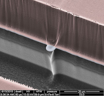
A scanning electron micrograph shows a nano-SPEAR suspended midway between layers of silicon (grey) and photoresist material (pink) that form a recording chamber for immobilized nematodes. The high-throughput technology developed at Rice University can be adapted for other small animals and could enhance data-gathering for disease characterization and drug interactions. Courtesy of the Robinson Lab

Here’s a link to and a citation for the paper,

Scalable electrophysiology in intact small animals with nanoscale suspended electrode arrays by Daniel L. Gonzales, Krishna N. Badhiwala, Daniel G. Vercosa, Benjamin W. Avants, Zheng Liu, Weiwei Zhong, & Jacob T. Robinson. Nature Nanotechnology (2017) doi:10.1038/nnano.2017.55 Published online 17 April 2017

This paper is behind a paywall.

Replicating brain’s neural networks with 3D nanoprinting

An announcement about European Union funding for a project to reproduce neural networks by 3D nanoprinting can be found in a June 10, 2016 news item on Nanowerk,

The MESO-BRAIN consortium has received a prestigious award of €3.3million in funding from the European Commission as part of its Future and Emerging Technology (FET) scheme. The project aims to develop three-dimensional (3D) human neural networks with specific biological architecture, and the inherent ability to interrogate the network’s brain-like activity both electrophysiologically and optically. It is expected that the MESO-BRAIN will facilitate a better understanding of human disease progression, neuronal growth and enable the development of large-scale human cell-based assays to test the modulatory effects of pharmacological and toxicological compounds on neural network activity. The use of more physiologically relevant human models will increase drug screening efficiency and reduce the need for animal testing.

A June 9, 2016 Institute of Photonic Sciences (ICFO) press release (also on EurekAlert), which originated the news item, provides more detail,

About the MESO-BRAIN project

The MESO-BRAIN project’s cornerstone will use human induced pluripotent stem cells (iPSCs) that have been differentiated into neurons upon a defined and reproducible 3D scaffold to support the development of human neural networks that emulate brain activity. The structure will be based on a brain cortical module and will be unique in that it will be designed and produced using nanoscale 3D-laser-printed structures incorporating nano-electrodes to enable downstream electrophysiological analysis of neural network function. Optical analysis will be conducted using cutting-edge light sheet-based, fast volumetric imaging technology to enable cellular resolution throughout the 3D network. The MESO-BRAIN project will allow for a comprehensive and detailed investigation of neural network development in health and disease.

Prof Edik Rafailov, Head of the MESO-BRAIN project (Aston University) said: “What we’re proposing to achieve with this project has, until recently, been the stuff of science fiction. Being able to extract and replicate neural networks from the brain through 3D nanoprinting promises to change this. The MESO-BRAIN project has the potential to revolutionise the way we are able to understand the onset and development of disease and discover treatments for those with dementia or brain injuries. We cannot wait to get started!”

The MESO-BRAIN project will launch in September 2016 and research will be conducted over three years.

About the MESO-BRAIN consortium

Each of the consortium partners have been chosen for the highly specific skills & knowledge that they bring to this project. These include technologies and expertise in stem cells, photonics, physics, 3D nanoprinting, electrophysiology, molecular biology, imaging and commercialisation.

Aston University (UK) Aston Institute of Photonic Technologies (School of Engineering and Applied Science) is one of the largest photonic groups in UK and an internationally recognised research centre in the fields of lasers, fibre-optics, high-speed optical communications, nonlinear and biomedical photonics. The Cell & Tissue Biomedical Research Group (Aston Research Centre for Healthy Ageing) combines collective expertise in genetic manipulation, tissue engineering and neuronal modelling with the electrophysiological and optical analysis of human iPSC-derived neural networks. Axol Bioscience Ltd. (UK) was founded to fulfil the unmet demand for high quality, clinically relevant human iPSC-derived cells for use in biomedical research and drug discovery. The Laser Zentrum Hannover (Germany) is a leading research organisation in the fields of laser development, material processing, laser medicine, and laser-based nanotechnologies. The Neurophysics Group (Physics Department) at University of Barcelona (Spain) are experts in combing experiments with theoretical and computational modelling to infer functional connectivity in neuronal circuits. The Institute of Photonic Sciences (ICFO) (Spain) is a world-leading research centre in photonics with expertise in several microscopy techniques including light sheet imaging. KITE Innovation (UK) helps to bridge the gap between the academic and business sectors in supporting collaboration, enterprise, and knowledge-based business development.

For anyone curious about the FET funding scheme, there’s this from the press release,

Horizon 2020 aims to ensure Europe produces world-class science by removing barriers to innovation through funding programmes such as the FET. The FET (Open) funds forward-looking collaborations between advanced multidisciplinary science and cutting-edge engineering for radically new future technologies. The published success rate is below 1.4%, making it amongst the toughest in the Horizon 2020 suite of funding schemes. The MESO-BRAIN proposal scored a perfect 5/5.

You can find out more about the MESO-BRAIN project on its ICFO webpage.

They don’t say anything about it but I can’t help wondering if the scientists aren’t also considering the possibility of creating an artificial brain.