Tag Archives: gene therapy

Reversing lower limb paralysis

This regenerative treatment is at a very early stage, which means the Swiss researchers have tried it on mice as you can see in the following video (runtime: 2 mins. 15 secs.). Towards the end of the video, researcher Grégoire Courtine cautions there are many hurdles before this could be used in humans, if ever,

A September 22, 2023 Ecole Polytechnique Fédérale de Lausanne (EPFL) press release (also on EurekAlert but published September 21, 2023) by Emmanuel Barraud, describes the work in more detail,

When the spinal cords of mice and humans are partially damaged, the initial paralysis is followed by the extensive, spontaneous recovery of motor function. However, after a complete spinal cord injury, this natural repair of the spinal cord doesn’t occur and there is no recovery. Meaningful recovery after severe injuries requires strategies that promote the regeneration of nerve fibers, but the requisite conditions for these strategies to successfully restore motor function have remained elusive.

“Five years ago, we demonstrated that nerve fibers can be regenerated across anatomically complete spinal cord injuries,” says Mark Anderson, a senior author of the study. “But we also realized this wasn’t enough to restore motor function, as the new fibers failed to connect to the right places on the other side of the lesion.” Anderson is the director of Central Nervous System Regeneration at .NeuroRestore and a scientist at the Wyss Center for Bio and Neuroengineering.

Working in tandem with peers at UCLA [University of California at Los Angeles] and Harvard Medical School, the scientists used state-of-the-art equipment at EPFL’s Campus Biotech facilities in Geneva to run in-depth analyses and identity which type of neuron is involved in natural spinal-cord repair after partial spinal cord injury. “Our observations using single-cell nuclear RNA sequencing not only exposed the specific axons that must regenerate, but also revealed that these axons must reconnect to their natural targets to restore motor function,” says Jordan Squair, the study’s first author. The team’s findings appear in the 22 September 2023 issue of Science.

Towards a combination of approaches

Their discovery informed the design of a multipronged gene therapy. The scientists activated growth programs in the identified neurons in mice to regenerate their nerve fibers, upregulated specific proteins to support the neurons’ growth through the lesion core, and administered guidance molecules to attract the regenerating nerve fibers to their natural targets below the injury. “We were inspired by nature when we designed a therapeutic strategy that replicates the spinal-cord repair mechanisms occurring spontaneously after partial injuries,” says Squair.

Mice with anatomically complete spinal cord injuries regained the ability to walk, exhibiting gait patterns that resembled those quantified in mice that resumed walking naturally after partial injuries. This observation revealed a previously unknown condition for regenerative therapies to be successful in restoring motor function after neurotrauma. “We expect that our gene therapy will act synergistically with our other procedures involving electrical stimulation of the spinal cord,” says Grégoire Courtine, a senior author of the study who also heads .NeuroRestore together with Jocelyne Bloch. “We believe a complete solution for treating spinal cord injury will require both approaches – gene therapy to regrow relevant nerve fibers, and spinal stimulation to maximize the ability of both these fibers and the spinal cord below the injury to produce movement.”

While many obstacles must still be overcome before this gene therapy can be applied in humans, the scientists have taken the first steps towards developing the technology necessary to achieve this feat in the years to come.

Here’s a link to and a citation for the paper,

Recovery of walking after paralysis by regenerating characterized neurons to their natural target region by Jordan W. Squair, Marco Milano, Alexandra de Coucy, Matthieu Gautier, Michael A. Skinnider, Nicholas D. James, Newton Cho, Anna Lasne, Claudia Kathe,Thomas H. Hutson, Steven Ceto, Laetitia Baud, Katia Galan, Viviana Aureli, Achilleas Laskaratos, Quentin Barraud, Timothy J. Deming, Richie E. Kohman, Bernard L. Schneider, Zhigang He, Jocelyne Bloch, Michael V. Sofroniew, Gregoire Courtine, and Mark A. Anderson. Science 21 Sep 2023 Vol 381, Issue 6664 pp. 1338-1345 DOI: 10.1126/science.adi641

This paper is behind a paywall.

This March 25, 2015 posting, “Spinal cords, brains, implants, and remote control,” features some research from EPFL researchers whose names you might recognize from this posting’s research paper.

Mentioned in the press release, the Swiss research centre website for NeuroRestore is here.

Gold nanoparticle loaded with CRISPR used to edit genes

CRISPR (clustered regularly interspaced short palindromic repeats) gene editing is usually paired with a virus (9, 12a, etc.) but this time scientists are using a gold nanoparticle. From a May 27, 2019 news item on Nanowerk (Note: Links have been removed),

Scientists at Fred Hutchinson Cancer Research Center took a step toward making gene therapy more practical by simplifying the way gene-editing instructions are delivered to cells. Using a gold nanoparticle instead of an inactivated virus, they safely delivered gene-editing tools in lab models of HIV and inherited blood disorders, as reported in Nature Materials (“Targeted homology-directed repair in blood stem and progenitor cells with CRISPR nanoformulations”).

A May 27, 2019 Fred Hutchinson Cancer Research Center news release (also on EurekAlert) by Jake Siegel, which originated the news item, expands on the theme, provides more detail,

It’s the first time that a gold nanoparticle loaded with CRISPR has been used to edit genes in a rare but powerful subset of blood stem cells, the source of all blood cells. The CRISPR-carrying gold nanoparticle led to successful gene editing in blood stem cells with no toxic effects.

“As gene therapies make their way through clinical trials and become available to patients, we need a more practical approach,” said senior author Dr. Jennifer Adair, an assistant member of the Clinical Research Division at Fred Hutch, adding that current methods of performing gene therapy are inaccessible to millions of people around the world. “I wanted to find something simpler, something that would passively deliver gene editing to blood stem cells.”

While CRISPR has made it faster and easier to precisely deliver genetic modifications to the genome, it still has challenges. Getting cells to accept CRISPR gene-editing tools involves a small electric shock that can damage and even kill the cells. And if precise gene edits are required, then additional molecules must be engineered to deliver them –adding cost and time.

Gold nanoparticles are a promising alternative because the surface of these tiny spheres (around 1 billionth the size of a grain of table salt) allows other molecules to easily stick to them and stay adhered.

“We engineered the gold nanoparticles to quickly cross the cell membrane, dodge cell organelles that seek to destroy them and go right to the cell nucleus to edit genes,” said Dr. Reza Shahbazi, a Fred Hutch postdoctoral researcher who has worked with gold nanoparticles for drug and gene delivery for seven years.

Shahbazi made the gold particles from laboratory-grade gold that is purified and comes as a liquid in a small lab bottle. He mixed the purified gold into a solution that causes the individual gold ions to form tiny particles, which the researchers then measured for size.

They found that a particular size – 19 nanometers wide – was the best for being big and sticky enough to add gene-editing materials to the surface of the particles, while still being small enough for cells to absorb them.

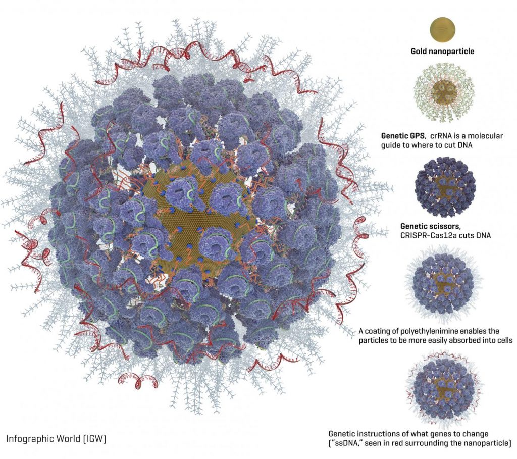
Packed onto the gold particles, the Fred Hutch team added these gene-editing components (diagram available [see below]):

A type of molecular guide called crRNA acts as a genetic GPS to show the CRISPR complex where in the genome to make the cut.

CRISPR nuclease protein, often called “genetic scissors,” makes the cut in the DNA. The CRISPR nuclease protein most often used is Cas9. But the Fred Hutch researchers also studied Cas12a (formerly called Cpf1) because Cas12a makes a staggered cut in DNA. The researchers hoped this would allow the cells to more efficiently repair the cut and while so doing embed the new genetic instructions into the cell. Another advantage of Cas12a over Cas9 is that it only requires one molecular guide, which is important because of space constraints on the nanoparticles. Cas9 requires two molecular guides.

Instructions for what genetic changes to make (“ssDNA”). The Fred Hutch team chose two inherited genetic changes that bestow protection from disease: CCR5, which protects against HIV, and gamma hemoglobin, which protects against blood disorders such as sickle cell disease and thalassemia.

A coating of a polyethylenimine swarms the surface of the particles to give them a more positive charge, which enables them to more readily be absorbed into cells. This is an improvement over another method of getting cells to take up gene editing tools, called electroporation, which involves lightly shocking the cells to get them to open and allow the genetic instructions to enter.

Then the researchers isolated blood stem cells with a protein marker on their surface called CD34. These CD34-positive cells contain the blood-making progenitor cells that give rise to the entire blood and immune system.

“These cells replenish blood in the body every day, making them a good candidate for one-time gene therapy because it will last a lifetime as the cells replace themselves,” Adair said.

Observing human blood stem cells in a lab dish, the researchers found that their fully loaded gold nanoparticles were taken up naturally by cells within six hours of being added and within 24 to 48 hours they could see gene editing happening. They observed that the Cas12a CRISPR protein partner was better at delivering very precise genetic edits to the cells than the more commonly used cas9 protein partner.

The gene-editing effect reached a peak eight weeks after the researchers injected the cells into mouse models; 22 weeks after injection the edited cells were still there. The Fred Hutch researchers also found edited cells in the bone marrow, spleen and thymus of the mouse models, a sign that the dividing blood cells in those organs could carry on the treatment without the mice having to be treated again.

“We believe we have a good candidate for two diseases — HIV and hemoglobinopathies — though we are also evaluating other disease targets where small genetic changes can have a big impact, as well as ways to make bigger genetic changes,” Adair said. “The next step is to increase how much gene editing happens in each cell, which is definitely doable. That will make it closer to being an effective therapy.”

In the study, the researchers report 10 to 20 percent of cells took on the gene edits, which is a promising start, but the researchers would like to aim for 50% or more of the cells being edited, which they believe will have a good chance of combatting these diseases.

###

Adair and Shahbazi are looking for commercial partners to develop the technology into therapies for people. They hope to begin clinical trials within a few years.

Here’s the diagram of a gold nanoparticle loaded with CRISPR,

Caption: Graphic of a fully loaded gold nanoparticle with CRISPR and other gene editing tools. Credit: Image courtesy of the Adair lab at Fred Hutch.

Here’s a link to and a citation for the paper,

Targeted homology-directed repair in blood stem and progenitor cells with CRISPR nanoformulations by Reza Shahbazi, Gabriella Sghia-Hughes, Jack L. Reid, Sara Kubek, Kevin G. Haworth, Olivier Humbert, Hans-Peter Kiem & Jennifer E. Adair. Nature Materials (2019) DOI https://doi.org/10.1038/s41563-019-0385-5Published 27 May 2019

This paper is behind a paywall.

Non-viral ocular gene therapy with gold nanoparticles and femtosecond lasers

I love the stylistic choice the writer made (pay special attention to the second paragraph) when producing this November 19, 2018 Polytechnique Montréal news release (also on EurekAlert),

A scientific breakthrough by Professor Michel Meunier of Polytechnique Montréal and his collaborators offers hope for people with glaucoma, retinitis or macular degeneration.

In January 2009, the life of engineer Michel Meunier, a professor at Polytechnique Montréal, changed dramatically. Like others, he had observed that the extremely short pulse of a femtosecond laser (0.000000000000001 second) could make nanometre-sized holes appear in silicon when it was covered by gold nanoparticles. But this researcher, recognized internationally for his skills in laser and nanotechnology, decided to go a step further with what was then just a laboratory curiosity. He wondered if it was possible to go from silicon to living matter, from inorganic to organic. Could the gold nanoparticles and the femtosecond laser, this “light scalpel,” reproduce the same phenomenon with living cells?

A very pretty image illustrating the work,

Caption: Gold nanoparticles, which act like “nanolenses,” concentrate the energy produced by the extremely short pulse of a femtosecond laser to create a nanoscale incision on the surface of the eye’s retina cells. This technology, which preserves cell integrity, can be used to effectively inject drugs or genes into specific areas of the eye, offering new hope to people with glaucoma, retinitis or macular degeneration. Credit and Copyright: Polytechnique Montréal

The news release goes on to describe the technology in more detail,

Professor Meunier started working on cells in vitro in his Polytechnique laboratory. The challenge was to make a nanometric incision in the cells’ extracellular membrane without damaging it. Using gold nanoparticles that acted as “nanolenses,” Professor Meunier realized that it was possible to concentrate the light energy coming from the laser at a wavelength of 800 nanometres. Since there is very little energy absorption by the cells at this wavelength, their integrity is preserved. Mission accomplished!

Based on this finding, Professor Meunier decided to work on cells in vivo, cells that are part of a complex living cell structure, such as the eye for example.

The eye and the light scalpel

In April 2012, Professor Meunier met Przemyslaw Sapieha, an internationally renowned eye specialist, particularly recognized for his work on the retina. “Mike”, as he goes by, is a professor in the Department of Ophthalmology at Université de Montréal and a researcher at Centre intégré universitaire de santé et de services sociaux (CIUSSS) de l’Est-de-l’Île-de-Montréal. He immediately saw the potential of this new technology and everything that could be done in the eye if you could block the ripple effect that occurs following a trigger that leads to glaucoma or macular degeneration, for example, by injecting drugs, proteins or even genes.

Using a femtosecond laser to treat the eye–a highly specialized and fragile organ–is very complex, however. The eye is part of the central nervous system, and therefore many of the cells or families of cells that compose it are neurons. And when a neuron dies, it does not regenerate like other cells do. Mike Sapieha’s first task was therefore to ensure that a femtosecond laser could be used on one or several neurons without affecting them. This is what is referred to as “proof of concept.”

Proof of concept

Mike and Michel called on biochemistry researcher Ariel Wilson, an expert in eye structures and vision mechanisms, as well as Professor Santiago Costantino and his team from the Department of Ophthalmology at Université de Montréal and the CIUSSS de l’Est-de-l’Île-de-Montréal for their expertise in biophotonics. The team first decided to work on healthy cells, because they are better understood than sick cells. They injected gold nanoparticles combined with antibodies to target specific neuronal cells in the eye, and then waited for the nanoparticles to settle around the various neurons or families of neurons, such as the retina. Following the bright flash generated by the femtosecond laser, the expected phenomenon occurred: small holes appeared in the cells of the eye’s retina, making it possible to effectively inject drugs or genes in specific areas of the eye. It was another victory for Michel Meunier and his collaborators, with these conclusive results now opening the path to new treatments.

The key feature of the technology developed by the researchers from Polytechnique and CIUSSS de l’Est-de-l’Île-de-Montréal is its extreme precision. With the use of functionalized gold nanoparticles, the light scalpel makes it possible to precisely locate the family of cells where the doctor will have to intervene.

Having successfully demonstrated proof of concept, Professor Meunier and his team filed a patent application in the United States. This tremendous work was also the subject of a paper reviewed by an impressive reading committee and published in the renowned journal Nano Letters in October 2018.

While there is still a lot of research to be done–at least 10 years’ worth, first on animals and then on humans–this technology could make all the difference in an aging population suffering from eye deterioration for which there are still no effective long-term treatments. It also has the advantage of avoiding the use of viruses commonly employed in gene therapy. These researchers are looking at applications of this technology in all eye diseases, but more particularly in glaucoma, retinitis and macular degeneration.

This light scalpel is unprecedented.

Here’s a link to and a citation for the paper,

In Vivo Laser-Mediated Retinal Ganglion Cell Optoporation Using KV1.1 Conjugated Gold Nanoparticles by Ariel M. Wilson, Javier Mazzaferri, Éric Bergeron, Sergiy Patskovsky, Paule Marcoux-Valiquette, Santiago Costantino, Przemyslaw Sapieha, Michel Meunier. Nano Lett.201818116981-6988 DOI: https://doi.org/10.1021/acs.nanolett.8b02896 Publication Date: October 4, 2018  Copyright © 2018 American Chemical Society

This paper is behind a paywall.

Immune to CRISPR?

I guess if you’re going to use bacteria as part of your gene editing technology (CRISPR [clustered regularly interspaced short palindromic repeats]/Cas9) then, you might half expect the body’s immune system may have developed some defenses. A Jan. 9, 2018 article by Sarah Zhang for The Atlantic provides some insight into what the new research suggests (Note: Links have been removed),

2018 is supposed to be the year of CRISPR in humans. The first U.S. and European clinical trials that test the gene-editing tool’s ability to treat diseases—such as sickle-cell anemia, beta thalassemia, and a type of inherited blindness—are slated to begin this year.

But the year has begun on a cautionary note. On Friday [January 5, 2018], Stanford researchers posted a preprint (which has not been peer reviewed) to the website biorXiv highlighting a potential obstacle to using CRISPR in humans: Many of us may already be immune to it. That’s because CRISPR actually comes from bacteria that often live on or infect humans, and we have built up immunity to the proteins from these bacteria over our lives.

Not all CRISPR therapies in humans will be doomed. “We don’t think this is the end of the story. This is the start of the story,” says Porteus [Matthew Porteus, a pediatrician and stem-cell researcher at Stanford]. There are likely ways around the problem of immunity to CRISPR proteins, and many of the early clinical trials appear to be designed around this problem.

Porteus and his colleagues focused on two versions of Cas9, the bacterial protein mostly commonly used in CRISPR gene editing. One comes from Staphylococcus aureus, which often harmlessly lives on skin but can sometimes causes staph infections, and another from Streptococcus pyogenes, which causes strep throat but can also become “flesh-eating bacteria” when it spreads to other parts of the body. So yeah, you want your immune system to be on guard against these bacteria.

The human immune system has a couple different ways of recognizing foreign proteins, and the team tested for both. First, they looked to see if people have molecules in their blood called antibodies that can specifically bind to Cas9. Among 34 people they tested, 79 percent had antibodies against the staph Cas9 and 65 percent against the strep Cas9.

The Stanford team only tested for preexisting immunity against Cas9, but anytime you inject a large bacterial protein into the human body, it can provoke an immune response. After all, that’s how the immune system learns to fight off bacteria it’s never seen before. (Preexisting immunity can make the response faster and more robust, though.)

The danger of the immune system turning on a patient’s body hangs over a lot of research into correcting genes. In the late 1990s and 2000s, research into gene therapy was derailed by the death of 18-year-old Jesse Gelsinger, who died from an immune reaction to the virus used to deliver the corrected gene. This is the worst-case scenario that the CRISPR world hopes to avoid.

Here’s a link to and a citation for the preprint,

Identification of Pre-Existing Adaptive Immunity to Cas9 Proteins in Humans by Carsten Trevor Charlesworth, Priyanka S Deshpande, Daniel P Dever, Beruh Dejene, Natalia Gomez-Ospina, Sruthi Mantri, Mara Pavel-Dinu, Joab Camarena, Kenneth I Weinberg, Matthew H Porteus. bioRxiv posted January 5, 2018 doi: https://doi.org/10.1101/243345

This article is a preprint and has not been peer-reviewed …

This preprint (not yet published paper) is open access and open for feedback.

Meanwhile, the year of CRISPR takes off (from a January 10, 2018 American Chemical Society news release on EurekAlert),

This year could be a defining one for CRISPR, the gene editing technique, which has been hailed as an important breakthrough in laboratory research. That’s because the first company-sponsored clinical studies will be conducted to see if it can help treat diseases in humans, according to an article in Chemical & Engineering News (C&EN), the weekly newsmagazine of the American Chemical Society.

C&EN Assistant Editor Ryan Cross reports that a big push is coming from industry, specifically from three companies that are each partly founded by one of the three inventors of the method. They are zeroing in on the blood diseases called sickle-cell anemia and β-thalassemia, mostly because their precise cause is known. In these diseases, hemoglobin doesn’t function properly, leading to severe health issues in some people. Crispr Therapeutics and Intellia Therapeutics plan to test the technique to boost levels of an alternative version of healthy hemoglobin. Editas Medicine, however, will also use CRISPR to correct mutations in the faulty hemoglobin gene. Labs led by university researchers are also joining the mix, starting or continuing clinical trials with the approach in 2018.

Because CRISPR is being used to cut a cell’s DNA and insert a new sequence, concerns have been raised about the potential for accidents. A cut in the wrong place could mean introducing a new mutation that could be benign — or cancerous. But according to proponents of the method, researchers are conducting extensive computer predictions and in vitro tests to help avoid this outcome.

The January 8, 2018 Chemical and Engineering News (C&EN) open access article by Ryan Cross is here.

Finally, if you are interested in how this affects research as it’s being developed, there’s University of British Columbia researcher Rosie Redfield’s January 16, 2018 posting on RRResearch blog,

Thursday’s [January 11, 2018] post described the hypothesis that bacteria might use gene transfer agent particles to inoculate other cells in the population with fragments of phage DNA, and outlined an experiment to test this.  Now I’m realizing that I need to know a lot more about the kind of immunity I should expect to see if this GTA-as-vaccine hypothesis is correct.

That should give you some idea of what I meant by “research as it’s being developed.” Redfield’s blog is not for the mildly interested.

Redfield is well-known internationally as being one of the first to refute research which suggested the existence of an ‘arsenic bacterium’ (see my Dec. 8, 2010 posting: My apologies for arsenic blooper. She’s first mentioned in the second excerpt, second paragraph.) The affair was known online as #arseniclife. There’s a May 27, 2011 essay by Carl Zimmer on Slate titled: The Discovery of Arsenic-Based Twitter: How #arseniclife changed science.

Inhale the drugs for Parkinson’s disease

The news out of Northeastern University’s Dr. Barbara Waszczak’s lab is exciting but it’s a single high point in a larger narrative.  First, here’s the high point described in the Apr. 24, 2013 news item on Azonano,

Researchers at Northeastern University in Boston have developed a gene therapy approach that may one day stop Parkinson’s disease (PD) in it tracks, preventing disease progression and reversing its symptoms. The novelty of the approach lies in the nasal route of administration and nanoparticles containing a gene capable of rescuing dying neurons in the brain.

The Apr. 21, 2013 news release on EurekAlert, which originated the news item, provides some information about Parkinson’s disease,

Parkinson’s is a devastating neurodegenerative disorder caused by the death of dopamine neurons in a key motor area of the brain, the substantia nigra (SN). Loss of these neurons leads to the characteristic tremor and slowed movements of PD, which get increasingly worse with time. Currently, more than 1% of the population over age 60 has PD and approximately 60,000 Americans are newly diagnosed every year. The available drugs on the market for PD mimic or replace the lost dopamine but do not get to the heart of the problem, which is the progressive loss of the dopamine neurons.

Here’s how the disease got its name, from the Wikipedia essay: Parkinson’s disease (Note: Links have been removed),

The disease is named after the English doctor James Parkinson, who published the first detailed description in An Essay on the Shaking Palsy in 1817. Several major organizations promote research and improvement of quality of life of those with the disease and their families. Public awareness campaigns include Parkinson’s disease day (on the birthday of James Parkinson, April 11) and the use of a red tulip as the symbol of the disease. People with parkinsonism who have increased the public’s awareness include Michael J. Fox and Muhammad Ali.

Now for some information about the background work leading up to this new, exciting, high point (from the news release on EurekAlert),

The focus of Dr. Barbara Waszczak’s lab at Northeastern University in Boston is to find a way to harvest the potential of glial cell line-derived neurotrophic factor (GDNF) as a treatment for PD. GDNF is a protein known to nourish dopamine neurons by activating survival and growth-promoting pathways inside the cells. Not surprisingly, GDNF is able to protect dopamine neurons from injury and restore the function of damaged and dying neurons in many animal models of PD. However, the action of GDNF is limited by its inability to cross the blood-brain barrier (BBB), thus requiring direct surgical injection into the brain. To circumvent this problem, Waszczak’s lab is investigating intranasal delivery as a way to bypass the BBB. Their previous work showed that intranasal delivery of GDNF protects dopamine neurons from damage by the neurotoxin, 6-hydroxydopamine (6-OHDA), a standard rat model of PD.

According to the Michael J. Fox Foundation, this research work dates from 2007 (at least), from the Intranasal Delivery of GDNF for Parkinson’s Disease: Next Steps grant page,

FINAL OUTCOME

The results of this Drug Delivery 2008 project confirm and extend the conclusions reached under a previous 2007 Rapid Response Innovation Award. The research team has demonstrated that intranasal administration of GDNF has neuroprotective efficacy in a preclinical model of Parkinson’s disease, that the protein gets into the brain and reaches target structures (the striatum and substantia nigra) within an hour of nasal administration, and that the nasal route causes no apparent toxicity in the nose. Longer term efficacy and toxicology studies will be necessary in other relevant preclinical models before testing can be initiated in humans.

The results of this work strongly supports pursuit of intranasal administration as a promising approach for harvesting the therapeutic potential of GDNF. Such an approach could ultimately provide an effective, non-invasive means of delivering GDNF to the brain for the treatment of Parkinson’s disease.

Here’s the 2013 innovation on intranasal delivery of GDNF therapy (from the news release on EurekAlert),

Taking this work a step further, Brendan Harmon, working in Waszczak’s lab, has adapted the intranasal approach so that cells in the brain can continuously produce GDNF. His work utilized nanoparticles, developed by Copernicus Therapeutics, Inc., which are able to transfect brain cells with an expression plasmid carrying the gene for GDNF (pGDNF). When given intranasally to rats, these pGDNF nanoparticles increase GDNF production throughout the brain for long periods, avoiding the need for frequent re-dosing. Now, in new research presented on April 20 at 12:30 pm during Experimental Biology 2013 in Boston, MA, Harmon reports that intranasal administration of Copernicus’ pGDNF nanoparticles results in GDNF expression sufficient to protect SN dopamine neurons in the 6-OHDA model of PD.

Waszczak and Harmon believe that intranasal delivery of Copernicus’ nanoparticles may provide an effective and non-invasive means of GDNF gene therapy for PD, and an avenue for transporting other gene therapy vectors to the brain. This work, which was funded in part by the Michael J. Fox Foundation for Parkinson’s Research and Northeastern University, has the potential to greatly expand treatment options for PD and many other central nervous system disorders.

For the curious, there’s more about Copernicus Therapeutics at the company website.

Congratulations to Harmon and Waszczak! I imagine the next step will be human clinical trials.

Gold unzips your DNA but not in a sexy way

The animation that the scientists from North Carolina have provided makes the gold nanoparticles look downright mean as that DNA definitely does not want to be unzipped but perhaps your mileage varies,

The June 20, 2012 news item on Nanowerk provides more detail,

New research from North Carolina State University finds that gold nanoparticles with a slight positive charge work collectively to unravel DNA’s double helix. This finding has ramifications for gene therapy research and the emerging field of DNA-based electronics.

The research team introduced gold nanoparticles, approximately 1.5 nanometers in diameter, into a solution containing double-stranded DNA. The nanoparticles were coated with organic molecules called ligands. Some of the ligands held a positive charge, while others were hydrophobic – meaning they were repelled by water.

Because the gold nanoparticles had a slight positive charge from the ligands, and DNA is always negatively charged, the DNA and nanoparticles were pulled together into complex packages.

“However, we found that the DNA was actually being unzipped by the gold nanoparticles,” Melechko [Dr. Anatoli Melechko, an associate professor of materials science and engineering at NC State and co-author of the paper] says. The positively-charged ligands on the nanoparticles attached to the DNA as predicted, but the hydrophobic ligands of the nanoparticles became tangled with each other. As this tangling pulled the nanoparticles into clusters, the nanoparticles pulled the DNA apart.

The implications for this ‘unzipping’ are,

“We think gold nanoparticles still hold promise for gene therapy,” says Dr. Yaroslava Yingling, an assistant professor of materials science and engineering at NC State and co-author of the paper. “But it’s clear that we need to tailor the ligands, charge and chemistry of these materials to ensure the DNA’s structural integrity is not compromised.”

The finding is also relevant to research on DNA-based electronics, which hopes to use DNA as a template for creating nanoelectronic circuits. Because some work in that field involves placing metal nanoparticles on DNA, this finding indicates that researchers will have to pay close attention to the characteristics of those nanoparticles – or risk undermining the structural integrity of the DNA.

Overpromising and underdelivering: genome, stem cells, gene therapy and nano food

When people talk about overpromising (aka hype/hyperbole) and science, they’re usually referring to overexcited marketing collateral and/or a public relations initiative and/or news media coverage.  Scientists themselves don’t tend to be identified as one of the sources for hype even when that’s clearly the case. That’s right, scientists are people too and sometimes they get carried away by their enthusiasms as Emily Yoffe notes in her excellent Slate essay, The Medical Revolution; Where are the cures promised by stem cells, gene therapy, and the human genome? From Yoffe’s essay,

Dr. J. William Langston has been researching Parkinson’s disease for 25 years. At one time, it seemed likely he’d have to find another disease to study, because a cure for Parkinson’s looked imminent. In the late 1980s, the field of regenerative medicine seemed poised to make it possible for doctors to put healthy tissue in a damaged brain, reversing the destruction caused by the disease.

Langston was one of many optimists. In 1999, the then-head of the National Institute of Neurological Disorders and Stroke, Dr. Gerald Fischbach, testified before the Senate that with “skill and luck,” Parkinson’s could be cured in five to 10 years. Now Langston, who is 67, doesn’t think he’ll see a Parkinson’s cure in his professional lifetime. He no longer uses “the C word” and acknowledges he and others were naive. [emphasis mine] He understands the anger of patients who, he says, “are getting quite bitter” that they remain ill, long past the time when they thought they would have been restored to health.

The disappointments are so acute in part because the promises have been so big. Over the past two decades, we’ve been told that a new age of molecular medicine—using gene therapy, stem cells, and the knowledge gleaned from unlocking the human genome—would bring us medical miracles. [emphasis mine] Just as antibiotics conquered infectious diseases and vaccines eliminated the scourges of polio and smallpox, the ability to manipulate our cells and genes is supposed to vanquish everything from terrible inherited disorders, such as Huntington’s and cystic fibrosis, to widespread conditions like cancer, diabetes, and heart disease.

Yoffe goes on to outline the problems that researchers encounter when trying to ‘fix’ what’s gone wrong.

Parkinson’s disease was long held out as the model for new knowledge and technologies eradicating illnesses. Instead, it has become the model for its unforeseen consequences. [emphasis mine]

Langston, head of the Parkinson’s Institute and Clinical Center, explains that scientists believed the damage to patients took place in a discrete part of the brain, the substantia nigra. “It was a small target. All we’d have to do was replace the missing cells, do it once, and that would cure the disease,” Langston says. “We were wrong about that. This disease hits many other areas of the brain. You can’t just put transplants here and there. The brain is not a pincushion.”

Disease of all kinds have proven to be infinitely more complex than first realized. Disease is not ’cause and effect’ driven so much as it is a process with an infinite number of potential inputs and any number of potential outcomes. Take for example gene therapy (Note: the human genome project was supposed to yield gene therapies),

In some ways, gene therapy for boys with a deadly immune disorder, X-linked severe combined immune deficiency, also known as “bubble boy” disease, is the miracle made manifest. Inserting good genes into these children has allowed some to live normal lives. Unfortunately, within a few years of treatment, a significant minority have developed leukemia. The gene therapy, it turns out, activated existing cancer-causing genes in these children. This results in what the co-discoverer of the structure of DNA, James Watson, calls “the depressing calculus” of curing an invariably fatal disease—and hoping it doesn’t cause a sometimes-fatal one.

For me, it seems that that the human genome project was akin to taking a clock apart. Looking at the constituent parts and replacing broken ones does not guarantee that you will be able assemble a more efficient working version unless you know how the clock worked in the first place. We still don’t understand the basic parts, the genes,  interact with each other, within their environment, or with external inputs.

The state of our ignorance is illustrated by the recent sequencing of the genome of Bishop Desmond Tutu and four Bushmen. Three of the Bushmen had a gene mutation associated with a liver disease that kills people while young. But the Bushmen are all over 80—which means either the variation doesn’t actually cause the disease, or there are other factors protecting the Bushmen.

As for the pressures acting on the scientists themselves,

There are forces, both external and internal, on scientists that almost require them to oversell. Without money, there’s no science. Researchers must constantly convince administrators who control tax dollars, investors, and individual donors that the work they are doing will make a difference. Nancy Wexler says that in order to get funding, “You have to promise cures, that you’ll meet certain milestones within a certain time frame.”

The infomercial-level hype for both gene therapy and stem cells is not just because scientists are trying to convince funders, but because they want to believe. [emphases mine]

Scientific advances as one of Yoffe’s interview subjects points out involve a process dogged with failure and setbacks requiring an attitude of humility laced with patience and practiced over decades before an ‘overnight success’ occurs, if it ever does.

I was reminded of Yoffe’s article after reading a nano food article recently written by Kate Kelland for Reuters,

In a taste of things to come, food scientists say they have cooked up a way of using nanotechnology to make low-fat or fat-free foods just as appetizing and satisfying as their full-fat fellows.

The implications could be significant in combating the spread of health problems such as obesity, diabetes and heart disease.

There are two promising areas of research. First, they are looking at ways to slow digestion,

One thing they might look into is work by scientists at Britain’s Institute of Food Research (IFR), who said last month they had found an unexpected synergy that helped break down fat and might lead to new ways of slowing digestion, and ultimately to creating foods that made consumers feel fuller.

“Much of the fat in processed foods is eaten in the form of emulsions such as soups, yoghurt, ice cream and mayonnaise,” said the IFR’s Peter Wilde. “We are unpicking the mechanisms of digestion used to break them down so we can design fats in a rational way that are digested more slowly.”

The idea is that if digestion is slower, the final section of the intestine called the ileum will be put on its “ileal brake,” sending a signal to the consumer that means they feel full even though they have eaten less fat

This sounds harmless and it’s even possible it’s a good idea but then replacing diseased tissue with healthy tissue, as they tried with Parkinson’s Disease gene therapies, seemed like a good idea too. Just how well is the digestive process understood?

As for the second promising area of research,

Experts see promise in another nano technique which involves encapsulating nutrients in bubble-like structures known as vesicles that can be engineered to break down and release their contents at specific stages in the digestive system.

According to Vic Morris, a nano expert at the IFR, this technique in a larger form, micro-encapsulation, was well established in the food industry. The major difference with nano-encapsulation was that the smaller size might be able to take nutrients further or deliver them to more appropriate places. [emphasis mine]

They’ve been talking about trying to encapsulate and target medicines to more appropriate places and, as far as I’m aware, to no avail. I sense a little overenthusiasm on the experts’ part. Kelland does try to counterbalance this by discussing other issues with nanofood such as secretiveness about the food companies’ research, experts’ concerns over nanoparticles, and public concerns over genetically modified food. Still the allure of ‘all you can eat with no consequences’ is likely to overshadow any journalist’s attempt at balanced reporting with resulting disappointment when somebody realizes it’s all much more complicated than we thought.

Dexter Johnson’s Sept. 22, 2010 posting ( Protein-based Nanotubes Pass Electrical Signals Between Cells) on his Nanoclast blog offers more proof that we still have a lot to learn about basic biological processes,

A few years back, scientists led by Hans-Hermann Gerdes at the University of Bergen noticed that there were nanoscale tubes connecting cells sometimes over significant distances. This discovery launched a field known somewhat by the term in the biological community as the “nanotube field.”

Microbiologists remained somewhat skeptical on what this phenomenon was and weren’t entirely pleased with some explanations offered because they seemed to fall outside “existing biological concepts.”

So let’s start summing up.  The team notices nanotubes that connect cells over distances which microbiologists have difficulty accepting as “they [seem] to fall outside existing biological concepts. [emphasis mine] Now the team has published a paper which suggests that electrical signals pass through the nanotubes and that a ‘gap junction’ enables transmission to nonadjacent cells.  (Dexter’s description provides  more technical detail in an accessible writing style.)

As Dexter notes,

Another key biological question it helps address–or complicate, as the case may be–is the complexity of the human brain. This research makes the brain drastically more complex than originally thought, according to Gerdes. [emphasis mine]

Getting back to where I started, scientists are people too. They have their enthusiasms as well as pressure to get grants and produce results for governments and other investors, not to mention their own egos.  And while I’ve focused on the biological and medical sciences in this article, I think that all the sciences yield more questions than answers and that everything is far more complicated and  interconnected than we have yet to realize.