A September 28, 2022 news item on Nanowerk announces research into combining nasal sprays and brain stimulation in efforts to improve stroke recovery (Note: A link has been removed),
In a recent study (Materials Today Chemistry, “Enhancing non-invasive brain stimulation with non-invasively delivered nanoparticles for improving stroke recovery”), researchers from Xi’an Jiaotong-Liverpool University and other universities in China have reported that brain stimulation combined with a nose spray containing nanoparticles can improve recovery after ischemic stroke in an animal model.
The nasal spray is a non-invasive method for delivering magnetic nanoparticles into the brain that the study finds can increase the benefits of transcranial magnetic stimulation (TMS). TMS is a method of non-invasive brain stimulation already used clinically or in clinical trials to treat neurological conditions like stroke, Parkinson’s disease, Alzheimer’s disease, depression, and addiction.
…
I have two previous posts about nasal sprays and nanoparticles (links to previous posts follow at the end) but this item is the first to include brain stimulation. From a September 27, 2022 Xi’an Jiaotong-Liverpool University press release (also on EurekAlert but published on September 28, 2022), which originated the news item,
Rats that were given combined nanoparticle and TMS treatment every 24 hours for 14 days after an ischemic stroke had better overall health, put on weight more quickly and had improved cognitive and motor functions compared to those treated with TMS alone.
During TMS treatment, an electrical current runs through an electric coil placed outside the skull, producing a magnetic field that stimulates brain cells by inducing a further electrical current inside the brain. However, the stimulation is often not intense enough to penetrate far enough into the brain to reach the areas needing treatment.
In this new study, the researchers show that magnetic nanoparticles, administered intranasally, can make neurons more responsive and amplify the magnetic signal from TMS to reach deeper brain tissue, aiding recovery. The finding offers new opportunities for treating neurological disorders.
From impossible to possible
The research answers a key question in nanomedicine – whether it is possible to enhance TMS by using nanoparticles that are non-invasively delivered into the brain. Leading figures in the field previously stated that it was almost impossible because of the blood-brain barrier. This physical barrier separates the brain from the rest of the body’s bloodstream.
However, the team of researchers overcame this by guiding the magnetic nanoparticles closer to the correct area with a large magnet near the head.
Dr Gang Ruan, a corresponding author of the study, says: “We were able to overcome the blood-brain barrier and send enough nanoparticles into the brain to use in combination with TMS simulation to improve recovery from stroke.
“TMS devices are already used for the clinical treatment of neurological disorders but have severe limitations in terms of stimulation strength and depths of the brain they can penetrate.
“By non-invasively putting magnetic nanoparticles into the brain, we can amplify and enhance the TMS stimulation effects on neurons, making the treatment more effective,” Dr Ruan adds.
“Showing it is possible to use nanoparticles in this way paves the way for medical applications of nanoparticles for other neurological disorders.”
Crossing barriers
The iron oxide nanoparticles used in the study are already prescribed to treat iron deficiency as they are non-toxic and biodegradable. The team also modified the nanoparticles by coating them with various non-toxic substances.
Dr Ruan says: “The coating causes the nanoparticles to stick to the blood-brain barrier, increasing their chances of passing through it. Without this coating, the particles just bounce back from the barrier instead of crossing it.
“The modifications of the iron oxide particles also ensure that the nanoparticles can stick to the neurons and increase their responsiveness to TMS stimulation.”
The safety of using the modified nanoparticles needs to be assessed in clinical trials but has the potential to be used in combination with TMS, and other methods such as brain imaging, to gain more insight into how the brain works and improve the treatment of neurological disorders.
“Many scientists still think it is impossible to non-invasively send enough nanoparticles into the brain to affect brain function. Yet we have shown that it is possible,” says Dr Ruan.
“We combined the expertise on our team in four different disciplines, materials science, biophysics, neuroscience, and medical science, to push the boundaries of our knowledge and challenge what is currently thought in the field.”
One final note, “Xi’an Jiaotong-Liverpool University (XJTLU) is an international university formed in partnership between the University of Liverpool and Xi’an Jiaotong University in China. Find out more about XJTLU“
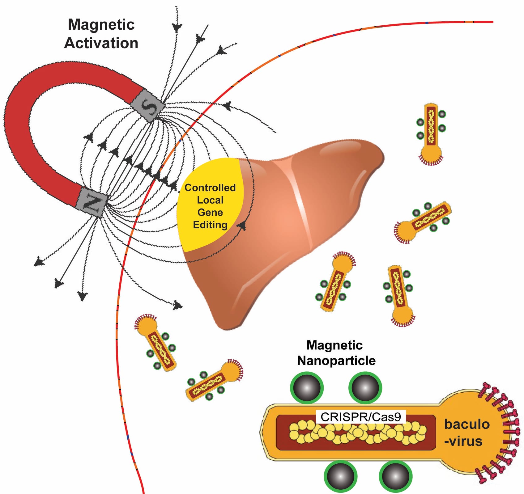
Rice University bioengineers use a magnetic field to activate nanoparticle-attached baculoviruses in a tissue. The viruses, which normally infect alfalfa looper moths, are modified to deliver gene-editing DNA code only to cells that are targeted with magnetic field-induced local transduction. Courtesy of the Laboratory of Biomolecular Engineering and Nanomedicine
Kudos to whomever put that diagram together! That’s a lot of well conveyed information.
Now for the details about how this technology might save lives. From a November 13, 2018 news item on Nanowerk,
A new technology that relies on a moth-infecting virus and nanomagnets could be used to edit defective genes that give rise to diseases like sickle cell, muscular dystrophy and cystic fibrosis.
Rice University bioengineer Gang Bao has combined magnetic nanoparticles with a viral container drawn from a particular species of moth to deliver CRISPR/Cas9 payloads that modify genes in a specific tissue or organ with spatial control.
Because magnetic fields are simple to manipulate and, unlike light, pass easily through tissue, Bao and his colleagues want to use them to control the expression of viral payloads in target tissues by activating the virus that is otherwise inactivated in blood.
The research appears in Nature Biomedical Engineering. In nature, CRISPR/Cas9 bolsters microbes’ immune systems by recording the DNA of invaders. That gives microbes the ability to recognize and attack returning invaders, but scientists have been racing to adapt CRISPR/Cas9 to repair mutations that cause genetic diseases and to manipulate DNA in laboratory experiments.
CRISPR/Cas9 has the potential to halt hereditary disease – if scientists can get the genome-editing machinery to the right cells inside the body. But roadblocks remain, especially in delivering the gene-editing payloads with high efficiency.
Bao said it will be necessary to edit cells in the body to treat many diseases. “But efficiently delivering genome-editing machinery into target tissue in the body with spatial control remains a major challenge,” Bao said. “Even if you inject the viral vector locally, it can leak to other tissues and organs, and that could be dangerous.”
The delivery vehicle developed by Bao’s group is based on a virus that infects Autographa californica, aka the alfalfa looper, a moth native to North America. The cylindrical baculovirus vector (BV), the payload-carrying part of the virus, is considered large at up to 60 nanometers in diameter and 200-300 nanometers in length. That’s big enough to transport more than 38,000 base pairs of DNA, which is enough to supply multiple gene-editing units to a target cell, Bao said.
He said the inspiration to combine BV and magnetic nanoparticles came from discussions with Rice postdoctoral researcher and co-lead author Haibao Zhu, who learned about the virus during a postdoctoral stint in Singapore but knew nothing about magnetic nanoparticles until he joined the Bao lab. The Rice team had previous experience using iron oxide nanoparticles and an applied magnetic field to open blood vessel walls just enough to let large-molecule drugs pass through.
“We really didn’t know if this would work for gene editing or not, but we thought, ‘worth a shot,'” Bao said.
The researchers use the magnetic nanoparticles to activate BV and deliver gene-editing payloads only where they’re needed. To do this, they take advantage of an immune-system protein called C3 that normally inactivates baculoviruses.
“If we combine BV with magnetic nanoparticles, we can overcome this deactivation by applying the magnetic field,” Bao said. “The beauty is that when we deliver it, gene editing occurs only at the tissue, or the part of the tissue, where we apply the magnetic field.”
Application of the magnetic field allows BV transduction, the payload-delivery process that introduces gene-editing cargo into the target cell. The payload is also DNA, which encodes both a reporter gene and the CRISPR/Cas9 system.
In tests, the BV was loaded with green fluorescent proteins or firefly luciferase. Cells with the protein glowed brightly under a microscope, and experiments showed the magnets were highly effective at targeted delivery of BV cargoes in both cell cultures and lab animals.
Bao noted his and other labs are working on the delivery of CRISPR/Cas9 with adeno-associated viruses (AAV), but he said BV’s capacity for therapeutic cargo is roughly eight times larger. “However, it is necessary to make BV transduction into target cells more efficient,” he said.
Identification of the precise 3-D coordinates of iron, shown in red, and platinum atoms in an iron-platinum nanoparticle.. Courtesy of Colin Ophus and Florian Nickel/Berkeley Lab
The image of the iron-platinum nanoparticle (referenced in the headline) reminds of foetal ultrasound images. A Feb. 1, 2017 news item on ScienceDaily tells us more,
In the world of the very tiny, perfection is rare: virtually all materials have defects on the atomic level. These imperfections — missing atoms, atoms of one type swapped for another, and misaligned atoms — can uniquely determine a material’s properties and function. Now, UCLA [University of California at Los Angeles] physicists and collaborators have mapped the coordinates of more than 23,000 individual atoms in a tiny iron-platinum nanoparticle to reveal the material’s defects.
The results demonstrate that the positions of tens of thousands of atoms can be precisely identified and then fed into quantum mechanics calculations to correlate imperfections and defects with material properties at the single-atom level.
Jianwei “John” Miao, a UCLA professor of physics and astronomy and a member of UCLA’s California NanoSystems Institute, led the international team in mapping the atomic-level details of the bimetallic nanoparticle, more than a trillion of which could fit within a grain of sand.
“No one has seen this kind of three-dimensional structural complexity with such detail before,” said Miao, who is also a deputy director of the Science and Technology Center on Real-Time Functional Imaging. This new National Science Foundation-funded consortium consists of scientists at UCLA and five other colleges and universities who are using high-resolution imaging to address questions in the physical sciences, life sciences and engineering.
Miao and his team focused on an iron-platinum alloy, a very promising material for next-generation magnetic storage media and permanent magnet applications.
By taking multiple images of the iron-platinum nanoparticle with an advanced electron microscope at Lawrence Berkeley National Laboratory and using powerful reconstruction algorithms developed at UCLA, the researchers determined the precise three-dimensional arrangement of atoms in the nanoparticle.
“For the first time, we can see individual atoms and chemical composition in three dimensions. Everything we look at, it’s new,” Miao said.
The team identified and located more than 6,500 iron and 16,600 platinum atoms and showed how the atoms are arranged in nine grains, each of which contains different ratios of iron and platinum atoms. Miao and his colleagues showed that atoms closer to the interior of the grains are more regularly arranged than those near the surfaces. They also observed that the interfaces between grains, called grain boundaries, are more disordered.
“Understanding the three-dimensional structures of grain boundaries is a major challenge in materials science because they strongly influence the properties of materials,” Miao said. “Now we are able to address this challenge by precisely mapping out the three-dimensional atomic positions at the grain boundaries for the first time.”
The researchers then used the three-dimensional coordinates of the atoms as inputs into quantum mechanics calculations to determine the magnetic properties of the iron-platinum nanoparticle. They observed abrupt changes in magnetic properties at the grain boundaries.
“This work makes significant advances in characterization capabilities and expands our fundamental understanding of structure-property relationships, which is expected to find broad applications in physics, chemistry, materials science, nanoscience and nanotechnology,” Miao said.
In the future, as the researchers continue to determine the three-dimensional atomic coordinates of more materials, they plan to establish an online databank for the physical sciences, analogous to protein databanks for the biological and life sciences. “Researchers can use this databank to study material properties truly on the single-atom level,” Miao said.
Miao and his team also look forward to applying their method called GENFIRE (GENeralized Fourier Iterative Reconstruction) to biological and medical applications. “Our three-dimensional reconstruction algorithm might be useful for imaging like CT scans,” Miao said. Compared with conventional reconstruction methods, GENFIRE requires fewer images to compile an accurate three-dimensional structure.
That means that radiation-sensitive objects can be imaged with lower doses of radiation.
The US Dept. of Energy (DOE) Lawrence Berkeley National Laboratory issued their own Feb. 1, 2017 news release (also on EurekAlert) about the work with a focus on how their equipment made this breakthrough possible (it repeats a little of the info. from the UCLA news release),
Scientists used one of the world’s most powerful electron microscopes to map the precise location and chemical type of 23,000 atoms in an extremely small particle made of iron and platinum.
The 3-D reconstruction reveals the arrangement of atoms in unprecedented detail, enabling the scientists to measure chemical order and disorder in individual grains, which sheds light on the material’s properties at the single-atom level. Insights gained from the particle’s structure could lead to new ways to improve its magnetic performance for use in high-density, next-generation hard drives.
What’s more, the technique used to create the reconstruction, atomic electron tomography (which is like an incredibly high-resolution CT scan), lays the foundation for precisely mapping the atomic composition of other useful nanoparticles. This could reveal how to optimize the particles for more efficient catalysts, stronger materials, and disease-detecting fluorescent tags.
Microscopy data was obtained and analyzed by scientists from the Department of Energy’s Lawrence Berkeley National Laboratory (Berkeley Lab) at the Molecular Foundry, in collaboration with Foundry users from UCLA, Oak Ridge National Laboratory, and the United Kingdom’s University of Birmingham. …
Atoms are the building blocks of matter, and the patterns in which they’re arranged dictate a material’s properties. These patterns can also be exploited to greatly improve a material’s function, which is why scientists are eager to determine the 3-D structure of nanoparticles at the smallest scale possible.
“Our research is a big step in this direction. We can now take a snapshot that shows the positions of all the atoms in a nanoparticle at a specific point in its growth. This will help us learn how nanoparticles grow atom by atom, and it sets the stage for a materials-design approach starting from the smallest building blocks,” says Mary Scott, who conducted the research while she was a Foundry user, and who is now a staff scientist. Scott and fellow Foundry scientists Peter Ercius and Colin Ophus developed the method in close collaboration with Jianwei Miao, a UCLA professor of physics and astronomy.
Their nanoparticle reconstruction builds on an achievement they reported last year in which they measured the coordinates of more than 3,000 atoms in a tungsten needle to a precision of 19 trillionths of a meter (19 picometers), which is many times smaller than a hydrogen atom. Now, they’ve taken the same precision, added the ability to distinguish different elements, and scaled up the reconstruction to include tens of thousands of atoms.
Importantly, their method maps the position of each atom in a single, unique nanoparticle. In contrast, X-ray crystallography and cryo-electron microscopy plot the average position of atoms from many identical samples. These methods make assumptions about the arrangement of atoms, which isn’t a good fit for nanoparticles because no two are alike.
“We need to determine the location and type of each atom to truly understand how a nanoparticle functions at the atomic scale,” says Ercius.
A TEAM approach
The scientists’ latest accomplishment hinged on the use of one of the highest-resolution transmission electron microscopes in the world, called TEAM I. It’s located at the National Center for Electron Microscopy, which is a Molecular Foundry facility. The microscope scans a sample with a focused beam of electrons, and then measures how the electrons interact with the atoms in the sample. It also has a piezo-controlled stage that positions samples with unmatched stability and position-control accuracy.
The researchers began growing an iron-platinum nanoparticle from its constituent elements, and then stopped the particle’s growth before it was fully formed. They placed the “partially baked” particle in the TEAM I stage, obtained a 2-D projection of its atomic structure, rotated it a few degrees, obtained another projection, and so on. Each 2-D projection provides a little more information about the full 3-D structure of the nanoparticle.
They sent the projections to Miao at UCLA, who used a sophisticated computer algorithm to convert the 2-D projections into a 3-D reconstruction of the particle. The individual atomic coordinates and chemical types were then traced from the 3-D density based on the knowledge that iron atoms are lighter than platinum atoms. The resulting atomic structure contains 6,569 iron atoms and 16,627 platinum atoms, with each atom’s coordinates precisely plotted to less than the width of a hydrogen atom.
Translating the data into scientific insights
Interesting features emerged at this extreme scale after Molecular Foundry scientists used code they developed to analyze the atomic structure. For example, the analysis revealed chemical order and disorder in interlocking grains, in which the iron and platinum atoms are arranged in different patterns. This has large implications for how the particle grew and its real-world magnetic properties. The analysis also revealed single-atom defects and the width of disordered boundaries between grains, which was not previously possible in complex 3-D boundaries.
“The important materials science problem we are tackling is how this material transforms from a highly randomized structure, what we call a chemically-disordered structure, into a regular highly-ordered structure with the desired magnetic properties,” says Ophus.
To explore how the various arrangements of atoms affect the nanoparticle’s magnetic properties, scientists from DOE’s Oak Ridge National Laboratory ran computer calculations on the Titan supercomputer at ORNL–using the coordinates and chemical type of each atom–to simulate the nanoparticle’s behavior in a magnetic field. This allowed the scientists to see patterns of atoms that are very magnetic, which is ideal for hard drives. They also saw patterns with poor magnetic properties that could sap a hard drive’s performance.
“This could help scientists learn how to steer the growth of iron-platinum nanoparticles so they develop more highly magnetic patterns of atoms,” says Ercius.
Adds Scott, “More broadly, the imaging technique will shed light on the nucleation and growth of ordered phases within nanoparticles, which isn’t fully theoretically understood but is critically important to several scientific disciplines and technologies.”
The folks at the Berkeley Lab have created a video (notice where the still image from the beginning of this post appears),
The Oak Ridge National Laboratory (ORNL), not wanting to be left out, has been mentioned in a Feb. 3, 2017 news item on ScienceDaily,
… researchers working with magnetic nanoparticles at the University of California, Los Angeles (UCLA), and the US Department of Energy’s (DOE’s) Lawrence Berkeley National Laboratory (Berkeley Lab) approached computational scientists at DOE’s Oak Ridge National Laboratory (ORNL) to help solve a unique problem: to model magnetism at the atomic level using experimental data from a real nanoparticle.
“These types of calculations have been done for ideal particles with ideal crystal structures but not for real particles,” said Markus Eisenbach, a computational scientist at the Oak Ridge Leadership Computing Facility (OLCF), a DOE Office of Science User Facility located at ORNL.
A Feb. 2, 2017 ORNL news release on EurekAlert, which originated the news item, elucidates further on how their team added to the research,
Eisenbach develops quantum mechanical electronic structure simulations that predict magnetic properties in materials. Working with Paul Kent, a computational materials scientist at ORNL’s Center for Nanophase Materials Sciences, the team collaborated with researchers at UCLA and Berkeley Lab’s Molecular Foundry to combine world-class experimental data with world-class computing to do something new–simulate magnetism atom by atom in a real nanoparticle.
Using the new data from the research teams on the West Coast, Eisenbach and Kent were able to precisely model the measured atomic structure, including defects, from a unique iron-platinum (FePt) nanoparticle and simulate its magnetic properties on the 27-petaflop Titan supercomputer at the OLCF.
Electronic structure codes take atomic and chemical structure and solve for the corresponding magnetic properties. However, these structures are typically derived from many 2-D electron microscopy or x-ray crystallography images averaged together, resulting in a representative, but not true, 3-D structure.
“In this case, researchers were able to get the precise 3-D structure for a real particle,” Eisenbach said. “The UCLA group has developed a new experimental technique where they can tell where the atoms are–the coordinates–and the chemical resolution, or what they are — iron or platinum.”
The ORNL news release goes on to describe the work from the perspective of the people who ran the supercompute simulationsr,
A Supercomputing Milestone
Magnetism at the atomic level is driven by quantum mechanics — a fact that has shaken up classical physics calculations and called for increasingly complex, first-principle calculations, or calculations working forward from fundamental physics equations rather than relying on assumptions that reduce computational workload.
For magnetic recording and storage devices, researchers are particularly interested in magnetic anisotropy, or what direction magnetism favors in an atom.
“If the anisotropy is too weak, a bit written to the nanoparticle might flip at room temperature,” Kent said.
To solve for magnetic anisotropy, Eisenbach and Kent used two computational codes to compare and validate results.
To simulate a supercell of about 1,300 atoms from strongly magnetic regions of the 23,000-atom nanoparticle, they used the Linear Scaling Multiple Scattering (LSMS) code, a first-principles density functional theory code developed at ORNL.
“The LSMS code was developed for large magnetic systems and can tackle lots of atoms,” Kent said.
As principal investigator on 2017, 2016, and previous INCITE program awards, Eisenbach has scaled the LSMS code to Titan for a range of magnetic materials projects, and the in-house code has been optimized for Titan’s accelerated architecture, speeding up calculations more than 8 times on the machine’s GPUs. Exceptionally capable of crunching large magnetic systems quickly, the LSMS code received an Association for Computing Machinery Gordon Bell Prize in high-performance computing achievement in 1998 and 2009, and developments continue to enhance the code for new architectures.
Working with Renat Sabirianov at the University of Nebraska at Omaha, the team also ran VASP, a simulation package that is better suited for smaller atom counts, to simulate regions of about 32 atoms.
“With both approaches, we were able to confirm that the local VASP results were consistent with the LSMS results, so we have a high confidence in the simulations,” Eisenbach said.
Computer simulations revealed that grain boundaries have a strong effect on magnetism. “We found that the magnetic anisotropy energy suddenly transitions at the grain boundaries. These magnetic properties are very important,” Miao said.
In the future, researchers hope that advances in computing and simulation will make a full-particle simulation possible — as first-principles calculations are currently too intensive to solve small-scale magnetism for regions larger than a few thousand atoms.
Also, future simulations like these could show how different fabrication processes, such as the temperature at which nanoparticles are formed, influence magnetism and performance.
“There’s a hope going forward that one would be able to use these techniques to look at nanoparticle growth and understand how to optimize growth for performance,” Kent said.
Finally, here’s a link to and a citation for the paper,
Deciphering chemical order/disorder and material properties at the single-atom level by Yongsoo Yang, Chien-Chun Chen, M. C. Scott, Colin Ophus, Rui Xu, Alan Pryor, Li Wu, Fan Sun, Wolfgang Theis, Jihan Zhou, Markus Eisenbach, Paul R. C. Kent, Renat F. Sabirianov, Hao Zeng, Peter Ercius, & Jianwei Miao. Nature 542, 75–79 (02 February 2017) doi:10.1038/nature21042 Published online 01 February 2017
Given the drive to legalize marijuana in Canada and in the US and the current crop of marijuana dispensaries in Vancouver (if nowhere else), this new ‘potalyzer’ test from Stanford University (California, US) seems quite timely and destined for popularity in police departments everywhere. From a Sept. 13, 2016 news item on Nanowerk,
This November [2016], several states will vote whether to legalize marijuana use, joining more than 20 states that already allow some form of cannabis use. This has prompted a need for effective tools for police to determine on the spot whether people are driving under the influence. Cars stopped while police interview drivers
Stanford researchers have devised a potential solution, applying magnetic nanotechnology, previously used as a cancer screen, to create what could be the first practical roadside test for marijuana intoxication.
While police are trying out potential tools, no device currently on the market has been shown to quickly provide a precise measurement of a driver’s marijuana intoxication as effectively as a breathalyzer gauges alcohol intoxication. THC, the drug’s most potent psychoactive agent, is commonly screened for in laboratory blood or urine tests – not very helpful for an officer in the field.
The Stanford device might function as a practical “potalyzer” because it can quickly detect not just the presence of THC in a person’s saliva, but also measure its concentration.
Led by Shan Wang, a professor of materials science and engineering and of electrical engineering, the Stanford team created a mobile device that uses magnetic biosensors to detect tiny THC molecules in saliva. Officers could collect a spit sample with a cotton swab and read the results on a smartphone or laptop in as little as three minutes.
Researchers tackling the “potalyzer” problem have zeroed in on saliva because testing it is less invasive and because THC in saliva may correlate with impairment better than THC in urine or blood. The big challenge is that these spit tests may be called upon to detect superlatively tiny concentrations of THC. Some states have no set limit of THC in the body for drivers, while others set a limit of 0 or 5 nanograms (a billionth of a gram) per milliliter of blood.
Wang’s device can detect concentrations of THC in the range of 0 to 50 nanograms per milliliter of saliva. While there’s still no consensus on how much THC in a driver’s system is too much, previous studies have suggested a cutoff between 2 and 25 ng/mL, well within the capability of Wang’s device.
Repurposing biomedical tools
The researchers achieved such precision by harnessing the behavior of magnetism in nanoparticles, which measure just a few tens of billionths of a meter.
The Wang Group has been exploring magnetic nanotechnology for years, using it to attack such diverse problems as in vitro cancer diagnostics and magnetic information storage. In this case, they’re combining magnetic nanotechnology with the time-tested biochemical technique of the immunoassay. Immunoassays detect a certain molecule in a solution by introducing an antibody that will bind only to that molecule.
In the test, saliva is mixed with THC antibodies, which bind to any THC molecules in the sample. Then the sample is placed on a disposable chip cartridge, which contains magnetoresistive (GMR) sensors pre-coated with THC, and inserted into the handheld reader.
This sets in motion a “competition” between the THC pre-coated on the sensor and THC in the saliva to bind with the antibodies; the more THC in the saliva, the fewer antibodies will be available to bind to the THC on the sensor surface.
The number of antibodies bound to THC molecules on the sensor tells the device how many antibodies the THC in the sample used up, and therefore how many THC molecules were present in the sample.
Next, magnetic nanoparticles, specially made to bind only to the antibodies, are introduced to the sample. Each nanoparticle binds onto a THC-antibody pair like a sticky beacon, but only the molecules on the sensor surface will be close enough to trip the GMR biosensors in the reader. The device then uses Bluetooth to communicate results to the screen of a smartphone or laptop.
“To the best of our knowledge, this is the first demonstration that GMR biosensors are capable of detecting small molecules,” Wang wrote in a paper describing the device, published in Analytical Chemistry.
Beyond marijuana
The platform has potential usefulness beyond THC. Just as they do with THC, the GMR biosensors in the device could detect any small molecule, meaning that the platform could also test for morphine, heroin, cocaine or other drugs.
In fact, with 80 sensors built into it, the GMR biosensor chip could screen a single sample for multiple substances. The team has already tried screening for morphine with promising results.
Students are currently working on creating a user-friendly form factor for the device, which would need to go through field tests and be approved by regulators before it can be deployed by police.
Another thing that would have to happen before the device would be useful to law enforcement: State laws must set limits for the concentration of THC allowed in a driver’s saliva.
Here too, the Wang Group’s device could be helpful. For example, the next generation of the device could screen both the blood and saliva of a subject to establish an understanding of the correlation between blood THC level and saliva THC level at the same degree of intoxication.
A very exuberant announcement has been made about cancer drug delivery by precise nanorobots, which have been tested in mice, in an Aug. 15, 2016 news item on ScienceDaily,
Researchers from Polytechnique Montréal, Université de Montréal and McGill University have just achieved a spectacular breakthrough in cancer research. They have developed new nanorobotic agents capable of navigating through the bloodstream to administer a drug with precision by specifically targeting the active cancerous cells of tumours. This way of injecting medication ensures the optimal targeting of a tumour and avoids jeopardizing the integrity of organs and surrounding healthy tissues. As a result, the drug dosage that is highly toxic for the human organism could be significantly reduced.
This scientific breakthrough has just been published in the prestigious journal Nature Nanotechnology in an article titled “Magneto-aerotactic bacteria deliver drug-containing nanoliposomes to tumour hypoxic regions.” The article notes the results of the research done on mice, which were successfully administered nanorobotic agents into colorectal tumours.
“These legions of nanorobotic agents were actually composed of more than 100 million flagellated bacteria – and therefore self-propelled – and loaded with drugs that moved by taking the most direct path between the drug’s injection point and the area of the body to cure,” explains Professor Sylvain Martel, holder of the Canada Research Chair in Medical Nanorobotics and Director of the Polytechnique Montréal Nanorobotics Laboratory, who heads the research team’s work. “The drug’s propelling force was enough to travel efficiently and enter deep inside the tumours.”
When they enter a tumour, the nanorobotic agents can detect in a wholly autonomous fashion the oxygen-depleted tumour areas, known as hypoxic zones, and deliver the drug to them. This hypoxic zone is created by the substantial consumption of oxygen by rapidly proliferative tumour cells. Hypoxic zones are known to be resistant to most therapies, including radiotherapy.
But gaining access to tumours by taking paths as minute as a red blood cell and crossing complex physiological micro-environments does not come without challenges. So Professor Martel and his team used nanotechnology to do it.
Bacteria with compass
To move around, bacteria used by Professor Martel’s team rely on two natural systems. A kind of compass created by the synthesis of a chain of magnetic nanoparticles allows them to move in the direction of a magnetic field, while a sensor measuring oxygen concentration enables them to reach and remain in the tumour’s active regions. By harnessing these two transportation systems and by exposing the bacteria to a computer-controlled magnetic field, researchers showed that these bacteria could perfectly replicate artificial nanorobots of the future designed for this kind of task.
“This innovative use of nanotransporters will have an impact not only on creating more advanced engineering concepts and original intervention methods, but it also throws the door wide open to the synthesis of new vehicles for therapeutic, imaging and diagnostic agents,” Professor Martel adds. “Chemotherapy, which is so toxic for the entire human body, could make use of these natural nanorobots to move drugs directly to the targeted area, eliminating the harmful side effects while also boosting its therapeutic effectiveness.”
This news contrasts somewhat with research at the University of Toronto (my April 27, 2016 posting) investigating how many drug-carrying nanoparticles find the cancer tumours they are intended for. The answer was that less than 1% make their way to the tumour and the conclusion those scientists reached was that we don’t know enough about how materials are delivered to the cells. My question, are the bacteria/nanorobots better at finding the tumours/cells? It’s not clear from the news release.
Here’s a link to and a citation for the paper,
Magneto-aerotactic bacteria deliver drug-containing nanoliposomes to tumour hypoxic regions by Ouajdi Felfoul, Mahmood Mohammadi, Samira Taherkhani, Dominic de Lanauze, Yong Zhong Xu, Dumitru Loghin, Sherief Essa, Sylwia Jancik, Daniel Houle, Michel Lafleur, Louis Gaboury, Maryam Tabrizian, Neila Kaou, Michael Atkin, Té Vuong, Gerald Batist, Nicole Beauchemin, Danuta Radzioch, & Sylvain Martel. Nature Nanotechnology (2016) doi:10.1038/nnano.2016.137 Published online 15 August 2016
This July 22, 2016 news item (on Nanowerk) about bacteria and marine oil spill remediation was a little challenging (for me) to read (Note: A link has been removed),
Bionanotechnology research is targeted on functional structures synergistically combining macromolecules, cells, or multicellular assemblies with a wide range of nanomaterials. Providing micrometer-sized cells with tiny nanodevices expands the uses of the cultured microorganisms and requires nanoassembly on individual live cells (“Nanoshell Assembly for Magnet-Responsive Oil-Degrading Bacteria”).
Surface engineering functionalizes the cell walls with polymer layers and/or nanosized particles and has been widely employed to modify the intrinsic properties of microbial cells. Cell encapsulation allows fabricating live microbial cells with magnetic nanoparticles onto cell walls, which mimics natural magnetotactic bacteria.
For this study researchers from Kazan Federal University and Louisiana Tech University chose Alcanivorax borkumensis marine bacteria as a target microorganism for cell surface engineering with magnetic nanoparticles for the following reasons: (1) these hydrocarbon-degrading bacteria are regarded as an important tool in marine oil spill remediation and potentially can be used in industrial oil-processing bioreactors, therefore the external magnetic manipulations with these cells seems to be practically relevant; (2) A. borkumensis are marine Gram-negative species having relatively fragile and thin cell walls, which makes cell wall engineering of these bacteria particularly challenging.
Rendering oil-degrading bacteria with artificially added magnetic functionality is important to attenuate their properties and to expand their practical use.
[downloaded from http://pubs.acs.org/doi/abs/10.1021/acs.langmuir.6b01743]
Cell surface engineering was performed using polycation-coated magnetic nanoparticles, which is a fast and straightforward process utilizing the direct deposition of positively charged iron oxide nanoparticles onto microbial cells during a brief incubation in excessive concentrations of nanoparticles. Gram-negative bacteria cell walls are built from the thin peptidoglycan layer sandwiched between the outer membrane and inner plasma membrane, with lipopolysaccharides rendering the overall negative cell charge, therefore cationic particles will attach to the cell walls due to electrostatic interactions.
Rod-like 0.5-μm diameter Gram-negative bacteria A. borkumensis were coated with 70?100 nm [sic] magnetite shells. The deposition of nanoparticles was performed with extreme care to ensure the survival of magnetized cells.
The development of biofilms on hydrophobic surface is a very important feature of A. borkumensis cells because this is how these cells attach to the oil droplets in natural environments. Consequently, any cell surface modification should not reduce their ability to attach and proliferate as biofilms. Here, at all concentrations of PAH- magnetite nanoparticles investigated, authors of the study detected the similar biofilm growth patterns. Overall, the magnetized cells were able to proliferate and exhibited normal physiological activity.
The next generations of the bacteria have a tendency to remove the artificial shell returning to the native form. Such magnetic nanoencapsulation may be used for the A. borkumensis transportation in the bioreactors to enhance the spill oil decomposition at certain locations.
If I read this rightly, the idea, in future iterations of this research, is to destroy the oil once it’s been gathered by the biofilm. This seems a different approach where other oil spill remediation techniques have hydrophobic/oleophilic sponges absorbing the oil, which could potentially be used in the future. There are carbon nanotube sponges (my April 17, 2012 posting) and boron nitride sponges (my Dec. 7, 2015 posting).
A Sept. 9, 2015 news item on Nanotechnology Now highlights a new technology for diagnosing the Ebola virus,
A new Ebola test that uses magnetic nanoparticles could help curb the spread of the disease in western Africa. Research published in Biosensors and Bioelectronics shows that the new test is 100 times more sensitive than the current test, and easier to use. Because of this, the new test makes it easier and cheaper to diagnose cases, enabling healthcare workers to isolate patients and prevent the spread of Ebola.
The authors of the study, from the Chinese Academy of Sciences, say their new technology could be applied to the detection of any biological molecules, making it useful to diagnose other infectious diseases, like flu, and potentially detect tumors and even contamination in wastewater.
The Ebola virus causes an acute illness that is deadly in half of all cases, on average. The current outbreak of Ebola, which started in March 2014, affects countries in west Africa. In the most severely affected countries, like Guinea, Liberia and Sierra Leone, resources are limited, making control of the outbreak challenging. There is no vaccine for Ebola, so detecting the virus is key to controlling the outbreak: with an accurate diagnosis, patients can be isolated and treated properly, reducing the risk of spread.
“In west Africa, resources are under pressure, so complicated, expensive tests are not very helpful,” said Professor Xiyun Yan, one of the authors of the study from the Chinese Academy of Sciences. “Our new strip test is a simple, one-step test that is cheap and easy to use, and provides a visible signal, which means people don’t need training to use it. We think it will be especially helpful in rural areas, where technical equipment and skills are not available.”
Currently there are two ways to test for the Ebola virus: using a method called polymerase chain reaction (PCR), which makes copies of the molecules for detection, and with antibody-capture enzyme-linked immunosorbent assay (ELISA), which gives a visual indication when a given molecule is in a sample. PCR is very sensitive, but is expensive and complicated, requiring special skills and technical equipment. The ELISA – or gold strip test – is cheaper but sensitivity is very low, which means it often gives the wrong results.
The new test, called the nanozyme test, uses magnetic nanoparticles, which work like enzymes to make the signal stronger, giving a clearer result you can see with the naked eye. The test can detect much smaller amounts of the virus, and is 100 times more sensitive than the gold strip test.
“People have loved the strip test for many years, but it has a major weakness: it’s not sensitive enough. We’re very excited about our new nanozyme test, as it is much more sensitive and you don’t need any specialist equipment to get a quick, accurate result,” said Dr. Yan.
Strip tests work by attaching molecules called antibodies to gold particles to look for a particular molecule in a sample. When they attach to the molecule you’re looking for, in this case a virus, they produce a signal, such as a color change. In order to find the virus, the particles need to be labelled with enzymes, which speed up detection and signalling.
With the new nanozyme test, the researchers applied magnetic nanoparticles as a nanozyme probe in place of gold nanoparticles. After labeling with an antibody that attaches to the Ebola virus, this novel probe is able to recognize and separate the virus in a sample. The nanoparticles are magnetic, so to concentrate the virus particles in a sample, all you need to do is hold the sample against a magnet; no expensive equipment is needed.
The nanozyme test is 100 times more sensitive than the gold strip test, detecting molecules called glycoproteins on the surface of the Ebola virus at concentrations as low as 1 nanogram per milliliter.
The researchers have applied for a patent for the new test, which is currently being taken to west Africa by the CDC to use in the field. The researchers are also collaborating with clinical teams to apply the technology to other diseases, and with a company that treats wastewater to see if it can help remove environmental contamination.
Here’s a link to and a citation for the paper,
Nanozyme-strip for rapid local diagnosis of Ebola by Demin Duan, Kelong Fan, Dexi Zhang, Shuguang Tan, Mifang Liang, Yang Liu, Jianlin Zhang, Panhe Zhang, Wei Liu, Xiangguo Qiu, Gary P. Kobinger, George Fu Gao, Xiyun Yan. Biosensors and Bioelectronics Volume 74, 15 December 2015, Pages 134–141 doi:10.1016/j.bios.2015.05.025
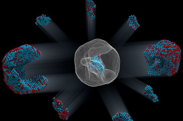
Scientists have taken inspiration from sandcastles to build robots made of nanoparticles. From an Aug. 5, 2015 news item on ScienceDaily,
If you want to form very flexible chains of nanoparticles in liquid in order to build tiny robots with flexible joints or make magnetically self-healing gels, you need to revert to childhood and think about sandcastles.
In a paper published this week in Nature Materials, researchers from North Carolina State University and the University of North Carolina-Chapel Hill show that magnetic nanoparticles encased in oily liquid shells can bind together in water, much like sand particles mixed with the right amount of water can form sandcastles.
“Because oil and water don’t mix, the oil wets the particles and creates capillary bridges between them so that the particles stick together on contact,” said Orlin Velev, INVISTA Professor of Chemical and Biomolecular Engineering at NC State and the corresponding author of the paper.
“We then add a magnetic field to arrange the nanoparticle chains and provide directionality,” said Bhuvnesh Bharti, research assistant professor of chemical and biomolecular engineering at NC State and first author of the paper.
Chilling the oil is like drying the sandcastle. Reducing the temperature from 45 degrees Celsius to 15 degrees Celsius freezes the oil and makes the bridges fragile, leading to breaking and fragmentation of the nanoparticle chains. Yet the broken nanoparticles chains will re-form if the temperature is raised, the oil liquefies and an external magnetic field is applied to the particles.
“In other words, this material is temperature responsive, and these soft and flexible structures can be pulled apart and rearranged,” Velev said. “And there are no other chemicals necessary.”
…
The paper is also co-authored by Anne-Laure Fameau, a visiting researcher from INRA [French National Institute for Agricultural Research or Institut National de la Recherche Agronomique], France. …
In the spirit of full disclosure, the March 25, 2014 news item on ScienceDaily describing the research about breeching the blood-brain barrier uses the term nanorobotic agents rather than nanobots, a term which makes my headline a lot catchier although less accurate. Getting back to the research,
Magnetic nanoparticles can open the blood-brain barrier and deliver molecules directly to the brain, say researchers from the University of Montreal, Polytechnique Montréal, and CHU Sainte-Justine. This barrier runs inside almost all vessels in the brain and protects it from elements circulating in the blood that may be toxic to the brain. The research is important as currently 98% of therapeutic molecules are also unable to cross the blood-brain barrier.
“The barrier is temporary [sic] opened at a desired location for approximately 2 hours by a small elevation of the temperature generated by the nanoparticles when exposed to a radio-frequency field,” explained first author and co-inventor Seyed Nasrollah Tabatabaei. “Our tests revealed that this technique is not associated with any inflammation of the brain. This new result could lead to a breakthrough in the way nanoparticles are used in the treatment and diagnosis of brain diseases,” explained the co-investigator, Hélène Girouard. “At the present time, surgery is the only way to treat patients with brain disorders. Moreover, while surgeons are able to operate to remove certain kinds of tumors, some disorders are located in the brain stem, amongst nerves, making surgery impossible,” added collaborator and senior author Anne-Sophie Carret.
Although the technology was developed using murine models and has not yet been tested in humans, the researchers are confident that future research will enable its use in people. “Building on earlier findings and drawing on the global effort of an interdisciplinary team of researchers, this technology proposes a modern version of the vision described almost 40 years ago in the movie Fantastic Voyage, where a miniature submarine navigated in the vascular network to reach a specific region of the brain,” said principal investigator Sylvain Martel. In earlier research, Martel and his team had managed to manipulate the movement of nanoparticles through the body using the magnetic forces generated by magnetic resonance imaging (MRI) machines.
To open the blood-brain barrier, the magnetic nanoparticles are sent to the surface of the blood-brain barrier at a desired location in the brain. Although it was not the technique used in this study, the placement could be achieved by using the MRI technology described above. Then, the researchers generated a radio-frequency field. The nanoparticles reacted to the radio-frequency field by dissipating heat thereby creating a mechanical stress on the barrier. This allows a temporary and localized opening of the barrier for diffusion of therapeutics into the brain.
The technique is unique in many ways. “The result is quite significant since we showed in previous experiments that the same nanoparticles can also be used to navigate therapeutic agents in the vascular network using a clinical MRI scanner,” Martel remarked. “Linking the navigation capability with these new results would allow therapeutics to be delivered directly to a specific site of the brain, potentially improving significantly the efficacy of the treatment while avoiding systemic circulation of toxic agents that affect healthy tissues and organs,” Carret added. “While other techniques have been developed for delivering drugs to the blood-brain barrier, they either open it too wide, exposing the brain to great risks, or they are not precise enough, leading to scattering of the drugs and possible unwanted side effect,” Martel said.
Although there are many hurdles to overcome before the technology can be used to treat humans, the research team is optimistic. “Although our current results are only proof of concept, we are on the way to achieving our goal of developing a local drug delivery mechanism that will be able to treat oncologic, psychiatric, neurological and neurodegenerative disorders, amongst others,” Carret concluded.
A Nov. 24, 2014 news item on ScienceDaily heralds some bone regeneration research which was published back in Sept. 2014,
Researchers in bone tissue regeneration believe they have made a significant breakthrough for sufferers of bone trauma, disease or defects such as osteoporosis.
Medical researchers from Keele University and Nottingham University have found that magnetic nanoparticles coated with targeting proteins can stimulate stem cells to regenerate bone. Researchers were also able to deliver the cells directly to the injured area, remotely controlling the nanoparticles to generate mechanical forces and maintain the regeneration process through staged releases of a protein growth stimulant.
The current method for repairing bone that can’t heal itself is through a graft taken from the patient. Unfortunately, this can be a painful, invasive procedure, and when the area that needs repair is too large or the patient has a skeletal disorder such as there can sometimes be a lack of healthy bone for grafting.
For this reason, spurring the growth of new bone through injected stem cells is an area of great interest to medical researchers. Much progress has been made, but a major hurdle remains – finding an appropriate means to stimulate the differentiation of the stem cells so they become the quality of bone tissue needed in a quantity large enough to treat patients effectively.
James Henstock, Ph.D. led the Biotechnology and Biological Sciences Research Council (BBSRC)-funded study, alongside Alicia El Haj, Ph.D., and colleagues at Keele University’s Institute for Science and Technology in Medicine, as well as Kevin Shakesheff, Ph.D., from the University of Nottingham’s School of Pharmacy.
James Henstock said: “Injectable therapies for regenerative medicine show great potential as a minimally invasive route for introducing therapeutic stem cells, drug delivery vehicles and biomaterials efficiently to wound sites.”
“In our investigation we coated magnetic nanoparticles with specific targeting proteins then controlled them remotely with an external magnetic field to simulate exercise. We wanted to learn how this might affect the injected stem cells and their ability to restore functional bone.”
The team of researchers conducted their test using two models: chicken foetal femurs and tissue-engineered collagen hydrogels. In both instances the results showed an increase in bone formation and density without causing any mechanical stress to the construct or surrounding tissue.
“This work demonstrates that providing the appropriate mechanical cues in conjunction with controlled release of growth factors to these injectable cell therapies can have a significant impact on improving bone growth. It also could potentially improve tissue engineering approaches for translational medicine” Dr. Henstock said.
Here’s a link to and a citation for the published paper,
![[downloaded from http://pubs.acs.org/doi/abs/10.1021/acs.langmuir.6b01743]](http://www.frogheart.ca/wp-content/uploads/2016/07/KazanUni_Oil-degradingBacteria.gif)