Monthly Archives: September 2016

Nanotubes tunnel between neurons in Parkinson’s disease

An Aug. 22, 2016 news item on ScienceDaily describes how scientists from the Institut Pasteur (France) have developed insight into one of the processes in Parkinson’s disease,

Scientists have demonstrated the role of lysosomal vesicles in transporting alpha-synuclein aggregates, responsible for Parkinson’s and other neurodegenerative diseases, between neurons. These proteins move from one neuron to the next in lysosomal vesicles which travel along the ‘tunneling nanotubes’ between cells.

An Aug. 22, 2016 Institut Pasteur press release (also on EurekAlert), expands on the theme,

Synucleinopathies, a group of neurodegenerative diseases including Parkinson’s disease, are characterized by the pathological deposition of aggregates of the misfolded α-synuclein protein into inclusions throughout the central and peripheral nervous system. Intercellular propagation (from one neuron to the next) of α-synuclein aggregates contributes to the progression of the neuropathology, but little was known about the mechanism by which spread occurs.

In this study, scientists from the Membrane Traffic and Pathogenesis Unit, directed by Chiara Zurzolo at the Institut Pasteur, used fluorescence microscopy to demonstrate that pathogenic α-synuclein fibrils travel between neurons in culture, inside lysosomal vesicles through tunneling nanotubes (TNTs), a new mechanism of intercellular communication.

After being transferred via TNTs, α-synuclein fibrils are able to recruit and induce aggregation of the soluble α-synuclein protein in the cytosol of cells receiving the fibrils, thus explaining the propagation of the disease. The scientists propose that cells overloaded with α-synuclein aggregates in lysosomes dispose of this material by hijacking TNT-mediated intercellular trafficking. However, this results in the disease being spread to naive neurons.

This study demonstrates that TNTs play a significant part in the intercellular transfer of α-synuclein fibrils and reveals the specific role of lysosomes in this process. This represents a major breakthrough in understanding the mechanisms underlying the progression of synucleinopathies.

These compelling findings, together with previous reports from the same team, point to the general role of TNTs in the propagation of prion-like proteins in neurodegenerative diseases and identify TNTs as a new therapeutic target to combat the progression of these incurable diseases.

Here’s a link to and a citation for the paper,

Tunneling nanotubes spread fibrillar α‐synuclein by intercellular trafficking of lysosomes by Saïda Abounit, Luc Bousset, Frida Loria, Seng Zhu, Fabrice de Chaumont, Laura Pieri, Jean-Christophe Olivo-Marin, Ronald Melki, Chiara Zurzolo. The EMBO Journal (2016) e201593411 DOI 10.15252/embj.201593411 Published online 22.08.2016

This paper is behind a paywall.

Making magnetic rust behave like gold and the nanoscale

Researchers at the University of Georgia (US) have found a way to combine gold nanoparticles with magnetic rust nanoparticles for a hybrid structure that behaves with the properties of both types of nanoparticles. From a Sept. 15, 2016 news item on ScienceDaily,

Researchers from the University of Georgia are giving new meaning to the phrase “turning rust into gold”—and making the use of gold in research settings and industrial applications far more affordable.

The research is akin to a type of modern-day alchemy, said Simona Hunyadi Murph, adjunct professor in the UGA Franklin College of Arts and Sciences department of physics and astronomy. Researchers combine small amounts of gold nanoparticles with magnetic rust nanoparticles to create a hybrid nanostructure that retains both the properties of gold and rust.

A Sept. 15, 2016 University of Georgia news release by Jessica Luton, which originated the news item, expands on the theme,

“Medieval alchemists tried to create gold from other metals,” she said. “That’s kind of what we did with our research. It’s not real alchemy, in the medieval sense, but it is a sort of 21st century version.”

Gold has long been a valuable resource for industry, medicine, dentistry, computers, electronics and aerospace, among others, due to unique physical and chemical properties that make it inert and resistant to oxidation. But because of its high cost and limited supply, large scale projects using gold can be prohibitive. At the nanoscale, however, using a very small amount of gold is far more affordable.

In the new study published this summer in the Journal of Physical Chemistry C, the researchers used solution chemistry to reduce gold ions into a metallic gold structure using sodium citrate. In this process, if other ingredients-rust in this case-are present in the reaction pot during the transformation process, the metallic gold structures nucleate and grow on these “ingredients,” otherwise known as supports.

“We are really excited to share our new discoveries. When researchers are looking at gold as a potential material for research, we talk about how expensive gold is. For the first time ever, we’ve been able to create a new class of cheaper, highly efficient, nontoxic, magnetically reusable hybrid nanomaterials that contain a far more abundant material-rust-than the typical noble metal gold,” said Murph, who is also a principal scientist in the National Security Directorate at the Savannah River National Laboratory in Aiken, South Carolina.

When materials are broken down in size to reach nanometer scale dimensions-1-100 nanometers, which is approximately 100,000 times smaller than the diameter of human hair-these substances can take on new properties. For example, bulk gold does not display catalytic properties; however, at the nanoscale, gold is an efficient catalyst, accelerating chemical change for many reactions including oxidation, hydrogen production or reduction of aromatic nitro compounds.

Gold nanoparticles of different sizes and shapes display different colors when impinged by light because they absorb and scatter light at specific wavelengths, known as plasmonic resonances. These plasmonic resonances are of particular interest for biological applications. If someone shines light on the gold nanoparticles, the absorbed light can be converted to heat in the surrounding media, and if bacteria or cancerous cells are in the vicinity of such gold nanoparticles, they can be destroyed by using light of appropriate wavelength. This phenomenon is known as photothermal therapy.

By replacing some of the nano-gold with magnetic nano-rust, researchers show that the hybrid gold and rust nanostructures are able to photothermally heat the surrounding media as efficiently as pure gold nanoparticles, even with a significantly smaller concentration of gold.

“In a way, we’ve done a little better than alchemy,” said George Larsen, co-investigator and postdoctoral researcher in the Group for Innovation and Advancements in Nano-Technology Sciences at the Savannah River National Laboratory, “because these new hybrid nanoparticles not only behave better than gold in some cases, but also have magnetic functionality.”

Murph and her team looked at three different shapes of hybrid nanoparticles in this research-spheres, rings and tubes.

“A differently shaped nanoparticle means that the atoms are arranged differently-into cubes, hexagons or triangles, for example,” she said. “A different atom arrangement means different packing densities, spacing between atoms, defects, surface area and surface energies. Different shapes lead to an increased atom area that is exposed to catalyze a chemical reaction. Scientifically speaking, different shape means different crystallographic facets and surface energy that could lead to higher catalytic activity and different catalytic products.

“The results of our research showed that the ring- and tube-shaped hybrid nanoparticles proved to be better catalytic materials than the sphere-shaped nanoparticles because of the way the atoms are arranged in the structure at this nanoscale. More importantly, the hybrid nanoparticles of gold and rust are better catalysts than gold nanoparticles alone, even with a significantly smaller amount of gold.

When these different shaped hybrid nanoparticles were exposed to light of specific wavelength, the spheres heated the solution up to slightly higher temperatures than the ring- or tube-shaped nanoparticles.

“This could have a variety of biological applications such as tracking, drug delivery or imaging inside the body,” Murph said. “If you feed these gold nanoparticles to bacteria and shine the light on them, you could destroy these by just using light.”

The hybrid structures could also be used for new application [sic], such as sensing, hyperthermia treatment, environmental cleaning and protection medical imaging applications including magnetic resonance imaging contrast agents, product detection and manipulation.

Here’s a link to and a citation for the paper/video,

Multifunctional Hybrid Fe2O3-Au Nanoparticles for Efficient Plasmonic Heating by Simona E. Hunyadi Murph, George K. Larsen, Robert J. Lascola. Journal of Visualized Experiments, 2016; (108) DOI: 10.3791/53598

This paper/video appears to be open access.

Doing math in a test tube using analog DNA

Basically, scientists at Duke University (US) have created an analog computer at the nanoscale, which can perform basic arithmetic. From an Aug. 23, 2016 news item on ScienceDaily,

Often described as the blueprint of life, DNA contains the instructions for making every living thing from a human to a house fly.

But in recent decades, some researchers have been putting the letters of the genetic code to a different use: making tiny nanoscale computers.

In a new study, a Duke University team led by professor John Reif created strands of synthetic DNA that, when mixed together in a test tube in the right concentrations, form an analog circuit that can add, subtract and multiply as they form and break bonds.

Rather than voltage, DNA circuits use the concentrations of specific DNA strands as signals.

An Aug. 23, 2016 Duke University news release (also on EurekAlert), which originated the news item, describes how most DNA-based circuits operate and what makes the one from Duke different,

Other teams have designed DNA-based circuits that can solve problems ranging from calculating square roots to playing tic-tac-toe. But most DNA circuits are digital, where information is encoded as a sequence of zeroes and ones.

Instead, the new Duke device performs calculations in an analog fashion by measuring the varying concentrations of specific DNA molecules directly, without requiring special circuitry to convert them to zeroes and ones first.

Unlike the silicon-based circuits used in most modern day electronics, commercial applications of DNA circuits are still a long way off, Reif said.

For one, the test tube calculations are slow. It can take hours to get an answer.

“We can do some limited computing, but we can’t even begin to think of competing with modern-day PCs or other conventional computing devices,” Reif said.

But DNA circuits can be far tinier than those made of silicon. And unlike electronic circuits, DNA circuits work in wet environments, which might make them useful for computing inside the bloodstream or the soupy, cramped quarters of the cell.

The technology takes advantage of DNA’s natural ability to zip and unzip to perform computations. Just like Velcro and magnets have complementary hooks or poles, the nucleotide bases of DNA pair up and bind in a predictable way.

The researchers first create short pieces of synthetic DNA, some single-stranded and some double-stranded with single-stranded ends, and mix them in a test tube.

When a single strand encounters a perfect match at the end of one of the partially double-stranded ones, it latches on and binds, displacing the previously bound strand and causing it to detach, like someone cutting in on a dancing couple.

The newly released strand can in turn pair up with other complementary DNA molecules downstream in the circuit, creating a domino effect.

The researchers solve math problems by measuring the concentrations of specific outgoing strands as the reaction reaches equilibrium.

To see how their circuit would perform over time as the reactions proceeded, Reif and Duke graduate student Tianqi Song used computer software to simulate the reactions over a range of input concentrations. They have also been testing the circuit experimentally in the lab.

Besides addition, subtraction and multiplication, the researchers are also designing more sophisticated analog DNA circuits that can do a wider range of calculations, such as logarithms and exponentials.

Conventional computers went digital decades ago. But for DNA computing, the analog approach has its advantages, the researchers say. For one, analog DNA circuits require fewer strands of DNA than digital ones, Song said.

Analog circuits are also better suited for sensing signals that don’t lend themselves to simple on-off, all-or-none values, such as vital signs and other physiological measurements involved in diagnosing and treating disease.

The hope is that, in the distant future, such devices could be programmed to sense whether particular blood chemicals lie inside or outside the range of values considered normal, and release a specific DNA or RNA — DNA’s chemical cousin — that has a drug-like effect.

Reif’s lab is also beginning to work on DNA-based devices that could detect molecular signatures of particular types of cancer cells, and release substances that spur the immune system to fight back.

“Even very simple DNA computing could still have huge impacts in medicine or science,” Reif said.

Here’s a link to and a citation for the paper,

Analog Computation by DNA Strand Displacement Circuits by Tianqi Song, Sudhanshu Garg, Reem Mokhtar, Hieu Bui, and John Reif. ACS Synth. Biol., 2016, 5 (8), pp 898–912 DOI: 10.1021/acssynbio.6b00144 Publication Date (Web): July 01, 2016

Copyright © 2016 American Chemical Society

This paper is behind a paywall.

Creating quantum dots (artificial atoms) in graphene

An Aug. 22, 2016 news item on phys.org describes some recent work on artificial atoms and graphene from the Technical University of Vienna (Austria) and partners in Germany and the UK,

In a tiny quantum prison, electrons behave quite differently as compared to their counterparts in free space. They can only occupy discrete energy levels, much like the electrons in an atom – for this reason, such electron prisons are often called “artificial atoms”. Artificial atoms may also feature properties beyond those of conventional ones, with the potential for many applications for example in quantum computing. Such additional properties have now been shown for artificial atoms in the carbon material graphene. The results have been published in the journal Nano Letters, the project was a collaboration of scientists from TU Wien (Vienna, Austria), RWTH Aachen (Germany) and the University of Manchester (GB).

“Artificial atoms open up new, exciting possibilities, because we can directly tune their properties”, says Professor Joachim Burgdörfer (TU Wien, Vienna). In semiconductor materials such as gallium arsenide, trapping electrons in tiny confinements has already been shown to be possible. These structures are often referred to as “quantum dots”. Just like in an atom, where the electrons can only circle the nucleus on certain orbits, electrons in these quantum dots are forced into discrete quantum states.

Even more interesting possibilities are opened up by using graphene, a material consisting of a single layer of carbon atoms, which has attracted a lot of attention in the last few years. “In most materials, electrons may occupy two different quantum states at a given energy. The high symmetry of the graphene lattice allows for four different quantum states. This opens up new pathways for quantum information processing and storage” explains Florian Libisch from TU Wien. However, creating well-controlled artificial atoms in graphene turned out to be extremely challenging.

Florian Libisch, explaining the structure of graphene. Courtesy Technical University of Vienna

Florian Libisch, explaining the structure of graphene. Courtesy Technical University of Vienna

An Aug. 22, 2016 Technical University of Vienna press release (also on EurekAlert), which originated the news item, provides more detail,

There are different ways of creating artificial atoms: The simplest one is putting electrons into tiny flakes, cut out of a thin layer of the material. While this works for graphene, the symmetry of the material is broken by the edges of the flake which can never be perfectly smooth. Consequently, the special four-fold multiplicity of states in graphene is reduced to the conventional two-fold one.

Therefore, different ways had to be found: It is not necessary to use small graphene flakes to capture electrons. Using clever combinations of electrical and magnetic fields is a much better option. With the tip of a scanning tunnelling microscope, an electric field can be applied locally. That way, a tiny region is created within the graphene surface, in which low energy electrons can be trapped. At the same time, the electrons are forced into tiny circular orbits by applying a magnetic field. “If we would only use an electric field, quantum effects allow the electrons to quickly leave the trap” explains Libisch.

The artificial atoms were measured at the RWTH Aachen by Nils Freitag and Peter Nemes-Incze in the group of Professor Markus Morgenstern. Simulations and theoretical models were developed at TU Wien (Vienna) by Larisa Chizhova, Florian Libisch and Joachim Burgdörfer. The exceptionally clean graphene sample came from the team around Andre Geim and Kostya Novoselov from Manchester (GB) – these two researchers were awarded the Nobel Prize in 2010 for creating graphene sheets for the first time.

The new artificial atoms now open up new possibilities for many quantum technological experiments: “Four localized electron states with the same energy allow for switching between different quantum states to store information”, says Joachim Burgdörfer. The electrons can preserve arbitrary superpositions for a long time, ideal properties for quantum computers. In addition, the new method has the big advantage of scalability: it should be possible to fit many such artificial atoms on a small chip in order to use them for quantum information applications.

Here’s a link to and a citation for the paper,

Electrostatically Confined Monolayer Graphene Quantum Dots with Orbital and Valley Splittings by Nils M. Freitag, Larisa A. Chizhova, Peter Nemes-Incze, Colin R. Woods, Roman V. Gorbachev, Yang Cao, Andre K. Geim, Kostya S. Novoselov, Joachim Burgdörfer, Florian Libisch, and Markus Morgenstern. Nano Lett., Article ASAP DOI: 10.1021/acs.nanolett.6b02548 Publication Date (Web): July 28, 2016

Copyright © 2016 American Chemical Society

This paper is behind a paywall.

Dexter Johnson in an Aug. 23, 2016 post on his Nanoclast blog (on the IEEE [Institute of Electrical and Electronics Engineers] website) provides some additional insight into the world of quantum dots,

Quantum dots made from semiconductor materials, like silicon, are beginning to transform the display market. While it is their optoelectronic properties that are being leveraged in displays, the peculiar property of quantum dots that allows their electrons to be forced into discrete quantum states has long held out the promise of enabling quantum computing.

If you have time to read it, Dexter’s post features an email interview with Florian Libisch where they further discuss quantum dots and quantum computing.

Canada’s Ingenuity Lab receives a $1.7M grant to develop oil recovery system for oil spills

A Sept. 15, 2016 news item on Benzinga.com describes the reasons for the $1.7M grant for Alberta’s (Canada) Ingenuity Lab to develop an oil spill recovery system,

Since 2010’s tragic events, which saw BP’s Deepwater Horizon disaster desecrate the Gulf of Mexico, oil safety has been on the forefront of the environmental debate and media outrage. In line with the mounting concerns continuing to pique public attention, at the end of this month [Sept. 2016], Hollywood will release its own biopic of the event. As can be expected, more questions will be raised about what exactly went wrong, in addition to fresh criticism aimed at the entire industry.

One question that is likely to emerge is how do we prevent such a calamity from ever happening again? Fortunately, some of the brightest minds in science have been preparing for such an answer.

One team that has been focusing on this dilemma is Alberta-based, multi-disciplinary research initiative Ingenuity Lab. The institution has just secured $1.7m in project funding for developing a highly advanced system for recovering oil from oil spills. This injection of capital will enable Ingenuity Lab to conduct new research and develop commercial production processes for recovering heavy oil spills in marine environments. The technology is centred on cutting edge nanowire-based stimuli-responsive membranes and devices that are capable for recovering oil.

A Sept. 15, 2016 Ingenuity Lab news release on MarketWired, which originated the news item, provides more insight into the oil spill situation,

Oil is a common pollutant in our oceans; more than three million metric tonnes contaminate the sea each year. When crude oil is accidentally released into a body of water by an oil tanker, refinery, storage facility, underwater pipeline or offshore oil-drilling rig, it is an environmental emergency of the most urgent kind.

Depending on the location, oil spills can be highly hazardous, as well as environmentally destructive. Consequently, a timely clean up is absolutely crucial in order to protect the integrity of the water, the shoreline and the numerous creatures that depend on these habitats.

Due to increased scrutiny of the oil industry with regard to its unseemly environmental track record, attention must be focused on the development of new materials and technologies for removing organic contaminants from waterways. Simply put, existing methods are not sufficiently robust.

Fortuitously, however, nanotechnology has opened the door for the development of sophisticated new tools that use specifically designed materials with properties that are ideally suited to enable complex separations, including the separation of crude oil from water.

Ingenuity Lab’s project focuses on the efficient recovery of oil through the development of this novel technology using a variety of stimuli-responsive nanomaterials. When the time comes for scale up production for this technology, Ingenuity Lab will work closely with industry trendsetters, Tortech Nanofibers.

This project forms a strong element of the Oil Spill Response Science (OSRS), which is part of Canada’s world-class tanker safety system for Responsible Resource Development. Through this programme, the Canadian Government ensures that the country’s resource wealth can be safely developed and transported to market, thus creating new jobs and economic growth for all Canadians.

From a communications standpoint, the news release is well written and well strategized to underline the seriousness of the situation and to take advantage of renewed interest in the devastating (people’s lives were lost and environmental damage is still being assessed) 2010 BP oil spill in the Gulf of Mexico due to the upcoming movie titled, Deepwater Horizon. A little more information about the team (how many people, who’s leading the research, are there international and/or interprovincial collaborators?), plans for the research (have they already started? what work, if any, are they building on? what challenges are they facing?) and some technical details would have been welcome.

Regardless, it’s good to hear about this initiative and I wish them great success with it.

You can find our more about Ingenuity Lab here and Tortech Nanofibers here. Interestingly, Tortech is a joint venture between Israel’s Plasan Sasa and the UK’s Q-Flo. (Q-Flo is a spinoff from Cambridge University.) One more thing, Tortech Nanofibers produces materials made of carbon nanotubes (CNTs). Presumably Ingenuity’s “nanowire-based stimuli-responsive membranes” include carbon nanotubes.

Oil-absorbing hairy fern leaves lead to ‘nanofur’ for oil spill cleanups

German researchers have developed a biomimetic material branded as ‘nanofur’ for oil spill cleanups, according to an Aug. 23, 2016 news item on Nanowerk (Note: A link has been removed),

Some water ferns can absorb large volumes of oil within a short time, because their leaves are strongly water-repellent and, at the same time, highly oil-absorbing. Researchers of Karlsruhe Institute of Technology, together with colleagues of Bonn University, have found that the oil-binding capacity of the water plant results from the hairy microstructure of its leaves (Bioinspiration & Biomimetics, “Microstructures of superhydrophobic plant leaves – inspiration for efficient oil spill cleanup materials”). It is now used as a model to further develop the new Nanofur material for the environmentally friendly cleanup of oil spills.

An Aug.(?) 23 (?), 2016 Karlsruhe Institute of Technology (KIT) press release on EurekAlert, which originated the news item, explains the interest in improving technology for oil spill cleanups and provides insight into this  innovation,

Damaged pipelines, oil tanker disasters, and accidents on oil drilling and production platforms may result in pollutions [sic] of water with crude or mineral oil. Conventional methods to clean up the oil spill are associated with specific drawbacks. Oil combustion or the use of chemical substances to accelerate oil decomposition cause secondary environmental pollution. Many natural materials to take up the oil, such as sawdust or plant fibers, are hardly effective, because they also absorb large amounts of water. On their search for an environmentally friendly alternative to clean up oil spills, the researchers compared various species of aquatic ferns. “We already knew that the leaves of these plants repel water, but for the first time now, we have studied their capacity to absorb oil,” Claudia Zeiger says. She conducted the project at KIT’s Institute of Microstructure Technology.

Damaged pipelines, oil tanker disasters, and accidents on oil drilling and production platforms may result in pollutions of water with crude or mineral oil. Conventional methods to clean up the oil spill are associated with specific drawbacks. Oil combustion or the use of chemical substances to accelerate oil decomposition cause secondary environmental pollution. Many natural materials to take up the oil, such as sawdust or plant fibers, are hardly effective, because they also absorb large amounts of water. On their search for an environmentally friendly alternative to clean up oil spills, the researchers compared various species of aquatic ferns. “We already knew that the leaves of these plants repel water, but for the first time now, we have studied their capacity to absorb oil,” Claudia Zeiger says. She conducted the project at KIT’s Institute of Microstructure Technology.

Aquatic ferns originally growing in tropical and subtropical regions can now also be found in parts of Europe. As they reproduce strongly, they are often considered weed. However, they have a considerable potential as low-cost, rapid, and environmentally friendly oil absorbers, which is obvious from a short video (see below). ”The plants might be used in lakes to absorb accidental oil spills,” Zeiger says. After less than 30 seconds, the leaves reach maximum absorption and can be skimmed off together with the absorbed oil. The water plant named salvinia has trichomes on the leaf surface – hairy extensions of 0.3 to 2.5 mm in length. Comparison of different salvinia species revealed that leaves with the longest hairs did not absorb the largest amounts of oil. “Oil-absorbing capacity is determined by the shape of the hair ends,” Zeiger emphasizes. The largest quantity of oil was absorbed by leaves of the water fern salvinia molesta, whose hair ends are shaped like an eggbeater.

Based on this new knowledge on the relationship between surface structure of leaves and their oil-absorbing capacity, the researchers improved the ‘Nanofur’ material developed at their institute. This plastic nanofur mimics the water-repellent and oil-absorbing effect of salvinia to separate oil and water. “We study nanostructures and microstructures in nature for potential technical developments,” says Hendrik Hölscher, Head of the Biomimetic Surfaces Group of the Institute of Microstructure Technology of KIT. He points out that different properties of plants made of the same material frequently result from differences of their finest structures.

Here’s a link to and a citation for the paper,

Microstructures of superhydrophobic plant leaves – inspiration for efficient oil spill cleanup materials by Claudia Zeiger, Isabelle C Rodrigues da Silva, Matthias Mail, Maryna N Kavalenka, Wilhelm Barthlott, and Hendrik Hölscher. Bioinspiration & Biomimetics, Volume 11, Number 5 DOI: http://dx.doi.org/10.1088/1748-3190/11/5/056003

Published 16 August 2016, © 2016 IOP Publishing Ltd

This article appears to be open access.

There is also a video demonstration of the material,

Enjoy!

Just swallow your battery, eh? Ingestible batteries

Christopher Bettinger, Ph.D., is developing an edible battery made with melanin and dissolvable materials. Courtesy of: Bettinger lab

Christopher Bettinger, Ph.D., is developing an edible battery made with melanin and dissolvable materials. Courtesy of: Bettinger lab

An Aug. 23, 2016 news item on phys.org describes a session at the 252nd American Chemical Society (ACS) meeting held Aug. 21 – 25, 2016 in Philadelphia,

Non-toxic, edible batteries could one day power ingestible devices for diagnosing and treating disease. One team reports new progress toward that goal with their batteries made with melanin pigments, naturally found in the skin, hair and eyes.

“For decades, people have been envisioning that one day, we would have edible electronic devices to diagnose or treat disease,” says Christopher Bettinger, Ph.D. “But if you want to take a device every day, you have to think about toxicity issues. That’s when we have to think about biologically derived materials that could replace some of these things you might find in a RadioShack.”

An Aug. 23, 2016 ACS news release (also on EurekAlert), which originated the news item, further describes the work featured in the ACS meeting session,

About 20 years ago, scientists did develop a battery-operated ingestible camera as a complementary tool to endoscopies. It can image places in the digestive system that are inaccessible to the traditional endoscope. But it is designed to pass through the body and be excreted. For a single use, the risk that the camera with a conventional battery will get stuck in the gastrointestinal tract is small. But the chances of something going wrong would increase unacceptably if doctors wanted to use it more frequently on a single patient.

The camera and some implantable devices such as pacemakers run on batteries containing toxic components that are sequestered away from contact with the body. But for low-power, repeat applications such as drug-delivery devices that are meant to be swallowed, non-toxic and degradable batteries would be ideal.

“The beauty is that by definition an ingestible, degradable device is in the body for no longer than 20 hours or so,” Bettinger says. “Even if you have marginal performance, which we do, that’s all you need.”

While he doesn’t have to worry about longevity, toxicity is an issue. To minimize the potential harm of future ingestible devices, Bettinger’s team at Carnegie Mellon University (CMU) decided to turn to melanins and other naturally occurring compounds. In our skin, hair and eyes, melanins absorb ultraviolet light to quench free radicals and protect us from damage. They also happen to bind and unbind metallic ions. “We thought, this is basically a battery,” Bettinger says.

Building on this idea, the researchers experimented with battery designs that use melanin pigments at either the positive or negative terminals; various electrode materials such as manganese oxide and sodium titanium phosphate; and cations such as copper and iron that the body uses for normal functioning.

“We found basically that they work,” says Hang-Ah Park, Ph.D., a post-doctoral researcher at CMU. “The exact numbers depend on the configuration, but as an example, we can power a 5 milliWatt device for up to 18 hours using 600 milligrams of active melanin material as a cathode.”

Although the capacity of a melanin battery is low relative to lithium-ion, it would be high enough to power an ingestible drug-delivery or sensing device. For example, Bettinger envisions using his group’s battery for sensing gut microbiome changes and responding with a release of medicine, or for delivering bursts of a vaccine over several hours before degrading.

In parallel with the melanin batteries, the team is also making edible batteries with other biomaterials such as pectin, a natural compound from plants used as a gelling agent in jams and jellies. Next, they plan on developing packaging materials that will safely deliver the battery to the stomach.

When these batteries will be incorporated into biomedical devices is uncertain, but Bettinger has already found another application for them. His lab uses the batteries to probe the structure and chemistry of the melanin pigments themselves to better understand how they work.

I previously wrote about an ingestible battery in a November 23, 2015 posting featuring work from MIT (Massachusetts Institute of Technology).

New electrochromic material for ‘smart’ windows

Given that it’s summer, I seem to be increasingly obsessed with windows that help control the heat from the sun. So, this Aug. 22, 2016 news item on ScienceDaily hit my sweet spot,

Researchers in the Cockrell School of Engineering at The University of Texas at Austin have invented a new flexible smart window material that, when incorporated into windows, sunroofs, or even curved glass surfaces, will have the ability to control both heat and light from the sun. …

Delia Milliron, an associate professor in the McKetta Department of Chemical Engineering, and her team’s advancement is a new low-temperature process for coating the new smart material on plastic, which makes it easier and cheaper to apply than conventional coatings made directly on the glass itself. The team demonstrated a flexible electrochromic device, which means a small electric charge (about 4 volts) can lighten or darken the material and control the transmission of heat-producing, near-infrared radiation. Such smart windows are aimed at saving on cooling and heating bills for homes and businesses.

An Aug. 22, 2016 University of Texas at Austin news release (also on EurekAlert), which originated the news item, describes the international team behind this research and offers more details about the research itself,

The research team is an international collaboration, including scientists at the European Synchrotron Radiation Facility and CNRS in France, and Ikerbasque in Spain. Researchers at UT Austin’s College of Natural Sciences provided key theoretical work.

Milliron and her team’s low-temperature process generates a material with a unique nanostructure, which doubles the efficiency of the coloration process compared with a coating produced by a conventional high-temperature process. It can switch between clear and tinted more quickly, using less power.

The new electrochromic material, like its high-temperature processed counterpart, has an amorphous structure, meaning the atoms lack any long-range organization as would be found in a crystal. However, the new process yields a unique local arrangement of the atoms in a linear, chain-like structure. Whereas conventional amorphous materials produced at high temperature have a denser three-dimensionally bonded structure, the researchers’ new linearly structured material, made of chemically condensed niobium oxide, allows ions to flow in and out more freely. As a result, it is twice as energy efficient as the conventionally processed smart window material.

At the heart of the team’s study is their rare insight into the atomic-scale structure of the amorphous materials, whose disordered structures are difficult to characterize. Because there are few techniques for characterizing the atomic-scale structure sufficiently enough to understand properties, it has been difficult to engineer amorphous materials to enhance their performance.

“There’s relatively little insight into amorphous materials and how their properties are impacted by local structure,” Milliron said. “But, we were able to characterize with enough specificity what the local arrangement of the atoms is, so that it sheds light on the differences in properties in a rational way.”

Graeme Henkelman, a co-author on the paper and chemistry professor in UT Austin’s College of Natural Sciences, explains that determining the atomic structure for amorphous materials is far more difficult than for crystalline materials, which have an ordered structure. In this case, the researchers were able to use a combination of techniques and measurements to determine an atomic structure that is consistent in both experiment and theory.

“Such collaborative efforts that combine complementary techniques are, in my view, the key to the rational design of new materials,” Henkelman said.

Milliron believes the knowledge gained here could inspire deliberate engineering of amorphous materials for other applications such as supercapacitors that store and release electrical energy rapidly and efficiently.

The Milliron lab’s next challenge is to develop a flexible material using their low-temperature process that meets or exceeds the best performance of electrochromic materials made by conventional high-temperature processing.

“We want to see if we can marry the best performance with this new low-temperature processing strategy,” she said.

Here’s a link to and a citation for the paper,

Linear topology in amorphous metal oxide electrochromic networks obtained via low-temperature solution processing by Anna Llordés, Yang Wang, Alejandro Fernandez-Martinez, Penghao Xiao, Tom Lee, Agnieszka Poulain, Omid Zandi, Camila A. Saez Cabezas, Graeme Henkelman, & Delia J. Milliron. Nature Materials (2016)  doi:10.1038/nmat4734 Published online 22 August 2016

This paper is behind a paywall.

Counteracting chemotherapy resistance with nanoparticles that mimic salmonella

Given the reputation that salmonella (for those who don’t know, it’s a toxin you don’t want to find in your food) has, a nanoparticle which mimics its effects has a certain cachet. An Aug. 22, 2016 news item on Nanowerk,

Researchers at the University of Massachusetts Medical School have designed a nanoparticle that mimics the bacterium Salmonella and may help to counteract a major mechanism of chemotherapy resistance.

Working with mouse models of colon and breast cancer, Beth McCormick, Ph.D., and her colleagues demonstrated that when combined with chemotherapy, the nanoparticle reduced tumor growth substantially more than chemotherapy alone.

Credit: Rocky Mountain Laboratories,NIAID,NIHColor-enhanced scanning electron micrograph showing Salmonella typhimurium (red) invading cultured human cells.

Credit: Rocky Mountain Laboratories,NIAID,NIHColor-enhanced scanning electron micrograph showing Salmonella typhimurium (red) invading cultured human cells.

An Aug. 22, 2016 US National Institute of Cancer news release, which originated the news item, explains the research in more detail,

A membrane protein called P-glycoprotein (P-gp) acts like a garbage chute that pumps waste, foreign particles, and toxins out of cells. P-gp is a member of a large family of transporters, called ATP-binding cassette (ABC) transporters, that are active in normal cells but also have roles in cancer and other diseases. For instance, cancer cells can co-opt P-gp to rid themselves of chemotherapeutic agents, severely limiting the efficacy of these drugs.

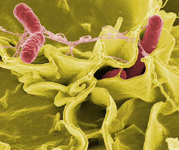
In previous work, Dr. McCormick and her colleagues serendipitously discovered that Salmonella enterica, a bacterium that causes food poisoning, decreases the amount of P-gp on the surface of intestinal cells. Because Salmonella has the capacity to grow selectively in cancer cells, the researchers wondered whether there was a way to use the bacterium to counteract chemotherapy resistance caused by P-gp.

“While trying to understand how Salmonella invades the human host, we made this other observation that may be relevant to cancer therapeutics and multidrug resistance,” explained Dr. McCormick.

Salmonella and Cancer Cells

To determine the specific bacterial component responsible for reducing P-gp levels, the researchers engineered multiple Salmonella mutant strains and tested their effect on P-gp levels in colon cells. They found that a Salmonella strain lacking the bacterial protein SipA was unable to reduce P-gp levels in the colon of mice or in a human colon cancer cell line. Salmonella secretes SipA, along with other proteins, to help the bacterium invade human cells.

The researchers then showed that treatment with SipA protein alone decreased P-gp levels in cell lines of human colon cancer, breast cancer, bladder cancer, and lymphoma.

Because P-gp can pump drugs out of cells, the researchers next sought to determine whether SipA treatment would prevent cancer cells from expelling chemotherapy drugs.

When they treated human colon cancer cells with the chemotherapy agents doxorubicin or vinblastine, with or without SipA, they found that the addition of SipA increased drug retention inside the cells. SipA also increased the cancer cells’ sensitivity to both drugs, suggesting that it could possibly be used to enhance chemotherapy.

“Through millions of years of co-evolution, Salmonella has figured out a way to remove this transporter from the surface of intestinal cells to facilitate host infection,” said Dr. McCormick. “We capitalized on the organism’s ability to perform that function.”

A Nanoparticle Mimic

It would not be feasible to infect people with the bacterium, and SipA on its own will likely deteriorate quickly in the bloodstream, coauthor Gang Han, Ph.D., of the University of Massachusetts Medical School, explained in a press release. The researchers therefore fused SipA to gold nanoparticles, generating what they refer to as a nanoparticle mimic of Salmonella. They designed the nanoparticle to enhance the stability of SipA, while retaining its ability to interact with other proteins.

In an effort to target tumors without harming healthy tissues, the researchers used a nanoparticle of specific size that should only be able to access the tumor tissue due to its “leaky” architecture. “Because of this property, we are hoping to be able to avoid negative effects to healthy tissues,” said Dr. McCormick. Another benefit of this technology is that the nanoparticle can be modified to enhance tumor targeting and minimize the potential for side effects, she added.

The researchers showed that this nanoparticle was 100 times more effective than SipA protein alone at reducing P-gp levels in a human colon cancer cell line. The enhanced function of the nanoparticle is likely due to stabilization of SipA, explained the researchers.

The team then tested the nanoparticle in a mouse model of colon cancer, because this cancer type is known to express high levels of P-gp. When they treated tumor-bearing mice with the nanoparticle plus doxorubicin, P-gp levels dropped and the tumors grew substantially less than in mice treated with the nanoparticle or doxorubicin alone. The researchers observed similar results in a mouse model of human breast cancer.

There are concerns about the potential effect of nanoparticles on normal tissues. “P-gp has evolved as a defense mechanism” to rid healthy cells of toxic molecules, said Suresh Ambudkar, Ph.D., deputy chief of the Laboratory of Cell Biology in NCI’s Center for Cancer Research. It plays an important role in protecting cells of the blood-brain barrier, liver, testes, and kidney. “So when you try to interfere with that, you may create problems,” he said.

The researchers, however, found no evidence of nanoparticle accumulation in the brain, heart, kidney, or lungs of mice, nor did it appear to cause toxicity. They did observe that the nanoparticles accumulated in the liver and spleen, though this was expected because these organs filter the blood, said Dr. McCormick.

Moving Forward

The research team is moving forward with preclinical studies of the SipA nanoparticle to test its safety and toxicity, and to establish appropriate dosage levels.

However, Dr. Ambudkar noted, “the development of drug resistance in cancer cells is a multifactorial process. In addition to the ABC transporters, other phenomena are involved, such as drug metabolism.” And because there is a large family of ABC transporters, one transporter can compensate if another is blocked, he explained.

For the last 25 years, clinical trials with drugs that inhibit P-gp have failed to overcome chemotherapy resistance, Dr. Ambudkar said. Tackling the issue of multidrug resistance in cancer, he continued, “is not something that can be solved easily.”

Dr. McCormick and her team are also pursuing research to better characterize and understand the biology of SipA. “We are not naïve about the complexity of the problem,” she said. “However, if we know more about the biology, we believe we can ultimately make a better drug.”

Here’s a link to and a citation for the paper,

A Salmonella nanoparticle mimic overcomes multidrug resistance in tumours by Regino Mercado-Lubo, Yuanwei Zhang, Liang Zhao, Kyle Rossi, Xiang Wu, Yekui Zou, Antonio Castillo, Jack Leonard, Rita Bortell, Dale L. Greiner, Leonard D. Shultz, Gang Han, & Beth A. McCormick. Nature Communications 7, Article number: 12225  doi:10.1038/ncomms12225 Published 25 July 2016

This paper is open access.

Nanoparticles could make blood clot faster

It was the 252nd meeting for the American Chemical Society from Aug. 21 – 25, 2016 and that meant a flurry of news about the latest research. From an Aug. 23, 2016 news item on Nanowerk,

Whether severe trauma occurs on the battlefield or the highway, saving lives often comes down to stopping the bleeding as quickly as possible. Many methods for controlling external bleeding exist, but at this point, only surgery can halt blood loss inside the body from injury to internal organs. Now, researchers have developed nanoparticles that congregate wherever injury occurs in the body to help it form blood clots, and they’ve validated these particles in test tubes and in vivo [animal testing].

The researchers will present their work today [Aug. 22, 2016] at the 252nd National Meeting & Exposition of the American Chemical Society (ACS). ACS, the world’s largest scientific society, is holding the meeting here through Thursday. It features more than 9,000 presentations on a wide range of science topics.

An Aug. 22, 2016 American Chemical Society (ACS) news release (also on EurekAlert), which originated the news item, provided more detail,

“When you have uncontrolled internal bleeding, that’s when these particles could really make a difference,” says Erin B. Lavik, Sc.D. “Compared to injuries that aren’t treated with the nanoparticles, we can cut bleeding time in half and reduce total blood loss.”

Trauma remains a top killer of children and younger adults, and doctors have few options for treating internal bleeding. To address this great need, Lavik’s team developed a nanoparticle that acts as a bridge, binding to activated platelets and helping them join together to form clots. To do this, the nanoparticle is decorated with a molecule that sticks to a glycoprotein found only on the activated platelets.

Initial studies suggested that the nanoparticles, delivered intravenously, helped keep rodents from bleeding out due to brain and spinal injury, Lavik says. But, she acknowledges, there was still one key question: “If you are a rodent, we can save your life, but will it be safe for humans?”

As a step toward assessing whether their approach would be safe in humans, they tested the immune response toward the particles in pig’s blood. If a treatment triggers an immune response, it would indicate that the body is mounting a defense against the nanoparticle and that side effects are likely. The team added their nanoparticles to pig’s blood and watched for an uptick in complement, a key indicator of immune activation. The particles triggered complement in this experiment, so the researchers set out to engineer around the problem.

“We made a battery of particles with different charges and tested to see which ones didn’t have this immune-response effect,” Lavik explains. “The best ones had a neutral charge.” But neutral nanoparticles had their own problems. Without repulsive charge-charge interactions, the nanoparticles have a propensity to aggregate even before being injected. To fix this issue, the researchers tweaked their nanoparticle storage solution, adding a slippery polymer to keep the nanoparticles from sticking to each other.

Lavik also developed nanoparticles that are stable at higher temperatures, up to 50 degrees Celsius (122 degrees Fahrenheit). This would allow the particles to be stored in a hot ambulance or on a sweltering battlefield.

In future studies, the researchers will test whether the new particles activate complement in human blood. Lavik also plans to identify additional critical safety studies they can perform to move the research forward. For example, the team needs to be sure that the nanoparticles do not cause non-specific clotting, which could lead to a stroke. Lavik is hopeful though that they could develop a useful clinical product in the next five to 10 years.

It’s not unusual for scientists to give an estimate of 5 – 10 years before their science reaches the market.  Another popular range is 3 – 5 years.