Tag Archives: EPFL

Capturing the particle and the wave: photographing light

On returning to school to get a bachelor’s degree, I registered in a communications course and my first paper was about science, light, and communication. The particle/wave situation still fascinates me (and I imagine many others).

A March 2, 2015 news item on phys.org describes the first successful photography of light as both particle and wave,

Light behaves both as a particle and as a wave. Since the days of Einstein, scientists have been trying to directly observe both of these aspects of light at the same time. Now, scientists at EPFL [École polytechnique fédérale de Lausanne in Switzerland] have succeeded in capturing the first-ever snapshot of this dual behavior.

Quantum mechanics tells us that light can behave simultaneously as a particle or a wave. However, there has never been an experiment able to capture both natures of light at the same time; the closest we have come is seeing either wave or particle, but always at different times. Taking a radically different experimental approach, EPFL scientists have now been able to take the first ever snapshot of light behaving both as a wave and as a particle. The breakthrough work is published in Nature Communications.

A March 2, 2015 EPFL press release (also on EurekAlert), which originated the news item, describes the science and the research,

When UV light hits a metal surface, it causes an emission of electrons. Albert Einstein explained this “photoelectric” effect by proposing that light – thought to only be a wave – is also a stream of particles. Even though a variety of experiments have successfully observed both the particle- and wave-like behaviors of light, they have never been able to observe both at the same time.

A research team led by Fabrizio Carbone at EPFL has now carried out an experiment with a clever twist: using electrons to image light. The researchers have captured, for the first time ever, a single snapshot of light behaving simultaneously as both a wave and a stream of particles particle.

The experiment is set up like this: A pulse of laser light is fired at a tiny metallic nanowire. The laser adds energy to the charged particles in the nanowire, causing them to vibrate. Light travels along this tiny wire in two possible directions, like cars on a highway. When waves traveling in opposite directions meet each other they form a new wave that looks like it is standing in place. Here, this standing wave becomes the source of light for the experiment, radiating around the nanowire.

This is where the experiment’s trick comes in: The scientists shot a stream of electrons close to the nanowire, using them to image the standing wave of light. As the electrons interacted with the confined light on the nanowire, they either sped up or slowed down. Using the ultrafast microscope to image the position where this change in speed occurred, Carbone’s team could now visualize the standing wave, which acts as a fingerprint of the wave-nature of light.

While this phenomenon shows the wave-like nature of light, it simultaneously demonstrated its particle aspect as well. As the electrons pass close to the standing wave of light, they “hit” the light’s particles, the photons. As mentioned above, this affects their speed, making them move faster or slower. This change in speed appears as an exchange of energy “packets” (quanta) between electrons and photons. The very occurrence of these energy packets shows that the light on the nanowire behaves as a particle.

“This experiment demonstrates that, for the first time ever, we can film quantum mechanics – and its paradoxical nature – directly,” says Fabrizio Carbone. In addition, the importance of this pioneering work can extend beyond fundamental science and to future technologies. As Carbone explains: “Being able to image and control quantum phenomena at the nanometer scale like this opens up a new route towards quantum computing.”

This work represents a collaboration between the Laboratory for Ultrafast Microscopy and Electron Scattering of EPFL, the Department of Physics of Trinity College (US) and the Physical and Life Sciences Directorate of the Lawrence Livermore National Laboratory. The imaging was carried out EPFL’s ultrafast energy-filtered transmission electron microscope – one of the two in the world.

For anyone who prefers videos, the EPFL researchers have  prepared a brief description (loaded with some amusing images) of their work,


Here’s a link to and a citation for the research paper,

Simultaneous observation of the quantization and the interference pattern of a plasmonic near-field by L Piazza, T.T.A. Lummen, E Quiñonez, Y Murooka, B.W. Reed, B Barwick & F Carbone. Nature Communications 6, Article number: 6407 doi:10.1038/ncomms7407 Published 02 March 2015

This is an open access paper.

The science behind a firefly’s glow

A Dec. 17, 2014 news item on Nanotechnology Now describes research into the phenomenon of bioluminescence and fireflies,

 Fireflies used rapid light flashes to communicate. This “bioluminescence” is an intriguing phenomenon that has many potential applications, from drug testing and monitoring water contamination, and even lighting up streets using glow-in-dark trees and plants. Fireflies emit light when a compound called luciferin breaks down. We know that this reaction needs oxygen, but what we don’t know is how fireflies actually supply oxygen to their light-emitting cells. Using state-of-the-art imaging techniques, scientists from Switzerland and Taiwan have determined how fireflies control oxygen distribution to light up their cells. The work is published in Physical Review Letters.

A Dec. 17, 2014 EPFL (Ecole Polytechnique Fédérale de Lausanne) news release on EurekAlert provides more description of the work,

The firefly’s light-producing organ is called the “lantern”, and it is located in the insect’s abdomen. It looks like a series of tubes progressing into smaller ones and so one, like a tree’s branches growing into twigs. The function of these tubes, called, is to supply oxygen to the cells of the lantern, which contain luciferase and can produce light. However, the complexity of the firefly’s lantern has made it difficult to study this mechanism in depth, and reproduce it for technological applications.

Giorgio Margaritondo at EPFL, Yeukuang Hwu at the Academia Sinica and their colleagues at the National Tsing Hua University in Taiwan have successfully used two sophisticated imaging techniques to overcome the complexity of the firefly lantern and map out how oxygen is supplied to light-emitting cells. The techniques are called synchrotron phase contrast microtomography and transmission x-ray microscopy. They can scan down to the level of a single cell, even allowing researchers to look inside it.

By applying these techniques on live fireflies, the scientists were able to see the entire structure of the lantern for the first time, and to also make quantitative evaluations of oxygen distribution.

The imaging showed that the firefly diverts oxygen from other cellular functions and puts it into the reaction that breaks up luciferin. Specifically, the researchers found that oxygen consumption in the cell decreased, slowing down energy production. At the same time, oxygen supply switched to light-emission.

The study is the first to ever show the firefly’s lantern in such detail, while also providing clear evidence that it is optimized for light emission thanks to the state-of-the-art techniques used by the scientists. But Margaritondo points out another innovation: “The techniques we used have an advantage over, say, conventional x-ray techniques, which cannot easily distinguish between soft tissues. By using an approach based on changes in light intensity (phase-contrast) as opposed to light absorption (x-rays), we were able to achieve high-resolution imaging of the delicate firefly lantern.”

Here’s an image illustrating the work,

Tomographic Reconstruction of Part of the Firefly Lantern;  This detailed microimage shows larger channels branching into smaller ones, supplying oxygen for the firefly's light emission. The smallest channels are ten thousand times smaller than a millimeter and therefore invisible to other experimental probes: this has prevented scientists so far to unlock the mystery of firefly light flashes. Credit: Giorgio Margaritondo/EPFL

Tomographic Reconstruction of Part of the Firefly Lantern; This detailed microimage shows larger channels branching into smaller ones, supplying oxygen for the firefly’s light emission. The smallest channels are ten thousand times smaller than a millimeter and therefore invisible to other experimental probes: this has prevented scientists so far to unlock the mystery of firefly light flashes. Credit: Giorgio Margaritondo/EPFL

Here’s a link to and a citation for the paper,

Firefly Light Flashing: Oxygen Supply Mechanism by Yueh-Lin Tsai, Chia-Wei Li, Tzay-Ming Hong, Jen-Zon Ho, En-Cheng Yang, Wen-Yen Wu, G. Margaritondo, Su-Ting Hsu, Edwin B. L. Ong, and Y. Hwu. Phys. Rev. Lett. 113, 258103 – Published 17 December 2014 DOI:  http://dx.doi.org/10.1103/PhysRevLett.113.258103

This paper is behind a paywall.

Solar cells and ‘tinkertoys’

A Nov. 3, 2014 news item on Nanowerk features a project researchers hope will improve photovoltaic efficiency and make solar cells competitive with other sources of energy,

 Researchers at Sandia National Laboratories have received a $1.2 million award from the U.S. Department of Energy’s SunShot Initiative to develop a technique that they believe will significantly improve the efficiencies of photovoltaic materials and help make solar electricity cost-competitive with other sources of energy.

The work builds on Sandia’s recent successes with metal-organic framework (MOF) materials by combining them with dye-sensitized solar cells (DSSC).

“A lot of people are working with DSSCs, but we think our expertise with MOFs gives us a tool that others don’t have,” said Sandia’s Erik Spoerke, a materials scientist with a long history of solar cell exploration at the labs.

A Nov. 3, 2014 Sandia National Laboratories news release, which originated the news item, describes the project and the technology in more detail,

Sandia’s project is funded through SunShot’s Next Generation Photovoltaic Technologies III program, which sponsors projects that apply promising basic materials science that has been proven at the materials properties level to demonstrate photovoltaic conversion improvements to address or exceed SunShot goals.

The SunShot Initiative is a collaborative national effort that aggressively drives innovation with the aim of making solar energy fully cost-competitive with traditional energy sources before the end of the decade. Through SunShot, the Energy Department supports efforts by private companies, universities and national laboratories to drive down the cost of solar electricity to 6 cents per kilowatt-hour.

DSSCs provide basis for future advancements in solar electricity production

Dye-sensitized solar cells, invented in the 1980s, use dyes designed to efficiently absorb light in the solar spectrum. The dye is mated with a semiconductor, typically titanium dioxide, that facilitates conversion of the energy in the optically excited dye into usable electrical current.

DSSCs are considered a significant advancement in photovoltaic technology since they separate the various processes of generating current from a solar cell. Michael Grätzel, a professor at the École Polytechnique Fédérale de Lausanne in Switzerland, was awarded the 2010 Millennium Technology Prize for inventing the first high-efficiency DSSC.

“If you don’t have everything in the DSSC dependent on everything else, it’s a lot easier to optimize your photovoltaic device in the most flexible and effective way,” explained Sandia senior scientist Mark Allendorf. DSSCs, for example, can capture more of the sun’s energy than silicon-based solar cells by using varied or multiple dyes and also can use different molecular systems, Allendorf said.

“It becomes almost modular in terms of the cell’s components, all of which contribute to making electricity out of sunlight more efficiently,” said Spoerke.

MOFs’ structure, versatility and porosity help overcome DSSC limitations

Though a source of optimism for the solar research community, DSSCs possess certain challenges that the Sandia research team thinks can be overcome by combining them with MOFs.

Allendorf said researchers hope to use the ordered structure and versatile chemistry of MOFs to help the dyes in DSSCs absorb more solar light, which he says is a fundamental limit on their efficiency.

“Our hypothesis is that we can put a thin layer of MOF on top of the titanium dioxide, thus enabling us to order the dye in exactly the way we want it,” Allendorf explained. That, he said, should avoid the efficiency-decreasing problem of dye aggregation, since the dye would then be locked into the MOF’s crystalline structure.

MOFs are highly-ordered materials that also offer high levels of porosity, said Allendorf, a MOF expert and 29-year veteran of Sandia. He calls the materials “Tinkertoys for chemists” because of the ease with which new structures can be envisioned and assembled. [emphasis mine]

Allendorf said the unique porosity of MOFs will allow researchers to add a second dye, placed into the pores of the MOF, that will cover additional parts of the solar spectrum that weren’t covered with the initial dye. Finally, he and Spoerke are convinced that MOFs can help improve the overall electron charge and flow of the solar cell, which currently faces instability issues.

“Essentially, we believe MOFs can help to more effectively organize the electronic and nano-structure of the molecules in the solar cell,” said Spoerke. “This can go a long way toward improving the efficiency and stability of these assembled devices.”

In addition to the Sandia team, the project includes researchers at the University of Colorado-Boulder, particularly Steve George, an expert in a thin film technology known as atomic layer deposition.

The technique, said Spoerke, is important in that it offers a pathway for highly controlled materials chemistry with potentially low-cost manufacturing of the DSSC/MOF process.

“With the combination of MOFs, dye-sensitized solar cells and atomic layer deposition, we think we can figure out how to control all of the key cell interfaces and material elements in a way that’s never been done before,” said Spoerke. “That’s what makes this project exciting.”

Here’s a picture showing an early Tinkertoy set,

Original Tinkertoy, Giant Engineer #155. Questor Education Products Co., c.1950 [downloaded from http://en.wikipedia.org/wiki/Tinkertoy#mediaviewer/File:Tinkertoy_300126232168.JPG]

Original Tinkertoy, Giant Engineer #155. Questor Education Products Co., c.1950 [downloaded from http://en.wikipedia.org/wiki/Tinkertoy#mediaviewer/File:Tinkertoy_300126232168.JPG]

The Tinkertoy entry on Wikipedia has this,

The Tinkertoy Construction Set is a toy construction set for children. It was created in 1914—six years after the Frank Hornby’s Meccano sets—by Charles H. Pajeau and Robert Pettit and Gordon Tinker in Evanston, Illinois. Pajeau, a stonemason, designed the toy after seeing children play with sticks and empty spools of thread. He and Pettit set out to market a toy that would allow and inspire children to use their imaginations. At first, this did not go well, but after a year or two over a million were sold.

Shrinky Dinks, tinkertoys, Lego have all been mentioned here in conjunction with lab work. I’m always delighted to see scientists working with or using children’s toys as inspiration of one type or another.

Gold on the brain, a possible nanoparticle delivery system for drugs

A July 21, 2014 news item on Nanowerk describes special gold nanoparticles that could make drug delivery to cells easier,

A special class of tiny gold particles can easily slip through cell membranes, making them good candidates to deliver drugs directly to target cells.

A new study from MIT materials scientists reveals that these nanoparticles enter cells by taking advantage of a route normally used in vesicle-vesicle fusion, a crucial process that allows signal transmission between neurons.

A July 21, 2014 MIT (Massachusetts Institute of Technology) news release (also on EurekAlert), which originated the news item, provides more details,

The findings suggest possible strategies for designing nanoparticles — made from gold or other materials — that could get into cells even more easily.

“We’ve identified a type of mechanism that might be more prevalent than is currently known,” says Reid Van Lehn, an MIT graduate student in materials science and engineering and one of the paper’s lead authors. “By identifying this pathway for the first time it also suggests not only how to engineer this particular class of nanoparticles, but that this pathway might be active in other systems as well.”

The paper’s other lead author is Maria Ricci of École Polytechnique Fédérale de Lausanne (EPFL) in Switzerland. The research team, led by Alfredo Alexander-Katz, an associate professor of materials science and engineering, and Francesco Stellacci from EPFL, also included scientists from the Carlos Besta Institute of Neurology in Italy and Durham University in the United Kingdom.

Most nanoparticles enter cells through endocytosis, a process that traps the particles in intracellular compartments, which can damage the cell membrane and cause cell contents to leak out. However, in 2008, Stellacci, who was then at MIT, and Darrell Irvine, a professor of materials science and engineering and of biological engineering, found that a special class of gold nanoparticles coated with a mix of molecules could enter cells without any disruption.

“Why this was happening, or how this was happening, was a complete mystery,” Van Lehn says.

Last year, Alexander-Katz, Van Lehn, Stellacci, and others discovered that the particles were somehow fusing with cell membranes and being absorbed into the cells. In their new study, they created detailed atomistic simulations to model how this happens, and performed experiments that confirmed the model’s predictions.

Gold nanoparticles used for drug delivery are usually coated with a thin layer of molecules that help tune their chemical properties. Some of these molecules, or ligands, are negatively charged and hydrophilic, while the rest are hydrophobic. The researchers found that the particles’ ability to enter cells depends on interactions between hydrophobic ligands and lipids found in the cell membrane.

Cell membranes consist of a double layer of phospholipid molecules, which have hydrophobic lipid tails and hydrophilic heads. The lipid tails face in toward each other, while the hydrophilic heads face out.

In their computer simulations, the researchers first created what they call a “perfect bilayer,” in which all of the lipid tails stay in place within the membrane. Under these conditions, the researchers found that the gold nanoparticles could not fuse with the cell membrane.

However, if the model membrane includes a “defect” — an opening through which lipid tails can slip out — nanoparticles begin to enter the membrane. When these lipid protrusions occur, the lipids and particles cling to each other because they are both hydrophobic, and the particles are engulfed by the membrane without damaging it.

In real cell membranes, these protrusions occur randomly, especially near sites where proteins are embedded in the membrane. They also occur more often in curved sections of membrane, because it’s harder for the hydrophilic heads to fully cover a curved area than a flat one, leaving gaps for the lipid tails to protrude.

“It’s a packing problem,” Alexander-Katz says. “There’s open space where tails can come out, and there will be water contact. It just makes it 100 times more probable to have one of these protrusions come out in highly curved regions of the membrane.”

This phenomenon appears to mimic a process that occurs naturally in cells — the fusion of vesicles with the cell membrane. Vesicles are small spheres of membrane-like material that carry cargo such as neurotransmitters or hormones.

The similarity between absorption of vesicles and nanoparticle entry suggests that cells where a lot of vesicle fusion naturally occurs could be good targets for drug delivery by gold nanoparticles. The researchers plan to further analyze how the composition of the membranes and the proteins embedded in them influence the absorption process in different cell types. “We want to really understand all the constraints and determine how we can best design nanoparticles to target particular cell types, or regions of a cell,” Van Lehn says.

Here’s a link to and a citation for the paper,

Lipid tail protrusions mediate the insertion of nanoparticles into model cell membranes by Reid C. Van Lehn, Maria Ricci, Paulo H.J. Silva, Patrizia Andreozzi, Javier Reguera, Kislon Voïtchovsky, Francesco Stellacci, & Alfredo Alexander-Katz. Nature Communications 5, Article number: 4482 doi:10.1038/ncomms5482 Published 21 July 2014

This article is behind a paywall but there is a free preview available via ReadCube Access.

I last featured this multi-country team’s work on gold nanoparticles in an Aug. 23, 2013 posting.

Data transmisstion at 1.44 terabits per second

It’s not only the amount of data we have which is increasing but the amount of data we want to transmit from one place to another. An April 14, 2014 news item on ScienceDaily describes a new technique designed to increase data transmission rates,

Miniaturized optical frequency comb sources allow for transmission of data streams of several terabits per second over hundreds of kilometers — this has now been demonstrated by researchers of Karlsruhe Institute of Technology (KIT) and the Swiss École Polytechnique Fédérale de Lausanne (EPFL) in a experiment presented in the journal Nature Photonics. The results may contribute to accelerating data transmission in large computing centers and worldwide communication networks.

In the study presented in Nature Photonics, the scientists of KIT, together with their EPFL colleagues, applied a miniaturized frequency comb as optical source. They reached a data rate of 1.44 terabits per second and the data was transmitted over a distance of 300 km. This corresponds to a data volume of more than 100 million telephone calls or up to 500,000 high-definition (HD) videos. For the first time, the study shows that miniaturized optical frequency comb sources are suited for coherent data transmission in the terabit range.

The April (?) 2014 KIT news release, which originated the news item, describes some of the current transmission technology’s constraints,

The amount of data generated and transmitted worldwide is growing continuously. With the help of light, data can be transmitted rapidly and efficiently. Optical communication is based on glass fibers, through which optical signals can be transmitted over large distances with hardly any losses. So-called wavelength division multiplexing (WDM) techniques allow for the transmission of several data channels independently of each other on a single optical fiber, thereby enabling extremely high data rates. For this purpose, the information is encoded on laser light of different wavelengths, i.e. different colors. However, scalability of such systems is limited, as presently an individual laser is required for each transmission channel. In addition, it is difficult to stabilize the wavelengths of these lasers, which requires additional spectral guard bands between the data channels to prevent crosstalk.

The news release goes on to further describe the new technology using ‘combs’,

Optical frequency combs, for the development of which John Hall and Theodor W. Hänsch received the 2005 Nobel Prize in Physics, consist of many densely spaced spectral lines, the distances of which are identical and exactly known. So far, frequency combs have been used mainly for highly precise optical atomic clocks or optical rulers measuring optical frequencies with utmost precision. However, conventional frequency comb sources are bulky and costly devices and hence not very well suited for use in data transmission. Moreover, spacing of the spectral lines in conventional frequency combs often is too small and does not correspond to the channel spacing used in optical communications, which is typically larger than 20 GHz.

In their joint experiment, the researchers of KIT and the EPFL have now demonstrated that integrated optical frequency comb sources with large line spacings can be realized on photonic chips and applied for the transmission of large data volumes. For this purpose, they use an optical microresonator made of silicon nitride, into which laser light is coupled via a waveguide and stored for a long time. “Due to the high light intensity in the resonator, the so-called Kerr effect can be exploited to produce a multitude of spectral lines from a single continuous-wave laser beam, hence forming a frequency comb,” explains Jörg Pfeifle, who performed the transmission experiment at KIT. This method to generate these so-called Kerr frequency combs was discovered by Tobias Kippenberg, EPFL, in 2007. Kerr combs are characterized by a large optical bandwidth and can feature line spacings that perfectly meet the requirements of data transmission. The underlying microresonators are produced with the help of complex nanofabrication methods by the EPFL Center of Micronanotechnology. “We are among the few university research groups that are able to produce such samples,” comments Kippenberg. Work at EPFL was funded by the Swiss program “NCCR Nanotera” and the European Space Agency [ESA].

Scientists of KIT’s Institute of Photonics and Quantum Electronics (IPQ) and Institute of Microstructure Technology (IMT) are the first to use such Kerr frequency combs for high-speed data transmission. “The use of Kerr combs might revolutionize communication within data centers, where highly compact transmission systems of high capacity are required most urgently,” Christian Koos says.

Here’s a link to and a citation for the paper,

Coherent terabit communications with microresonator Kerr frequency combs by Joerg Pfeifle, Victor Brasch, Matthias Lauermann, Yimin Yu, Daniel Wegner, Tobias Herr, Klaus Hartinger, Philipp Schindler, Jingshi Li, David Hillerkuss, Rene Schmogrow, Claudius Weimann, Ronald Holzwarth, Wolfgang Freude, Juerg Leuthold, Tobias J. Kippenberg, & Christian Koos. Nature Photonics (2014) doi:10.1038/nphoton.2014.57 Published online 13 April 2014

This paper is behind a paywall.

A complete medical checkup in a stapler-sized laboratory

I find this device strangely attractive,

© 2014 EPFL

A March 4, 2014 news item on Azonano provides more information,

About the size of a stapler, this new handheld device developed at EFPL [École polytechnique fédérale de Lausanne] is able to test a large number of proteins in our body all at once-a subtle combination of optical science and engineering.

Could it be possible one day to do a complete checkup without a doctor’s visit? EPFL’s latest discovery is headed in that direction. Professor Hatice Altug and postoctoral fellow Arif Cetin, in collaboration with Prof. Aydogan Ozcan from UCLA [University of California at Los Angeles], have developed an “optical lab on a chip.” Compact and inexpensive, it could offer to quickly analyze up to 170,000 different molecules in a blood sample. This method could simultaneously identify insulin levels, cancer and Alzheimer markers, or even certain viruses. “We were looking to build an interface similar to a car’s dashboard, which is able to indicate gas and oil levels as well as let you know if your headlights are on or if your engine is working correctly,” explains Altug.

A March 3, 2014 EPFL news release, which originated the news item, describes the technique and the device in detail,

Nanoholes on the gold substrates are compartmented into arrays of different sections, where each section functions as an independent sensor. Sensors are coated with special biofilms that are specifically attracting targeted proteins. Consequently, multiple different proteins in the biosamples could be captured at different places on the platform and monitored simultaneously.

The diode then allows for detection of the trapped proteins almost immediately. The light shines on the platform, passes through the nano-openings and its properties are recorded onto the CMOS chip. Since light going through the nanoscaled holes changes its properties depending on the presence of biomolecules, it is possible to easily deduce the number of particles trapped on the sensors.

Laboratories normally observe the difference between the original wavelength and the resulting one, but this requires using bulky spectrometers. Hatice Altug’s ingenuity consists in choosing to ignore the light’s wavelength, or spectrum, and focus on changes in the light’s intensity instead. This method is possible by tuning into the “surface plasmonic resonance” – the collective oscillation of electrons when in contact with light. And this oscillation is very different depending on the presence or absence of a particular protein. Then, the CMOS chip only needs to record the intensity of the oscillation.

The size, price and efficiency of this new multi-analyze device make it a highly promising invention for a multiplicity of uses. “Recent studies have shown that certain illness like cancer or Alzheimer’s are better diagnosed and false positive results avoided when several parameters can be analyzed at once,” says Hatice Altug. “Moreover, it is possible to remove the substrate and then replace it with another one, allowing to be adapted for a wide range of biomedical and environmental research requiring monitoring of biomolecules, chemicals and bioparticles.” The research team foresees collaborating with local hospitals in the near future to find the best way to use this new technology.

A new science magazine edited and peer-reviewed by children: Frontiers for Young Minds

November 15, 2013 article by Alice Truong about Frontiers for Young Minds (for Fast Company), profiles a new journal meant to be read by children and edited and peer-reviewed by children. Let’s start with an excerpt from the Truong article as an introduction to the Frontiers for Young Minds journal (Note: Links have been removed),

Frontiers for Young Minds is made up of editors ages 8 to 18 who learn the ropes of peer review from working scientists. With 18 young minds and 38 adult authors and associate editors lending their expertise, the journal–an offshoot of the academic research network Frontiers …

With a mission to engage a budding generation of scientists, UC [University of California at] Berkeley professor Robert Knight created the kid-friendly version of Frontiers and serves as its editor-in-chief. The young editors review and approve submissions, which are written so kids can understand them–“clearly, concisely and with enthusiasm!” the guidelines suggest. Many of the scientists who provide guidance are academics, hailing from Harvard to Rio de Janeiro’s D’Or Institute for Research and Education. The pieces are peer reviewed by one of the young editors, but to protect their identities only their first names are published along with the authors’ names.

Great idea and bravo to all involved in the project! Here’s an excerpt from the Frontiers for Young Minds About webpage,

Areas in Development now include:

  • The Brain and Friends (social neuroscience)
  • The Brain and Fun (emotion)
  • The Brain and Magic (perception, sensation)
  • The Brain and Allowances (neuroeconomics)
  • The Brain and School (attention, decision making)
  • The Brain and Sports (motor control, action)
  • The Brain and Life (memory)
  • The Brain and Talking/Texting (language)
  • The Brain and Growing (neurodevelopment)
  • The Brain and Math (neural organization of math, computational neuroscience)
  • The Brain and Health (neurology, psychiatry)
  • The Brain and Robots (brain machine interface)
  • The Brain and Music (music!)
  • The Brain and Light (optogenetics)
  • The Brain and Gaming (Fun, Action, Learning)
  • The Brain and Reading
  • The Brain and Pain
  • The Brain and Tools (basis of brain measurements)
  • The Brain and History (the story of brain research)
  • The Brain and Drugs (drugs)
  • The Brain and Sleep

I believe the unofficial title for this online journal is Frontiers (in Neuroscience) for Young Minds. I guess they were trying to make the title less cumbersome which, unfortunately, results in a bit of confusion.

At any rate, there’s a quite a range of young minds at work as editors and reviewers, from the Editorial Team’s webpage,

Sacha
14 years old
Amsterdam, Netherlands

When I was just a few weeks old, we moved to Bennekom, a small town close to Arnhem (“a bridge too far”). I am now 14 and follow the bilingual stream in secondary school, receiving lessons in English and Dutch. I hope to do the International Bacquelaurate before I leave school. In my spare time, I like to play football and hang out with my mates. Doing this editing interested me for three reasons: I really wanted to understand more about my dad’s work; I like the idea of this journal that helps us understand what our parents do; and I also like the idea of being an editor!

Abby
11 years old
Israel

I currently live in Israel, but I lived in NYC and I loved it. I like wall climbing, dancing, watching TV, scuba diving, and I love learning new things about how our world works. Oh, I also love the Weird-but-True books. You should try reading them too.

Caleb
14 years old
Canada

I enjoy reading and thinking about life. I have a flair for the dramatic. Woe betide the contributor who falls under my editorial pen. I am in several theatrical productions and I like to go camping in the Canadian wilds. My comment on brains: I wish I had one.

Darius
10 years old
Lafayette, CA, USA

I am in fifth grade. In my free time I enjoy reading and computer programming. As a hobby, I make useful objects and experiment with devices. I am very interested in the environment and was one of the founders of my school’s green committee. I enjoy reading about science, particularly chemistry, biology, and neuroscience.

Marin
8 years old
Cambridge, MA, USA

3rd grader who plays the piano and loves to sing and dance. She participates in Science Club for Girls and she and her Mom will be performing in their second opera this year.

Eleanor
8 years old
Champaign, IL, USA

I like reading and drawing. My favorite colors are blue, silver, pink, and purple. My favorite food is creamed spinach. I like to go shopping with my Mom.

….

At age 8, I would have been less Marin and more Eleanor. I hated opera; my father made us listen every Sunday afternoon during the winters.

Here’s something from an article about brain-machine interfaces for the final excerpt from the website (from the articles webpage),

[downloaded from http://kids.frontiersin.org/articles/brain-machine_interfaces/7/]

[downloaded from http://kids.frontiersin.org/articles/brain-machine_interfaces/7/]

Brain-Machine Interfaces (BMI), or brain-computer interfaces (BCI), is an exciting multidisciplinary field that has grown tremendously during the last decade. In a nutshell, BMI is about transforming thought into action and sensation into perception. In a BMI system, neural signals recorded from the brain are fed into a decoding algorithm that translates these signals into motor output. This includes controlling a computer cursor, steering a wheelchair, or driving a robotic arm. A closed control loop is typically established by providing the subject with visual feedback of the prosthetic device. BMIs have tremendous potential to greatly improve the quality of life of millions of people suffering from spinal cord injury, stroke, amyotrophic lateral sclerosis, and other severely disabling conditions.6

I think this piece written by Jose M. Carmena and José del R. Millán and reviewed by Bhargavi, 13 years old, is a good beginner’s piece for any adults who might be interested, as well as,, the journal’s target audience. This illustration the scientists have provided is very helpful to anyone who, for whatever reason, isn’t that knowledgeable about this area of research,

Figure 1 - Your brain in action: the different components of a BMI include the recording system, the decoding algorithm, device to be controlled, and the feedback delivered to the user (modified from Heliot and Carmena, 2010).

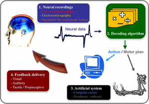
Figure 1 – Your brain in action:
the different components of a BMI include the recording system, the decoding algorithm, device to be controlled, and the feedback delivered to the user (modified from Heliot and Carmena, 2010).

As for getting information about basic details, here’s some of what I unearthed. The parent organization, ‘Frontiers in’ is based in Switzerland and describes itself this way on its About page,

Frontiers is a community-oriented open-access academic publisher and research network.

Our grand vision is to build an Open Science platform that empowers researchers in their daily work and where everybody has equal opportunity to seek, share and generate knowledge.

Frontiers is at the forefront of building the ultimate Open Science platform. We are driving innovations and new technologies around peer-review, article and author impact metrics, social networking for researchers, and a whole ecosystem of open science tools. We are the first – and only – platform that combines open-access publishing with research networking, with the goal to increase the reach of publications and ultimately the impact of articles and their authors.

Frontiers was launched as a grassroots initiative in 2007 by scientists from the Swiss Federal Institute of Technology in Lausanne, Switzerland, out of the collective desire to improve the publishing options and provide better tools and services to researchers in the Internet age. Since then, Frontiers has become the fastest-growing open-access scholarly publisher, with a rapidly growing number of community-driven journals, more than 25,000 of high-impact researchers across a wide range of academic fields serving on the editorial boards and more than 4 million monthly page views.

As of a Feb. 27, 2013 news release, Frontiers has partnered with the Nature Publishing Group (NPG), Note: Links have been removed,

Emerging publisher Frontiers is joining Nature Publishing Group (NPG) in a strategic alliance to advance the global open science movement.

NPG, publisher of Nature, today announces a majority investment in the Swiss-based open access (OA) publisher Frontiers.

NPG and Frontiers will work together to empower researchers to change the way science is communicated, through open access publication and open science tools. Frontiers, led by CEO and neuroscientist Kamila Markram, will continue to operate with its own platform, brands, and policies.

Founded by scientists from École Polytechnique Fédérale de Lausanne (EPFL) in 2007, Frontiers is one of the fastest growing open access publishers, more than doubling articles published year on year. Frontiers now has a portfolio of open access journals in 14 fields of science and medicine, and published over 5,000 OA articles in 2012.

Working with NPG, the journal series “Frontiers in” will significantly expand in 2013-2014. Currently, sixty-three journals published by NPG offer open access options or are open access and NPG published over 2000 open access articles in 2012. Bilateral links between nature.com and frontiersin.org will ensure that open access papers are visible on both sites.

Frontiers and NPG will also be working together on innovations in open science tools, networking, and publication processes.

Frontiers is based at EPFL in Switzerland, and works out of Innovation Square, a technology park supporting science start-ups, and hosting R&D divisions of large companies such as Logitech & Nestlé.

As for this new venture, Frontiers for Young Minds, this appears to have been launched on Nov. 11, 2013. At least, that’s what I understand from this notice on Frontier’s Facebook page (Note: Links have been removed,

Frontiers
November 11 [2013?]
Great news for kids, parents, teachers and neuroscientists! We have just launched the first Frontiers for Young Minds!

Frontiers in #Neuroscience for Young Minds is an #openaccess scientific journal that involves young people in the review of articles.

This has the double benefit of bringing kids into the world of science and offering scientists a platform for reaching out to the broadest of all audiences.

Frontiers for Young Minds is science edited for kids, by kids. Learn more and spread the word! http://bit.ly/1dijipy #sfn13

I am glad to see this effort and I wish all the parties involved the best of luck.

Europe’s flagshop projects (Graphene and Human Brain) take sail

Nine months after (my Jan. 28, 2013 posting) announcing that  the Future and Emerging Technologies 1B Euro research prizes were being awarded to the Graphene flagship and the Human Brain Project flagship, the two endeavours have been officially launched,  According to an Oct. 7, 2013 news item on Nanowerk,, the Graphene Flagship project is being launched at Chalmers University in Sweden (Note: Links have been removed),,

After years of preparations it is time for Europe to launch The Graphene Flagship – a 10-year, 1,000 million euro research and innovation initiative on graphene and related layered materials.


Now, on October 10-11, graphene researchers from all over Europe – from 74 research partners in 17 countries – will gather in Gothenburg, Sweden, for kick-off. Their mission is to take the supermaterial graphene and related ultra-thin layered materials from academic laboratories to society, revolutionize multiple industries and create economic growth and new jobs in Europe.

Here’s more about the Graphene Flagship kickoff event being held on Oct. 10, 2013 (i couldn’t find anything about events on Oct. 11, 2013)  at Sweden’s Chalmers University. The Human Brain Project kickoff event has started (Oct. 6 – 13, 2013) in EPFL (École Polytechnque Fédérale de Lausanne) campus in Switzerland. You can find more  links and more information about both projects on the FET (Future and Emerging Technologies) Flagship Initiatives webpage on the CORDIS website.

So, why do gold nanoparticles facilitate cell penetration without damage to cell walls?

Researchers at the Massachusetts Institute of Technology (MIT) and l’École Polytechnique Fédérale de Lausanne (EPFL) in Switerzerland have found an answer to the question about why gold nanoparticles facilitate cell penetration without damage to the cell walls. Apparently it has nothing to with the gold; it’s all in the coating according to an Aug. 23, 2013 MIT news release by David L. Chandler (also on EurekAlert),

Cells are very good at protecting their precious contents — and as a result, it’s very difficult to penetrate their membrane walls to deliver drugs, nutrients or biosensors without damaging or destroying the cell. One effective way of doing so, discovered in 2008, is to use nanoparticles of pure gold, coated with a thin layer of a special polymer. But nobody knew exactly why this combination worked so well, or how it made it through the cell wall.

Now, researchers at MIT and the Ecole Polytechnique de Lausanne in Switzerland have figured out how the process works, and the limits on the sizes of particles that can be used. …

Until now, says Van Lehn, the paper’s lead author [Reid Van Lehn], “the mechanism was unknown. … In this work, we wanted to simplify the process and understand the forces” that allow gold nanoparticles to penetrate cell walls without permanently damaging the membranes or rupturing the cells. The researchers did so through a combination of lab experiments and computer simulations.

The news release goes on to provide details about the research,

The team demonstrated that the crucial first step in the process is for coated gold nanoparticles to fuse with the lipids — a category of natural fats, waxes and vitamins — that form the cell wall. The scientists also demonstrated an upper limit on the size of such particles that can penetrate the cell wall — a limit that depends on the composition of the particle’s coating. [emphases mine]

The coating applied to the gold particles consists of a mix of hydrophobic and hydrophilic components that form a monolayer — a layer just one molecule thick — on the particle’s surface. Any of several different compounds can be used, the researchers explain. [emphases mine]

“Cells tend to engulf things on the surface,” says Alexander-Katz, an associate professor of materials science and engineering at MIT, but it’s “very unusual” for materials to cross that membrane into the cell’s interior without causing major damage. Irvine and Stellacci demonstrated in 2008 that monolayer-coated gold nanoparticles could do so; they have since been working to better understand why and how that works.

Since the nanoparticles themselves are completely coated, the fact that they are made of gold doesn’t have any direct effect, except that gold nanoparticles are an easily prepared model system, the researchers say. However, there is some evidence that the gold particles have therapeutic properties, which could be a side benefit.

Gold particles are also very good at capturing X-rays — so if they could be made to penetrate cancer cells, and were then heated by a beam of X-rays, they could destroy those cells from within. “So the fact that it’s gold may be useful,” says Irvine, a professor of materials science and engineering and biological engineering and member of the Koch Institute for Integrative Cancer Research.

Significantly, the mechanism that allows the nanoparticles to pass through the membrane seems also to seal the opening as soon as the particle has passed. “They would go through without allowing even small molecules to leak through behind them,” Van Lehn says.

Irvine says that his lab is also interested in harnessing this cell-penetrating mechanism as a way of delivering drugs to the cell’s interior, by binding them to the surface coating material. One important step in making that a useful process, he says, is finding ways to allow the nanoparticle coatings to be selective about what types of cells they attach to. “If it’s all cells, that’s not very useful,” he says, but if the coatings can be targeted to a particular cell type that is the target of a drug, that could be a significant benefit.

Another potential application of this work could be in attaching or inserting biosensing molecules on or into certain cells, Van Lehn says. In this way, scientists could detect or monitor specific biochemical markers, such as proteins that indicate the onset or decline of a disease or a metabolic process.

The research paper can be found here,

Effect of Particle Diameter and Surface Composition on the Spontaneous Fusion of Monolayer-Protected Gold Nanoparticles with Lipid Bilayers by Reid C. Van Lehn, Prabhani U. Atukorale, Randy P. Carney, Yu-Sang Yang, Francesco Stellacci, Darrell J. Irvine, and Alfredo Alexander-Katz. Nano Lett., Article ASAP DOI: 10.1021/nl401365n Publication Date (Web): August 5, 2013
Copyright © 2013 American Chemical Society

It is behind a paywall.

There once was a champion … it was nano-rust

Swiss and Israeli scientists have discovered water and nano iron oxide (rust) can be used to produce solar hydrogen cheaply. From the July 7, 2013 news release on EurekAlert,

In the quest for the production of renewable and clean energy, photoelectrochemical cells (PECs) constitute a sort of a Holy Grail. PECs are devices able of splitting water molecules into hydrogen and oxygen in a single operation, thanks to solar radiation. “As a matter of fact, we’ve already discovered this precious chalice, says Michael Grätzel, Director of the Laboratory of Photonics and Interfaces (LPI) at EPFL [Ecole Polytechnique Fédérale de Lausanne] and inventor of dye-sensitized photoelectrochemical cells. Today we have just reached an important milestone on the path that will lead us forward to profitable industrial applications.”

This week, Nature Materials is indeed publishing a groundbreaking article on the subject. EPFL researchers, working with Avner Rotschild from Technion (Israel), have managed to accurately characterize the iron oxide nanostructures to be used in order to produce hydrogen at the lowest possible cost. “The whole point of our approach is to use an exceptionally abundant, stable and cheap material: rust,” adds Scott C. Warren, first author of the article.

The EFFL July 9, 2013 news release by Emmanuel Barraud about this research provides more details,

At the end of last year, Kevin Sivula, one of the collaborators at the LPI laboratory, presented a prototype electrode based on the same principle. Its efficiency was such that gas bubbles emerged as soon as it was under a light stimulus. Without a doubt, the potential of such cheap electrodes was demonstrated, even if there was still room for improvement.

By using transmission electron microscopy (TEM) techniques, researchers were able to precisely characterize the movement of the electrons through the cauliflower-looking nanostructures forming the iron oxide particles, laid on electrodes during the manufacturing process. “These measures have helped us understand the reason why we get performance differences depending on the electrodes manufacturing process”, says Grätzel.

By comparing several electrodes, whose manufacturing method is now mastered, scientists were able to identify the “champion” structure. A 10×10 cm prototype has been produced and its effectiveness is in line with expectations. The next step will be the development of the industrial process to large-scale manufacturing. A European funding and the Swiss federal government could provide support for this last part.

Evidently, the long-term goal is to produce hydrogen – the fuel of the future – in an environmentally friendly and especially competitive way. For Michael Grätzel, “current methods, in which a conventional photovoltaic cell is coupled to an electrolyzer for producing hydrogen, cost 15 € per kilo at their cheapest. We’re aiming at a € 5 charge per kilo”.

Here’s a link to and a citation for the published research paper,

Identifying champion nanostructures for solar water-splitting by Scott C. Warren, Kislon Voïtchovsky, Hen Dotan, Celine M. Leroy, Maurin Cornuz, Francesco Stellacci, Cécile Hébert, Avner Rothschild & Michael Grätzel. Nature Materials (2013) doi:10.1038/nmat3684
Published online 07 July 2013

This paper is behind a paywall.