Tag Archives: nanocrystals

Organic-inorganic nanohybrids: organic ligands attached to colloidal inorganic nanocrystals

A May 24, 2023 news item on phys.org introduces organic-inorganic nanohybrids for optoelectronic devices,

When designing optoelectronic devices, such as solar cells, photocatalysts, and photodetectors, scientists usually prioritize materials that are stable and possess tunable properties. This allows them precise control over optical characteristics of the materials and ensures retention of their properties over time, despite varying environmental conditions.

Organic-inorganic nanohybrids, which are made up of organic ligands attached to the surface of colloidal inorganic nanocrystals via coordinate bonds, are promising in this regard. They are known to exhibit enhanced stability owing to the formation of a protective layer by organic ligands around the reactive inorganic nanocrystal. However, the incorporation of organic ligands has been found to lower the conductivity and photon absorption efficiency of inorganic nanocrystals.

In a breakthrough study on ligand-nanocrystal interactions, researchers from Japan now demonstrate a quasi-reversible displacement of organic ligands on the surface of nanocrystals. Their findings, published in ACS Nano, provide a new perspective to the common belief that the organic ligands are anchored to the surface of the nanocrystals.

A May 22, 2023 Ritsumeikan University (Japan) press release (also on EurekAlert but published May 24, 2023), which originated the news item, provides more detail,

… The research team, led by Professor Yoichi Kobayashi from Ritsumeikan University, Japan, found that the coordination bond between perylene bisimide with a carboxyl group (PBI) and inorganic zinc sulfide (ZnS) nanocrystals can be reversibly displaced by exposing the material to visible light.

Shedding light on this novel behavior of organic-inorganic nanohybrids, Prof. Kobayashi says, “We explored the ligand properties of organic-inorganic nanohybrid systems by using perylene bisimide with a carboxyl group (PBI)-coordinated zinc sulfide (ZnS) NCs (PBI–ZnS) as a model system. Our findings provide the first example of photoinduced displacement of aromatic ligands with semiconductor nanocrystals.”

In their study, the researchers carried out both theoretical analysis and experimental investigations to understand the material’s unique photoinducible characteristics. They first conducted density functional theory calculations to study the structure and orbitals of PBI–ZnS ([PBI-Zn25S31]) in both its ground and first excited states. Next, they performed time-resolved impulsive stimulated Raman spectroscopy to excite the sample with an ultrafast laser. This helped them analyze the corresponding Raman spectrum that revealed the nature of the excited state of PBI–ZnS.

The experimental observations and calculations showed that, upon photoexcitation, an electron is excited from the PBI molecule, and the corresponding “hole”(the vacancy formed due to the absence of the electron) rapidly moves from the aromatic ligand (PBI) to ZnS. This results in a long-lived, negatively-charged PBI ion that is displaced from the surface of the ZnS nanocrystal. Over time, however, the displaced ligands recombine with the surface defects of the ZnS nanocrystal, leading to a quasi-reversible photoinduced displacement of coordinated PBI. Notably, the dynamic behavior of coordinated ligand molecules observed in this study is different from that observed for typical photoinduced charge transfer processes in which the hole typically remains on the donor molecule, enabling it to recombine with the electron quickly.

Explaining the significance of these findings, Prof. Kobayashi says, “The precise understanding of ligand-nanocrystal interaction is important not only for fundamental nanoscience but also for developing advanced photofunctional materials using nanomaterials. These include photocatalysts for the decomposition of persistent chemicals using visible light and photoconductive microcircuit patterning for wearable devices.”

Indeed, the results of this study present a promising avenue for enhancing the tunability and functionality of inorganic materials with aromatic molecules. This, in turn, could significantly impact the field of fundamental nanoscience and photochemistry in the times to come.

Here’s a link to and a citation for the paper,

Quasi-Reversible Photoinduced Displacement of Aromatic Ligands from Semiconductor Nanocrystals by Daisuke Yoshioka, Yusuke Yoneda, I-Ya Chang, Hikaru Kuramochi, Kim Hyeon-Deuk, and Yoichi Kobayashi. ACS Nano 2023, 17, 12, 11309–11317 DOI: https://doi.org/10.1021/acsnano.2c12578 Publication Date:May 9, 2023 Copyright © 2023 American Chemical Society

This paper is behind a paywall.
Shaving the ‘hairs’ off nanocrystals for more efficient electronics

A March 24, 2022 news item on phys.org announced research into nanoscale crystals and how they might be integrated into electronic devices, Note: A link has been removed,

You can carry an entire computer in your pocket today because the technological building blocks have been getting smaller and smaller since the 1950s. But in order to create future generations of electronics—such as more powerful phones, more efficient solar cells, or even quantum computers—scientists will need to come up with entirely new technology at the tiniest scales.

One area of interest is nanocrystals. These tiny crystals can assemble themselves into many configurations, but scientists have had trouble figuring out how to make them talk to each other.  

A new study introduces a breakthrough in making nanocrystals function together electronically. Published March 25 [2022] in Science, the research may open the doors to future devices with new abilities. 

A March 25, 2022 University of Chicago news release (also on EurekAlert but published on March 24, 2022), which originated the news item, expands on the possibilities the research makes possible, Note: Links have been removed,

“We call these super atomic building blocks, because they can grant new abilities—for example, letting cameras see in the infrared range,” said University of Chicago Prof. Dmitri Talapin, the corresponding author of the paper. “But until now, it has been very difficult to both assemble them into structures and have them talk to each other. Now for the first time, we don’t have to choose. This is a transformative improvement.”  

In their paper, the scientists lay out design rules which should allow for the creation of many different types of materials, said Josh Portner, a Ph.D. student in chemistry and one of the first authors of the study. 

A tiny problem

Scientists can grow nanocrystals out of many different materials: metals, semiconductors, and magnets will each yield different properties. But the trouble was that whenever they tried to assemble these nanocrystals together into arrays, the new supercrystals would grow with long “hairs” around them. 

These hairs made it difficult for electrons to jump from one nanocrystal to another. Electrons are the messengers of electronic communication; their ability to move easily along is a key part of any electronic device. 

The researchers needed a method to reduce the hairs around each nanocrystal, so they could pack them in more tightly and reduce the gaps in between. “When these gaps are smaller by just a factor of three, the probability for electrons to jump across is about a billion times higher,” said Talapin, the Ernest DeWitt Burton Distinguished Service Professor of Chemistry and Molecular Engineering at UChicago and a senior scientist at Argonne National Laboratory. “It changes very strongly with distance.”

To shave off the hairs, they sought to understand what was going on at the atomic level. For this, they needed the aid of powerful X-rays at the Center for Nanoscale Materials at Argonne and the Stanford Synchrotron Radiation Lightsource at SLAC National Accelerator Laboratory, as well as powerful simulations and models of the chemistry and physics at play. All these allowed them to understand what was happening at the surface—and find the key to harnessing their production.

Part of the process to grow supercrystals is done in solution—that is, in liquid. It turns out that as the crystals grow, they undergo an unusual transformation in which gas, liquid and solid phases all coexist. By precisely controlling the chemistry of that stage, they could create crystals with harder, slimmer exteriors which could be packed in together much more closely. “Understanding their phase behavior was a massive leap forward for us,” said Portner. 

The full range of applications remains unclear, but the scientists can think of multiple areas where the technique could lead. “For example, perhaps each crystal could be a qubit in a quantum computer; coupling qubits into arrays is one of the fundamental challenges of quantum technology right now,” said Talapin. 

Portner is also interested in exploring the unusual intermediate state of matter seen during supercrystal growth: “Triple phase coexistence like this is rare enough that it’s intriguing to think about how to take advantage of this chemistry and build new materials.”

The study included scientists with the University of Chicago, Technische Universität Dresden, Northwestern University, Arizona State University, SLAC, Lawrence Berkeley National Laboratory, and the University of California, Berkeley.

Here’s a link to and a citation for the paper,

Self-assembly of nanocrystals into strongly electronically coupled all-inorganic supercrystals by Igor Coropceanu, Eric M. Janke, Joshua Portner, Danny Haubold, Trung Dac Nguyen, Avishek Das, Christian P. N. Tanner, James K. Utterback, Samuel W. Teitelbaum¸ Margaret H. Hudson, Nivedina A. Sarma, Alex M. Hinkle, Christopher J. Tassone, Alexander Eychmüller, David T. Limmer, Monica Olvera de la Cruz, Naomi S. Ginsberg and Dmitri V. Talapin. Science • 24 Mar 2022 • Vol 375, Issue 6587 • pp. 1422-1426 • DOI: 10.1126/science.abm6753

This paper is behind a paywall.

Eradicating bacteria biofilm with nanocrystals

A January 8, 2021 news item on ScienceDaily announces new work from South Korea’s Pohang University of Science & Technology (POSTECH),

The COVID-19 pandemic is raising fears of new pathogens such as new viruses or drug-resistant bacteria. To this, a Korean research team has recently drawn attention for developing the technology for removing antibiotic-resistant bacteria by controlling the surface texture of nanomaterials.

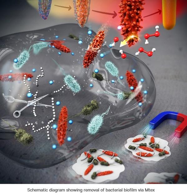
A joint research team from POSTECH and UNIST [Ulsan National Institute of Science and Technology] has introduced mixed-FeCo-oxide-based surface-textured nanostructures (MTex) as highly efficient magneto-catalytic platform in the international journal Nano Letters. The team consisted of professors In Su Lee and Amit Kumar with Dr. Nitee Kumari of POSTECH’s Department of Chemistry and Professor Yoon-Kyung Cho and Dr. Sumit Kumar of UNIST’s Department of Biomedical Engineering.

Caption: Schematic diagram showing removal of bacterial biofilm via Mtex Credit: POSTECH

A January 8, 2021 POSTECH press release (also on EurkeAlert), which originated the news item, delves further,

First, the researchers synthesized smooth surface nanocrystals in which various metal ions were wrapped in an organic polymer shell and heated them at a very high temperature. While annealing the polymer shell, a high-temperature solid-state chemical reaction induced mixing of other metal ions on the nanocrystal surface, creating a number of few-nm-sized branches and holes on it. This unique surface texture was found to catalyze a chemical reaction that produced reactive oxygen species (ROS) that kills the bacteria. It was also confirmed to be highly magnetic and easily attracted toward the external magnetic field. The team had discovered a synthetic strategy for converting normal nanocrystals without surface features into highly functional mixed-metal-oxide nanocrystals.

The research team named this surface topography – with branches and holes that resembles that of a ploughed field – “MTex.” This unique surface texture has been verified to increase the mobility of nanoparticles to allow efficient penetration into biofilm matrix while showing high activity in generating reactive oxygen species (ROS) that are lethal to bacteria.

This system produces ROS over a broad pH range and can effectively diffuse into the biofilm and kill the embedded bacteria resistant to antibiotics. And since the nanostructures are magnetic, biofilm debris can be scraped out even from the hard-to-reach microchannels.

“This newly developed MTex shows high catalytic activity, distinct from the stable smooth-surface of the conventional spinel forms,” explained Dr. Amit Kumar, one of the corresponding authors of the paper. “This characteristic is very useful in infiltrating biofilms even in small spaces and is effective in killing the bacteria and removing biofilms.”

“This research allows to regulate the surface nanotexturization, which opens up possibilities to augment and control the exposure of active sites,” remarked Professor In Su Lee who led the research. “We anticipate the nanoscale-textured surfaces to contribute significantly in developing a broad array of new enzyme-like properties at the nano-bio interface.”

Here’s a link to and a citation for the paper,

Surface-Textured Mixed-Metal-Oxide Nanocrystals as Efficient Catalysts for ROS Production and Biofilm Eradication by Nitee Kumari, Sumit Kumar, Mamata Karmacharya, Sateesh Dubbu, Taewan Kwon, Varsha Singh, Keun Hwa Chae, Amit Kumar, Yoon-Kyoung Cho, and In Su Lee. Nano Lett. 2021, 21, 1, 279–287 DOI: https://doi.org/10.1021/acs.nanolett.0c03639 Publication Date: December 11, 2020 Copyright © 2020 American Chemical Society

This paper is behind a paywall.

Blue quantum dots and your television screen

Scientists used equipment at the Canadian Light Source (CLS; synchrotron in Saskatoon, Saskatchewan, Canada) in the quest for better glowing dots on your television (maybe computers and telephones, too?) screen. From an August 20, 2020 news item on Nanowerk,

There are many things quantum dots could do, but the most obvious place they could change our lives is to make the colours on our TVs and screens more pristine. Research using the Canadian Light Source (CLS) at the University of Saskatchewan is helping to bring this technology closer to our living rooms.

An August 19, 2020 CLS news release (also received via email) by Victoria Martinez, which originated the news item, explains what quantum dots are and fills in with technical details about this research,

Quantum dots are nanocrystals that glow, a property that scientists have been working with to develop next-generation LEDs. When a quantum dot glows, it creates very pure light in a precise wavelength of red, blue or green. Conventional LEDs, found in our TV screens today, produce white light that is filtered to achieve desired colours, a process that leads to less bright and muddier colours.

Until now, blue-glowing quantum dots, which are crucial for creating a full range of colour, have proved particularly challenging for researchers to develop. However, University of Toronto (U of T) researcher Dr. Yitong Dong and collaborators have made a huge leap in blue quantum dot fluorescence, results they recently published in Nature Nanotechnology.

“The idea is that if you have a blue LED, you have everything. We can always down convert the light from blue to green and red,” says Dong. “Let’s say you have green, then you cannot use this lower-energy light to make blue.”

The team’s breakthrough has led to quantum dots that produce green light at an external quantum efficiency (EQE) of 22% and blue at 12.3%. The theoretical maximum efficiency is not far off at 25%, and this is the first blue perovskite LED reported as achieving an EQE higher than 10%.

The Science

Dong has been working in the field of quantum dots for two years in Dr. Edward Sargent’s research group at the U of T. This astonishing increase in efficiency took time, an unusual production approach, and overcoming several scientific hurdles to achieve.

CLS techniques, particularly GIWAXS [grazing incidence wide-angle X-ray scattering] on the HXMA beamline [hard X-ray micro-analysis (HXMA)], allowed the researchers to verify the structures achieved in their quantum dot films. This validated their results and helped clarify what the structural changes achieve in terms of LED performance.

“The CLS was very helpful. GIWAXS is a fascinating technique,” says Dong.

The first challenge was uniformity, important to ensuring a clear blue colour and to prevent the LED from moving towards producing green light.

“We used a special synthetic approach to achieve a very uniform assembly, so every single particle has the same size and shape. The overall film is nearly perfect and maintains the blue emission conditions all the way through,” says Dong.

Next, the team needed to tackle the charge injection needed to excite the dots into luminescence. Since the crystals are not very stable, they need stabilizing molecules to act as scaffolding and support them. These are typically long molecule chains, with up to 18 carbon-non-conductive molecules at the surface, making it hard to get the energy to produce light.

“We used a special surface structure to stabilize the quantum dot. Compared to the films made with long chain molecules capped quantum dots, our film has 100 times higher conductivity, sometimes even 1000 times higher.”

This remarkable performance is a key benchmark in bringing these nanocrystal LEDs to market. However, stability remains an issue and quantum dot LEDs suffer from short lifetimes. Dong is excited about the potential for the field and adds, “I like photons, these are interesting materials, and, well, these glowing crystals are just beautiful.”

Here’s a link to and a citation for the paper,

Bipolar-shell resurfacing for blue LEDs based on strongly confined perovskite quantum dots by Yitong Dong, Ya-Kun Wang, Fanglong Yuan, Andrew Johnston, Yuan Liu, Dongxin Ma, Min-Jae Choi, Bin Chen, Mahshid Chekini, Se-Woong Baek, Laxmi Kishore Sagar, James Fan, Yi Hou, Mingjian Wu, Seungjin Lee, Bin Sun, Sjoerd Hoogland, Rafael Quintero-Bermudez, Hinako Ebe, Petar Todorovic, Filip Dinic, Peicheng Li, Hao Ting Kung, Makhsud I. Saidaminov, Eugenia Kumacheva, Erdmann Spiecker, Liang-Sheng Liao, Oleksandr Voznyy, Zheng-Hong Lu, Edward H. Sargent. Nature Nanotechnology volume 15, pages668–674(2020) DOI: https://doi.org/10.1038/s41565-020-0714-5 Published: 06 July 2020 Issue Date: August 2020

This paper is behind a paywall.

If you search “Edward Sargent,” he’s the last author listed in the citation, here on this blog, you will find a number of postings that feature work from his laboratory at the University of Toronto.

Colo(u)r-changing nanolaser inspired by chameleons

Caption: Novel nanolaser leverages the same color-changing mechanism that a chameleon uses to camouflage its skin. Credit: Egor Kamelev Courtesy: Northwestern University

I wish there was some detail included about how those colo(u)rs were achieved in that photograph. Strangely, Northwestern University (Chicago, Illinois, US) is more interested in describing the technology that chameleons have inspired. A June 20, 2018 news item on ScienceDaily announces the research,

As a chameleon shifts its color from turquoise to pink to orange to green, nature’s design principles are at play. Complex nano-mechanics are quietly and effortlessly working to camouflage the lizard’s skin to match its environment.

Inspired by nature, a Northwestern University team has developed a novel nanolaser that changes colors using the same mechanism as chameleons. The work could open the door for advances in flexible optical displays in smartphones and televisions, wearable photonic devices and ultra-sensitive sensors that measure strain.

A June 20, 2018 Northwestern University news release (also on EurekAlert) by Amanda Morris, which originated the news item, expands on the theme,

“Chameleons can easily change their colors by controlling the spacing among the nanocrystals on their skin, which determines the color we observe,” said Teri W. Odom, Charles E. and Emma H. Morrison Professor of Chemistry in Northwestern’s Weinberg College of Arts and Sciences. “This coloring based on surface structure is chemically stable and robust.”

The research was published online yesterday [June 19, 2018] in the journal Nano Letters. Odom, who is the associate director of Northwestern’s International Institute of Nanotechnology, and George C. Schatz, Charles E. and Emma H. Morrison Professor of Chemistry in Weinberg, served as the paper’s co-corresponding authors.

The same way a chameleon controls the spacing of nanocrystals on its skin, the Northwestern team’s laser exploits periodic arrays of metal nanoparticles on a stretchable, polymer matrix. As the matrix either stretches to pull the nanoparticles farther apart or contracts to push them closer together, the wavelength emitted from the laser changes wavelength, which also changes its color.

“Hence, by stretching and releasing the elastomer substrate, we could select the emission color at will,” Odom said.

The resulting laser is robust, tunable, reversible and has a high sensitivity to strain. These properties are critical for applications in responsive optical displays, on-chip photonic circuits and multiplexed optical communication.

Here’s a link to and a citation for the paper,

Stretchable Nanolasing from Hybrid Quadrupole Plasmons by Danqing Wang, Marc R. Bourgeois, Won-Kyu Lee, Ran Li, Dhara Trivedi, Michael P. Knudson, Weijia Wang, George C. Schatz, and Teri W. Odom. Nano Lett., Article ASAP DOI: 10.1021/acs.nanolett.8b01774 Publication Date (Web): June 18, 2018

Copyright © 2018 American Chemical Society

This paper is behind a paywall.
What do nanocrystals have in common with the earth’s crust?

The deformation properties of nanocrystals resemble those in the earth’s crust according to a Nov. 17, 2015 news item on Nanowerk,

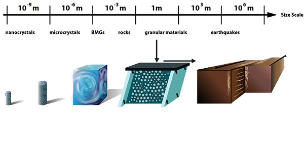
Apparently, size doesn’t always matter. An extensive study by an interdisciplinary research group suggests that the deformation properties of nanocrystals are not much different from those of the Earth’s crust.

“When solid materials such as nanocrystals, bulk metallic glasses, rocks, or granular materials are slowly deformed by compression or shear, they slip intermittently with slip-avalanches similar to earthquakes,” explained Karin Dahmen, a professor of physics at the University of Illinois at Urbana-Champaign. “Typically these systems are studied separately. But we found that the scaling behavior of their slip statistics agree across a surprisingly wide range of different length scales and material structures.”

There’s an illustration accompanying the research,

Courtesy of the University of Illinois

Caption: When solid materials such as nanocrystals, bulk metallic glasses, rocks, or granular materials are slowly deformed by compression or shear, they slip intermittently with slip-avalanches similar to earthquakes. Credit: University of Illinois

A Nov. 17, 2015 University of Illinois news release (also on EurekAlert) by Rick Kubetz, which originated the news item, provides more detail,

“Identifying agreement in aspects of the slip statistics is important, because it enables us to transfer results from one scale to another, from one material to another, from one stress to another, or from one strain rate to another,” stated Shivesh Pathak, a physics undergraduate at Illinois, and a co-author of the paper, “Universal Quake Statistics: From Compressed Nanocrystals to Earthquakes,” appearing in Scientific Reports. “The study shows how to identify and explain commonalities in the deformation mechanisms of different materials on different scales.

“The results provide new tools and methods to use the slip statistics to predict future materials deformation,” added Michael LeBlanc, a physics graduate student and co-author of the paper. “They also clarify which system parameters significantly affect the deformation behavior on long length scales. We expect the results to be useful for applications in materials testing, failure prediction, and hazard prevention.”

Researchers representing a broad a range of disciplines–including physics, geosciences, mechanical engineering, chemical engineering, and materials science–from the United States, Germany, and the Netherlands contributed to the study, comparing five different experimental systems, on several different scales, with model predictions.

As a solid is sheared, each weak spot is stuck until the local shear stress exceeds a random failure threshold. It then slips by a random amount until it re-sticks. The released stress is redistributed to all other weak spots. Thus, a slipping weak spot can trigger other spots to fail in a slip avalanche.

Using tools from the theory of phase transitions, such as the renormalization group, one can show that the slip statistics of the model do not depend on the details of the system.

“Although these systems span 13 decades in length scale, they all show the same scaling behavior for their slip size distributions and other statistical properties,” stated Pathak. “Their size distributions follow the same simple (power law) function, multiplied with the same exponential cutoff.”

The cutoff, which is the largest slip or earthquake size, grows with applied force for materials spanning length scales from nanometers to kilometers. The dependence of the size of the largest slip or quake on stress reflects “tuned critical” behavior, rather than so-called self-organized criticality, which would imply stress-independence.

“The agreement of the scaling properties of the slip statistics across scales does not imply the predictability of individual slips or earthquakes,” LeBlanc said. “Rather, it implies that we can predict the scaling behavior of average properties of the slip statistics and the probability of slips of a certain size, including their dependence on stress and strain-rate.”

Here’s a link to and a citation for the paper,

Universal Quake Statistics: From Compressed Nanocrystals to Earthquakes by Jonathan T. Uhl, Shivesh Pathak, Danijel Schorlemmer, Xin Liu, Ryan Swindeman, Braden A. W. Brinkman, Michael LeBlanc, Georgios Tsekenis, Nir Friedman, Robert Behringer, Dmitry Denisov, Peter Schall, Xiaojun Gu, Wendelin J. Wright, Todd Hufnagel, Andrew Jennings, Julia R. Greer, P. K. Liaw, Thorsten Becker, Georg Dresen, & Karin A. Dahmen.  Scientific Reports 5, Article number: 16493 (2015)  doi:10.1038/srep16493 Published online: 17 November 2015

This is an open access paper.

One final comment, this story reminds me of a few other pieces of research featured here, which focus on repeating patterns in nature. The research was mentioned in an Aug. 27, 2015 posting about white dwarf stars and heartbeats and in an April 14, 2015 posting about gold nanoparticles and their resemblance to the Milky Way. You can also find more in the Wikipedia entry titled ‘Patterns in nature‘.

Chameleons (male panther chameleons in particular)—colour revelation

Caption: These are male panther chameleons (Furcifer pardalis) photographed in Madagascar. Credit: © Michel Milinkovitch

Caption: These are male panther chameleons (Furcifer pardalis) photographed in Madagascar.
Credit: © Michel Milinkovitch

Researchers at Switzerland’s University of Geneva/Université de Genève (UNIGE) have revealed the mechanisms (note the plural) by which chameleons change their colour. From a March 10, 2015 news item on phys.org,

Many chameleons have the remarkable ability to exhibit complex and rapid color changes during social interactions. A collaboration of scientists within the Sections of Biology and Physics of the Faculty of Science from the University of Geneva (UNIGE), Switzerland, unveils the mechanisms that regulate this phenomenon.

In a study published in Nature Communications [March 10, 2015], the team led by professors Michel Milinkovitch and Dirk van der Marel demonstrates that the changes take place via the active tuning of a lattice of nanocrystals present in a superficial layer of dermal cells called iridophores. The researchers also reveal the existence of a deeper population of iridophores with larger and less ordered crystals that reflect the infrared light. The organisation of iridophores into two superimposed layers constitutes an evolutionary novelty and it allows the chameleons to rapidly shift between efficient camouflage and spectacular display, while providing passive thermal protection.

Male chameleons are popular for their ability to change colorful adornments depending on their behaviour. If the mechanisms responsible for a transformation towards a darker skin are known, those that regulate the transition from a lively color to another vivid hue remained mysterious. Some species, such as the panther chameleon, are able to carry out such a change within one or two minutes to court a female or face a competing male.

A March 10, 2015 University of Geneva press release on EurekAlert (French language version is here on the university website), which originated the news item, explains the chameleon’s ability as being due to its ability to display structural colour,

Besides brown, red and yellow pigments, chameleons and other reptiles display so-called structural colors. «These colors are generated without pigments, via a physical phenomenon of optical interference. They result from interactions between certain wavelengths and nanoscopic structures, such as tiny crystals present in the skin of the reptiles», says Michel Milinkovitch, professor at the Department of Genetics and Evolution at UNIGE. These nanocrystals are arranged in layers that alternate with cytoplasm, within cells called iridophores. The structure thus formed allows a selective reflection of certain wavelengths, which contributes to the vivid colors of numerous reptiles.

To determine how the transition from one flashy color to another one is carried out in the panther chameleon, the researchers of two laboratories at UNIGE worked hand in hand, combining their expertise in both quantum physics and in evolutionary biology. «We discovered that the animal changes its colors via the active tuning of a lattice of nanocrystals. When the chameleon is calm, the latter are organised into a dense network and reflect the blue wavelengths. In contrast, when excited, it loosens its lattice of nanocrystals, which allows the reflection of other colors, such as yellows or reds», explain the physicist Jérémie Teyssier and the biologist Suzanne Saenko, co-first authors of the article. This constitutes a unique example of an auto-organised intracellular optical system controlled by the chameleon.

The press release goes on to note that the iridophores have another function,

The scientists also demonstrated the existence of a second deeper layer of iridophores. «These cells, which contain larger and less ordered crystals, reflect a substantial proportion of the infrared wavelengths», states Michel Milinkovitch. This forms an excellent protection against the thermal effects of high exposure to sun radiations in low-latitude regions.

The organisation of iridophores in two superimposed layers constitutes an evolutionary novelty: it allows the chameleons to rapidly shift between efficient camouflage and spectacular display, while providing passive thermal protection.

In their future research, the scientists will explore the mechanisms that explain the development of an ordered nanocrystals lattice within iridophores, as well as the molecular and cellular mechanisms that allow chameleons to control the geometry of this lattice.

Here’s a link to and a citation for the paper,

Photonic crystals cause active colour change in chameleons by Jérémie Teyssier, Suzanne V. Saenko, Dirk van der Marel, & Michel C. Milinkovitch. Nature Communications 6, Article number: 6368 doi:10.1038/ncomms7368 Published 10 March 2015

This article is open access.

Purple promises and bioimaging from Singapore’s A*STAR

A May 7, 2014 news item on Nanowerk describes a promising new approach to bioimaging,

Labeling biomolecules with light-emitting nanoparticles is a powerful technique for observing cell movement and signaling under realistic, in vivo conditions. The small size of these probes, however, often limits their optical capabilities. In particular, many nanoparticles have trouble producing high-energy light with wavelengths in the violet to ultraviolet range, which can trigger critical biological reactions.

Now, an international team led by Xiaogang Liu from the A*STAR Institute of Materials Research and Engineering and the National University of Singapore has discovered a novel class of rare-earth nanocrystals that preserve excited energy inside their atomic framework, resulting in unusually intense violet emissions …

A May 7, 2014 A*STAR (Agency for Science, Technology and Research) news release (h/t Imagist), which originated the news item, describes the problems with current bioimaging techniques and the new approach in more detail (Note: Links have been removed)

Nanocrystals selectively infused, or ‘doped’, with rare-earth ions have attracted the attention of researchers, because of their low toxicity and ability to convert low-energy laser light into violet-colored luminescence emissions — a process known as photon upconversion. Efforts to improve the intensity of these emissions have focused on ytterbium (Yb) rare-earth dopants, as they are easily excitable with standard lasers. Unfortunately, elevated amounts of Yb dopants can rapidly diminish, or ‘quench’, the generated light.

This quenching probably arises from the long-range migration of laser-excited energy states from Yb and toward defects in the nanocrystal. Most rare-earth nanocrystals have relatively uniform dopant distributions, but Liu and co-workers considered that a different crystal arrangement — clustering dopants into multi-atom arrays separated by large distances — could produce localized excited states that do not undergo migratory quenching.

The team screened numerous nanocrystals with different symmetries before discovering a material that met their criteria: a potassium fluoride crystal doped with Yb and europium rare earths (KYb2F7:Eu). Experiments revealed that the isolated Yb ‘energy clusters’ inside this pill-shaped nanocrystal (see image) enabled substantially higher dopant concentrations than usual — Yb accounted for up to 98 per cent of the crystal’s mass — and helped initiate multiphoton upconversion that yielded violet light with an intensity eight times higher than previously seen.

The researchers then explored the biological applications of their nanocrystals by using them to detect alkaline phosphatases, enzymes that frequently indicate bone and liver diseases. When the team brought the nanocrystals close to an alkaline phosphate-catalyzed reaction, they saw the violet emissions diminish in direct proportion to a chemical indicator produced by the enzyme. This approach enables swift and sensitive detection of this critical biomolecule at microscale concentration levels.

“We believe that the fundamental aspects of these findings — that crystal structures can greatly influence luminescence properties — could allow upconversion nanocrystals to eventually outperform conventional fluorescent biomarkers,” says Liu.

Here’s a link to and a citation for the paper,

Enhancing multiphoton upconversion through energy clustering at sublattice level by Juan Wang, Renren Deng, Mark A. MacDonald, Bolei Chen, Jikang Yuan, Feng Wang, Dongzhi Chi, Tzi Sum Andy Hor, Peng Zhang, Guokui Liu, Yu Han, & Xiaogang Liu. Nature Materials 13, 157–162 (2014) doi:10.1038/nmat3804 Published online 24 November 2013

This paper is behind a paywall but there is a free preview via ReadCube Access.

Similarities between biological molecules and synthetic nanocrystals extend beyond size

Researchers at the University of Illinois at Urbana-Champaign have determined that there are more similarities between biological molecules and synthetic nanocrystals than formerly believed, according to a Dec. 17, 2013 news item on Nanowerk (Note: A link has been removed),

Researchers have long thought that biological molecules and synthetic nanocrystals were similar only in size. Now, University of Illinois at Urbana-Champaign chemists have found that they can add reactivity to the list of shared traits. Atoms in a nanocrystal can cooperate with each other to facilitate binding or switching, a phenomenon widely found in biological molecules.

The finding could catalyze manufacturing of nanocrystals for smart sensors, solar cells, tiny transistors for optical computers, and medical imaging. Led by chemistry professor Prashant Jain, the team published its findings in the journal Nature Communications (“Co-operativity in a nanocrystalline solid-state transition”).

A Dec. 16, 2013 University of Illinois at Urbana-Champaign news release, which originated the news item, explains why the scientists are so interested and how they went about their investigation,

“In geological, industrial and domestic environments, the nanoscale grains of any material undergo chemical transitions when they are put under reactive conditions,” Jain said. “Iron rusting over time and diamond forming from carbon are examples of two commonly occurring transitions. Understanding how these transitions occur on the scale of the tiniest grains of the material is a major motivation of our work.”

Scientists can exploit such transitions to make nanocrystals that conform to a particular structure. They can make a nanocrystal of one material and transform it into another material, essentially using the original nanocrystal framework as a template for creating a nanocrystal of the new material with the same size and shape. This lets researchers create nanocrystals of new materials in shapes and structures they may not be able to otherwise.

In the new study, the researchers transformed tiny crystals of the material cadmium selenide to crystals of copper selenide. Copper selenide nanocrystals have a number of interesting properties that can be used for solar energy harvesting, optical computing and laser surgery. Transformation from cadmium selenide creates nanocrystals with a purity difficult to attain from other methods.

The researchers, including graduate student Sarah White, used advanced microscopy and spectroscopy techniques to determine the dynamics of the atoms within the crystals during the transformation and found that the transformation occurs not as a slow diffusion process, but as a rapid switching thanks to co-operativity.

The researchers saw that once the cadmium-selenide nanocrystal has taken up a few initial copper “seed” impurities, atoms in the rest of the lattice can cooperate to rapidly swap out the rest of the cadmium for copper. Jain compares the crystals to hemoglobin, the molecule in red blood cells that carries oxygen. Once one oxygen molecule has bound to hemoglobin, other binding sites within hemoglobin slightly change conformation to more easily pick up more oxygen. He posits that similarly, copper impurities might cause a structural change in the nanocrystal, making it easier for more copper ions to infiltrate the nanocrystal in a rapid cascade.

The researchers reproduced the experiment with silver, in addition to copper, and saw similar, though slightly less speedy, cooperative behavior.

Now, Jain’s team is using its advanced imaging to watch transitions happen in single nanocrystals, in real time.

“We have a sophisticated optical microscope in our lab, which has now allowed us to catch a single nanocrystal in the act of making a transition,” Jain said. “This is allowing us to learn hidden details about how the transition actually proceeds. We are also learning how one nanocrystal behaves differently from another.”

Next, the researchers plan to explore biomolecule-like cooperative phenomena in other solid-state materials and processes. For example, co-operativity in catalytic processes could have major implications for solar energy or manufacturing of expensive specialty chemicals.

“In the long term, we are interested in exploiting the co-operative behavior to design artificial smart materials that respond in a switch-like manner like hemoglobin in our body does,” Jain said.

Here’s an image of the various forms of cadmium selenide used in research,

Nanocrystals of cadmium selenide, known for their brilliant luminescence, display intriguing chemical behavior resulting from positive cooperation between atoms, a behavior akin to that found in biomolecules.  Photo courtesy Prashant Jain

Nanocrystals of cadmium selenide, known for their brilliant luminescence, display intriguing chemical behavior resulting from positive cooperation between atoms, a behavior akin to that found in biomolecules. Photo courtesy
Prashant Jain

For the curious, here’s a link to and a citation for the paper,

Co-operativity in a nanocrystalline solid-state transition by Sarah L. White, Jeremy G. Smith, Mayank Behl, & Prashant K. Jain Nature Communications 4, Article number: 2933 doi:10.1038/ncomms3933 Published 12 December 2013

This article is behind a paywall.

Sea sponges inspire body armour of the future

A Mar. 15, 2013 news item on ScienceDaily features research inspired by sea sponges,

Scientists at Johannes Gutenberg University Mainz (JGU) and the Max Planck Institute for Polymer Research (MPI-P) in Germany have created a new synthetic hybrid material with a mineral content of almost 90 percent, yet extremely flexible. They imitated the structural elements found in most sea sponges and recreated the sponge spicules using the natural mineral calcium carbonate and a protein of the sponge. Natural minerals are usually very hard and prickly, as fragile as porcelain.

Amazingly, the synthetic spicules are superior to their natural counterparts in terms of flexibility, exhibiting a rubber-like flexibility. The synthetic spicules can, for example, easily be U-shaped without breaking or showing any signs of fracture. …

Spicules are structural elements found in most sea sponges. They provide structural support and deter predators. They are very hard, prickly, and even quite difficult to cut with a knife. The spicules of sponges thus offer a perfect example of a lightweight, tough, and impenetrable defense system, which may inspire engineers to create body armors of the future.

I found an image of a sea sponge (this may not be exactly the same type of sponge that inspired the latest work but I think there are enough similarities to the description the researchers give to  include it here) and more information in a Nov. 13, 2008 post by Ed Grabianows on IO9.com,

Downloaded from: http://io9.com/5085064/giant-deep-sea-sponges-evolved-fiber-optic-exoskeletons

Downloaded from: http://io9.com/5085064/giant-deep-sea-sponges-evolved-fiber-optic-exoskeletons

This gigantic sea sponge has an exoskeleton made of glass rods, and each rod can grow up to a meter in length. In the deep sea, these massive sponges contain a menagerie of other tiny lifeforms, all dependent on their sea sponge hosts for something in short supply far under the water. They need light – and some sponges have a [sic] evolved a way to provide it using fiber optics.Sea sponges are among the most primitive animals on Earth. …

Here’s more about the research (from the ScienceDaily news item),

 The researchers led by Wolfgang Tremel, Professor at Johannes Gutenberg University Mainz, and Hans-Jürgen Butt, Director at the Max Planck Institute for Polymer Research in Mainz, used these natural sponge spicules as a model to cultivate them in the lab. The synthetic spicules were made from calcite (CaCO3) and silicatein-α. The latter is a protein from siliceous sponges that, in nature, catalyzes the formation of silica, which forms the natural silica spicules of sponges. Silicatein-α was used in the lab setting to control the self-organization of the calcite spicules. The synthetic material was self-assembled from an amorphous calcium carbonate intermediate and silicatein and subsequently aged to the final crystalline material. After six months, the synthetic spicules consisted of calcite nanocrystals aligned in a brick wall fashion with the protein embedded like cement in the boundaries between the calcite nanocrystals. The spicules were of 10 to 300 micrometers in length with a diameter of 5 to 10 micrometers.

… the synthetic spicules have yet another special characteristic, i.e., they are able to transmit light waves even when they are bent.

The researchers have created a video animation to illustrate their work,

For those who would like to find out more about the research, there’s a citation for and a link to the researchers’ paper here.