Tag Archives: UC Davis

The CRISPR yogurt story and a hornless cattle update

Clustered regularly interspaced short palindromic repeats (CRISPR) does not and never has made much sense to me. I understand each word individually it’s just that I’ve never thought they made much sense strung together that way. It’s taken years but I’ve finally found out what the words (when strung together that way) mean and the origins for the phrase. Hint: it’s all about the phages.

Apparently, it all started with yogurt as Cynthia Graber and Nicola Twilley of Gastropod discuss on their podcast, “4 CRISPR experts on how gene editing is changing the future of food.” During the course of the podcast they explain the ‘phraseology’ issue, mention hornless cattle (I have an update to the information in the podcast later in this posting), and so much more.

CRISPR started with yogurt

You’ll find the podcast (almost 50 minutes long) here on an Oct. 11, 2019 posting on the Genetic Literacy Project. If you need a little more encouragement, here’s how the podcast is described,

To understand how CRISPR will transform our food, we begin our episode at Dupont’s yoghurt culture facility in Madison, Wisconsin. Senior scientist Dennis Romero tells us the story of CRISPR’s accidental discovery—and its undercover but ubiquitous presence in the dairy aisles today.

Jennifer Kuzma and Yiping Qi help us understand the technology’s potential, both good and bad, as well as how it might be regulated and labeled. And Joyce Van Eck, a plant geneticist at the Boyce Thompson Institute in Ithaca, New York, tells us the story of how she is using CRISPR, combined with her understanding of tomato genetics, to fast-track the domestication of one of the Americas’ most delicious orphan crops [groundcherries].

I featured Van Eck’s work with groundcherries last year in a November 28, 2018 posting and I don’t think she’s published any new work about the fruit since. As for Kuzma’s point that there should be more transparency where genetically modified food is concerned, Canadian consumers were surprised (shocked) in 2017 to find out that genetically modified Atlantic salmon had been introduced into the food market without any notification (my September 13, 2017 posting; scroll down to the Fish subheading; Note: The WordPress ‘updated version from Hell’ has affected some of the formatting on the page).

The earliest article on CRISPR and yogurt that I’ve found is a January 1, 2015 article by Kerry Grens for The Scientist,

Two years ago, a genome-editing tool referred to as CRISPR (clustered regularly interspaced short palindromic repeats) burst onto the scene and swept through laboratories faster than you can say “adaptive immunity.” Bacteria and archaea evolved CRISPR eons before clever researchers harnessed the system to make very precise changes to pretty much any sequence in just about any genome.

But life scientists weren’t the first to get hip to CRISPR’s potential. For nearly a decade, cheese and yogurt makers have been relying on CRISPR to produce starter cultures that are better able to fend off bacteriophage attacks. “It’s a very efficient way to get rid of viruses for bacteria,” says Martin Kullen, the global R&D technology leader of Health and Protection at DuPont Nutrition & Health. “CRISPR’s been an important part of our solution to avoid food waste.”

Phage infection of starter cultures is a widespread and significant problem in the dairy-product business, one that’s been around as long as people have been making cheese. Patrick Derkx, senior director of innovation at Denmark-based Chr. Hansen, one of the world’s largest culture suppliers, estimates that the quality of about two percent of cheese production worldwide suffers from phage attacks. Infection can also slow the acidification of milk starter cultures, thereby reducing creameries’ capacity by up to about 10 percent, Derkx estimates.
In the early 2000s, Philippe Horvath and Rodolphe Barrangou of Danisco (later acquired by DuPont) and their colleagues were first introduced to CRISPR while sequencing Streptococcus thermophilus, a workhorse of yogurt and cheese production. Initially, says Barrangou, they had no idea of the purpose of the CRISPR sequences. But as his group sequenced different strains of the bacteria, they began to realize that CRISPR might be related to phage infection and subsequent immune defense. “That was an eye-opening moment when we first thought of the link between CRISPR sequencing content and phage resistance,” says Barrangou, who joined the faculty of North Carolina State University in 2013.

One last bit before getting to the hornless cattle, scientist Yi Li has a November 15, 2018 posting on the GLP website about his work with gene editing and food crops,

I’m a plant geneticist and one of my top priorities is developing tools to engineer woody plants such as citrus trees that can resist the greening disease, Huanglongbing (HLB), which has devastated these trees around the world. First detected in Florida in 2005, the disease has decimated the state’s US$9 billion citrus crop, leading to a 75 percent decline in its orange production in 2017. Because citrus trees take five to 10 years before they produce fruits, our new technique – which has been nominated by many editors-in-chief as one of the groundbreaking approaches of 2017 that has the potential to change the world – may accelerate the development of non-GMO citrus trees that are HLB-resistant.

Genetically modified vs. gene edited

You may wonder why the plants we create with our new DNA editing technique are not considered GMO? It’s a good question.

Genetically modified refers to plants and animals that have been altered in a way that wouldn’t have arisen naturally through evolution. A very obvious example of this involves transferring a gene from one species to another to endow the organism with a new trait – like pest resistance or drought tolerance.

But in our work, we are not cutting and pasting genes from animals or bacteria into plants. We are using genome editing technologies to introduce new plant traits by directly rewriting the plants’ genetic code.

This is faster and more precise than conventional breeding, is less controversial than GMO techniques, and can shave years or even decades off the time it takes to develop new crop varieties for farmers.

There is also another incentive to opt for using gene editing to create designer crops. On March 28, 2018, U.S. Secretary of Agriculture Sonny Perdue announced that the USDA wouldn’t regulate new plant varieties developed with new technologies like genome editing that would yield plants indistinguishable from those developed through traditional breeding methods. By contrast, a plant that includes a gene or genes from another organism, such as bacteria, is considered a GMO. This is another reason why many researchers and companies prefer using CRISPR in agriculture whenever it is possible.

As the Gatropod’casters note, there’s more than one side to the gene editing story and not everyone is comfortable with the notion of cavalierly changing genetic codes when so much is still unknown.

Hornless cattle update

First mentioned here in a November 28, 2018 posting, hornless cattle have been in the news again. From an October 7, 2019 news item on ScienceDaily,

For the past two years, researchers at the University of California, Davis, have been studying six offspring of a dairy bull, genome-edited to prevent it from growing horns. This technology has been proposed as an alternative to dehorning, a common management practice performed to protect other cattle and human handlers from injuries.

UC Davis scientists have just published their findings in the journal Nature Biotechnology. They report that none of the bull’s offspring developed horns, as expected, and blood work and physical exams of the calves found they were all healthy. The researchers also sequenced the genomes of the calves and their parents and analyzed these genomic sequences, looking for any unexpected changes.

An October 7, 2019 UC Davis news release (also on EurekAlert), which originated the news item, provides more detail about the research (I have checked the UC Davis website here and the October 2019 update appears to be the latest available publicly as of February 5, 2020),

All data were shared with the U.S. Food and Drug Administration. Analysis by FDA scientists revealed a fragment of bacterial DNA, used to deliver the hornless trait to the bull, had integrated alongside one of the two hornless genetic variants, or alleles, that were generated by genome-editing in the bull. UC Davis researchers further validated this finding.

“Our study found that two calves inherited the naturally-occurring hornless allele and four calves additionally inherited a fragment of bacterial DNA, known as a plasmid,” said corresponding author Alison Van Eenennaam, with the UC Davis Department of Animal Science.

Plasmid integration can be addressed by screening and selection, in this case, selecting the two offspring of the genome-edited hornless bull that inherited only the naturally occurring allele.

“This type of screening is routinely done in plant breeding where genome editing frequently involves a step that includes a plasmid integration,” said Van Eenennaam.

Van Eenennaam said the plasmid does not harm the animals, but the integration technically made the genome-edited bull a GMO, because it contained foreign DNA from another species, in this case a bacterial plasmid.

“We’ve demonstrated that healthy hornless calves with only the intended edit can be produced, and we provided data to help inform the process for evaluating genome-edited animals,” said Van Eenennaam. “Our data indicates the need to screen for plasmid integration when they’re used in the editing process.”

Since the original work in 2013, initiated by the Minnesota-based company Recombinetics, new methods have been developed that no longer use donor template plasmid or other extraneous DNA sequence to bring about introgression of the hornless allele.

Scientists did not observe any other unintended genomic alterations in the calves, and all animals remained healthy during the study period. Neither the bull, nor the calves, entered the food supply as per FDA guidance for genome-edited livestock.

WHY THE NEED FOR HORNLESS COWS?

Many dairy breeds naturally grow horns. But on dairy farms, the horns are typically removed, or the calves “disbudded” at a young age. Animals that don’t have horns are less likely to harm animals or dairy workers and have fewer aggressive behaviors. The dehorning process is unpleasant and has implications for animal welfare. Van Eenennaam said genome-editing offers a pain-free genetic alternative to removing horns by introducing a naturally occurring genetic variant, or allele, that is present in some breeds of beef cattle such as Angus.

Here’s a link to and a citation for the paper,

Genomic and phenotypic analyses of six offspring of a genome-edited hornless bull by Amy E. Young, Tamer A. Mansour, Bret R. McNabb, Joseph R. Owen, Josephine F. Trott, C. Titus Brown & Alison L. Van Eenennaam. Nature Biotechnology (2019) DOI: https://doi.org/10.1038/s41587-019-0266-0 Published 07 October 2019

This paper is open access.

Agriculture and gene editing … shades of the AquAdvantage salmon

Salmon are not the only food animals being genetically altered (more about that later in this post) we can now add cows, pigs, and more.

This November 15, 2018 article by Candice Choi on the Huffington Post website illustrates some of the excitement and terror associated with gene editing farm animals,

A company wants to alter farm animals by adding and subtracting genetic traits in a lab. It sounds like science fiction, but Recombinetics sees opportunity for its technology in the livestock industry.

But first, it needs to convince regulators that gene-edited animals are no different than conventionally bred ones. To make the technology appealing and to ease any fears that it may be creating Franken-animals, [emphasis mine] Recombinetics isn’t starting with productivity. Instead, it’s introducing gene-edited traits as a way to ease animal suffering.

“It’s a better story to tell,” said Tammy Lee, CEO of the St. Paul, Minnesota-based company.

For instance, animal welfare advocates have long criticized the way farmers use caustic paste or hot irons to dehorn dairy cows so the animals don’t harm each other. Recombinetics snips out the gene for growing horns so the procedure is unnecessary. [emphases mine]

Last year, a bull gene-edited by Recombinetics to have the dominant hornless trait sired several offspring. All were born hornless as expected, and are being raised at the University of California, Davis. Once the female offspring starts lactating, its milk will be tested for any abnormalities.

Another Recombinetics project: castration-free pigs.

When male piglets go through puberty, their meat can take on an unpleasant odour, something known as “boar taint.” To combat it, farmers castrate pigs, a procedure animal welfare advocates say is commonly performed without painkillers. Editing genes so that pigs never go through puberty would make castration unnecessary.

Also in development are dairy cows that could withstand higher temperatures, so the animals don’t suffer in hotter climates. [emphasis mine]

..

Before food from gene-edited animals can land on dinner tables, however, Recombinetics has to overcome any public unease about the technology.

Beyond worries about “playing God,” it may be an uncomfortable reminder of how modern food production already treats animals, said Paul Thompson, a professor of agriculture at Michigan State University.

“There’s an ethical question that’s been debated for at least the last 20 years, of whether you need to change the animal or change the system,” Thompson said.

Support for gene editing will also likely depend on how the technology is used: whether it’s for animal welfare, productivity or disease resistance. In August, a Pew study found 43 per cent of Americans supported genetically engineered animals for more nutritious meat.

Choi has written an interesting article, which includes a picture of the hornless cows embedded in the piece. One note: Choi makes reference to a milk glut. As far as I’m aware that’s not the case in Canada (at this time) but it is a problem in the US where in 2015 (?) farmers dumped some 43  million gallons of milk (October 12, 2016 article by Martha C. White for Money magazine).

As for the salmon, I’ve covered that story a few times during its journey to being approved for human consumption i Canada (my May 20, 2016 posting) to the discovery in 2017 that the genetically modified product, AquAdvantage salmon, had been introduced into the market, (from my Sept. 13, 2017 posting; scroll down about 40R of the way),

“Since the 2016 approval, AquAdvantage salmon, 4.5M tonnes has been sold in Canada according to an Aug. 8, 2017 article by Sima Shakeri for Huffington Post …”

After decades of trying to get approval by in North America, genetically modified Atlantic salmon has been sold to consumers in Canada.

AquaBounty Technologies, an American company that produces the Atlantic salmon, confirmed it had sold 4.5 tonnes of the modified fish on August 4 [2017], the Scientific American reported.

The fish have been engineered with a growth hormone gene from Chinook salmon to grow faster than regular salmon and require less food. They take about 18 months to reach market size, which is much quicker than the 30 months or so for conventional salmon.

The Washington Post wrote AquaBounty’s salmon also contains a gene from the ocean pout that makes the salmon produce the growth hormone gene all-year-round.

The company produces the eggs in a facility in P.E.I. [Prince Edward Island; a province in Canada], which is currently being expanded, and then they’re shipped to Panama where the fish are raised.

….

There was a bit of a kerfuffle about the whole affair but it seems Canadians have gone on to embrace the genetically modified product. At least that’s Christine Blank’s perspective in her Sept. 13, 2018 article (Canada, US embrace AquAdvantage GMO salmon, Brazil and China may be next) for the Genetic Literacy Project website,

Genetically modified salmon firm AquaBounty has found “very enthusiastic” buyers in Canada, according to president and CEO Ronald Stotish.

The first sale of the Maynard, Massachusetts, U.S.A.-based firm’s AquAdvantage salmon was made last June [2017], when unnamed buyers in Canada bought five metric tons at the going rate of traditional farmed Atlantic salmon, according to the company. Since then, AquaBounty has sold 10 additional metric tons of its AquAdvantage salmon to buyers in Canada

Meanwhile, Stotish revealed that AquAdvantage will be sold in the U.S. through established distributors.

“Once [AquaBounty salmon] is established in the market, the option for branding as a ‘sustainably produced’ food item can be considered,” he told investors.

Alex Gillis’ June 5, 2018 article for Macleans magazine suggests that Canadians may be a bit more doubtful about GM (genetically modified) salmon than Stotish seems to be believe,

An Ipsos Reid poll conducted for the Canadian Biotechnology Action Network in 2015 suggested that Canadians are concerned about GM foods, in spite of government assurances that they’re safe. About 60 per cent of respondents opposed genetically modifying crops and animals for food; nearly half supported a ban on all GM food. More than 20 years of surveys indicate that the vast majority of Canadians want to know when they’re eating GMOs. Fully 88 per cent of those polled in the 2015 survey said they want mandatory labelling.

Their concern hasn’t escaped the notice of those who raise and sell much of the salmon consumed in this country. Five years ago, Marine Harvest, one of the world’s largest producers of farmed salmon, called for labelling of GMOs. Today, it says that it doesn’t grow, sell or research GM salmon, a policy it shares with major salmon producers in Canada. And most big grocery retailers have stated they don’t want GM salmon. When contacted by Maclean’s for this story, Metro, Sobeys, Wal-Mart and Loblaws—four of Canada’s five largest food retailers—declared that none of AquaBounty’s GM salmon from 2017 was sold in their stores, saying neither Sea Delight Canada nor Montreal Fish Co. supplied them with Atlantic salmon at the time.

“I’m happy to report that we don’t source salmon from these two companies,” says Geneviève Grégoire, communications adviser with Metro Richelieu Inc., which operates or supplies 948 food stores in Quebec and Ontario, including Metro, Super C, Food Basics, Adonis and Première Moisson. “As we said before, we didn’t and will not sell GM Atlantic salmon.”

If you’re looking for a more comprehensive and critical examination of the issue, read Lucy Sharratt’s Sept. 1, 2018 article for the Canadian Centre for Policy Alternatives (CCPA).

Growing shells atom-by-atom

The University of California at Davis (UC Davis) and the University of Washington (state) collaborated in research into fundamental questions on how aquatic animals grow. From an Oct. 24, 2016 news item on ScienceDaily,

For the first time scientists can see how the shells of tiny marine organisms grow atom-by-atom, a new study reports. The advance provides new insights into the mechanisms of biomineralization and will improve our understanding of environmental change in Earth’s past.

An Oct. 24, 2016 UC Davis news release by Becky Oskin, which originated the news item, provides more detail,

Led by researchers from the University of California, Davis and the University of Washington, with key support from the U.S. Department of Energy’s Pacific Northwest National Laboratory, the team examined an organic-mineral interface where the first calcium carbonate crystals start to appear in the shells of foraminifera, a type of plankton.

“We’ve gotten the first glimpse of the biological event horizon,” said Howard Spero, a study co-author and UC Davis geochemistry professor. …

Foraminifera’s Final Frontier

The researchers zoomed into shells at the atomic level to better understand how growth processes may influence the levels of trace impurities in shells. The team looked at a key stage — the interaction between the biological ‘template’ and the initiation of shell growth. The scientists produced an atom-scale map of the chemistry at this crucial interface in the foraminifera Orbulina universa. This is the first-ever measurement of the chemistry of a calcium carbonate biomineralization template, Spero said.

Among the new findings are elevated levels of sodium and magnesium in the organic layer. This is surprising because the two elements are not considered important architects in building shells, said lead study author Oscar Branson, a former postdoctoral researcher at UC Davis who is now at the Australian National University in Canberra. Also, the greater concentrations of magnesium and sodium in the organic template may need to be considered when investigating past climate with foraminifera shells.

Calibrating Earth’s Climate

Most of what we know about past climate (beyond ice core records) comes from chemical analyses of shells made by the tiny, one-celled creatures called foraminifera, or “forams.” When forams die, their shells sink and are preserved in seafloor mud. The chemistry preserved in ancient shells chronicles climate change on Earth, an archive that stretches back nearly 200 million years.

The calcium carbonate shells incorporate elements from seawater — such as calcium, magnesium and sodium — as the shells grow. The amount of trace impurities in a shell depends on both the surrounding environmental conditions and how the shells are made. For example, the more magnesium a shell has, the warmer the ocean was where that shell grew.

“Finding out how much magnesium there is in a shell can allow us to find out the temperature of seawater going back up to 150 million years,” Branson said.

But magnesium levels also vary within a shell, because of nanometer-scale growth bands. Each band is one day’s growth (similar to the seasonal variations in tree rings). Branson said considerable gaps persist in understanding what exactly causes the daily bands in the shells.

“We know that shell formation processes are important for shell chemistry, but we don’t know much about these processes or how they might have changed through time,” he said. “This adds considerable uncertainty to climate reconstructions.”

Atomic Maps

The researchers used two cutting-edge techniques: Time-of-Flight Secondary Ionization Mass Spectrometry (ToF-SIMS) and Laser-Assisted Atom Probe Tomography (APT). ToF-SIMS is a two-dimensional chemical mapping technique which shows the elemental composition of the surface of a polished sample. The technique was developed for the elemental analysis of complex polymer materials, and is just starting to be applied to natural samples like shells.

APT is an atomic-scale three-dimensional mapping technique, developed for looking at internal structures in advanced alloys, silicon chips and superconductors. The APT imaging was performed at the Environmental Molecular Sciences Laboratory, a U.S. Department of Energy Office of Science User Facility at the Pacific Northwest National Laboratory.

This foraminifera is just starting to form its adult spherical shell. The calcium carbonate spherical shell first forms on a thin organic template, shown here in white, around the dark juvenile skeleton. Calcium carbonate spines then extend from the juvenile skeleton through the new sphere and outward. The bright flecks are algae that the foraminifera “farm” for sustenance.Howard Spero/University of California, Davis

This foraminifera is just starting to form its adult spherical shell. The calcium carbonate spherical shell first forms on a thin organic template, shown here in white, around the dark juvenile skeleton. Calcium carbonate spines then extend from the juvenile skeleton through the new sphere and outward. The bright flecks are algae that the foraminifera “farm” for sustenance.Howard Spero/University of California, Davis

An Oct. 24, 2016 University of Washington (state) news release (also on EurekAlert) adds more information (there is a little repetition),

Unseen out in the ocean, countless single-celled organisms grow protective shells to keep them safe as they drift along, living off other tiny marine plants and animals. Taken together, the shells are so plentiful that when they sink they provide one of the best records for the history of ocean chemistry.

Oceanographers at the University of Washington and the University of California, Davis, have used modern tools to provide an atomic-scale look at how that shell first forms. Results could help answer fundamental questions about how these creatures grow under different ocean conditions, in the past and in the future. …

“There’s this debate among scientists about whether shelled organisms are slaves to the chemistry of the ocean, or whether they have the physiological capacity to adapt to changing environmental conditions,” said senior author Alex Gagnon, a UW assistant professor of oceanography.

The new work shows, he said, that they do exert some biologically-based control over shell formation.

“I think it’s just incredible that we were able to peer into the intricate details of those first moments that set how a seashell forms,” Gagnon said. “And that’s what sets how much of the rest of the skeleton will grow.”

The results could eventually help understand how organisms at the base of the marine food chain will respond to more acidic waters. And while the study looked at one organism, Orbulina universa, which is important for understanding past climate, the same method could be used for other plankton, corals and shellfish.

The study used tools developed for materials science and semiconductor research to view the shell formation in the most detail yet to see how the organisms turn seawater into solid mineral.

“We’re interested more broadly in the question ‘How do organisms make shells?'” said first author Oscar Branson, a former postdoctoral researcher at the University of California, Davis who is now at Australian National University in Canberra. “We’ve focused on a key stage in mineral formation — the interaction between biological template materials and the initiation of shell growth by an organism.”

These tiny single-celled animals, called foraminifera, can’t reproduce anywhere but in their natural surroundings, which prevents breeding them in captivity. The researchers caught juvenile foraminifera by diving in deep water off Southern California. Then they then raised them in the lab, using tiny pipettes to feed them brine shrimp during their weeklong lives.

Marine shells are made from calcium carbonate, drawing the calcium and carbon from surrounding seawater. But the animal first grows a soft template for the mineral to grow over. Because this template is trapped within the growing skeleton, it acts as a snapshot of the chemical conditions during the first part of skeletal growth.

To see this chemical picture, the authors analyzed tiny sections of foraminifera template with a technique called atom probe tomography at the Pacific Northwest National Laboratory. This tool creates an atom-by-atom picture of the organic template, which was located using a chemical tag.

Results show that the template contains more magnesium and sodium atoms than expected, and that this could influence how the mineral in the shell begins to grow around it.

“One of the key stages in growing a skeleton is when you make that first bit, when you build that first bit of structure. Anything that changes that process is a key control point,” Gagnon said.

The clumping suggests that magnesium and sodium play a role in the first stages of shell growth. If their availability changes for any reason, that could influence how the shell grows beyond what simple chemistry would predict.

“We can say who the players are — further experiments will have to tell us exactly how important each of them is,” Gagnon said.

Follow-up work will try to grow the shells and create models of their formation to see how the template affects growth under different conditions, such as more acidic water.

“Translating that into, ‘Can these forams survive ocean acidification?’ is still many steps down the line,” Gagnon cautioned. “But you can’t do that until you have a picture of what that surface actually looks like.”

The researchers also hope that by better understanding the exact mechanism of shell growth they could tease apart different aspects of seafloor remains so the shells can be used to reconstruct more than just the ocean’s past temperature. In the study, they showed that the template was responsible for causing fine lines in the shells — one example of the rich chemical information encoded in fossil shells.

“There are ways that you could separate the effects of temperature from other things and learn much more about the past ocean,” Gagnon said.

Here’s a link to and a citation for the paper,

Nanometer-Scale Chemistry of a Calcite Biomineralization Template: Implications for Skeletal Composition and Nucleation, Proceedings of the National Academy of Sciences, www.pnas.org/cgi/doi/10.1073/pnas.1522864113

This paper is behind a paywall.

A bioelectronic future made possible with DNA-based electromechanical switch

DNA-based electronics are discussed in the context of a Dec. 14, 2015 news item by Beth Ellison for Azonano about research into electromechanical switches at the University of California at Davis,

Researchers from the University of California, Davis (UC Davis) and the University of Washington have shown the possibility of using DNA-based electromechanical switches for nanoscale computing.

DNA is considered to be the molecule of life, and researchers have shown considerable interest in utilizing DNA as a nanoscale material in various applications.

A Dec. 14, 2015 UC Davis news release on EurekAlert, which originated the news item, provides more detail,

In their paper published in Nature Communications, the team demonstrated that changing the structure of the DNA double helix by modifying its environment allows the conductance (the ease with which an electric current passes) to be reversibly controlled. This ability to structurally modulate the charge transport properties may enable the design of unique nanodevices based on DNA. These devices would operate using a completely different paradigm than today’s conventional electronics.

“As electronics get smaller they are becoming more difficult and expensive to manufacture, but DNA-based devices could be designed from the bottom-up using directed self-assembly techniques such as ‘DNA origami’,” said Josh Hihath, assistant professor of electrical and computer engineering at UC Davis and senior author on the paper. DNA origami is the folding of DNA to create two- and three-dimensional shapes at the nanoscale level.

“Considerable progress has been made in understanding DNA’s mechanical, structural, and self-assembly properties and the use of these properties to design structures at the nanoscale. The electrical properties, however, have generally been difficult to control,” said Hihath.

New Twist on DNA? Possible Paradigms for Computing

In addition to potential advantages in fabrication at the nanoscale level, such DNA-based devices may also improve the energy efficiency of electronic circuits. The size of devices has been significantly reduced over the last 40 years, but as the size has decreased, the power density on-chip has increased. Scientists and engineers have been exploring novel solutions to improve the efficiency.

“There’s no reason that computation must be done with traditional transistors. Early computers were fully mechanical and later worked on relays and vacuum tubes,” said Hihath. “Moving to an electromechanical platform may eventually allow us to improve the energy efficiency of electronic devices at the nanoscale.”

This work demonstrates that DNA is capable of operating as an electromechanical switch and could lead to new paradigms for computing.

To develop DNA into a reversible switch, the scientists focused on switching between two stable conformations of DNA, known as the A-form and the B-form. In DNA, the B-form is the conventional DNA duplex that is commonly associated with these molecules. The A-form is a more compact version with different spacing and tilting between the base pairs. Exposure to ethanol forces the DNA into the A-form conformation resulting in an increased conductance. Similarly, by removing the ethanol, the DNA can switch back to the B-form and return to its original reduced conductance value.

One Step Toward Molecular Computing

In order to develop this finding into a technologically viable platform for electronics, the authors also noted that there is still a great deal of work to be done. Although this discovery provides a proof-of-principle demonstration of electromechanical switching in DNA, there are generally two major hurdles yet to be overcome in the field of molecular electronics. First, billions of active molecular devices must be integrated into the same circuit as is done currently in conventional electronics. Next, scientists must be able to gate specific devices individually in such a large system.

“Eventually, the environmental gating aspect of this work will have to be replaced with a mechanical or electrical signal in order to locally address a single device,” noted Hihath.

Here’s a link to and a citation for the paper,

Conformational gating of DNA conductance by Juan Manuel Artés, Yuanhui Li, Jianqing Qi, M. P. Anantram, & Joshua Hihath. Nature Communications 6, Article number: 8870 doi:10.1038/ncomms9870 Published 09 December 2015

This paper is open access.

Sponges made of nanoporous gold and DNA detection

This work from the University of California at Davis seems to represent a step forward for better detection of diseases and pathogens. From a Sept. 4, 2015 news item on ScienceDaily,

Sponge-like nanoporous gold could be key to new devices to detect disease-causing agents in humans and plants, according to UC Davis researchers.

In two recent papers in Analytical Chemistry, a group from the UC Davis Department of Electrical and Computer Engineering demonstrated that they could detect nucleic acids using nanoporous gold, a novel sensor coating material, in mixtures of other biomolecules that would gum up most detectors. This method enables sensitive detection of DNA [deoxyribonucleic acid] in complex biological samples, such as serum from whole blood.

A Sept. 4, 2015 UC Davis news release on EurekAlert, which originated the news item, offers more detail,

“Nanoporous gold can be imagined as a porous metal sponge with pore sizes that are a thousand times smaller than the diameter of a human hair,” said Erkin Şeker, assistant professor of electrical and computer engineering at UC Davis and the senior author on the papers. “What happens is the debris in biological samples, such as proteins, is too large to go through those pores, but the fiber-like nucleic acids that we want to detect can actually fit through them. It’s almost like a natural sieve.”

Rapid and sensitive detection of nucleic acids plays a crucial role in early identification of pathogenic microbes and disease biomarkers. Current sensor approaches usually require nucleic acid purification that relies on multiple steps and specialized laboratory equipment, which limit the sensors’ use in the field. The researchers’ method reduces the need for purification.

“So now we hope to have largely eliminated the need for extensive sample clean-up, which makes the process conducive to use in the field,” Şeker said.

The result is a faster and more efficient process that can be applied in many settings.

The researchers hope the technology can be translated into the development of miniature point-of-care diagnostic platforms for agricultural and clinical applications.

“The applications of the sensor are quite broad ranging from detection of plant pathogens to disease biomarkers,” said Şeker.

For example, in agriculture, scientists could detect whether a certain pathogen exists on a plant without seeing any symptoms. And in sepsis cases in humans, doctors might determine bacterial contamination much more quickly than at present, preventing any unnecessary treatments.

Here are links to and citations for two recent published papers about this work,

Effect of Nanoporous Gold Thin Film Morphology on Electrochemical DNA Sensing by Pallavi Daggumati, Zimple Matharu, and Erkin Şeker. Anal. Chem., 2015, 87 (16), pp 8149–8156 DOI: 10.1021/acs.analchem.5b00846 Publication Date (Web): April 30, 2015

Copyright © 2015 American Chemical Society

Biofouling-Resilient Nanoporous Gold Electrodes for DNA Sensing by Pallavi Daggumati, Zimple Matharu, Ling Wang, and Erkin Şeker. Anal. Chem., 2015, 87 (17), pp 8618–8622 DOI: 10.1021/acs.analchem.5b02969 Publication Date (Web): August 14, 2015

Copyright © 2015 American Chemical Society

These papers are behind a paywall.

Gold and your neurons

Should you need any electrode implants for your neurons at some point in the future, it’s possible they could be coated with gold. Researchers at the Lawrence Livermore National Laboratory (LLNL) and at the University of California at Davis (UC Davis) have discovered that electrodes covered in nanoporous gold could prevent scarring (from a May 5, 2015 news item on Azonano),

A team of researchers from Lawrence Livermore and UC Davis have found that covering an implantable neural electrode with nanoporous gold could eliminate the risk of scar tissue forming over the electrode’s surface.

The team demonstrated that the nanostructure of nanoporous gold achieves close physical coupling of neurons by maintaining a high neuron-to-astrocyte surface coverage ratio. Close physical coupling between neurons and the electrode plays a crucial role in recording fidelity of neural electrical activity.

An April 30, 2015 LLNL news release, which originated the news item, details the scarring issue and offers more information about the proposed solution,

Neural interfaces (e.g., implantable electrodes or multiple-electrode arrays) have emerged as transformative tools to monitor and modify neural electrophysiology, both for fundamental studies of the nervous system, and to diagnose and treat neurological disorders. These interfaces require low electrical impedance to reduce background noise and close electrode-neuron coupling for enhanced recording fidelity.

Designing neural interfaces that maintain close physical coupling of neurons to an electrode surface remains a major challenge for both implantable and in vitro neural recording electrode arrays. An important obstacle in maintaining robust neuron-electrode coupling is the encapsulation of the electrode by scar tissue.

Typically, low-impedance nanostructured electrode coatings rely on chemical cues from pharmaceuticals or surface-immobilized peptides to suppress glial scar tissue formation over the electrode surface, which is an obstacle to reliable neuron−electrode coupling.

However, the team found that nanoporous gold, produced by an alloy corrosion process, is a promising candidate to reduce scar tissue formation on the electrode surface solely through topography by taking advantage of its tunable length scale.

“Our results show that nanoporous gold topography, not surface chemistry, reduces astrocyte surface coverage,” said Monika Biener, one of the LLNL authors of the paper.

Nanoporous gold has attracted significant interest for its use in electrochemical sensors, catalytic platforms, fundamental structure−property studies at the nanoscale and tunable drug release. It also features high effective surface area, tunable pore size, well-defined conjugate chemistry, high electrical conductivity and compatibility with traditional fabrication techniques.

“We found that nanoporous gold reduces scar coverage but also maintains high neuronal coverage in an in vitro neuron-glia co-culture model,” said Juergen Biener, the other LLNL author of the paper. “More broadly, the study demonstrates a novel surface for supporting neuronal cultures without the use of culture medium supplements to reduce scar overgrowth.”

Here’s a link to and a citation for the paper,

Nanoporous Gold as a Neural Interface Coating: Effects of Topography, Surface Chemistry, and Feature Size by Christopher A. R. Chapman, Hao Chen, Marianna Stamou, Juergen Biener, Monika M. Biener, Pamela J. Lein, and Erkin Seker. ACS Appl. Mater. Interfaces, 2015, 7 (13), pp 7093–7100 DOI: 10.1021/acsami.5b00410 Publication Date (Web): February 23, 2015

Copyright © 2015 American Chemical Society

This paper is behind a paywall.

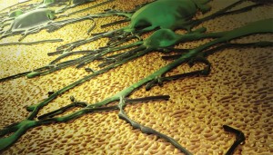
The researchers have provided this image to illustrate their work,

The image depicts a neuronal network growing on a novel nanotextured gold electrode coating. The topographical cues presented by the coating preferentially favor spreading of neurons as opposed to scar tissue. This feature has the potential to enhance the performance of neural interfaces. Image by Ryan Chen/LLNL.

The image depicts a neuronal network growing on a novel nanotextured gold electrode coating. The topographical cues presented by the coating preferentially favor spreading of neurons as opposed to scar tissue. This feature has the potential to enhance the performance of neural interfaces. Image by Ryan Chen/LLNL.