Tag Archives: University of Pennsylvania

Reversing Parkinson’s type symptoms in rats

Indian scientists have developed a technique for delivering drugs that could reverse Parkinson-like symptoms according to an April 22, 2015 news item on Nanowerk (Note: A link has been removed),

As baby boomers age, the number of people diagnosed with Parkinson’s disease is expected to increase. Patients who develop this disease usually start experiencing symptoms around age 60 or older. Currently, there’s no cure, but scientists are reporting a novel approach that reversed Parkinson’s-like symptoms in rats.

Their results, published in the journal ACS Nano (“Trans-Blood Brain Barrier Delivery of Dopamine-Loaded Nanoparticles Reverses Functional Deficits in Parkinsonian Rats”), could one day lead to a new therapy for human patients.

An April 22, 2015 American Chemical Society press pac news release (also on EurekAlert), which originated the news item, describes the problem the researchers were solving (Note: Links have been removed),

Rajnish Kumar Chaturvedi, Kavita Seth, Kailash Chand Gupta and colleagues from the CSIR-Indian Institute of Toxicology Research note that among other issues, people with Parkinson’s lack dopamine in the brain. Dopamine is a chemical messenger that helps nerve cells communicate with each other and is involved in normal body movements. Reduced levels cause the shaking and mobility problems associated with Parkinson’s. Symptoms can be relieved in animal models of the disease by infusing the compound into their brains. But researchers haven’t yet figured out how to safely deliver dopamine directly to the human brain, which is protected by something called the blood-brain barrier that keeps out pathogens, as well as many medicines. Chaturvedi and Gupta’s team wanted to find a way to overcome this challenge.

The researchers packaged dopamine in biodegradable nanoparticles that have been used to deliver other therapeutic drugs to the brain. The resulting nanoparticles successfully crossed the blood-brain barrier in rats, released its dopamine payload over several days and reversed the rodents’ movement problems without causing side effects.

The authors acknowledge funding from the Indian Department of Science and Technology as Woman Scientist and Ramanna Fellow Grant, and the Council of Scientific and Industrial Research (India).

Here’s a link to and citation for the paper,

Trans-Blood Brain Barrier Delivery of Dopamine-Loaded Nanoparticles Reverses Functional Deficits in Parkinsonian Rats by Richa Pahuja, Kavita Seth, Anshi Shukla, Rajendra Kumar Shukla, Priyanka Bhatnagar, Lalit Kumar Singh Chauhan, Prem Narain Saxena, Jharna Arun, Bhushan Pradosh Chaudhari, Devendra Kumar Patel, Sheelendra Pratap Singh, Rakesh Shukla, Vinay Kumar Khanna, Pradeep Kumar, Rajnish Kumar Chaturvedi, and Kailash Chand Gupta. ACS Nano, Article ASAP DOI: 10.1021/nn506408v Publication Date (Web): March 31, 2015
Copyright © 2015 American Chemical Society

This paper is open access.

Another recent example of breaching the blood-brain barrier, coincidentally, in rats, can be found in my Dec. 24, 2014 titled: Gelatin nanoparticles for drug delivery after a stroke. Scientists are also trying to figure out the the blood-brain barrier operates in the first place as per this April 22, 2015 University of Pennsylvania news release on EurekAlert titled, Penn Vet, Montreal and McGill researchers show how blood-brain barrier is maintained (University of Pennsylvania School of Veterinary Medicine, University of Montreal or Université de Montréal, and McGill University). You can find out more about CSIR-Indian Institute of Toxicology Research here.

‘Soft’ nanoparticles, 2D liquid, and fluid interfaces

There’s a story about University of Pennsylvania research on 2D liquids and ‘soft’ particles in an April 6, 2015 news item on Azonano,

Researchers at the University of Pennsylvania have used ‘soft’ nanoparticles to create a system that behaves as a 2D liquid. A 2D world exists at the place where oil and water meet. This interface has properties that could be useful for engineers and chemists.

Researchers have been able to make a soap molecule stay at the interface and make it behave in a predictable manner. However, they have not been able to make more complex molecules behave in the same manner.

An April 3, 2015 University of Pennsylvania news release (also on EurekAlert), which appears to have originated the news item, describes the research in detail,

Where water and oil meet, a two-dimensional world exists. This interface presents a potentially useful set of properties for chemists and engineers, but getting anything more complex than a soap molecule to stay there and behave predictably remains a challenge.

A University of Pennsylvania team has now shown how to make nanoparticles that are attracted to this interface but not to each other, creating a system that acts as a two-dimensional liquid. By measuring this liquid’s pressure and density, they have shown a way forward in using it for a variety of applications, such as in nanomanufacturing, catalysis and photonic devices.

By creating a system where these particles do not clump into clusters or skins, they have enabled a way of investigating the physical fundamentals of how nanoscale objects interact with one another in two dimensions.

“Things get stuck at the interface between oil and water,” Stebe said. “That’s of tremendous fundamental and technological interest, because we can think of that interface as a two-dimensional world. If we can start to understand the interactions of the things that accumulate there and learn how they are arranged, we can exploit them in a number of different applications.”

Getting nanoparticles to go to and stay at this interface is tricky, however. Their surface chemistry can easily be adapted to either water or oil, but balancing the two to get the particles to stay in this 2-D regime is more difficult.

“We understand how particles work in 3-D,” Crocker said. “If you put polymer chains on the surface that are attracted to the solvent, the particles will bounce off each other and make a nice suspension, meaning you can do work with them. However, people haven’t really done that in 2-D before.”

Even when particles are able to stay at the interface, they tend to clump together and form a skin that can’t be pulled apart into its constituent particles.

“All particles love themselves,” Stebe said. “Just due to Van der Waals interactions, if they can get close enough, they aggregate. But because our nanoparticles have protective ligand arms, they don’t clump together and form a liquid state. They’re in two-dimensional equilibrium.”

The team’s technique for surmounting this problem hinged on decorating their gold nanoparticles with surfactant, or soap-like, ligands. These ligands have a water-loving head and an oil-loving tail, and the way they are attached to the central particle allows them to contort themselves so both sides are happy when the particle is at an interface. This arrangement produces a “flying saucer” shape, with the ligands stretching out more at the interface than above or below. These ligand bumpers keeps the particles from clumping together.

“This is a very beautiful system,” Stebe said. “The ability to tune their packing means that we can now take everything we know about the equilibrium thermodynamics in two dimensions and start to pose questions about particle layers. Do these particles behave like we think they should? How can we manipulate them in the future?”

To get at the fundamentals of this system, the researchers needed to deduce the relationships of certain properties, such as how the pressure of their 2-D liquid changes as a function of the packing of the particles. They used a variation of the pendant drop method, in which an oil droplet formed in a suspension of particles in water.  Over time, particles attached to the oil-water interface, producing the 2-D liquid in a form where they could measure those traits.

“We can infer the pressure of this 2-D fluid by the shape of the drop,” Stebe said. “Once we compress the drop by pulling some of the oil back into the syringe, we can determine how the shape changes and relate it to the pressure in the layer.”

The researchers also needed to determine how densely the particles were packed. To do so, they wanted to take advantage of the fact that the drop became more opaque as the density of the particle increased when the drop was compressed. However, it was not possible to simply measure the amount of light that shone through the drop, as plasmonic behavior meant that the properties of the gold nanoparticles changed as they got closer together.

“Fortunately, we discovered another interesting feature of this nanoparticle system,” Garbin said. “If the drop was compressed too much, some particles would fall out of the interface because they didn’t fit anymore. This enabled us to measure the amount of particles that were in that falling plume, since the particles are farther apart from each other there. From that measurement, we could work backwards to the number of particles on the interface”

The smooth relationship between the particles’ packing and the pressure of the 2-D liquid they form provides the basis of universal rules that govern the physics of such systems.

”From this data,” Crocker said, “we can figure out the force versus distance of two nanoparticles. That means we can now make a model of how these particles behave in the 2-D liquid.”

Having these rules will allow researchers to develop functional nanoparticles with different traits, such as longer and more complex ligands that perform some chemical task.

“One application is interface catalysis,” Stebe said. “For example, if you have a reagent that’s in the oil phase, but its product is in the water phase, having a particle on the interface that can help move it from one to the other would be perfect.”

A better understanding of when and why particles get trapped in liquid-liquid interfaces could also underpin future work.

Here’s a link to and  a citation for the paper,

Interactions and Stress Relaxation in Monolayers of Soft Nanoparticles at Fluid-Fluid Interfaces by Valeria Garbin, Ian Jenkins, Talid Sinno, John C. Crocker, and Kathleen J. Stebe. Phys. Rev. Lett. 114, 108301 (Vol. 114, Iss. 10 — 13 March 2015) Published 9 March 2015 DOI: http://dx.doi.org/10.1103/PhysRevLett.114.108301

This paper is behind a paywall.

Graphene used to create electrodes one atom thick and transparent for brain research applications

It’s usually a ‘John Rogers (at the University of Illinois)’ story when there’s mention of transparent electronic devices but not this time. In an Oct. 20, 2014 news item on ScienceDaily, the University of Pennsylvania’s researchers are in the spotlight,

Researchers from the Perelman School of Medicine and School of Engineering at the University of Pennsylvania and The Children’s Hospital of Philadelphia have used graphene — a two-dimensional form of carbon only one atom thick — to fabricate a new type of microelectrode that solves a major problem for investigators looking to understand the intricate circuitry of the brain.

Pinning down the details of how individual neural circuits operate in epilepsy and other neurological disorders requires real-time observation of their locations, firing patterns, and other factors, using high-resolution optical imaging and electrophysiological recording. But traditional metallic microelectrodes are opaque and block the clinician’s view and create shadows that can obscure important details. In the past, researchers could obtain either high-resolution optical images or electrophysiological data, but not both at the same time.

The Center for NeuroEngineering and Therapeutics (CNT), under the leadership of senior author Brian Litt, PhD, has solved this problem with the development of a completely transparent graphene microelectrode that allows for simultaneous optical imaging and electrophysiological recordings of neural circuits. [emphasis mine] Their work was published this week in Nature Communications.

An Oct. 20, 2014 University of Pennsylvania news release (also on EurekAlert), which originated the news item, further describes the research,

“There are technologies that can give very high spatial resolution such as calcium imaging; there are technologies that can give high temporal resolution, such as electrophysiology, but there’s no single technology that can provide both,” says study co-first-author Duygu Kuzum, PhD. Along with co-author Hajime Takano, PhD, and their colleagues, Kuzum notes that the team developed a neuroelectrode technology based on graphene to achieve high spatial and temporal resolution simultaneously.

Aside from the obvious benefits of its transparency, graphene offers other advantages: “It can act as an anti-corrosive for metal surfaces to eliminate all corrosive electrochemical reactions in tissues,” Kuzum says. “It’s also inherently a low-noise material, which is important in neural recording because we try to get a high signal-to-noise ratio.”

While previous efforts have been made to construct transparent electrodes using indium tin oxide, they are expensive and highly brittle, making that substance ill-suited for microelectrode arrays. “Another advantage of graphene is that it’s flexible, so we can make very thin, flexible electrodes that can hug the neural tissue,” Kuzum notes.

In the study, Litt, Kuzum, and their colleagues performed calcium imaging of hippocampal slices in a rat model with both confocal and two-photon microscopy, while also conducting electrophysiological recordings. On an individual cell level, they were able to observe temporal details of seizures and seizure-like activity with very high resolution. The team also notes that the single-electrode techniques used in the Nature Communications study could be easily adapted to study other larger areas of the brain with more expansive arrays.

The graphene microelectrodes developed could have wider application. “They can be used in any application that we need to record electrical signals, such as cardiac pacemakers or peripheral nervous system stimulators,” says Kuzum. Because of graphene’s nonmagnetic and anti-corrosive properties, these probes “can also be a very promising technology to increase the longevity of neural implants.” Graphene’s nonmagnetic characteristics also allow for safe, artifact-free MRI reading, unlike metallic implants.

Kuzum emphasizes that the transparent graphene microelectrode technology was achieved through an interdisciplinary effort of CNT and the departments of Neuroscience, Pediatrics, and Materials Science at Penn and the division of Neurology at CHOP.

Ertugrul Cubukcu’s lab at Materials Science and Engineering Department helped with the graphene processing technology used in fabricating flexible transparent neural electrodes, as well as performing optical and materials characterization in collaboration with Euijae Shim and Jason Reed. The simultaneous imaging and recording experiments involving calcium imaging with confocal and two photon microscopy was performed at Douglas Coulter’s Lab at CHOP with Hajime Takano. In vivo recording experiments were performed in collaboration with Halvor Juul in Marc Dichter’s Lab. Somatasensory stimulation response experiments were done in collaboration with Timothy Lucas’s Lab, Julius De Vries, and Andrew Richardson.

As the technology is further developed and used, Kuzum and her colleagues expect to gain greater insight into how the physiology of the brain can go awry. “It can provide information on neural circuits, which wasn’t available before, because we didn’t have the technology to probe them,” she says. That information may include the identification of specific marker waveforms of brain electrical activity that can be mapped spatially and temporally to individual neural circuits. “We can also look at other neurological disorders and try to understand the correlation between different neural circuits using this technique,” she says.

It’s fascinating work and I hope it’s helpful but I can’t help noticing that these researchers, in common with most, tend to view the brain or whatever body part they’re examining in isolation from the rest of the body, whatever species is being examined. The answers as to why there are brain disorders and diseases may not lie wholly within the brain itself but within the totality of the organism in which the brain resides, i.e., the body. That reservation aside, there’s a link to and a citation for the research paper,

Transparent and flexible low noise graphene electrodes for simultaneous electrophysiology and neuroimaging by Duygu Kuzum, Hajime Takano, Euijae Shim, Jason C. Reed, Halvor Juul, Andrew G. Richardson, Julius de Vries, Hank Bink, Marc A. Dichter, Timothy H. Lucas, Douglas A. Coulter, Ertugrul Cubukcu, & Brian Litt. Nature Communications 5, Article number: 5259 doi:10.1038/ncomms6259 Published 20 October 2014

This paper is behind a paywall but there is a free preview available through ReadCube Access.

Graphene-based sensor mimics pain (mu-opioid) receptor

I once had a job where I had to perform literature searches and read papers on pain research as it related to morphine tolerance. Not a pleasant task, it has left me eager to encourage and write about alternatives to animal testing, a key component of pain research. So, with a ‘song in my heart’, I feature this research from the University of Pennsylvania written up in a May 12, 2014 news item on ScienceDaily,

Almost every biological process involves sensing the presence of a certain chemical. Finely tuned over millions of years of evolution, the body’s different receptors are shaped to accept certain target chemicals. When they bind, the receptors tell their host cells to produce nerve impulses, regulate metabolism, defend the body against invaders or myriad other actions depending on the cell, receptor and chemical type.

Now, researchers from the University of Pennsylvania have led an effort to create an artificial chemical sensor based on one of the human body’s most important receptors, one that is critical in the action of painkillers and anesthetics. In these devices, the receptors’ activation produces an electrical response rather than a biochemical one, allowing that response to be read out by a computer.

By attaching a modified version of this mu-opioid receptor to strips of graphene, they have shown a way to mass produce devices that could be useful in drug development and a variety of diagnostic tests. And because the mu-opioid receptor belongs to the most common class of such chemical sensors, the findings suggest that the same technique could be applied to detect a wide range of biologically relevant chemicals.

A May 6, 2014 University of Pennsylvania news release, which originated the news item, describes the main teams involved in this research along with why and how they worked together (Note: Links have been removed),

The study, published in the journal Nano Letters, was led by A.T. Charlie Johnson, director of Penn’s Nano/Bio Interface Center and professor of physics in Penn’s School of Arts & Sciences; Renyu Liu, assistant professor of anesthesiology in Penn’s Perelman School of Medicine; and Mitchell Lerner, then a graduate student in Johnson’s lab. It was made possible through a collaboration with Jeffery Saven, professor of chemistry in Penn Arts & Sciences. The Penn team also worked with researchers from the Seoul National University in South Korea.

Their study combines recent advances from several disciplines.

Johnson’s group has extensive experience attaching biological components to nanomaterials for use in chemical detectors. Previous studies have involved wrapping carbon nanotubes with single-stranded DNA to detect odors related to cancer and attaching antibodies to nanotubes to detect the presence of the bacteria associated with Lyme disease.

After Saven and Liu addressed these problems with the redesigned receptor, they saw that it might be useful to Johnson, who had previously published a study on attaching a similar receptor protein to carbon nanotubes. In that case, the protein was difficult to grow genetically, and Johnson and his colleagues also needed to include additional biological structures from the receptors’ natural membranes in order to keep them stable.

In contrast, the computationally redesigned protein could be readily grown and attached directly to graphene, opening up the possibility of mass producing biosensor devices that utilize these receptors.

“Due to the challenges associated with isolating these receptors from their membrane environment without losing functionality,” Liu said, “the traditional methods of studying them involved indirectly investigating the interactions between opioid and the receptor via radioactive or fluorescent labeled ligands, for example. This multi-disciplinary effort overcame those difficulties, enabling us to investigate these interactions directly in a cell free system without the need to label any ligands.”

With Saven and Liu providing a version of the receptor that could stably bind to sheets of graphene, Johnson’s team refined their process of manufacturing those sheets and connecting them to the circuitry necessary to make functional devices.

The news release provides more technical details about the graphene sensor,

“We start by growing a piece of graphene that is about six inches wide by 12 inches long,” Johnson said. “That’s a pretty big piece of graphene, but we don’t work with the whole thing at once. Mitchell Lerner, the lead author of the study, came up with a very clever idea to cut down on chemical contamination. We start with a piece that is about an inch square, then separate them into ribbons that are about 50 microns across.

“The nice thing about these ribbons is that we can put them right on top of the rest of the circuitry, and then go on to attach the receptors. This really reduces the potential for contamination, which is important because contamination greatly degrades the electrical properties we measure.”

Because the mechanism by which the device reports on the presence of the target molecule relies only on the receptor’s proximity to the nanostructure when it binds to the target, Johnson’s team could employ the same chemical technique for attaching the antibodies and other receptors used in earlier studies.

Once attached to the ribbons, the opioid receptors would produce changes in the surrounding graphene’s electrical properties whenever they bound to their target. Those changes would then produce electrical signals that would be transmitted to a computer via neighboring electrodes.

The high reliability of the manufacturing process — only one of the 193 devices on the chip failed — enables applications in both clinical diagnostics and further research. [emphasis mine]

“We can measure each device individually and average the results, which greatly reduces the noise,” said Johnson. “Or you could imagine attaching 10 different kinds of receptors to 20 devices each, all on the same chip, if you wanted to test for multiple chemicals at once.”

In the researchers’ experiment, they tested their devices’ ability to detect the concentration of a single type of molecule. They used naltrexone, a drug used in alcohol and opioid addiction treatment, because it binds to and blocks the natural opioid receptors that produce the narcotic effects patients seek.

“It’s not clear whether the receptors on the devices are as selective as they are in the biological context,” Saven said, “as the ones on your cells can tell the difference between an agonist, like morphine, and an antagonist, like naltrexone, which binds to the receptor but does nothing. By working with the receptor-functionalized graphene devices, however, not only can we make better diagnostic tools, but we can also potentially get a better understanding of how the bimolecular system actually works in the body.”

“Many novel opioids have been developed over the centuries,” Liu said. “However, none of them has achieved potent analgesic effects without notorious side effects, including devastating addiction and respiratory depression. This novel tool could potentially aid the development of new opioids that minimize these side effects.”

Wherever these devices find applications, they are a testament to the potential usefulness of the Nobel-prize winning material they are based on.

“Graphene gives us an advantage,” Johnson said, “in that its uniformity allows us to make 192 devices on a one-inch chip, all at the same time. There are still a number of things we need to work out, but this is definitely a pathway to making these devices in large quantities.”

There is no mention of animal research but it seems likely to me that this work could lead to a decreased use of animals in pain research.

This project must have been quite something as it involved collaboration across many institutions (from the news release),

Also contributing to the study were Gang Hee Han, Sung Ju Hong and Alexander Crook of Penn Arts & Sciences’ Department of Physics and Astronomy; Felipe Matsunaga and Jin Xi of the Department of Anesthesiology at the Perelman School of Medicine, José Manuel Pérez-Aguilar of Penn Arts & Sciences’ Department of Chemistry; and Yung Woo Park of Seoul National University. Mitchell Lerner is now at SPAWAR Systems Center Pacific, Felipe Matsunaga at Albert Einstein College of Medicine, José Manuel Pérez-Aguilar at Cornell University and Sung Ju Hong at Seoul National University.

Here’s a link to and a citation for the paper,

Scalable Production of Highly Sensitive Nanosensors Based on Graphene Functionalized with a Designed G Protein-Coupled Receptor by Mitchell B. Lerner, Felipe Matsunaga, Gang Hee Han, Sung Ju Hong, Jin Xi, Alexander Crook, Jose Manuel Perez-Aguilar, Yung Woo Park, Jeffery G. Saven, Renyu Liu, and A. T. Charlie Johnson.Nano Lett., Article ASAP
DOI: 10.1021/nl5006349 Publication Date (Web): April 17, 2014
Copyright © 2014 American Chemical Society

This paper is behind a paywall.

US Air Force wants to merge classical and quantum physics

The US Air Force wants to merge classical and quantum physics for practical purposes according to a May 5, 2014 news item on Azonano,

The Air Force Office of Scientific Research has selected the Harvard School of Engineering and Applied Sciences (SEAS) to lead a multidisciplinary effort that will merge research in classical and quantum physics and accelerate the development of advanced optical technologies.

Federico Capasso, Robert L. Wallace Professor of Applied Physics and Vinton Hayes Senior Research Fellow in Electrical Engineering, will lead this Multidisciplinary University Research Initiative [MURI] with a world-class team of collaborators from Harvard, Columbia University, Purdue University, Stanford University, the University of Pennsylvania, Lund University, and the University of Southampton.

The grant is expected to advance physics and materials science in directions that could lead to very sophisticated lenses, communication technologies, quantum information devices, and imaging technologies.

“This is one of the world’s strongest possible teams,” said Capasso. “I am proud to lead this group of people, who are internationally renowned experts in their fields, and I believe we can really break new ground.”

A May 1, 2014 Harvard University School of Engineering and Applied Sciences news release, which originated the news item, provides a description of project focus: nanophotonics and metamaterials along with some details of Capasso’s work in these areas (Note: Links have been removed),

The premise of nanophotonics is that light can interact with matter in unusual ways when the material incorporates tiny metallic or dielectric features that are separated by a distance shorter than the wavelength of the light. Metamaterials are engineered materials that exploit these phenomena, producing strange effects, enabling light to bend unnaturally, twist into a vortex, or disappear entirely. Yet the fabrication of thick, or bulk, metamaterials—that manipulate light as it passes through the material—has proven very challenging.

Recent research by Capasso and others in the field has demonstrated that with the right device structure, the critical manipulations can actually be confined to the very surface of the material—what they have dubbed a “metasurface.” These metasurfaces can impart an instantaneous shift in the phase, amplitude, and polarization of light, effectively controlling optical properties on demand. Importantly, they can be created in the lab using fairly common fabrication techniques.

At Harvard, the research has produced devices like an extremely thin, flat lens, and a material that absorbs 99.75% of infrared light. But, so far, such devices have been built to order—brilliantly suited to a single task, but not tunable.

This project, however,is focused on the future (Note: Links have been removed),

“Can we make a rapidly configurable metasurface so that we can change it in real time and quickly? That’s really a visionary frontier,” said Capasso. “We want to go all the way from the fundamental physics to the material building blocks and then the actual devices, to arrive at some sort of system demonstration.”

The proposed research also goes further. A key thrust of the project involves combining nanophotonics with research in quantum photonics. By exploiting the quantum effects of luminescent atomic impurities in diamond, for example, physicists and engineers have shown that light can be captured, stored, manipulated, and emitted as a controlled stream of single photons. These types of devices are essential building blocks for the realization of secure quantum communication systems and quantum computers. By coupling these quantum systems with metasurfaces—creating so-called quantum metasurfaces—the team believes it is possible to achieve an unprecedented level of control over the emission of photons.

“Just 20 years ago, the notion that photons could be manipulated at the subwavelength scale was thought to be some exotic thing, far fetched and of very limited use,” said Capasso. “But basic research opens up new avenues. In hindsight we know that new discoveries tend to lead to other technology developments in unexpected ways.”

The research team includes experts in theoretical physics, metamaterials, nanophotonic circuitry, quantum devices, plasmonics, nanofabrication, and computational modeling. Co-principal investigator Marko Lončar is the Tiantsai Lin Professor of Electrical Engineering at Harvard SEAS. Co-PI Nanfang Yu, Ph.D. ’09, developed expertise in metasurfaces as a student in Capasso’s Harvard laboratory; he is now an assistant professor of applied physics at Columbia. Additional co-PIs include Alexandra Boltasseva and Vladimir Shalaev at Purdue, Mark Brongersma at Stanford, and Nader Engheta at the University of Pennsylvania. Lars Samuelson (Lund University) and Nikolay Zheludev (University of Southampton) will also participate.

The bulk of the funding will support talented graduate students at the lead institutions.

The project, titled “Active Metasurfaces for Advanced Wavefront Engineering and Waveguiding,” is among 24 planned MURI awards selected from 361 white papers and 88 detailed proposals evaluated by a panel of experts; each award is subject to successful negotiation. The anticipated amount of the Harvard-led grant is up to $6.5 million for three to five years.

For anyone who’s not familiar (that includes me, anyway) with MURI awards, there’s this from Wikipedia (Note: links have been removed),

Multidisciplinary University Research Initiative (MURI) is a basic research program sponsored by the US Department of Defense (DoD). Currently each MURI award is about $1.5 million a year for five years.

I gather that in addition to the Air Force, the Army and the Navy also award MURI funds.

2013 International Science & Engineering Visualization Challenge Winners

Thanks to a RT from @coreyspowell I stumbled across a Feb. 7, 2014 article in Science (magazine) describing the 2013 International Science & Engineering Visualization Challenge Winners. I am highlighting a few of the entries here but there are more images in the article and a slideshow.

First Place: Illustration

Credit: Greg Dunn and Brian Edwards, Greg Dunn Design, Philadelphia, Pennsylvania; Marty Saggese, Society for Neuroscience, Washington, D.C.; Tracy Bale, University of Pennsylvania, Philadelphia; Rick Huganir, Johns Hopkins University, Baltimore, Maryland

Cortex in Metallic Pastels. Credit: Greg Dunn and Brian Edwards, Greg Dunn Design, Philadelphia, Pennsylvania; Marty Saggese, Society for Neuroscience, Washington, D.C.; Tracy Bale, University of Pennsylvania, Philadelphia; Rick Huganir, Johns Hopkins University, Baltimore, Maryland

From the article, a description of Greg Dunn and his work,

With a Ph.D. in neuroscience and a love of Asian art, it may have been inevitable that Greg Dunn would combine them to create sparse, striking illustrations of the brain. “It was a perfect synthesis of my interests,” Dunn says.

Cortex in Metallic Pastels represents a stylized section of the cerebral cortex, in which axons, dendrites, and other features create a scene reminiscent of a copse of silver birch at twilight. An accurate depiction of a slice of cerebral cortex would be a confusing mess, Dunn says, so he thins out the forest of cells, revealing the delicate branching structure of each neuron.

Dunn blows pigments across the canvas to create the neurons and highlights some of them in gold leaf and palladium, a technique he is keen to develop further.

“My eventual goal is to start an art-science lab,” he says. It would bring students of art and science together to develop new artistic techniques. He is already using lithography to give each neuron in his paintings a different angle of reflectance. “As you walk around, different neurons appear and disappear, so you can pack it with information,” he says.

People’s Choice:  Games & Apps

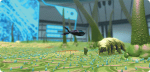
Meta!Blast: The Leaf. Credit: Eve Syrkin Wurtele, William Schneller, Paul Klippel, Greg Hanes, Andrew Navratil, and Diane Bassham, Iowa State University, Ames

Meta!Blast: The Leaf. Credit: Eve Syrkin Wurtele, William Schneller, Paul Klippel, Greg Hanes, Andrew Navratil, and Diane Bassham, Iowa State University, Ames

More from the article,

“Most people don’t expect a whole ecosystem right on the leaf surface,” says Eve Syrkin Wurtele, a plant biologist at Iowa State University. Meta!Blast: The Leaf, the game that Wurtele and her team created, lets high school students pilot a miniature bioship across this strange landscape, which features nematodes and a lumbering tardigrade. They can dive into individual cells and zoom around a chloroplast, activating photosynthesis with their ship’s search lamp. Pilots can also scan each organelle they encounter to bring up more information about it from the ship’s BioLog—a neat way to put plant biology at the heart of an interactive gaming environment.

This is a second recognition for Meta!Blast, which won an Honorable Mention in the 2011 visualization challenge for a version limited to the inside of a plant cell.

The Metablast website homepage describes the game,

The last remaining plant cell in existence is dying. An expert team of plant scientists have inexplicably disappeared. Can you rescue the lost team, discover what is killing the plant, and save the world?

Meta!Blast is a real-time 3D action-adventure game that puts you in the pilot’s seat. Shrink down to microscopic size and explore the vivid, dynamic world of a soybean plant cell spinning out of control. Interact with numerous characters, fight off plant pathogens, and discover how important plants are to the survival of the human race.

Enjoy!

A superhydrophobic coating for glass from the University of Pennsylvania (US) with promises that it’s better than others

Anyone who’s read this blog with any frequency has likely encountered my obsession with self-cleaning glass (specifically, windows). Frankly, I’ve almost given up hope of ever seeing the product in my lifetime several times and then I see another announcement such as this in a Nov. 26, 2013 news item on Nanowerk,

Hanging hundreds of feet off the ground to wash a skyscraper’s windows or pumping water out to a desert solar array to keep its panels and mirrors clean is more than just a hassle—it’s an expensive problem with serious ecological implications.

A spin-off company from Penn has found a way to solve the problem of keeping surfaces clean, while also keeping them transparent.

The undated University of Pennsylvania article by Evan Lerner, which originated the news item, describes both the university’s spin-off company and the research which it is exploiting (Note: Links have been removed),

Nelum Sciences, created under an UPstart program in Penn’s Center for Technology Transfer, has developed a superhydrophobic coating that can be sprayed onto any surface. The water-based solution contains nanoscopic particles that add a nearly invisible layer of roughness to a surface. This increases the contact angle of the material to which these particles are applied.

A contact angle is the angle the edges of a resting drop of liquid make with a surface. When the angle is low, a drop resembles a flattened hemisphere, with edges that are stuck to the surface. But as the angle increases, a drop begins to look more like a ball, until it literally rolls away instead of sticking.

When these balls of liquid roll off a superhydrophobic surface, they pick up any debris they encounter in their paths, keeping a surface clean.

Co-founded in 2011 by Shu Yang, professor of materials science and engineering in Penn’s School of Engineering and Applied Science, Nelum Science’s coating is based on her nanotechnology research. Fabricating the coating’s nanoparticles at sizes smaller than the wavelength of light—the quality that makes them transparent—is the product of cutting-edge laboratory techniques. The company’s inspiration, however, came from structures created by nature.

“Some plants, like lotuses, and other biological structures, like butterfly wings, have this kind of nano-roughness to keep them clean and dry,” Yang says. “That’s why we named the company after the lotus’ Latin name, nelumbo.”

Other superhydrophobic sprays have recently come on the market, but they give surfaces a hazy, frosted appearance, making them inappropriate for applications where cleanliness is critical, such as windows, lenses, safety goggles, and solar panels.

Here’s a University of Pennsylvanis video illustrating the technology,

I wasn’t able to find much information about Nelum Sciences but there is this page on the University of Pennsylvania’s Center for Technology Transfer website, which leads me to suspect I may not be seeing the product in the market place any time soon.

Nanotechnology research influences architecture: Krishna P. Singh Center for Nanotechnology (University of Pennsylvania)

Architects Bring Sunshine Into Nanotech Labs is the title for Sammy Medina’s Fast Company article about the University of Pennsylvania’s mind-blowing Krishna P. Singh Center for Nanotechnology,

(Photo © Albert Vecerka/Esto Courtesy: University of Pennsylvania

(Photo © Albert Vecerka/Esto Courtesy: University of Pennsylvania

That’s a very pretty image but here’s what makes it mind-blowing (from Medina’s article; Note: A link has been removed),

The building, by architecture office Wisss/Manfredi, “turns the paradigm of the laboratory inside out.” So says Marion Weiss, co-partner, along with Michael Manfredi, of the New York-based firm. “Most nanotechnology facilities are often in fairly remote locations, like Cornell’s,” Manfredi explains. “[O]r if they are in an urban campus, their signature spaces aren’t easily discovered.”

… The building’s most spectacular architectural moments are pushed to the fore, most notably in the 68-foot-deep cantilever that juts out over the ground-level lawn. The expansive facade glitters, in Weiss’s words, “like a cracked-open geode,” refracting scattered shards of light this way and that.

… Scientists can be seen from the courtyard through the structure’s three layers of glass, where they appear behind a wide amber glass partition ambling about like extras in a science fiction film. (As is standard, the laboratories themselves are wrapped in amber-tinted screens or windows, whose color protects against the sun’s rays. Only here, the architects tinted entire glass walls, rendering the lab space shockingly transparent.) Outside, in the atrium-like “galleria,” digital projections blow up microscopic images sourced from the scientists’ instruments and display them to the campus beyond. [emphasis mine]

This theme of scale is, naturally, very much inscribed in the project. The disparity between the building and the forms of nanotechnology it contains is ripe for creative interpretation … [emphasis mine]

The October 2, 2013 University of Pennsylvania news release about the Oct. 4, 2013 opening of the Krishna P. Singh Center for Nanotechnology provides details complementary to the information in Medina’s article (Note: Links have been removed),

Faculty from the School of Engineering and Applied Science, the School of Arts and Sciences, and across the University will make use of the Singh Center’s characterization and fabrication suites. Each of the two 10,000-square-foot facilities is filled with state-of-the-art equipment and designed to enable the high-precision techniques that research at the smallest scales necessitates.

The characterization facility is situated on bedrock, 18 feet below the surface, to help minimize vibrations that would interfere with its various atomic and electron microscopes. Its labs are also designed to be isolated from temperature fluctuations, atmospheric turbulence, and electromagnetic noise.

The fabrication facility on the Singh Center’s ground floor contains a next-generation cleanroom. Once in isolation garb, researchers will use its assembly tools to grow carbon nanotubes, deposit graphene, and etch microelectronic systems, among many other applications. The facility’s photolithography equipment is shielded from interfering ultraviolet light by a pane of marigold glass, which gives the Center its signature color.

I recommend reading Medina’s article if you want to find out more about how the architects approached the project and if you want to see more pictures.

POD (print-on-demand) robots

I’ve heard of print-on-demand (POD) books before but not robots as per the April 4, 2012 article on BBC News online (link to National Science Foundation removed),

Researchers aim to build a desktop technology that would allow an average person to design and print a machine within 24 hours.

The team says that making it easier to create specialised robots could have a “profound impact on society”.

The effort is being funded by a $10m (£6.3m) grant from the National Science Foundation [NSF].

The Virginia-based organization [NSF] described the move as a “game changing investment”.

“It has the potential to democratise and personalise automation to meet the needs of individual users – whether for search and rescue workers in remote areas of the world or educators in classrooms around the US – possibilities for social impact abound,” said spokeswoman Lisa-Joy Zgorski.

The April 3, 2012 MIT (Massachusetts Institute of Technology) news item by Abby Abazorius provides more detail,

“This research envisions a whole new way of thinking about the design and manufacturing of robots, and could have a profound impact on society,” says MIT Professor Daniela Rus, leader of the project and a principal investigator at the MIT Computer Science and Artificial Intelligence Laboratory (CSAIL). “We believe that it has the potential to transform manufacturing and to democratize access to robots.”

“Our goal is to develop technology that enables anyone to manufacture their own customized robot. This is truly a game changer,” says Professor Vijay Kumar, who is leading the team from the University of Pennsylvania. “It could allow for the rapid design and manufacture of customized goods, and change the way we teach science and technology in high schools.”

The five-year project, called “An Expedition in Computing for Compiling Printable Programmable Machines,” brings together a team of researchers from MIT, the University of Pennsylvania and Harvard University, and is funded as part of the NSF’s “Expeditions in Computing” program.

It currently takes years to produce, program and design a functioning robot, and is an extremely expensive process, involving hardware and software design, machine learning and vision, and advanced programming techniques. The new project would automate the process of producing functional 3-D devices and allow individuals to design and build functional robots from materials as easily accessible as a sheet of paper.


Researchers hope to create a platform that would allow an individual to identify a household problem that needs assistance; then head to a local printing store to select a blueprint, from a library of robotic designs; and then customize an easy-to-use robotic device that could solve the problem. Within 24 hours, the robot would be printed, assembled, fully programmed and ready for action.

By altering the way in which machines can be produced, designed and built, the project could have far reaching implications for a variety of fields.

“This project aims to dramatically reduce the development time for a variety of useful robots, opening the doors to potential applications in manufacturing, education, personalized health care and even disaster relief,” says Rob Wood, an associate professor at Harvard University.


Thus far, the research team has prototyped two machines for designing, printing and programming, including an insect-like robot that could be used for exploring a contaminated area and a gripper that could be used by people with limited mobility.

Here’s a video demonstrating a few of the prototypes the team has developed (an “insect-like robot that could be used for exploring a contaminated area and a gripper that could be used by people with limited mobility”).

You can find out more about the CSAIL project at MIT here.

Other research collaborators on the five-year NSF project include Visiting Scientist Martin Demaine, Associate Professor Wojciech Matusik, Professor Martin Rinard, and Assistant Professor Sangbae Kim of MIT. Besides Wood (Harvard) and Kumar (UPenn), the team also includes Associate Professor Andre DeHon, Professor Sanjeev Khanna and Professor Insup Lee, all from UPenn.

Rejecting creativity?

Most people know this from experience. We laud creativity in theory while attempting to crush it in practice. If you doubt this, try launching a new idea at a meeting. In fact, trying to launch a new idea anywhere at any time is difficult as anyone who’s tried will tell you. Frustratingly, people don’t necessarily believe you when you point it out. They’re more likely to announce that the idea couldn’t have been much good in the first place or perhaps laugh at you because the idea seems so crazy or there’s dead silence because they can’t understand what you’re talking about. (Yes, I’ve had a few bad experiences.) So I was thrilled to see a study that confirms my experience. From the Sept. 3, 2011 news item on Science Daily, Why We Crave Creativity but Reject Creative Ideas,

The next time your great idea at work elicits silence or eye rolls, you might just pity those co-workers. Fresh research indicates they don’t even know what a creative idea looks like and that creativity, hailed as a positive change agent, actually makes people squirm.

“How is it that people say they want creativity but in reality often reject it?” said Jack Goncalo, ILR [Industrial and Labor Relations] School [at Cornell University] assistant professor of organizational behavior and co-author of research to be published in an upcoming issue of the journal Psychological Science. The paper reports on two 2010 experiments at the University of Pennsylvania involving more than 200 people.

The studies’ findings include:

  • Creative ideas are by definition novel, and novelty can trigger feelings of uncertainty that make most people uncomfortable.
  • People dismiss creative ideas in favor of ideas that are purely practical — tried and true.
  • Objective evidence shoring up the validity of a creative proposal does not motivate people to accept it.
  • Anti-creativity bias is so subtle that people are unaware of it, which can interfere with their ability to recognize a creative idea.

For example, subjects had a negative reaction to a running shoe equipped with nanotechnology that adjusted fabric thickness to cool the foot and reduce blisters. [emphasis mine]

While I don’t always require a connection to nanotechnology in postings like this this, it’s nice to find one. And, I’m quite, quite surprised that people would not leap for joy at the thought of a shoe that would cool your foot and reduce the incidence of blisters. Who’d reject that? Apparently, more than one of us. Here’s more from the researchers,

“Our findings imply a deep irony,” wrote the authors, who also include Jennifer Mueller of the University of Pennsylvania and Shimul Melwani of the University of North Carolina, Chapel Hill. “Revealing the existence and nature of a bias against creativity can help explain why people might reject creative ideas and stifle scientific advancements, even in the face of strong intentions to the contrary.”

I hope I’ll have a chance to read the studies once they’re published and I hope the researchers will have the opportunity to tackle some other related questions such as why do we accept new ideas? If we rejected everything, we wouldn’t have agriculture, the wheel, money, etc. Plus, I also know that while I’m open to new ideas and have generated a few of my own, I have missed the boat on occasion. If I’d been in charge, there never would have been a camera included in a phone. By what means am I (or is anyone) more open to a new idea since the nanotechnology in the footwear wouldn’t be a problem for me but the phone camera was?