Tag Archives: heart disease

Powered by light: a battery-free pacemaker

I think it looks more like a potato than a heart but it does illustrate how this new battery-free pacemaker would wrap around a heart,

Caption: An artist’s rendering shows how a new pacemaker, designed by a UArizona-led team of researchers, is able to envelop the heart. The wireless, battery-free pacemaker could be implanted with a less invasive procedure than currently possible and would cause patients less pain. Credit: Philipp Gutruff

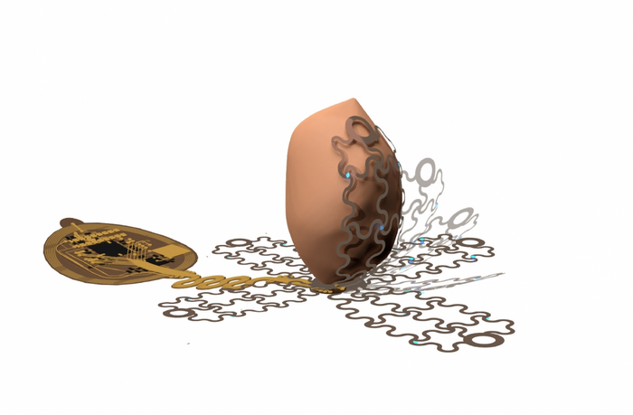
An October 27, 2022 news item on ScienceDaily announces a technology that could make life much easier for people with pacemakers (Comment: In the image, that looks more like a potato than a heart, to me),

University of Arizona engineers lead a research team that is developing a new kind of pacemaker, which envelops the heart and uses precise targeting capabilities to bypass pain receptors and reduce patient discomfort.

An October 27, 2022 University of Arizona news release (also on EurekAlert) by Emily Dieckman, which originated the news item, explains the reasons for the research and provides some technical details (Note: Links have been removed),

Pacemakers are lifesaving devices that regulate the heartbeats of people with chronic heart diseases like atrial fibrillation and other forms of arrhythmia. However, pacemaker implantation is an invasive procedure, and the lifesaving pacing the devices provide can be extremely painful. Additionally, pacemakers can only be used to treat a few specific types of disease.

In a paper published Wednesday [October 26, 2022] in Science Advances, a University of Arizona-led team of researchers detail the workings of a wireless, battery-free pacemaker they designed that could be implanted with a less invasive procedure than currently possible and would cause patients less pain. The study was helmed by researchers in the Gutruf Lab, led by biomedical engineering assistant professor and Craig M. Berge Faculty Fellow Philipp Gutruf.

Currently available pacemakers work by implanting one or two leads, or points of contact, into the heart with hooks or screws. If the sensors on these leads detect a dangerous irregularity, they send an electrical shock through the heart to reset the beat.

“All of the cells inside the heart get hit at one time, including the pain receptors, and that’s what makes pacing or defibrillation painful,” Gutruf said. “It affects the heart muscle as a whole.”

The device Gutruf’s team has developed, which has not yet been tested in humans, would allow pacemakers to send much more targeted signals using a new digitally manufactured mesh design that encompasses the entire heart. The device uses light and a technique called optogenetics.

Optogenetics modifies cells, usually neurons, sensitive to light, then uses light to affect the behavior of those cells. This technique only targets cardiomyocytes, the cells of the muscle that trigger contraction and make up the beat of the heart. This precision will not only reduce pain for pacemaker patients by bypassing the heart’s pain receptors, it will also allow the pacemaker to respond to different kinds of irregularities in more appropriate ways. For example, during atrial fibrillation, the upper and lower chambers of the heart beat asynchronously, and a pacemaker’s role is to get the two parts back in line.

“Whereas right now, we have to shock the whole heart to do this, these new devices can do much more precise targeting, making defibrillation both more effective and less painful,” said Igor Efimov, professor of biomedical engineering and medicine at Northwestern University, where the devices were lab-tested. “This technology could make life easier for patients all over the world, while also helping scientists and physicians learn more about how to monitor and treat the disease.”

Flexible mesh encompasses the heart

To ensure the light signals can reach many different parts of the heart, the team created a design that involves encompassing the organ, rather than implanting leads that provide limited points of contact.

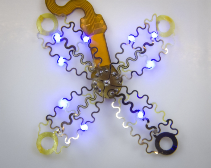
The new pacemaker model consists of four petallike structures made of thin, flexible film, which contain light sources and a recording electrode. The petals, specially designed to accommodate the way the heart changes shape as it beats, fold up around the sides of the organ to envelop it, like a flower closing up at night.

“Current pacemakers record basically a simple threshold, and they will tell you, ‘This is going into arrhythmia, now shock!'” Gutruf said. “But this device has a computer on board where you can input different algorithms that allow you to pace in a more sophisticated way. It’s made for research.”

Because the system uses light to affect the heart, rather than electrical signals, the device can continue recording information even when the pacemaker needs to defibrillate. In current pacemakers, the electrical signal from the defibrillation can interfere with recording capabilities, leaving physicians with an incomplete picture of cardiac episodes. Additionally, the device does not require a battery, which could save pacemaker patients from needing to replace the battery in their device every five to seven years, as is currently the norm.

Gutruf’s team collaborated with researchers at Northwestern University on the project. While the current version of the device has been successfully demonstrated in animal models, the researchers look forward to furthering their work, which could improve the quality of life for millions of people.

The prototype looks like this,

Caption: The device uses light and a technique called optogenetics, which modifies cells that are sensitive to light, then uses light to affect the behavior of those cells.. Credit: Philipp Gutruff

Here’s a link to and a citation for the paper,

Wireless, fully implantable cardiac stimulation and recording with on-device computation for closed-loop pacing and defibrillation by Jokubas Ausra, Micah Madrid, Rose T. Yin, Jessica Hanna, Suzanne Arnott, Jaclyn A. Brennan, Roberto Peralta, David Clausen, Jakob A. Bakall, Igor R. Efimov, and Philipp Gutruf. Science Advances 26 Oct 2022 Vol 8, Issue 43 DOI: 10.1126/sciadv.abq7469

This paper is open access.

Maybe spray-on technology can be used for heart repair?

Courtesy: University of Ottawa

That is a pretty stunning image and this March 15, 2022 news item on phys.org provides an explanation of what you see (Note: A link has been removed),

Could a spritz of super-tiny particles of gold and peptides on a damaged heart potentially provide minimally invasive, on-the-spot repair?

Cutting-edge research led by University of Ottawa Faculty of Medicine Associate Professors Dr. Emilio Alarcon and Dr. Erik Suuronen suggests a spray-on technology using customized nanoparticles of one of the world’s most precious metals offers tremendous therapeutic potential and could eventually help save many lives. Cardiovascular diseases are the leading cause of death globally, claiming roughly 18 million lives each year.

In a paper recently published online in ACS Nano, a peer-reviewed journal that highlighted the new research on its supplementary cover, Dr. Alarcon and his team of fellow investigators suggest that this approach might one day be used in conjunction with coronary artery bypass surgeries. That’s the most common type of heart surgery.

A March 15, 2021 University of Ottawa news release (also on EurekAlert) by David McFadden, which originated the news item, describes the research in more detail (Note: A link has been removed),

The therapy tested by the researchers – which was sprayed on the hearts of lab mice – used very low concentrations of peptide-modified particles of gold created in the laboratory. From the nozzle of a miniaturized spraying apparatus, the material can be evenly painted on the surface of a heart within a few seconds.

Gold nanoparticles have been shown to have some unusual properties and are highly chemically reactive. For years, researchers have been employing gold nanoparticles – so tiny they are undetectable by the human eye – in such a wide range of technologies that it’s become an area of intense research interest.

In this case, the custom-made nanogold modified with peptides—a short chain of amino acids —was sprayed on the hearts of lab mice. The research found that the spray-on therapy not only resulted in an increase in cardiac function and heart electrical conductivity but that there was no off-target organ infiltration by the tiny gold particles.

“That’s the beauty of this approach. You spray, then you wait a couple of weeks, and the animals are doing just fine compared to the controls,” says Dr. Alarcon, who is part of the Faculty of Medicine’s Department of Biochemistry, Microbiology and Immunology and also Director of the Bio-nanomaterials Chemistry and Engineering Laboratory at the University of the Ottawa Heart Institute.

Dr. Alarcon says that not only does the data suggest that the therapeutic action of the spray-on nanotherapeutic is highly effective, but its application is far simpler than other regenerative approaches for treating an infarcted heart.

At first, the observed improvement of cardiac function and electrical signal propagation in the hearts of tested mice was hard for the team to believe. But repeated experiments delivered the same positive results, according to Dr. Alarcon, who is part of the Faculty of Medicine’s Department of Biochemistry, Microbiology and Immunology and Director of the Bio-nanomaterials Chemistry and Engineering Laboratory at the University of Ottawa Heart Institute.

To validate the exciting findings in mice, the team is now seeking to adapt this technology to minimally invasive procedures that will expedite testing in large animal models, such as rabbits and pigs.

Dr. Alarcon praised the research culture at uOttawa and the Heart Institute, saying that the freedom to explore is paramount. “When you have an environment where you are allowed to make mistakes and criticize, that really drives discoveries,” he says.

The team involved in the paper includes researchers from uOttawa and the University of Talca in Chile. Part of the work was funded by the Canadian government’s New Frontiers in Research Fund, which was launched in 2018 and supports transformative high risk/high reward research led by Canadian researchers working with local and international partners.

Here’s a link to and a citation for the paper,

Nanoengineered Sprayable Therapy for Treating Myocardial Infarction by Marcelo Muñoz, Cagla Eren Cimenci, Keshav Goel, Maxime Comtois-Bona, Mahir Hossain, Christopher McTiernan, Matias Zuñiga-Bustos, Alex Ross, Brenda Truong, Darryl R. Davis, Wenbin Liang, Benjamin Rotstein, Marc Ruel, Horacio Poblete, Erik J. Suuronen, and Emilio I. Alarcon. ACS Nano 2022, 16, 3, 3522–3537 DOI: https://doi.org/10.1021/acsnano.1c08890 Publication Date: February 14, 2022 Copyright © 2022 The Authors. Published by American Chemical Society

This paper appears to be open access.

Metallic nanoparticles inside heart tissue mitochondria can cause damage

With all the focus on COVID-19, viruses , and aerosols, it’s easy to forget that there are other kinds of contaminated air too. The last time I featured work on nanoparticles and air pollution was in a May 31, 2017 posting, “Explaining the link between air pollution and heart disease?” where scientists announced they may have discovered how air pollution (nanoparticles) were making their way from lungs to the heart. Answer: the bloodstream.

A July 3, 2020 Lancaster University press release (also on EurekAlert) announces research into how air made toxic by metallic nanoparticles affects the heart in very specific ways (Note: A link has been removed),

Toxic metallic air pollution nanoparticles are getting inside the crucial, energy-producing structures within the hearts of people living in polluted cities, causing cardiac stress – a new study confirms.

The research team, led by Professors Barbara Maher of Lancaster University and Lilian Calderón-Garcidueñas of The University of Montana and the Universidad del Valle de Mexico, found the metallic nanoparticles, which included iron-rich nanoparticles and other pollution-derived metals such as titanium, inside the damaged heart cells of a 26-year-old and even a three-year-old toddler.

The hearts had belonged to people who had died in accidents and who had lived in highly-polluted Mexico City.

The findings shed new light on how air pollution can cause the development of heart disease, as the iron-rich particles were associated with damage to the cells, and increased cardiac oxidative stress, even in these very young hearts.

The repeated inhalation of these iron-rich nanoparticles, and their circulation by the bloodstream to the heart, may account for the well-established associations between exposure to particulate air pollution and increased cardiovascular disease, including heart attacks. The study indicates that heart disease can start in very early age, before progressing to full-blown cardiovascular illness later in life. This type of air pollution may thus be responsible for the ‘silent epidemic’ of heart disease, internationally. By causing pre-existing heart conditions, it may also account for some of the increased death rates from Covid-19 seen in areas with high levels of particulate air pollution.

Professor Maher said: “It’s been known for a long time that people with high exposure to particulate air pollution experience increased levels and severity of heart disease. Our new work shows that iron-rich nanoparticles from air pollution can get right inside the millions of mitochondria inside our hearts…the structures which generate the energy needed for our hearts to pump properly.

“That we found these metal particles inside the heart of even a three-year old indicates that we’re setting heart disease in train right from the earliest days, but only seeing its full, clinical effects in later life. It’s really urgent to reduce emissions of ultrafine particles from our vehicles and from industry, before we give heart disease to the next generation too.”

The researchers, using high-resolution transmission electron microscopy and energy-dispersive X-ray analysis, found that the mitochondria containing the iron-rich nanoparticles appeared to be damaged, with some cells showing deformities and others with ruptured membranes. Professor Calderón-Garcidueñas stated that increased levels of markers of cardiac oxidative stress are present in the very young cases examined.

The iron-rich nanoparticles found inside the heart cells are identical in size, shape and composition to those emitted from sources such as the exhausts, tyres and brakes of vehicles. These air pollution nanoparticles are also emitted by industrial sources as well as open fires in homes.

Some of the iron-rich nanoparticles are also strongly magnetic. This raises concerns about what might happen when people with millions of these nanoparticles in their hearts are using appliances with associated magnetic fields, such as hair dryers and mobile phones. People who work in industries that mean they are exposed to magnetic fields such as welders and power line engineers may also be at risk. This kind of exposure could potentially lead to heart electrical dysfunction and cell damage.

The findings builds on the researchers’ previous findings that show that the hearts of city dwellers contain billions of these nanoparticles and can be up to ten times more polluted than the hearts of people living in less polluted places.

The researchers say their study underlines the need for governments across the world to tackle ultrafine particulate pollution in their cities.

Professor Calderón-Garcidueñas said: “Exposure to such air pollution is a modifiable risk factor for cardiovascular disease, on a global scale, reinforcing the urgent need for individual and government actions not just to reduce PM2.5 but to monitor, regulate and reduce emissions of these specific, ultrafine components of the urban air pollution ‘cocktail’.”

Here’s a link to and a citation for the paper,

Iron-rich air pollution nanoparticles: An unrecognised environmental risk factor for myocardial mitochondrial dysfunction and cardiac oxidative stress by B.A.Maher, A.González-Maciel, R.Reynoso-Robles, R.Torres-Jardón, L.Calderón-Garcidueñas. Environmental Research Volume 188, September 2020, 109816 DOI: https://doi.org/10.1016/j.envres.2020.109816 Available online 21 June 2020

This paper appears to be open access (just keep scrolling down).

‘What becomes of the broken-hearted?’ Trinity College Dublin scientists may have an answer

While Valentine’s Day as celebrated here in Canada and elsewhere (but not everywhere) on February 14 of each year is usually marked in a purely joyous fashion,I’m going to focus on heartbreak. Here is one of the greatest versions of ‘What becomes of the broken-hearted?’ Then, repair follows in the context of some cardiac research coming out of Ireland,

Thank you Joan Osborne and the Funk Brothers. If you haven’t seen ‘Standing in the shadows of Motown’, you may want to make a point of it.

As for the musical question in the headline, researchers at Trinity College Dublin may have an answer of sorts. A February 13, 2020 Trinity College Dublin press release (also on EurekAlert) describes how broken hearts can be mended,

Bioengineers from Trinity College Dublin, Ireland, have developed a prototype patch that does the same job as crucial aspects of heart tissue.

Their patch withstands the mechanical demands and mimics the electrical signalling properties that allow our hearts to pump blood rhythmically round our bodies.

Their work essentially takes us one step closer to a functional design that could mend a broken heart.

One in six men and one in seven women in the EU will suffer a heart attack at some point in their lives. Worldwide, heart disease kills more women and men – regardless of race, than any other disease.

Cardiac patches lined with heart cells can be applied surgically to restore heart tissue in patients who have had damaged tissue removed after a heart attack and to repair congenital heart defects in infants and children. Ultimately, though, the goal is to create cell-free patches that can restore the synchronous beating of the heart cells, without impairing the heart muscle movement.

The bioengineers report their work, which takes us one step closer to such a reality, in the journal Advanced Functional Materials.

Michael Monaghan, ussher assistant professor in biomedical engineering at Trinity, and senior author on the paper, said:

“Despite some advances in the field, heart disease still places a huge burden on our healthcare systems and the life quality of patients worldwide. It affects all of us either directly or indirectly through family and friends. As a result, researchers are continuously looking to develop new treatments which can include stem cell treatments, biomaterial gel injections and assistive devices.”

“Ours is one of few studies that looks at a traditional material, and through effective design allows us to mimic the direction-dependent mechanical movement of the heart, which can be sustained repeatably. This was achieved through a novel method called ‘melt electrowriting’ and through close collaboration with the suppliers located nationally we were able to customise the process to fit our design needs.”

This work was performed in the Trinity Centre for Biomedical Engineering, based in the Trinity Biomedical Sciences Institute in collaboration with Spraybase®, a subsidiary of Avectas Ltd. It was funded by Enterprise Ireland through the Innovation Partnership Program (IPP).

Dr Gillian Hendy, director of Spraybase® is a co-author on the paper. Dr Hendy commended the team at Trinity on the work completed and advancements made on the Spraybase® Melt Electrowriting (MEW) System. The success achieved by the team highlights the potential applications of this novel technology in the cardiac field and succinctly captures the benefits of industry and academic collaboration, through platforms such as the IPP.

Engineering replacement materials for heart tissue is challenging since it is an organ that is constantly moving and contracting. The mechanical demands of heart muscle (myocardium) cannot be met using polyester-based thermoplastic polymers, which are predominantly the approved options for biomedical applications.

However, the functionality of thermoplastic polymers could be leveraged by its structural geometry. The bioengineers then set about making a patch that could control the expansion of a material in multiple directions and tune this using an engineering design approach.

The patches were manufactured via melt electrowriting – a core technology of Spraybase® – which is reproducible, accurate, and scalable. The patches were also coated with the electroconductive polymer polypyrrole to provide electrical conductivity while maintaining cell compatibility.

The patch withstood repeated stretching, which is a dominant concern for cardiac biomaterials, and showed good elasticity, to accurately mimic that key property of heart muscle.

Professor Monaghan added:

“Essentially, our material addresses a lot of requirements. The bulk material is currently approved for medical device use, the design accommodates the movement of the pumping heart, and has been functionalised to accommodate signaling between isolated contractile tissues.”

“This study currently reports the development of our method and design, but we are now looking forward to furthering the next generation of designs and materials with the eventual aim of applying this patch as a therapy for a heart attack.”

Dr Dinorath Olvera, Trinity, first author on the paper, added:

“Our electroconductive patches support electrical conduction between biological tissue in an ex vivo model. These results therefore represent a significant step towards generating a bioengineered patch capable of recapitulating aspects of heart tissue – namely its mechanical movement and electrical signalling.”

Here’s a link to and a citation for the paper,

Electroconductive Melt Electrowritten Patches Matching the Mechanical Anisotropy of Human Myocardium by Dinorath Olvera, Mina Sohrabi Molina, Gillian Hendy, Michael G. Monaghan. Advanced Functional Materials DOI: https://doi.org/10.1002/adfm.201909880 First published: 12 February 2020

This paper is behind a paywall.

Here are links, should you be interested in the company partnering with the researchers, Spraybase®, or its parent company, Avectas Ltd.

Finally, the singer who made ‘What becomes of the broken-hearted?’ a hit in 1965 was Jimmy Ruffin,

Enjoy.

Explaining the link between air pollution and heart disease?

An April 26, 2017 news item on Nanowerk announces research that may explain the link between heart disease and air pollution (Note: A link has been removed),

Tiny particles in air pollution have been associated with cardiovascular disease, which can lead to premature death. But how particles inhaled into the lungs can affect blood vessels and the heart has remained a mystery.

Now, scientists have found evidence in human and animal studies that inhaled nanoparticles can travel from the lungs into the bloodstream, potentially explaining the link between air pollution and cardiovascular disease. Their results appear in the journal ACS Nano (“Inhaled Nanoparticles Accumulate at Sites of Vascular Disease”).

An April 26, 2017 American Chemical Society news release on EurekAlert, which originated the news item,  expands on the theme,

The World Health Organization estimates that in 2012, about 72 percent of premature deaths related to outdoor air pollution were due to ischemic heart disease and strokes. Pulmonary disease, respiratory infections and lung cancer were linked to the other 28 percent. Many scientists have suspected that fine particles travel from the lungs into the bloodstream, but evidence supporting this assumption in humans has been challenging to collect. So Mark Miller and colleagues at the University of Edinburgh in the United Kingdom and the National Institute for Public Health and the Environment in the Netherlands used a selection of specialized techniques to track the fate of inhaled gold nanoparticles.

In the new study, 14 healthy volunteers, 12 surgical patients and several mouse models inhaled gold nanoparticles, which have been safely used in medical imaging and drug delivery. Soon after exposure, the nanoparticles were detected in blood and urine. Importantly, the nanoparticles appeared to preferentially accumulate at inflamed vascular sites, including carotid plaques in patients at risk of a stroke. The findings suggest that nanoparticles can travel from the lungs into the bloodstream and reach susceptible areas of the cardiovascular system where they could possibly increase the likelihood of a heart attack or stroke, the researchers say.

Here’s a link to and a citation for the paper,

Inhaled Nanoparticles Accumulate at Sites of Vascular Disease by Mark R. Miller, Jennifer B. Raftis, Jeremy P. Langrish, Steven G. McLean, Pawitrabhorn Samutrtai, Shea P. Connell, Simon Wilson, Alex T. Vesey, Paul H. B. Fokkens, A. John F. Boere, Petra Krystek, Colin J. Campbell, Patrick W. F. Hadoke, Ken Donaldson, Flemming R. Cassee, David E. Newby, Rodger Duffin, and Nicholas L. Mills. ACS Nano, Article ASAP DOI: 10.1021/acsnano.6b08551 Publication Date (Web): April 26, 2017

Copyright © 2017 American Chemical Society

This paper is behind a paywall.

Repairing a ‘broken’ heart with a 3D printed patch

The idea of using stem cells to help heal your heart so you don’t have scar tissue seems to be a step closer to reality. From an April 14, 2017 news item on ScienceDaily which announces the research and explains why scar tissue in your heart is a problem,

A team of biomedical engineering researchers, led by the University of Minnesota, has created a revolutionary 3D-bioprinted patch that can help heal scarred heart tissue after a heart attack. The discovery is a major step forward in treating patients with tissue damage after a heart attack.

According to the American Heart Association, heart disease is the No. 1 cause of death in the U.S. killing more than 360,000 people a year. During a heart attack, a person loses blood flow to the heart muscle and that causes cells to die. Our bodies can’t replace those heart muscle cells so the body forms scar tissue in that area of the heart, which puts the person at risk for compromised heart function and future heart failure.

An April 13, 2017 University of Minnesota news release (also on EurekAlert but dated April 14, 2017), which originated the news item, describes the work in more detail,

In this study, researchers from the University of Minnesota-Twin Cities, University of Wisconsin-Madison, and University of Alabama-Birmingham used laser-based 3D-bioprinting techniques to incorporate stem cells derived from adult human heart cells on a matrix that began to grow and beat synchronously in a dish in the lab.

When the cell patch was placed on a mouse following a simulated heart attack, the researchers saw significant increase in functional capacity after just four weeks. Since the patch was made from cells and structural proteins native to the heart, it became part of the heart and absorbed into the body, requiring no further surgeries.

“This is a significant step forward in treating the No. 1 cause of death in the U.S.,” said Brenda Ogle, an associate professor of biomedical engineering at the University of Minnesota. “We feel that we could scale this up to repair hearts of larger animals and possibly even humans within the next several years.”

Ogle said that this research is different from previous research in that the patch is modeled after a digital, three-dimensional scan of the structural proteins of native heart tissue.  The digital model is made into a physical structure by 3D printing with proteins native to the heart and further integrating cardiac cell types derived from stem cells.  Only with 3D printing of this type can we achieve one micron resolution needed to mimic structures of native heart tissue.

“We were quite surprised by how well it worked given the complexity of the heart,” Ogle said.  “We were encouraged to see that the cells had aligned in the scaffold and showed a continuous wave of electrical signal that moved across the patch.”

Ogle said they are already beginning the next step to develop a larger patch that they would test on a pig heart, which is similar in size to a human heart.

The researchers has made this video of beating heart cells in a petri dish available,

Date: Published on Apr 14, 2017

Caption: Researchers used laser-based 3D-bioprinting techniques to incorporate stem cells derived from adult human heart cells on a matrix that began to grow and beat synchronously in a dish in the lab. Credit: Brenda Ogle, University of Minnesota

Here’s a link to and a citation for the paper,

Myocardial Tissue Engineering With Cells Derived From Human-Induced Pluripotent Stem Cells and a Native-Like, High-Resolution, 3-Dimensionally Printed Scaffold by Ling Gao, Molly E. Kupfer, Jangwook P. Jung, Libang Yang, Patrick Zhang, Yong Da Sie, Quyen Tran, Visar Ajeti, Brian T. Freeman, Vladimir G. Fast, Paul J. Campagnola, Brenda M. Ogle, Jianyi Zhang. Circulation Research April 14, 2017, Volume 120, Issue 8 https://doi.org/10.1161/CIRCRESAHA.116.310277 Circulation Research. 2017;120:1318-1325 Originally published online] January 9, 2017

This paper appears to be open access.

Soy and cellulose come together for a bionano air filter

A Jan. 18, 2017 news item on Nanowerk describes research into an environmentally friendly air filter from Washington State University,

Washington State University researchers have developed a soy-based air filter that can capture toxic chemicals, such as carbon monoxide and formaldehyde, which current air filters can’t.

The research could lead to better air purifiers, particularly in regions of the world that suffer from very poor air quality. …

Working with researchers from the University of Science and Technology Beijing, the WSU team, including Weihong (Katie) Zhong, professor in the School of Mechanical and Materials Engineering, and graduate student Hamid Souzandeh, used a pure soy protein along with bacterial cellulose for an all-natural, biodegradable, inexpensive air filter.

Here’s an image the researchers have made available,

Bionano air filter before and after filtration. Courtesy: Washington State University

A Jan. 12, 2017 Washington State University news release by Tilda Hilding, which originated the news item, expands on the theme,

Poor air quality causes health problems worldwide and is a factor in diseases such as asthma, heart disease and lung cancer. Commercial air purifiers aim for removing the small particles that are present in soot, smoke or car exhaust because these damaging particles are inhaled directly into the lungs.

With many sources of pollution in some parts of the world, however, air pollution also can contain a mix of hazardous gaseous molecules, such as carbon monoxide, formaldehyde, sulfur dioxide and other volatile organic compounds.

Typical air filters, which are usually made of micron-sized fibers of synthetic plastics, physically filter the small particles but aren’t able to chemically capture gaseous molecules. Furthermore, they’re most often made of glass and petroleum products, which leads to secondary pollution, Zhong said.

Soy captures nearly all pollutants

The WSU and Chinese team developed a new kind of air filtering material that uses natural, purified soy protein and bacterial cellulose – an organic compound produced by bacteria. The soy protein and cellulose are cost effective and already used in numerous applications, such as adhesives, plastic products, tissue regeneration materials and wound dressings.

Soy contains a large number of functional chemical groups – it includes 18 types of amino groups. Each of the chemical groups has the potential to capture passing pollution at the molecular level. The researchers used an acrylic acid treatment to disentangle the very rigid soy protein, so that the chemical groups can be more exposed to the pollutants.

The resulting filter was able to remove nearly all of the small particles as well as chemical pollutants, said Zhong.

Filters are economical, biodegradable

Especially in very polluted environments, people might be breathing an unknown mix of pollutants that could prove challenging to purify. But, with its large number of functional groups, the soy protein is able to attract a wide variety of polluting molecules.

“We can take advantage from those chemical groups to grab the toxics in the air,” Zhong said.

The materials are also cost-effective and biodegradable. Soybeans are among the most abundant plants in the world, she added.

Zhong occasionally visits her native China and has personally experienced the heavy pollution in Beijing as sunny skies turn to gray smog within a few days.

“Air pollution is a very serious health issue,” she said. “If we can improve indoor air quality, it would help a lot of people.”

Patents filed on filters, paper towels

In addition to the soy-based filters, the researchers have also developed gelatin- and cellulose-based air filters. They are also applying the filter material on top of low-cost and disposable paper towel to reinforce it and to improve its performance. They have filed patents on the technology and are interested in commercialization opportunities.

Here’s a link to and a citation for the paper,

Soy protein isolate/bacterial cellulose composite membranes for high efficiency particulate air filtration by Xiaobing Liu, Hamid Souzandeh, Yudong Zheng, Yajie Xie, Wei-Hong Zhong, Cai Wang. Composites Science and Technology Volume 138, 18 January 2017, Pages 124–133         http://dx.doi.org/10.1016/j.compscitech.2016.11.022

This paper is behind a paywall.

Silicon dioxide nanoparticles may affect the heart

This is an interesting piece of research although it’s difficult to draw conclusions since the testing was ‘in vitro’, which literally means ‘in glass’ and in practice means testing cells in a test tube, a petri dish or, possibly, on a slide. That said, this work centering on silicon dioxide nanoparticles, which are increasingly used in biomedical applications, suggests further investigation is warranted. From a Jan. 9, 2015 news item on Azonano,

Nanoparticles, extremely tiny particles measured in billionths of a meter, are increasingly everywhere, and especially in biomedical products. Their toxicity has been researched in general terms, but now a team of Israeli scientists has for the first time found that exposure nanoparticles (NPs) of silicon dioxide (SiO2) can play a major role in the development of cardiovascular diseases when the NP cross tissue and cellular barriers and also find their way into the circulatory system.

A Jan. 8, 2015American Technion Society news release by Kevin Hattori, which originated the news item, describes the research in more detail,

“Environmental exposure to nanoparticles is becoming unavoidable due to the rapid expansion of nanotechnology,” says the study’s lead author, Prof. Michael Aviram, of the Technion Faculty of Medicine, “This exposure may be especially chronic for those employed in research laboratories and in high tech industry where workers handle, manufacture, use and dispose of nanoparticles. Products that use silica-based nanoparticles for biomedical uses, such as various chips, drug or gene delivery and tracking, imaging, ultrasound therapy, and diagnostics, may also pose an increased cardiovascular risk for consumers as well.” [emphasis mine]

In this study, researchers exposed cultured laboratory mouse cells resembling the arterial wall cells to NPs of silicon dioxide and investigated the effects. SiO2 NPs are toxic to and have significant adverse effects on macrophages. a type of white blood cell that take up lipids, leading to atherosclerotic lesion development and its consequent cardiovascular events, such as heart attack or stroke. Macrophages accumulation in the arterial wall under atherogenic conditions such as high cholesterol, triglycerides, oxidative stress – are converted into lipids, or laden “foam cells” which, in turn, accelerate atherosclerosis development.

“Macrophage foam cells accumulation in the arterial wall are a key cell type in the development of atherosclerosis, which is an inflammatory disease” says co-author Dr. Lauren Petrick. “The aims of our study were to gain additional insight into the cardiovascular risk associated with silicon dioxide nanoparticle exposure and discover the mechanisms behind Si02’s induced atherogenic effects on macrophages. We also wanted to use nanoparticles as a model for ultrafine particle (UFP) exposure as cardiovascular disease risk factors.”

Both NPs and UFPs can be inhaled and induce negative biological effects. [emphasis mine] However, until this study, their effect on the development of atherosclerosis has been largely unknown. Here, researchers have discovered for the first time that the toxicity of silicon dioxide nanoparticles has a “significant and substantial effect on the accumulation of triglycerides in the macrophages,” at all exposure concentrations analyzed, and that they also “increase oxidative stress and toxicity.”

A recent update from the American Heart Association also suggested that “fine particles” in air pollution leads to elevated risk for cardiovascular diseases. However, more research was needed to examine the role of “ultrafine particles” (which are much smaller than “fine particles”) on atherosclerosis development and cardiovascular risk.

“The number of nano-based consumer products has risen a thousand fold in recent years, with an estimated world market of $3 trillion by the year 2020,” conclude the researchers. “This reality leads to increased human exposure and interaction of silica-based nanoparticles with biological systems. Because our research demonstrates a clear cardiovascular health risk associated with this trend, steps need to be taken to help ensure that potential health and environmental hazards are being addressed at the same time as the nanotechnology is being developed.

Unfortunately, there seems to be a little exaggeration at work in this news release. For example, I’m not sure how a consumer would go about inhaling a computer chip or more specifically the silicon dioxide nanoparticles embedded in the chip although I can see how someone involved in the manufacture of the chip might be exposed and inhale silicon dioxide nanoparticles. I’m not trying to negate the research but do want to point out that it has limitations.

Here’s a link to and a citation for the paper,

Silicon dioxide nanoparticles increase macrophage atherogenicity: Stimulation of cellular cytotoxicity, oxidative stress, and triglycerides accumulation by Lauren Petrick, Mira Rosenblat, Nicole Paland, and Michael Aviram. Article first published online: 28 NOV 2014 DOI: 10.1002/tox.22084

Copyright © 2014 Wiley Periodicals, Inc.

This article is behind a paywall.