Tag Archives: Yonsei University

Pink rice: scientists dish up hybrid food by growing animal cells in rice grains

Beef-infused rice that’s pink? My father is rolling in his grave.

Caption: Growing animal muscle and fat cells inside rice grains. Credit: Yonsei University

A February 14, 2024 (which was in time for Valentine’s Day) news item on phys.org announced the hybrid rice,

From lab-grown chicken to cricket-derived protein, these innovative alternatives offer hope for a planet struggling with the environmental and ethical impacts of industrial agriculture. Now, Korean scientists add a new recipe to the list—cultured beef rice—by growing animal muscle and fat cells inside rice grains.

A February 14, 2024 Cell Press news release on EurekAlert, which originated the news item, provides more detail,

“Imagine obtaining all the nutrients we need from cell-cultured protein rice,” says first author Sohyeon Park, who conducted the study under the guidance of corresponding author Jinkee Hong at Yonsei University, South Korea. “Rice already has a high nutrient level, but adding cells from livestock can further boost it.”

In animals, biological scaffolds help guide and support the cells’ three-dimensional growth to form tissue and organs. To cultivate cell-cultured meat, the team mimicked this cellular environment—using rice. Rice grains are porous and have organized structures, providing a solid scaffold to house animal-derived cells in the nooks and crannies. Certain molecules found in rice can also nourish and promote the growth of these cells, making rice an ideal platform.

The team first coated rice with fish gelatin, a safe and edible ingredient that helps cells latch onto the rice better. Cow muscle and fat stem cells were then seeded into the rice and left to culture in the petri dish for 9 to 11 days. The harvested final product is a cell-cultured beef rice with main ingredients that meet food safety requirements and have a low risk of triggering food allergies.

To characterize the hybrid beef rice, the researchers steamed it and performed various food industry analyses, including nutritional value, odor, and texture. The findings revealed that hybrid rice has 8% more protein and 7% more fat than regular rice. Compared to the typical sticky and soft texture, the hybrid rice was firmer and brittler. Hybrid rice with higher muscle content had beef- and almond-related odor compounds, while those with higher fat content had compounds corresponding to cream, butter, and coconut oil. 

“We usually obtain the protein we need from livestock, but livestock production consumes a lot of resources and water and releases a lot of greenhouse gas,” says Park. The team’s product has a significantly smaller carbon footprint at a fraction of the price. For every 100 g of protein produced, hybrid rice is estimated to release less than 6.27 kg of CO2, while beef releases 49.89 kg. If commercialized, the hybrid rice could cost around $2.23 per kilogram, while beef costs $14.88. 

Given that the hybrid meat rice has low food safety risks and a relatively easy production process, the team is optimistic about commercializing the product. But before the rice makes its way to our stomachs, the team plans to create better conditions in the rice grain for both muscle and fat cells to thrive, which can further boost the nutritional value. 

“I didn’t expect the cells to grow so well in the rice,” says Park. “Now I see a world of possibilities for this grain-based hybrid food. It could one day serve as food relief for famine, military ration, or even space food.”

Here’s a link to and a citation for the paper,

Rice grains integrated with animal cells: A shortcut to a sustainable food system by Sohyeon Park, Milae Lee, Sungwon Jung, Hyun Lee, Bumgyu Choi, Moonhyun Choi, Jeong Min Lee, Ki Hyun Yoo, Dongoh Han, Seung Tae Lee, Won-Gun Koh, Geul Bang, Heeyoun Hwang, Sangmin Lee, Jinkee Hong. Matter Volume 7, ISSUE 3, P1292-1313, March 06, 2024 DOI: https://doi.org/10.1016/j.matt.2024.01.015 First published online: February 14, 2024

This paper is behind a paywall.

Human-machine interfaces and ultra-small nanoprobes

We’re back on the cyborg trail or what I sometimes refer to as machine/flesh. A July 3, 2019 news item on ScienceDaily describes the latest attempts to join machine with flesh,

Machine enhanced humans — or cyborgs as they are known in science fiction — could be one step closer to becoming a reality, thanks to new research Lieber Group at Harvard University, as well as scientists from University of Surrey and Yonsei University.

Researchers have conquered the monumental task of manufacturing scalable nanoprobe arrays small enough to record the inner workings of human cardiac cells and primary neurons.

The ability to read electrical activities from cells is the foundation of many biomedical procedures, such as brain activity mapping and neural prosthetics. Developing new tools for intracellular electrophysiology (the electric current running within cells) that push the limits of what is physically possible (spatiotemporal resolution) while reducing invasiveness could provide a deeper understanding of electrogenic cells and their networks in tissues, as well as new directions for human-machine interfaces.

The Lieber Group at Harvard University provided this image illustrating the work,

U-shaped nanowires can record electrical chatter inside a brain or heart cell without causing any damage. The devices are 100 times smaller than their biggest competitors, which kill a cell after recording. Courtesy: University of Surrey

A July 3, 2019 University of Surrey press release (also on EurekAlert), which originated the news item, provides more details about this UK/US/China collaboration,

In a paper published by Nature Nanotechnology, scientists from Surrey’s Advanced Technology Institute (ATI) and Harvard University detail how they produced an array of the ultra-small U-shaped nanowire field-effect transistor probes for intracellular recording. This incredibly small structure was used to record, with great clarity, the inner activity of primary neurons and other electrogenic cells, and the device has the capacity for multi-channel recordings.

Dr Yunlong Zhao from the ATI at the University of Surrey said: “If our medical professionals are to continue to understand our physical condition better and help us live longer, it is important that we continue to push the boundaries of modern science in order to give them the best possible tools to do their jobs. For this to be possible, an intersection between humans and machines is inevitable.

“Our ultra-small, flexible, nanowire probes could be a very powerful tool as they can measure intracellular signals with amplitudes comparable with those measured with patch clamp techniques; with the advantage of the device being scalable, it causes less discomfort and no fatal damage to the cell (cytosol dilation). Through this work, we found clear evidence for how both size and curvature affect device internalisation and intracellular recording signal.”

Professor Charles Lieber from the Department of Chemistry and Chemical Biology at Harvard University said: “This work represents a major step towards tackling the general problem of integrating ‘synthesised’ nanoscale building blocks into chip and wafer scale arrays, and thereby allowing us to address the long-standing challenge of scalable intracellular recording.

“The beauty of science to many, ourselves included, is having such challenges to drive hypotheses and future work. In the longer term, we see these probe developments adding to our capabilities that ultimately drive advanced high-resolution brain-machine interfaces and perhaps eventually bringing cyborgs to reality.”

Professor Ravi Silva, Director of the ATI at the University of Surrey, said: “This incredibly exciting and ambitious piece of work illustrates the value of academic collaboration. Along with the possibility of upgrading the tools we use to monitor cells, this work has laid the foundations for machine and human interfaces that could improve lives across the world.”

Dr Yunlong Zhao and his team are currently working on novel energy storage devices, electrochemical probing, bioelectronic devices, sensors and 3D soft electronic systems. Undergraduate, graduate and postdoc students with backgrounds in energy storage, electrochemistry, nanofabrication, bioelectronics, tissue engineering are very welcome to contact Dr Zhao to explore the opportunities further.

Here’s a link to and a citation for the paper,

Scalable ultrasmall three-dimensional nanowire transistor probes for intracellular recording by Yunlong Zhao, Siheng Sean You, Anqi Zhang, Jae-Hyun Lee, Jinlin Huang & Charles M. Lieber. Nature Nanotechnology (2019) DOI: https://doi.org/10.1038/s41565-019-0478-y Published 01 July 2019

The link I’ve provided leads to a paywall. However, I found a freely accessible version of the paper (this may not be the final published version) here.

Foldable glass (well, there’s some plastic too)

Michael Berger has written a fascinating Aug. 11, 2015 Nanowerk Spotlight article on folding glass,

Have you ever heard about foldable glass?

Exactly.

Glass is notorious for its brittleness. Although industry has developed ultra-thin (∼0.1 mm), flexible glass (like Corning’s Willow® Glass) that can be bent for applications liked curved TV and smartphone displays, fully foldable glass had not been demonstrated. Until now.

Khang [Dahl-Young Khang, an Associate Professor in the Department of Materials Science and Engineering at Yonsei University] and his group have now demonstrated substrate platforms of glass and plastics, which can be reversibly and repeatedly foldable at pre designed location(s) without any mechanical failure or deterioration in device performances.

“We have engineered the substrates to have thinned parts on which the folding deformation should occur,” Moon Jong Han, first author of the paper a graduate student in Khang’s lab, says. “This localizes the deformation strain on those thinned parts only.”

He adds that this approach to engineering substrates has another advantage regarding device materials: “There is no need to adopt any novel materials such as nanowires, carbon nanotubes, graphene, etc. Rather, all the conventional materials that have been used for high-performance devices can be directly applied on our engineered substrates.”

Intriguingly, even ITO (indium tin oxide), a very brittle transparent conducting oxide, can be used as electrode on this novel foldable glass platform.

What makes the approach especially intriguing is the ability to reverse the fold and that it doesn’t require special nanomaterials, such as carbon nanotubes, etc. From Berger’s Aug. 11, 2015 article,

The width of the thinned parts, the gap width, plays the key role in implementing dual foldability. The other key element is the asymmetric design of the gap width for the second folding.

The researchers achieved foldability, in part, by copying a technique used for folding mats and oriental hinge-less screens which have thinned areas to allow folding.

Here’s a link to and a citation for the paper,

Glass and Plastics Platforms for Foldable Electronics and Displays by Moon Jung Han and Dahl-Young Khang. Advanced Materials DOI: 10.1002/adma.201501060 First published: 21 July 2015

This paper is behind a paywall.

Berger’s article is not only fascinating, it is also illustrated with some images provided by the researchers.

Batteryfree cardiac pacemaker

This particular energy-havesting pacemaker has been tested ‘in vivo’ or, as some like to say, ‘on animal models’. From an Aug. 31, 2014 European Society of Cardiology news release (also on EurekAlert),

A new batteryless cardiac pacemaker based on an automatic wristwatch and powered by heart motion was presented at ESC Congress 2014 today by Adrian Zurbuchen from Switzerland. The prototype device does not require battery replacement.

Mr Zurbuchen, a PhD candidate in the Cardiovascular Engineering Group at ARTORG, University of Bern, Switzerland, said: “Batteries are a limiting factor in today’s medical implants. Once they reach a critically low energy level, physicians see themselves forced to replace a correctly functioning medical device in a surgical intervention. This is an unpleasant scenario which increases costs and the risk of complications for patients.”

Four years ago Professor Rolf Vogel, a cardiologist and engineer at the University of Bern, had the idea of using an automatic wristwatch mechanism to harvest the energy of heart motion. Mr Zurbuchen said: “The heart seems to be a very promising energy source because its contractions are repetitive and present for 24 hours a day, 7 days a week. Furthermore the automatic clockwork, invented in the year 1777, has a good reputation as a reliable technology to scavenge energy from motion.”

The researchers’ first prototype is based on a commercially available automatic wristwatch. All unnecessary parts were removed to reduce weight and size. In addition, they developed a custom-made housing with eyelets that allows suturing the device directly onto the myocardium (photo 1).

The prototype works the same way it would on a person’s wrist. When it is exposed to an external acceleration, the eccentric mass of the clockwork starts rotating. This rotation progressively winds a mechanical spring. After the spring is fully charged it unwinds and thereby spins an electrical micro-generator.

To test the prototype, the researchers developed an electronic circuit to transform and store the signal into a small buffer capacity. They then connected the system to a custom-made cardiac pacemaker (photo 2). The system worked in three steps. First, the harvesting prototype acquired energy from the heart. Second, the energy was temporarily stored in the buffer capacity. And finally, the buffered energy was used by the pacemaker to apply minute stimuli to the heart.

The researchers successfully tested the system in in vivo experiments with domestic pigs. The newly developed system allowed them for the first time to perform batteryless overdrive-pacing at 130 beats per minute.

Mr Zurbuchen said: “We have shown that it is possible to pace the heart using the power of its own motion. The next step in our prototype is to integrate both the electronic circuit for energy storage and the custom-made pacemaker directly into the harvesting device. This will eliminate the need for leads.”

He concluded: “Our new pacemaker tackles the two major disadvantages of today’s pacemakers. First, pacemaker leads are prone to fracture and can pose an imminent threat to the patient. And second, the lifetime of a pacemaker battery is limited. Our energy harvesting system is located directly on the heart and has the potential to avoid both disadvantages by providing the world with a batteryless and leadless pacemaker.”

This project seems the furthest along with regard to its prospects for replacing batteries in pacemakers (with leadlessness being a definite plus) but there are other projects such as Korea’s Professor Keon Jae Lee of KAIST and Professor Boyoung Joung, M.D. at Severance Hospital of Yonsei University who are working on a piezoelectric nanogenerator according to a June 26, 2014 article by Colin Jeffrey for Gizmodo.com,

… Unfortunately, the battery technology used to power these devices [cardiac pacemakers] has not kept pace and the batteries need to be replaced on average every seven years, which requires further surgery. To address this problem, a group of researchers from Korea Advanced Institute of Science and Technology (KAIST) has developed a cardiac pacemaker that is powered semi-permanently by harnessing energy from the body’s own muscles.

The research team, headed by Professor Keon Jae Lee of KAIST and Professor Boyoung Joung, M.D. at Severance Hospital of Yonsei University, has created a flexible piezoelectric nanogenerator that has been used to directly stimulate the heart of a live rat using electrical energy produced from small body movements of the animal.

… the team created their new high-performance flexible nanogenerator from a thin film semiconductor material. In this case, lead magnesium niobate-lead titanate (PMN-PT) was used rather than the graphene oxide and carbon nanotubes of previous versions. As a result, the new device was able to harvest up to 8.2 V and 0.22 mA of electrical energy as a result of small flexing motions of the nanogenerator. The resultant voltage and current generated in this way were of sufficient levels to stimulate the rat’s heart directly.

I gather this project too was tested on animal models, in this case, rats.

Gaining some attention at roughly the same time as the Korean researchers, a French team’s work with a ‘living battery’ is mentioned in a June 17, 2014 news item on the Open Knowledge website,

Philippe Cinquin, Serge Cosnier and their team at Joseph Fourier University in France have invented a ‘living battery.’ The device – a fuel cell and conductive wires modified with reactive enzymes – has the power to tap into the body’s endless supply of glucose and convert simple sugar, which constitutes the energy source of living cells, into electricity.

Visions of implantable biofuel cells that use the body’s natural energy sources to power pacemakers or artificial hearts have been around since the 1960s, but the French team’s innovations represents the closest anyone has ever come to harnessing this energy.

The French team was a finalist for the 2014 European Inventor Award. Here’s a description of how their invention works, from their 2014 European Inventor Award’s webpage,

Biofuel cells that harvest energy from glucose in the body function much like every-day batteries that conduct electricity through positive and negative terminals called anodes and cathodes and a medium conducive to electric charge known as the electrolyte. Electricity is produced via a series of electrochemical reactions between these three components. These reactions are catalysed using enzymes that react with glucose stored in the blood.

Bodily fluids, which contain glucose and oxygen, serve as the electrolyte. To create an anode, two enzymes are used. The first enzyme breaks down the sugar glucose, which is produced every time the animal or person consumes food. The second enzyme oxidises the simpler sugars to release electrons. A current then flows as the electrons are drawn to the cathode. A capacitor that is hooked up to the biofuel cell stores the electric charge produced.

I wish all the researchers good luck as they race towards a new means of powering pacemakers, deep brain stimulators, and other implantable devices that now rely on batteries which need to be changed thus forcing the patient to undergo major surgery.

Self-powered batteries for pacemakers, etc. have been mentioned here before:

April 3, 2009 posting

July 12, 2010 posting

March 8, 2013 posting

Cardiac pacemakers: Korea’s in vivo demonstration of a self-powered one* and UK’s breath-based approach

As i best I can determine ,the last mention of a self-powered pacemaker and the like on this blog was in a Nov. 5, 2012 posting (Developing self-powered batteries for pacemakers). This latest news from The Korea Advanced Institute of Science and Technology (KAIST) is, I believe, the first time that such a device has been successfully tested in vivo. From a June 23, 2014 news item on ScienceDaily,

As the number of pacemakers implanted each year reaches into the millions worldwide, improving the lifespan of pacemaker batteries has been of great concern for developers and manufacturers. Currently, pacemaker batteries last seven years on average, requiring frequent replacements, which may pose patients to a potential risk involved in medical procedures.

A research team from the Korea Advanced Institute of Science and Technology (KAIST), headed by Professor Keon Jae Lee of the Department of Materials Science and Engineering at KAIST and Professor Boyoung Joung, M.D. of the Division of Cardiology at Severance Hospital of Yonsei University, has developed a self-powered artificial cardiac pacemaker that is operated semi-permanently by a flexible piezoelectric nanogenerator.

A June 23, 2014 KAIST news release on EurekAlert, which originated the news item, provides more details,

The artificial cardiac pacemaker is widely acknowledged as medical equipment that is integrated into the human body to regulate the heartbeats through electrical stimulation to contract the cardiac muscles of people who suffer from arrhythmia. However, repeated surgeries to replace pacemaker batteries have exposed elderly patients to health risks such as infections or severe bleeding during operations.

The team’s newly designed flexible piezoelectric nanogenerator directly stimulated a living rat’s heart using electrical energy converted from the small body movements of the rat. This technology could facilitate the use of self-powered flexible energy harvesters, not only prolonging the lifetime of cardiac pacemakers but also realizing real-time heart monitoring.

The research team fabricated high-performance flexible nanogenerators utilizing a bulk single-crystal PMN-PT thin film (iBULe Photonics). The harvested energy reached up to 8.2 V and 0.22 mA by bending and pushing motions, which were high enough values to directly stimulate the rat’s heart.

Professor Keon Jae Lee said:

“For clinical purposes, the current achievement will benefit the development of self-powered cardiac pacemakers as well as prevent heart attacks via the real-time diagnosis of heart arrhythmia. In addition, the flexible piezoelectric nanogenerator could also be utilized as an electrical source for various implantable medical devices.”

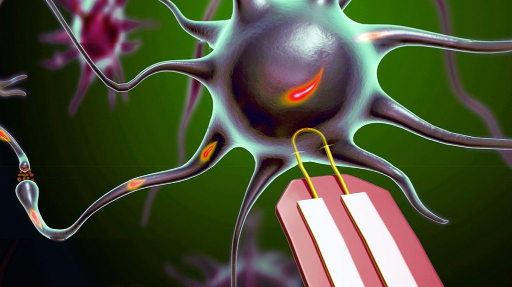
This image illustrating a self-powered nanogenerator for a cardiac pacemaker has been provided by KAIST,

This picture shows that a self-powered cardiac pacemaker is enabled by a flexible piezoelectric energy harvester. Credit: KAIST

This picture shows that a self-powered cardiac pacemaker is enabled by a flexible piezoelectric energy harvester.
Credit: KAIST

Here’s a link to and a citation for the paper,

Self-Powered Cardiac Pacemaker Enabled by Flexible Single Crystalline PMN-PT Piezoelectric Energy Harvester by Geon-Tae Hwang, Hyewon Park, Jeong-Ho Lee, SeKwon Oh, Kwi-Il Park, Myunghwan Byun, Hyelim Park, Gun Ahn, Chang Kyu Jeong, Kwangsoo No, HyukSang Kwon, Sang-Goo Lee, Boyoung Joung, and Keon Jae Lee. Advanced Materials DOI: 10.1002/adma.201400562
Article first published online: 17 APR 2014

© 2014 WILEY-VCH Verlag GmbH & Co. KGaA, Weinheim

This paper is behind a paywall.

There was a May 15, 2014 KAIST news release on EurekAlert announcing this same piece of research but from a technical perspective,

The energy efficiency of KAIST’s piezoelectric nanogenerator has increased by almost 40 times, one step closer toward the commercialization of flexible energy harvesters that can supply power infinitely to wearable, implantable electronic devices

NANOGENERATORS are innovative self-powered energy harvesters that convert kinetic energy created from vibrational and mechanical sources into electrical power, removing the need of external circuits or batteries for electronic devices. This innovation is vital in realizing sustainable energy generation in isolated, inaccessible, or indoor environments and even in the human body.

Nanogenerators, a flexible and lightweight energy harvester on a plastic substrate, can scavenge energy from the extremely tiny movements of natural resources and human body such as wind, water flow, heartbeats, and diaphragm and respiration activities to generate electrical signals. The generators are not only self-powered, flexible devices but also can provide permanent power sources to implantable biomedical devices, including cardiac pacemakers and deep brain stimulators.

However, poor energy efficiency and a complex fabrication process have posed challenges to the commercialization of nanogenerators. Keon Jae Lee, Associate Professor of Materials Science and Engineering at KAIST, and his colleagues have recently proposed a solution by developing a robust technique to transfer a high-quality piezoelectric thin film from bulk sapphire substrates to plastic substrates using laser lift-off (LLO).

Applying the inorganic-based laser lift-off (LLO) process, the research team produced a large-area PZT thin film nanogenerators on flexible substrates (2 cm x 2 cm).

“We were able to convert a high-output performance of ~250 V from the slight mechanical deformation of a single thin plastic substrate. Such output power is just enough to turn on 100 LED lights,” Keon Jae Lee explained.

The self-powered nanogenerators can also work with finger and foot motions. For example, under the irregular and slight bending motions of a human finger, the measured current signals had a high electric power of ~8.7 μA. In addition, the piezoelectric nanogenerator has world-record power conversion efficiency, almost 40 times higher than previously reported similar research results, solving the drawbacks related to the fabrication complexity and low energy efficiency.

Lee further commented,

“Building on this concept, it is highly expected that tiny mechanical motions, including human body movements of muscle contraction and relaxation, can be readily converted into electrical energy and, furthermore, acted as eternal power sources.”

The research team is currently studying a method to build three-dimensional stacking of flexible piezoelectric thin films to enhance output power, as well as conducting a clinical experiment with a flexible nanogenerator.

In addition to the 2012 posting I mentioned earlier, there was also this July 12, 2010 posting which described research on harvesting biomechanical movement ( heart beat, blood flow, muscle stretching, or even irregular vibration) at the Georgia (US) Institute of Technology where the lead researcher observed,

…  Wang [Professor Zhong Lin Wang at Georgia Tech] tells Nanowerk. “However, the applications of the nanogenerators under in vivo and in vitro environments are distinct. Some crucial problems need to be addressed before using these devices in the human body, such as biocompatibility and toxicity.”

Bravo to the KAIST researchers for getting this research to the in vivo testing stage.

Meanwhile at the University of Bristol and at the University of Bath, researchers have received funding for a new approach to cardiac pacemakers, designed them with the breath in mind. From a June 24, 2014 news item on Azonano,

Pacemaker research from the Universities of Bath and Bristol could revolutionise the lives of over 750,000 people who live with heart failure in the UK.

The British Heart Foundation (BHF) is awarding funding to researchers developing a new type of heart pacemaker that modulates its pulses to match breathing rates.

A June 23, 2014 University of Bristol press release, which originated the news item, provides some context,

During 2012-13 in England, more than 40,000 patients had a pacemaker fitted.

Currently, the pulses from pacemakers are set at a constant rate when fitted which doesn’t replicate the natural beating of the human heart.

The normal healthy variation in heart rate during breathing is lost in cardiovascular disease and is an indicator for sleep apnoea, cardiac arrhythmia, hypertension, heart failure and sudden cardiac death.

The device is then briefly described (from the press release),

The novel device being developed by scientists at the Universities of Bath and Bristol uses synthetic neural technology to restore this natural variation of heart rate with lung inflation, and is targeted towards patients with heart failure.

The device works by saving the heart energy, improving its pumping efficiency and enhancing blood flow to the heart muscle itself.  Pre-clinical trials suggest the device gives a 25 per cent increase in the pumping ability, which is expected to extend the life of patients with heart failure.

One aim of the project is to miniaturise the pacemaker device to the size of a postage stamp and to develop an implant that could be used in humans within five years.

Dr Alain Nogaret, Senior Lecturer in Physics at the University of Bath, explained“This is a multidisciplinary project with strong translational value.  By combining fundamental science and nanotechnology we will be able to deliver a unique treatment for heart failure which is not currently addressed by mainstream cardiac rhythm management devices.”

The research team has already patented the technology and is working with NHS consultants at the Bristol Heart Institute, the University of California at San Diego and the University of Auckland. [emphasis mine]

Professor Julian Paton, from the University of Bristol, added: “We’ve known for almost 80 years that the heart beat is modulated by breathing but we have never fully understood the benefits this brings. The generous new funding from the BHF will allow us to reinstate this natural occurring synchrony between heart rate and breathing and understand how it brings therapy to hearts that are failing.”

Professor Jeremy Pearson, Associate Medical Director at the BHF, said: “This study is a novel and exciting first step towards a new generation of smarter pacemakers. More and more people are living with heart failure so our funding in this area is crucial. The work from this innovative research team could have a real impact on heart failure patients’ lives in the future.”

Given some current events (‘Tesla opens up its patents’, Mike Masnick’s June 12, 2014 posting on Techdirt), I wonder what the situation will be vis à vis patents by the time this device gets to market.

* ‘one’ added to title on Aug. 13, 2014.