Category Archives: medicine

Georges Canguilhem’s influence on life sciences philosophy and ‘it’s all about Kant’

This July 5, 2023 Fundação de Amparo à Pesquisa do Estado de São Paulo (FAPESP) press release by José Tadeu Arantes (also on EurekAlert but published on July 3, 2023) slow walks us through a listing of French intellectuals and some history (which I enjoyed) before making a revelation,

The constitution of the World Health Organization (WHO) defines health as “a state of complete physical, mental and social well-being and not merely the absence of disease or infirmity”. The definition dates from the 1940s, but even then the thinking behind it was hardly novel. Similar concepts can be found in antiquity, in Eastern as well as Western societies, but in Europe, the cradle of Western culture, the view that mental well-being was part of health enjoyed little prestige in the eighteenth and nineteenth centuries owing to a reductionist understanding of disease as strictly somatic (relating only to the body). This outlook eventually began to be questioned. One of its leading critics in the twentieth century was French philosopher and physician Georges Canguilhem (1904-1995).

A disciple of Gaston Bachelard (1884-1962), a colleague of Jean-Paul Sartre (1905-1980), Paul Nizan (1905-1940) and Raymond Aron (1905-1983), and a major influence on Michel Foucault (1926-1984), Canguilhem was one of the foremost French intellectuals of the postwar years. Jacques Lacan (1901-1981), Gilles Deleuze (1925-1995) and Jacques Derrida (1930-2004) were among the thinkers who took inspiration from his ideas.

Canguilhem began studying medicine in the mid-thirties and earned his medical doctorate in 1943, by which time he had already taught philosophy in high schools for many years (having qualified in 1927). Another significant tack in his life course occurred during World War Two. He had long been both a pacifist and an antifascist. Following the French surrender in 1940, he refused to continue teaching under the Vichy regime and joined the Resistance, fighting with the rural guerrillas of the Maquis. In this historically and politically complex period, he apparently set out to train as a physician in order to have practical experience as well as book learning and to work on the history of the life sciences. He was awarded the Croix de Guerre and the Médaille da la Résistance for organizing a field hospital while under attack in the Auvergne.

In an article published in the journal History and Philosophy of the Life Sciences, Emiliano Sfara, who has a PhD in philosophy from the University of Montpellier and was a postdoctoral fellow at the University of São Paulo (USP) in Brazil from 2018 to 2022, argues that Canguilhem’s concepts of “technique”, “technical activity” and “practice” derived from Immanuel Kant’s Critique of Judgment (1790) and influenced Canguilhem’s decision to study medicine.

“Earlier historiographical research showed how Kant influenced Canguilhem, especially the concept of ‘knowledge’ developed in Kant’s Critique of Pure Reason as the unification of heterogeneous data by an organizing intellect, and the idea of the ‘organism’ as a totality of interdependent and interacting parts, inspired by the Critique of Judgment. I tried to show in the article the importance, and roots in Kant, of a third cluster of ideas relating to the concept of ‘technique’ in Canguilhem’s work, beginning in mid-thirties,” said Sfara, currently a researcher at the National Institute of Science and Technology for Interdisciplinary and Transdisciplinary Studies in Ecology and Evolution (INCT IN-TREE), hosted by the Federal University of Bahia (UFBA).

“Section 43 of Kant’s Critique of Judgment makes a distinction between technical capacity and science as a theoretical faculty. Technique is the subject’s concrete practice operating in a certain context, a vital movement of construction or manufacturing of objects and tools that enable a person to live in their environment. This is not reducible to science. Analogously, Canguilhem postulates that science is posterior to technique. Practice comes first; theory arises later. This movement is evident in art. True, the artist starts out with a project. But the development of the artwork isn’t confined to the project, which is reconstructed as the process unfolds. This practical element of the subject’s interaction with the environment, which has its roots in Kant’s theories, was very important to the evolution of Canguilhem’s thought. It even influenced his decision to study medicine, as well as the conception of medicine he developed.”

Sfara explained that while Canguilhem espoused the values of the Parti Radical in his youth, in the mid-thirties he moved left, without becoming a pro-Soviet Stalinist. Later on, according to some scholars who knew him and are still active (such as the Moroccan philosopher and mathematician Hourya Benis Sinaceur), Canguilhem gave primacy to the egalitarian principles symbolized by the French Republic’s motto Liberté, Egalité, Fraternité.

His main contributions were to medicine and philosophy of science. His most important work, The Normal and the Pathological (1966), is basically an expansion of his 1943 doctoral thesis. “In his original thesis, Canguilhem broke with part of eighteenth- and nineteenth-century French medical tradition and formulated ideas that are very much part of medicine today. [emphasis mine] Taking a purely analytical and quantitative approach, physicians like François Broussais (1772-1838) believed disease resulted from a surplus or lack of some organic substance, such as blood. Bloodletting was regularly used as a form of treatment. France imported 33 million leeches from southern Europe in the first half of the nineteenth century. Canguilhem saw the organism as a totality that interacted with its environment [emphasis mine] rather than a mere aggregation of parts whose functioning depended only on a ‘normal’ amount of the right organic substances,” Safra said.

In Canguilhem, the movement changes. Instead of transiting from the part to the whole, he moves from the whole to the part (as does Kant in the Critique of Judgment). He views the organism not as a machine but as an integral self-regulating totality. Life cannot be deduced from physical and chemical laws. One must start from the living being to understand life. Practice is the bridge that connects this totality to the environment. At the same time as it changes the environment, practice changes the organism and helps determine its physiological states.

“So Canguilhem implies that in order to find a state called normal, i.e. healthy, a given organism has to adapt its own operating rules to the outside world in the course of interacting concretely and practically with the environment. A human organism, for example, is in a ‘normal’ state when its pulse slows sharply after a period of long daily running. A case in point is the long-distance runner, who has to train every day,” Safra said.

“For Canguilhem, disease is due to inadaptation between the part, the organism and the environment, and often manifests itself as a feeling of malaise. Adaptive mechanisms in the organism can correct pathological dysfunctions.”

The article resulted from Sfara’s postdoctoral research supervised by Márcio Suzuki and supported by FAPESP.

The article “From technique to normativity: the influence of Kant on Georges Canguilhem’s philosophy of life” is at: link.springer.com/article/10.1007/s40656-023-00573-8.

This text was originally published by FAPESP Agency according to Creative Commons license CC-BY-NC-ND. Read the original here.https://agencia.fapesp.br/republicacao_frame?url=https://agencia.fapesp.br/study-shows-kants-influence-on-georges-canguilhem-who-anticipated-concepts-current-in-medicine-today/41794/&utm_source=republish&utm_medium=republish&utm_content=https://agencia.fapesp.br/study-shows-kants-influence-on-georges-canguilhem-who-anticipated-concepts-current-in-medicine-today/41794/

Even though you can find a link to the paper in the press release, here’s my version of a citation complete with link,

From technique to normativity: the influence of Kant on Georges Canguilhem’s philosophy of life by Emiliano Sfara .History and Philosophy of the Life Sciences volume 45, Article number: 16 (2023) DOI: https://doi.org/10.1007/s40656-023-00573-8 Published: 06 April 2023

This paper is open access.
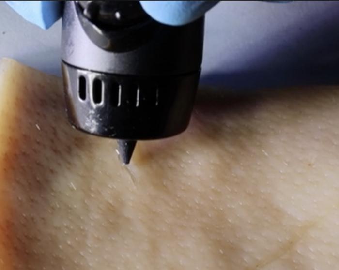
PAINT wound-healing ink into your cuts with a 3D-printing pen

This screams tattoo to me but it’s not,

Caption This 3-D printing pen is painting a gel that can help wounds of all shapes heal quickly and effectively. Credit: Adapted from ACS Applied Materials & Interfaces, 2023, DOI: 10.1021/acsami.3c03630

A June 1, 2023 American Chemical Society (ACS) news release (also on EurekAlert), announces a new approach to wound healing,

The body is pretty good at healing itself, though more severe wounds can require bandages or stitches. But researchers publishing in ACS Applied Materials & Interfaces have developed a wound-healing ink that can actively encourage the body to heal by exposing the cut to immune-system vesicles. The ink can be spread into a cut of any shape using a 3D-printing pen, and in mice, the technology nearly completely repaired wounds in just 12 days.

When the skin is cut or torn, the body’s natural “construction crew” kicks in to fix it back up — clearing out any bacterial invaders, regrowing broken blood vessels and eventually forming a scar. Many techniques used to heal wounds can’t do much beyond helping the body do its job better. Bandages or stitches are used to prevent further bleeding, while antibiotics work to prevent complications from infections. But by adding members of the construction crew to a wound-healing treatment or bandage, it could actually accelerate the natural healing process. Specifically, white blood cells or the extracellular vesicles (EVs) secreted from them play important roles in promoting blood vessel formation and reducing inflammation during healing. So, Dan Li, Xianguang Ding and Lianhui Wang wanted to incorporate these EVs into a hydrogel-based wound healing ink that could be painted into cuts of any shape.

The team developed a system called PAINT, or “portable bioactive ink for tissue healing,” using EVs secreted from macrophages combined with sodium alginate. These components were combined in a 3D-printing pen, where they mixed at the pen’s tip and formed a sturdy gel at the site of injury within three minutes. The EVs promoted blood vessel formation and reduced inflammatory markers in human epithelial cells, shifting them into the “proliferative,” or growth, phase of healing. PAINT was also tested on injured mice, where it promoted collagen fiber formation. Mice treated with PAINT had almost healed completely from a large wound after 12 days, compared to mice that didn’t receive the treatment, who were not nearly as far along in the healing process at this time point. The researchers say that this work could help heal a wide variety of cuts quickly and easily, without the need for complex procedures.

The authors acknowledge funding from the Leading-Edge Technology Programme of Jiangsu Natural Science Foundation, the Natural Science Foundation, the Natural Science Foundation of Jiangsu Province, the CAS [Chinese Academy of Sciences] Key Laboratory of Nano-Bio Interface, the Key Laboratory of Nanodevices and Applications, and the Postgraduate Research & Practice Innovation Program of Jiangsu Province.

The American Chemical Society (ACS) is a nonprofit organization chartered by the U.S. Congress. ACS’ mission is to advance the broader chemistry enterprise and its practitioners for the benefit of Earth and all its people. The Society is a global leader in promoting excellence in science education and providing access to chemistry-related information and research through its multiple research solutions, peer-reviewed journals, scientific conferences, eBooks and weekly news periodical Chemical & Engineering News. ACS journals are among the most cited, most trusted and most read within the scientific literature; however, ACS itself does not conduct chemical research. As a leader in scientific information solutions, its CAS division partners with global innovators to accelerate breakthroughs by curating, connecting and analyzing the world’s scientific knowledge. ACS’ main offices are in Washington, D.C., and Columbus, Ohio.

Here’s a link to and a citation for the paper,

Paintable Bioactive Extracellular Vesicle Ink for Wound Healing by Li Li, Zhiyu Wang, Kepeng Wang, Siyuan Fu, Dan Li, Mao Wang, Yi Cao, Houjuan Zhu, Ziyan Li, Lixing Weng, Zhiyang Li, Xianguang Ding, and Lianhui Wang. ACS Appl. Mater. Interfaces 2023, 15, 21, 25427–25436 DOI: https://doi.org/10.1021/acsami.3c03630 Publication Date:May 19, 2023 Copyright © 2023 American Chemical Society

This paper is behind a paywall.

A new lipid nanoparticle (LNP) delivery system for CRISPR-Cas9) gene editing

The first time lipid nanoparticles were mentioned here as a delivery system for CRISPR-Cas9 editing was in a January 26, 2018 posting featuring work at the Massachusetts Institute of Technology (MIT). This latest research on the topic comes from Japan according to a March 2, 2023 news item on phys.org,

Gene therapy is a potential mode of treatment for a wide variety of diseases caused by genetic mutations. While it has been an area of diverse and intense research, historically, only a very few patients have been treated using gene therapy—and fewer still cured. The advent of the genetic modification technique called CRISPR-Cas9 in 2012 has revolutionized gene therapy—as well as biology as a whole—and it has recently entered clinical trials for the treatment of some diseases in humans.

Haruno Onuma, Yusuke Sato and Hideyoshi Harashima at Hokkaido University have developed a new delivery system for CRISPR-Cas9, based on lipid nanoparticles (LNPs), that could greatly increases the efficiency of in vivo gene therapy. Their findings were published in the Journal of Controlled Release.

A March 2, 2023 Hokkaido University press release (also on EurekAlert), which originated the news item, provides details about the researchers’ new technique,

“There are broadly two ways of treating diseases with gene therapy,” Sato explained, “ex vivo, where cells are subjected to the desired modifications in the laboratory and then introduced into the patient, and in vivo, where the treatment is administered to the patient to change the cells in their body. Safe and effective in vivo treatment is the ultimate aspiration of gene therapy, as it would be a straightforward process for patients and healthcare providers. LNPs can function as a vehicle for the safe and effective delivery of such therapies.”

CRISPR-Cas9 consists of a large molecule composed of the Cas9 protein and guide RNA. The guide RNA binds to a specific, complementary DNA sequence, and the Cas9 protein cuts that sequence, allowing it to be modified. The guide RNA can be altered to target specific DNA sequences to be modified.

“In a previous study, we discovered that additional DNA molecules, called ssODNs, ensure that the CRISPR-Cas9 molecule is loaded into the LNPs (CRISPR-LNPs),” Harashima elucidated. “In this study,  we again used ssODNs, but they were carefully designed so that they would not inhibit the function of the guide RNA.” 

Using a guide RNA targeting the expression of a protein called transthyretin, they evaluated the effectiveness of the CRISPR-LNPs in mice models. CRISPR-LNPs with ssODNs that dissociated from the guide RNA at room temperature were most effective at reducing serum transthyretin: two consecutive doses, one day apart, reduced it by 80%.

“We have demonstrated the optimal ssODN sequence affinity that ensures the loading and the release of CRISPR-Cas9 at the target location; and that this system can be used to edit cells in vivo,” concluded Onuma. “We will continue to improve the design of ssODNs, as well as to develop optimal lipid formulations to increase the effectiveness of delivery.” 

The image and caption helped me with better understanding the technique described in the press release,

The RNP-ssODN is designed to ensure the CRISPR-Cas9 molecule is encapsulated by the LNP. Once inside the cells, the ssODN dissociates and CRISPR-Cas9 can carry out its effect. (Haruno Onuma, Yusuke Sato, Hideyoshi Harashima. Journal of Controlled Release. February 10, 2023).

Here’s a link to and a citation for the paper,

Lipid nanoparticle-based ribonucleoprotein delivery for in vivo genome editing by Haruno Onuma, Yusuke Sato, Hideyoshi Harashima. Journal of Controlled Release Volume 355, March 2023, Pages 406-416 DOI: https://doi.org/10.1016/j.jconrel.2023.02.008

This paper is behind a paywall.

Biodegradable electronics: a seaweed biosensor

By combining seaweed and graphene, scientists have been able to create sensors that can be worn like a ‘second skin’ and outperform other similar biosensors, according to a March 3, 2023 news item on ScienceDaily,

Scientists at the University of Sussex have successfully trialed new biodegradable health sensors that could change the way we experience personal healthcare and fitness monitoring technology.

The team at Sussex have developed the new health sensors — such as those worn by runners or patients to monitor heart rate and temperature — using natural elements like rock salt, water and seaweed, combined with graphene. Because they are solely made with ingredients found in nature, the sensors are fully biodegradable, making them more environmentally friendly than commonly used rubber and plastic-based alternatives. Their natural composition also places them within the emerging scientific field of edible electronics — electronic devices that are safe for a person to consume.

Better still, the researchers found that their sustainable seaweed-based sensors actually outperform existing synthetic based hydrogels and nanomaterials, used in wearable health monitors, in terms of sensitivity. Therefore, improving the accuracy, as the more sensitive a sensor, the more accurately it will record a person’s vital signs.

A March 2, 2023 University of Sussex press release (also on EurekAlert) by Poppy Luckett, which originated the news item, describes the inspiration for the research,

Dr Conor Boland, a materials physics lecturer in the School of Mathematical and Physical Sciences, said:  “I was first inspired to use seaweed in the lab after watching MasterChef during lockdown. Seaweed, when used to thicken deserts, gives them a soft and bouncy structure – favored by vegans and vegetarians as an alternative to gelatin. It got me thinking: “what if we could do that with sensing technology?”.

“For me, one of the most exciting aspects to this development is that we have a sensor that is both fully biodegradable and highly effective. The mass production of unsustainable rubber and plastic based health technology could, ironically, pose a risk to human health through microplastics leeching into water sources as they degrade.  

“As a new parent, I see it as my responsibility to ensure my research enables the realisation of a cleaner world for all our children.” 

Seaweed is first and foremost an insulator, but by adding a critical amount of graphene to a seaweed mixture the scientists were able to create an electrically conductive film. When soaked in a salt bath, the film rapidly absorbs water, resulting in a soft, spongy, electrically conductive hydrogel.  

The development has the potential to revolutionise health monitoring technology, as future applications of the clinical grade wearable sensors would look something like a second skin or a temporary tattoo: lightweight, easy to apply, and safe, as they are made with all natural ingredients. This would significantly improve the overall patient experience, without the need for more commonly used and potentially invasive hospital instruments, wires and leads.  

Dr Sue Baxter, Director of Innovation and Business Partnerships at the University of Sussex, is excited about the potential benefits of this technology:  “At the University of Sussex, we are committed to protecting the future of the planet through sustainability research, expertise and innovation. What’s so exciting about this development from Dr Conor Boland and his team is that it manages to be all at once truly sustainable, affordable, and highly effective – out-performing synthetic alternatives.  

“What’s also remarkable for this stage of research – and I think this speaks to the meticulous ground-work that Dr Boland and his team put in when they created their blueprint – is that it’s more than a proof of principle development. Our Sussex scientists have created a device that has real potential for industry development into a product from which you or I could benefit in the relatively near future.” 

This latest  research breakthrough follows the publication of a blueprint for nanomaterial development from the Sussex scientists in 2019, which presented a method for researchers to follow in order to optimise the development of nanomaterial sensors.  

Kevin Doty, a Masters student in the School of Mathematical and Physical Sciences, at the University of Sussex, said:  “I taught chemistry previously, but decided I wanted to learn more about nanoscience. My gamble paid off, and not only did I enjoy it more than I expected, but I also ended up with an opportunity to utilize the information I had learned to work on a novel idea that has evolved into a first author publication as an MSc student. Learning about nanoscience showed me just how varied and multidisciplinary the field is. Any science background can bring knowledge that can be applied to this field in a unique way. This has led to further studies in a PhD studentship, opening up an all new career path I could not have previously considered.” 

Here’s a link to and a citation for the paper,

Food-Inspired, High-Sensitivity Piezoresistive Graphene Hydrogels by Adel A. K. Aljarid, Kevin L. Doty, Cencen Wei, Jonathan P. Salvage, and Conor S. Boland. ACS Sustainable Chem. Eng. 2023, 11, 5, 1820–1827 DOI: https://doi.org/10.1021/acssuschemeng.2c06101 Publication Date:January 25, 2023 Copyright © 2023 The Authors. Published by American Chemical Society

This paper appears to be open access.

Artificially-grown mini-brains (organoids)—without animal components— offer opportunities for neuroscience

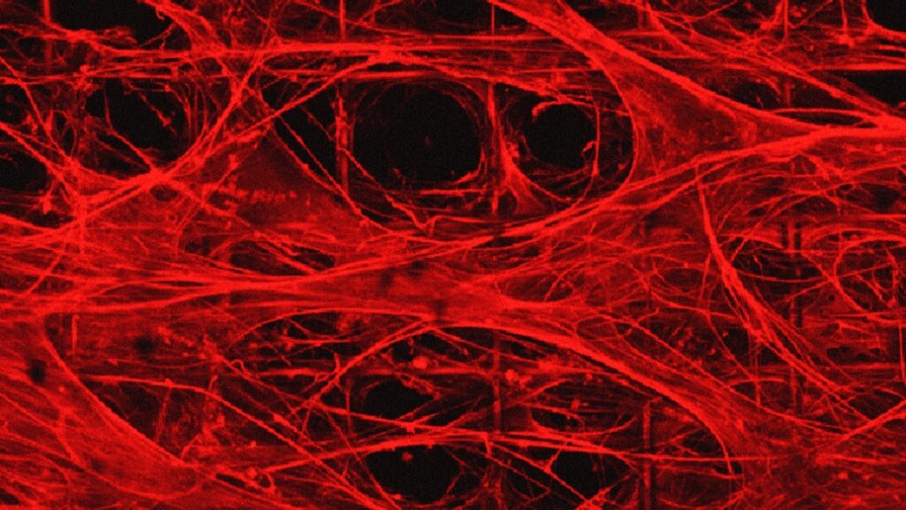
There’s a good (brief) description of how these fibres become organoids in the photo caption,

Engineered extracellular matrices composed of fibrillar fibronectin are suspended over a porous polymer framework and provide the niche for stem cells to attach, differentiate, and mature into organoids. Credit: Ayse Muñiz Courtesy: Michigan Medicine – University of Michigan

A July 13 ,2023 University of Michigan (Michigan Medicine) news release by Noah Fromson (also on EurekAlert) announces ‘kinder, gentler’ brain organoids. Coincidentally, these organoids more closely resemble human brains, Note: Links have been removed,

Researchers at University of Michigan developed a method to produce artificially grown miniature brains — called human brain organoids — free of animal cells that could greatly improve the way neurodegenerative conditions are studied and, eventually, treated.

Over the last decade of researching neurologic diseases, scientists have explored the use of human brain organoids as an alternative to mouse models. These self-assembled, 3D tissues derived from embryonic or pluripotent stem cells more closely model the complex brain structure compared to conventional two-dimensional cultures.

Until now, the engineered network of proteins and molecules that give structure to the cells in brain organoids, known as extracellular matrices, often used a substance derived from mouse sarcomas called Matrigel. That method suffers significant disadvantages, with a relatively undefined composition and batch-to-batch variability.

The latest U-M research, published in Annals of Clinical and Translational Neurology, offers a solution to overcome Matrigel’s weaknesses. Investigators created a novel culture method that uses an engineered extracellular matrix for human brain organoids — without the presence of animal components – and enhanced the neurogenesis of brain organoids compared to previous studies.

“This advancement in the development of human brain organoids free of animal components will allow for significant strides in the understanding of neurodevelopmental biology,” said senior author Joerg Lahann, Ph.D., director of the U-M Biointerfaces Institute and Wolfgang Pauli Collegiate Professor of Chemical Engineering at U-M.

“Scientists have long struggled to translate animal research into the clinical world, and this novel method will make it easier for translational research to make its way from the lab to the clinic.”

The foundational extracellular matrices of the research team’s brain organoids were comprised of human fibronectin, a protein that serves as a native structure for stem cells to adhere, differentiate and mature. They were supported by a highly porous polymer scaffold.

The organoids were cultured for months, while lab staff was unable to enter the building due to the COVID 19-pandemic.

Using proteomics, researchers found their brain organoids developed cerebral spinal fluid, a clear liquid that flows around healthy brain and spinal cords. This fluid more closely matched human adult CSF compared to a landmark study of human brain organoids developed in Matrigel.

“When our brains are naturally developing in utero, they are of course not growing on a bed of extracellular matrix produced by mouse cancer cells,” said first author Ayşe Muñiz, Ph.D., who was a graduate student in the U-M Macromolecular Science and Engineering Program at the time of the work.  

“By putting cells in an engineered niche that more closely resembles their natural environment, we predicted we would observe differences in organoid development that more faithfully mimics what we see in nature.”

The success of these xenogeneic-free human brain organoids opens the door for reprogramming with cells from patients with neurodegenerative diseases, says co-author Eva Feldman, M.D., Ph.D., director of the ALS Center of Excellence at U-M and James W. Albers Distinguished Professor of Neurology at U-M Medical School.

“There is a possibility to take the stem cells from a patient with a condition such as ALS or Alzheimer’s and, essentially, build an avatar mini brain of that patients to investigate possible treatments or model how their disease will progress,” Feldman said. “These models would create another avenue to predict disease and study treatment on a personalized level for conditions that often vary greatly from person to person.”

Here’s a link to and a citation for the paper,

Engineered extracellular matrices facilitate brain organoids from human pluripotent stem cells by Ayşe J. Muñiz, Tuğba Topal, Michael D. Brooks, Angela Sze, Do Hoon Kim, Jacob Jordahl, Joe Nguyen, Paul H. Krebsbach, Masha G. Savelieff, Eva L. Feldman, Joerg Lahann. Annals of Clinical and Translational Neurology DOI: https://doi.org/10.1002/acn3.51820 First published: 07 June 2023

This paper is open access.

Growing electrodes in your brain?

This isn’t for everybody. From a February 23, 2023 news item on Nanowerk, Note: A link has been removed,

The boundaries between biology and technology are becoming blurred. Researchers at Linköping, Lund, and Gothenburg universities in Sweden have successfully grown electrodes in living tissue using the body’s molecules as triggers. The result, published in the journal Science (“Metabolite-induced in vivo fabrication of substrate-free organic bioelectronics”), paves the way for the formation of fully integrated electronic circuits in living organisms.

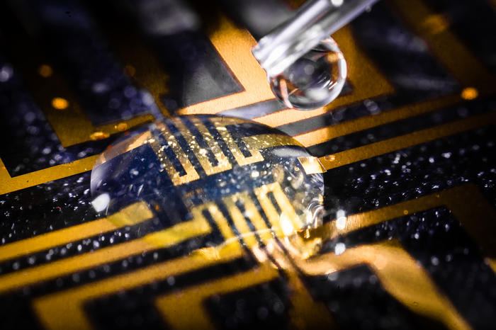
Caption: The injectable gel being tested on a microfabricated circuit. Credit: Thor Balkhed

I have two news releases for this research. First, the February 23, 2023 American Association for the Advancement of Science (AAAS) news release on EurekAlert,

Researchers have developed a way to make bioelectronics directly inside living tissues, an approach they tested by making electrodes in the brain, heart, and fin tissue of living zebrafish, as well as in isolated mammalian muscle tissues. According to the authors, the new method paves the way for in vivo fabrication of fully integrated electronic circuits within the nervous system and other living tissue. “Safety and stability analyses over long periods will be essential to determining whether such technology is useful for chronic implantations,” writes Sahika Inal in a related Perspective. “However, the strategy … suggests that any living tissue can turn into electronic matter and brings the field closer to generating seamless biotic-abiotic interfaces with a potentially long lifetime and minimum harm to tissues.” Implantable electronic devices that can interface with soft biological neural tissues offer a valuable approach to studying the complex electrical signaling of the nervous system and enable the therapeutic modulation of neural circuitry to prevent or treat various diseases and disorders. However, conventional bioelectronic implants often require the use of rigid electronic substrates that are incompatible with delicate living tissues and can provoke injury and inflammation that can affect a device’s electrical properties and long-term performance. Overcoming the incompatibility between static, solid-state electronic materials and dynamic, soft biological tissues has proven challenging. Here, Xenofon Strakosas and colleagues present a method to fabricate polymer-based, substrate-free electronic conducting materials directly inside a tissue. Strakosas et al. developed a complex molecular precursor cocktail that, when injected into a tissue, uses endogenous metabolites (glucose and lactate) to induce polymerization of organic precursors to form conducting polymer gels. To demonstrate the approach, the authors “grew” gel electrodes in the brain, heart, and fin tissue of living zebrafish, with no signs of tissue damage, and in isolated mammalian muscle tissues, including beef, pork and chicken. In medicinal leeches, they showed how the conducting gel could interface nervous tissue with electrodes on a tiny flexible probe.

The second is the February 23, 2023 Linköping University press release on EurekAlert, which originated the news item, and it provides further insight,

“For several decades, we have tried to create electronics that mimic biology. Now we let biology create the electronics for us,” says Professor Magnus Berggren at the Laboratory for Organic Electronics, LOE, at Linköping University.

Linking electronics to biological tissue is important to understand complex biological functions, combat diseases in the brain, and develop future interfaces between man and machine. However, conventional bioelectronics, developed in parallel with the semiconductor industry, have a fixed and static design that is difficult, if not impossible, to combine with living biological signal systems.

To bridge this gap between biology and technology, researchers have developed a method for creating soft, substrate-free, electronically conductive materials in living tissue. By injecting a gel containing enzymes as the “assembly molecules”, the researchers were able to grow electrodes in the tissue of zebrafish and medicinal leeches.

“Contact with the body’s substances changes the structure of the gel and makes it electrically conductive, which it isn’t before injection. Depending on the tissue, we can also adjust the composition of the gel to get the electrical process going,” says Xenofon Strakosas, researcher at LOE and Lund University and one of the study’s main authors.

The body’s endogenous molecules are enough to trigger the formation of electrodes. There is no need for genetic modification or external signals, such as light or electrical energy, which has been necessary in previous experiments. The Swedish researchers are the first in the world to succeed in this.

Their study paves the way for a new paradigm in bioelectronics. Where it previously took implanted physical objects to start electronic processes in the body, injection of a viscous gel will be enough in the future.

In their study, the researchers further show that the method can target the electronically conducting material to specific biological substructures and thereby create suitable interfaces for nerve stimulation. In the long term, the fabrication of fully integrated electronic circuits in living organisms may be possible.

In experiments conducted at Lund University, the team successfully achieved electrode formation in the brain, heart, and tail fins of zebrafish and around the nervous tissue of medicinal leeches. The animals were not harmed by the injected gel and were otherwise not affected by the electrode formation. One of the many challenges in these trials was to take the animals’ immune system into account.

“By making smart changes to the chemistry, we were able to develop electrodes that were accepted by the brain tissue and immune system. The zebrafish is an excellent model for the study of organic electrodes in brains,” says Professor Roger Olsson at the Medical Faculty at Lund University, who also has a chemistry laboratory at the University of Gothenburg.

It was Professor Roger Olsson who took the initiative for the study, after he read about the electronic rose developed by researchers at Linköping University in 2015. One research problem, and an important difference between plants and animals, was the difference in cell structure. Whereas plants have rigid cell walls which allow for the formation of electrodes, animal cells are more like a soft mass. Creating a gel with enough structure and the right combination of substances to form electrodes in such surroundings was a challenge that took many years to solve.

“Our results open up for completely new ways of thinking about biology and electronics. We still have a range of problems to solve, but this study is a good starting point for future research,” says Hanne Biesmans, PhD student at LOE and one of the main authors.

Here’s a link to and a citation for the paper,

Metabolite-induced in vivo fabrication of substrate-free organic bioelectronics by Xenofon Strakosas, Hanne Biesmans, Tobias Abrahamsson, Karin Hellman, Malin Silverå Ejneby, Mary J. Donahue, Peter Ekström, Fredrik Ek, Marios Savvakis, Martin Hjort, David Bliman, Mathieu Linares, Caroline Lindholm, Eleni Stavrinidou, Jennifer Y. Gerasimov, Daniel T. Simon, Roger Olsson, and Magnus Berggren. Science 23 Feb 2023 Vol 379, Issue 6634 pp. 795-802 DOI: 10.1126/science.adc9998

This paper is behind a paywall.

Photonic cellulose nanocrystals (CNC) for flexible sweat sensor

It turns out there’s also a hydrogel aspect to this story about a flexible sweat sensor. As for cellulose nanocrystals (CNC), Canada played a leading role in the development of this nanomaterial and I have a bit more about the Canadian CNC scene later in this posting following the link and citation for the research paper. On to the research,

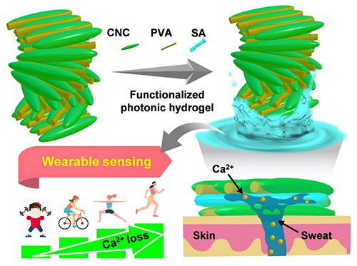
Highly elastic hydrogels constructed by heat-induced hydrogen bond remodeling can switch between wet and dry states (Image by ZHANG Fusheng and LI Qiongya)

A May 8, 2023 news item on phys.org features this work from the Dalian Institute of Chemical Physics of the Chinese Academy Sciences,

Cellulose nanocrystal (CNC), an emerging bio-based material, has been widely applied in fields such as electronics, bioplastics and energy. However, the functional failure of such materials in wet or liquid environments inevitably impairs their development in biomedicine, membrane separation, environmental monitoring, and wearable devices.

Now, a research group led by Prof. Qing Guangyan from the Dalian Institute of Chemical Physics (DICP) of the Chinese Academy of Sciences [CAS] reported a sustainable, insoluble, and chiral photonic cellulose nanocrystal patch for calcium ion (Ca2+) sensing in sweat.

A May 4, 2023 Dalian Institute of Chemical Physics of the Chinese Academy Sciences press release (also on EurekAlert but published May 8, 2023), which originated the news item, provides more detail about the work,

The researchers developed a simple and efficient method to fabricate insoluble CNC-based hydrogels. They found that by utilizing intermolecular hydrogen bond reconstruction, thermal dehydration enabled the optimized CNC composite photonic film to form a stable hydrogel network in an aqueous solution. Moreover, they indicated that the hydrogel could be reversibly switched between dry and wet states, which was convenient for specific functionalization.

The introduction of functionalized molecules by adsorption swelling in a liquid environment resulted in a hydrogel with freeze resistance (–20°C), strong adhesion, good biocompatibility, and high sensitivity to Ca2+.

“This work is expected to facilitate the application of sustainable cellulose sensors to monitor other metabolites (i.e., glucose, urea, and vitamins, etc.),” said Prof. QING. “It also lays foundation for digitally controlled hydrogel systems operating in environment monitoring, membrane separation, and wearable devices.”

Here’s a link to and a citation for the paper,

Sustainable, Insoluble, and Photonic Cellulose Nanocrystal Patches for Calcium Ion Sensing in Sweat by Qiongya Li, Chenchen He, Cunli Wang, Yuxiao Huang, Jiaqi Yu, Chunbo Wang, Wei Li, Xin Zhang, Fusheng Zhang, Guangyan Qing. small DOI: https://doi.org/10.1002/smll.202207932 First published online: 13 April 2023

This paper is behind a paywall.

FPInnovations is a Canadian research and development (R&D) not-for profit organization that was instrumental in the development of CNC. (If memory serves, they are a spinoff from the University of British Columbia.) There are two Canadian CNC production facilities (that I know of): CelluForce in Québec and Blue Goose Biorefineries in Saskatchewan. I get more information about research into applications for CNC from other parts of the world while the Canadian scene remains mostly silent.

Using touch (bionic fingers) instead of x-rays

This is not the most exciting video but it is weirdly fascinating (thank you to ScientifiCult),

A February 15, 2023 news item on Nanowerk provides a textual description for what you’re seeing in the video, Note: A link has been removed,

What if, instead of using X-rays or ultrasound, we could use touch to image the insides of human bodies and electronic devices? In a study publishing in the journal Cell Reports Physical Science (“A smart bionic finger for subsurface tactile-tomography”), researchers present a bionic finger that can create 3D maps of the internal shapes and textures of complex objects by touching their exterior surface.

“We were inspired by human fingers, which have the most sensitive tactile perception that we know of,” says senior author Jianyi Luo, a professor at Wuyi University. “For example, when we touch our own bodies with our fingers, we can sense not only the texture of our skin, but also the outline of the bone beneath it.”

“Our bionic finger goes beyond previous artificial sensors that were only capable of recognizing and discriminating between external shapes, surface textures, and hardness,” says co-author Zhiming Chen, a lecturer at Wuyi University.

The bionic finger “scans” an object by moving across it and applying pressure—think of a constant stream of pokes or prods. With each poke, the carbon fibers compress, and the degree to which they compress provides information about the relative stiffness or softness of the object. Depending on the object’s material, it might also compress when poked by the bionic finger: rigid objects hold their shape, while soft objects will deform when enough pressure is applied. This information, along with the location at which it was recorded, is relayed to a personal computer and displayed onscreen as a 3D map.

A February 13, 2023 Cell Press news release on EurekAlert, which originated the news item, provides more details about the research and some hints at what the researchers may do next,

The researchers tested the bionic finger’s ability to map out the internal and external features of complex objects made of multiple types of material, such as a rigid “letter A” buried under a layer of soft silicon, as well as more abstractly shaped objects. When they used it to scan a small compound object made of three different materials—a rigid internal material, a soft internal material, and a soft outer coating—the bionic finger was able to discriminate between not only the soft outer coating and the internal hard ridges, but it could also tell the difference between the soft outer coating and the soft material that filled the internal grooves.

Next, the researchers tested the finger’s ability to sense and image simulated human tissue. They created this tissue— consisting of a skeletal component, made of three layers of hard polymer, and a soft silicone “muscle” layer—using 3D printing. The bionic finger was able to reproduce a 3D profile of the tissue’s structure and locate a simulated blood vessel beneath the muscle layer.

The team also explored the bionic finger’s ability to diagnose issues in electronic devices without opening them up. By scanning the surface of a defective electronic device with the bionic finger, the researchers were able to create a map of its internal electrical components and pinpoint the location at which the circuit was disconnected, as well as a mis-drilled hole, without breaking through the encapsulating layer.

“This tactile technology opens up a non-optical way for the nondestructive testing of the human body and flexible electronics,” says Luo. “Next, we want to develop the bionic finger’s capacity for omnidirectional detection with different surface materials.”

Here’s a link to and a citation for the paper,

A smart bionic finger for subsurface tactile tomography by Yizhou Li, Zhiming Chen, Youbin Chen, Hao Yang, Junyong Lu, Zhennan Li, Yongyao Chen, Dongyi Ding, Cuiying Zeng, Bingpu Zhou, Hongpeng Liang, Xingpeng Huang, Jiajia Hu, Jingcheng Huang, Jinxiu Wen, Jianyi Luo. Volume 4, Issue 2, 15 February 2023, 101257 DOI: https://doi.org/10.1016/j.xcrp.2023.101257 Published online: February 15, 2023 Version of Record 15 February 2023.

This paper is open access.

Sound waves for wearable patches that deliver drugs painlessly

While watching this video I started wondering if they were testing their research on students but that’s not the case; these wearable patches were tested on porcine (pig) skin, which is quite similar to human skin, Note: They tested a B vitamin called niacinamide so, it’s highly unlikely the pigs suffered from it,

An April 20, 2023 news item on ScienceDaily announces the research into using ultrasonic waves for drug delivery,

The skin is an appealing route for drug delivery because it allows drugs to go directly to the site where they’re needed, which could be useful for wound healing, pain relief, or other medical and cosmetic applications. However, delivering drugs through the skin is difficult because the tough outer layer of the skin prevents most small molecules from passing through it.

In hopes of making it easier to deliver drugs through the skin, MIT [Massachusetts Institute of Technology] researchers have developed a wearable patch that applies painless ultrasonic waves to the skin, creating tiny channels that drugs can pass through. This approach could lend itself to delivery of treatments for a variety of skin conditions, and could also be adapted to deliver hormones, muscle relaxants, and other drugs, the researchers say.

An April 20, 2023 Massachusetts Institute of Technology (MIT) news release (also on EurekAlert), which originated the news item, provides technical details about the research, Note: A link has been removed,

“The ease-of-use and high-repeatability offered by this system provides a game-changing alternative to patients and consumers suffering from skin conditions and premature skin aging,” says Canan Dagdeviren, an associate professor in MIT’s Media Lab and the senior author of the study. “Delivering drugs this way could offer less systemic toxicity and is more local, comfortable, and controllable.”

MIT research assistants Chia-Chen Yu and Aastha Shah are the lead authors of the paper, which appears in Advanced Materials, as part of the journal’s “Rising Stars” series, which showcases the outstanding work of researchers in the early stages of their independent careers. Other MIT authors include Research Assistant Colin Marcus and postdoc Md Osman Goni Nayeem. Nikta Amiri, Amit Kumar Bhayadia, and Amin Karami of the University of Buffalo are also authors of the paper.

A boost from sound waves

The researchers began this project as an exploration of alternative ways to deliver drugs. Most drugs are delivered orally or intravenously, but the skin is a route that could offer much more targeted drug delivery for certain applications.

“The main benefit with skin is that you bypass the whole gastrointestinal tract. With oral delivery, you have to deliver a much larger dose in order to account for the loss that you would have in the gastric system,” Shah says. “This is a much more targeted, focused modality of drug delivery.”

Ultrasound exposure has been shown to enhance the skin’s permeability to small-molecule drugs, but most of the existing techniques for performing this kind of drug delivery require bulky equipment. The MIT team wanted to come up with a way to perform this kind of transdermal drug delivery with a lightweight, wearable patch, which could make it easier to use for a variety of applications.

The device that they designed consists of a patch embedded with several disc-shaped piezoelectric transducers, which can convert electric currents into mechanical energy. Each disc is embedded in a polymeric cavity that contains the drug molecules dissolved in a liquid solution. When an electric current is applied to the piezoelectric elements, they generate pressure waves in the fluid, creating bubbles that burst against the skin. These bursting bubbles produce microjets of fluid that can penetrate through the skin’s tough outer layer, the stratum corneum.

“This works open the door to using vibrations to enhance drug delivery. There are several parameters that result in generation of different kinds of waveform patterns. Both mechanical and biological aspects of drug delivery can be improved by this new toolset,” Karami says.

The patch is made of PDMS, a silicone-based polymer that can adhere to the skin without tape. In this study, the researchers tested the device by delivering a B vitamin called niacinamide, an ingredient in many sunscreens and moisturizers.

In tests using pig skin, the researchers showed that when they delivered niacinamide using the ultrasound patch, the amount of drug that penetrated the skin was 26 times greater than the amount that could pass through the skin without ultrasonic assistance.

The researchers also compared the results from their new device to microneedling, a technique sometimes used for transdermal drug delivery, which involves puncturing the skin with miniature needles. The researchers found that their patch was able to deliver the same amount of niacinamide in 30 minutes that could be delivered with microneedles over a six-hour period.

Local delivery

With the current version of the device, drugs can penetrate a few millimeters into the skin, making this approach potentially useful for drugs that act locally within the skin. These could include niacinamide or vitamin C, which is used to treat age spots or other dark spots on the skin, or topical drugs used to heal burns.

With further modifications to increase the penetration depth, this technique could also be used for drugs that need to reach the bloodstream, such as caffeine, fentanyl, or lidocaine. Dagdeviren also envisions that this kind of patch could be useful for delivering hormones such as progesterone. In addition, the researchers are now exploring the possibility of implanting similar devices inside the body to deliver drugs to treat cancer or other diseases.

The researchers are also working on further optimizing the wearable patch, in hopes of testing it soon on human volunteers. They also plan to repeat the lab experiments they did in this study, with larger drug molecules.

“After we characterize the drug penetration profiles for much larger drugs, we would then see which candidates, like hormones or insulin, can be delivered using this technology, to provide a painless alternative for those who are currently bound to self-administer injections on a daily basis,” Shah says.

Here’s a link to and a citation for the paper,

A Conformable Ultrasound Patch for Cavitation-Enhanced Transdermal Cosmeceutical Delivery by Chia-Chen Yu, Aastha Shah, Nikta Amiri, Colin Marcus, Md Osman Goni Nayeem, Amit Kumar Bhayadia, Amin Karami, Canan Dagdeviren. Advanced Materials Volume35, Issue 23 June 8, 2023 2300066 DOI: https://doi.org/10.1002/adma.202300066 First published online: 19 March 2023

This paper is open access.