Tag Archives: Gerd Binnig

More memory, less space and a walk down the cryptocurrency road

Libraries, archives, records management, oral history, etc. there are many institutions and names for how we manage collective and personal memory. You might call it a peculiarly human obsession stretching back into antiquity. For example, there’s the Library of Alexandria (Wikipedia entry) founded in the third, or possibly 2nd, century BCE (before the common era) and reputed to store all the knowledge in the world. It was destroyed although accounts differ as to when and how but its loss remains a potent reminder of memory’s fragility.

These days, the technology community is terribly concerned with storing ever more bits of data on materials that are reaching their limits for storage.I have news of a possible solution,  an interview of sorts with the researchers working on this new technology, and some very recent research into policies for cryptocurrency mining and development. That bit about cryptocurrency makes more sense when you read what the response to one of the interview questions.

Memory

It seems University of Alberta researchers may have found a way to increase memory exponentially, from a July 23, 2018 news item on ScienceDaily,

The most dense solid-state memory ever created could soon exceed the capabilities of current computer storage devices by 1,000 times, thanks to a new technique scientists at the University of Alberta have perfected.

“Essentially, you can take all 45 million songs on iTunes and store them on the surface of one quarter,” said Roshan Achal, PhD student in Department of Physics and lead author on the new research. “Five years ago, this wasn’t even something we thought possible.”

A July 23, 2018 University of Alberta news release (also on EurekAlert) by Jennifer-Anne Pascoe, which originated the news item, provides more information,

Previous discoveries were stable only at cryogenic conditions, meaning this new finding puts society light years closer to meeting the need for more storage for the current and continued deluge of data. One of the most exciting features of this memory is that it’s road-ready for real-world temperatures, as it can withstand normal use and transportation beyond the lab.

“What is often overlooked in the nanofabrication business is actual transportation to an end user, that simply was not possible until now given temperature restrictions,” continued Achal. “Our memory is stable well above room temperature and precise down to the atom.”

Achal explained that immediate applications will be data archival. Next steps will be increasing readout and writing speeds, meaning even more flexible applications.

More memory, less space

Achal works with University of Alberta physics professor Robert Wolkow, a pioneer in the field of atomic-scale physics. Wolkow perfected the art of the science behind nanotip technology, which, thanks to Wolkow and his team’s continued work, has now reached a tipping point, meaning scaling up atomic-scale manufacturing for commercialization.

“With this last piece of the puzzle now in-hand, atom-scale fabrication will become a commercial reality in the very near future,” said Wolkow. Wolkow’s Spin-off [sic] company, Quantum Silicon Inc., is hard at work on commercializing atom-scale fabrication for use in all areas of the technology sector.

To demonstrate the new discovery, Achal, Wolkow, and their fellow scientists not only fabricated the world’s smallest maple leaf, they also encoded the entire alphabet at a density of 138 terabytes, roughly equivalent to writing 350,000 letters across a grain of rice. For a playful twist, Achal also encoded music as an atom-sized song, the first 24 notes of which will make any video-game player of the 80s and 90s nostalgic for yesteryear but excited for the future of technology and society.

As noted in the news release, there is an atom-sized song, which is available in this video,

As for the nano-sized maple leaf, I highlighted that bit of whimsy in a June 30, 2017 posting.

Here’s a link to and a citation for the paper,

Lithography for robust and editable atomic-scale silicon devices and memories by Roshan Achal, Mohammad Rashidi, Jeremiah Croshaw, David Churchill, Marco Taucer, Taleana Huff, Martin Cloutier, Jason Pitters, & Robert A. Wolkow. Nature Communicationsvolume 9, Article number: 2778 (2018) DOI: https://doi.org/10.1038/s41467-018-05171-y Published 23 July 2018

This paper is open access.

For interested parties, you can find Quantum Silicon (QSI) here. My Edmonton geography is all but nonexistent, still, it seems to me the company address on Saskatchewan Drive is a University of Alberta address. It’s also the address for the National Research Council of Canada. Perhaps this is a university/government spin-off company?

The ‘interview’

I sent some questions to the researchers at the University of Alberta who very kindly provided me with the following answers. Roshan Achal passed on one of the questions to his colleague Taleana Huff for her response. Both Achal and Huff are associated with QSI.

Unfortunately I could not find any pictures of all three researchers (Achal, Huff, and Wolkow) together.

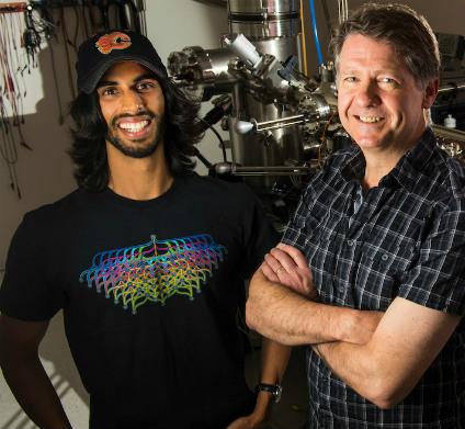
Roshan Achal (left) used nanotechnology perfected by his PhD supervisor, Robert Wolkow (right) to create atomic-scale computer memory that could exceed the capacity of today’s solid-state storage drives by 1,000 times. (Photo: Faculty of Science)

(1) SHRINKING THE MANUFACTURING PROCESS TO THE ATOMIC SCALE HAS
ATTRACTED A LOT OF ATTENTION OVER THE YEARS STARTING WITH SCIENCE
FICTION OR RICHARD FEYNMAN OR K. ERIC DREXLER, ETC. IN ANY EVENT, THE
ORIGINS ARE CONTESTED SO I WON’T PUT YOU ON THE SPOT BY ASKING WHO
STARTED IT ALL INSTEAD ASKING HOW DID YOU GET STARTED?

I got started in this field about 6 years ago, when I undertook a MSc
with Dr. Wolkow here at the University of Alberta. Before that point, I
had only ever heard of a scanning tunneling microscope from what was
taught in my classes. I was aware of the famous IBM logo made up from
just a handful of atoms using this machine, but I didn’t know what
else could be done. Here, Dr. Wolkow introduced me to his line of
research, and I saw the immense potential for growth in this area and
decided to pursue it further. I had the chance to interact with and
learn from nanofabrication experts and gain the skills necessary to
begin playing around with my own techniques and ideas during my PhD.

(2) AS I UNDERSTAND IT, THESE ARE THE PIECES YOU’VE BEEN
WORKING ON: (1) THE TUNGSTEN MICROSCOPE TIP, WHICH MAKE[s] (2) THE SMALLEST
QUANTUM DOTS (SINGLE ATOMS OF SILICON), (3) THE AUTOMATION OF THE
QUANTUM DOT PRODUCTION PROCESS, AND (4) THE “MOST DENSE SOLID-STATE
MEMORY EVER CREATED.” WHAT’S MISSING FROM THE LIST AND IS THAT WHAT
YOU’RE WORKING ON NOW?

One of the things missing from the list, that we are currently working
on, is the ability to easily communicate (electrically) from the
macroscale (our world) to the nanoscale, without the use of a scanning
tunneling microscope. With this, we would be able to then construct
devices using the other pieces we’ve developed up to this point, and
then integrate them with more conventional electronics. This would bring
us yet another step closer to the realization of atomic-scale
electronics.

(3) PERHAPS YOU COULD CLARIFY SOMETHING FOR ME. USUALLY WHEN SOLID STATE
MEMORY IS MENTIONED, THERE’S GREAT CONCERN ABOUT MOORE’S LAW. IS
THIS WORK GOING TO CREATE A NEW LAW? AND, WHAT IF ANYTHING DOES
;YOUR MEMORY DEVICE HAVE TO DO WITH QUANTUM COMPUTING?

That is an interesting question. With the density we’ve achieved,
there are not too many surfaces where atomic sites are more closely
spaced to allow for another factor of two improvement. In that sense, it
would be difficult to improve memory densities further using these
techniques alone. In order to continue Moore’s law, new techniques, or
storage methods would have to be developed to move beyond atomic-scale
storage.

The memory design itself does not have anything to do with quantum
computing, however, the lithographic techniques developed through our
work, may enable the development of certain quantum-dot-based quantum
computing schemes.

(4) THIS MAY BE A LITTLE OUT OF LEFT FIELD (OR FURTHER OUT THAN THE
OTHERS), COULD;YOUR MEMORY DEVICE HAVE AN IMPACT ON THE
DEVELOPMENT OF CRYPTOCURRENCY AND BLOCKCHAIN? IF SO, WHAT MIGHT THAT
IMPACT BE?

I am not very familiar with these topics, however, co-author Taleana
Huff has provided some thoughts:

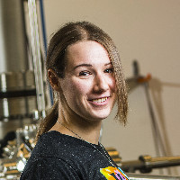
Taleana Huff (downloaded from https://ca.linkedin.com/in/taleana-huff]

“The memory, as we’ve designed it, might not have too much of an
impact in and of itself. Cryptocurrencies fall into two categories.
Proof of Work and Proof of Stake. Proof of Work relies on raw
computational power to solve a difficult math problem. If you solve it,
you get rewarded with a small amount of that coin. The problem is that
it can take a lot of power and energy for your computer to crunch
through that problem. Faster access to memory alone could perhaps
streamline small parts of this slightly, but it would be very slight.
Proof of Stake is already quite power efficient and wouldn’t really
have a drastic advantage from better faster computers.

Now, atomic-scale circuitry built using these new lithographic
techniques that we’ve developed, which could perform computations at
significantly lower energy costs, would be huge for Proof of Work coins.
One of the things holding bitcoin back, for example, is that mining it
is now consuming power on the order of the annual energy consumption
required by small countries. A more efficient way to mine while still
taking the same amount of time to solve the problem would make bitcoin
much more attractive as a currency.”

Thank you to Roshan Achal and Taleana Huff for helping me to further explore the implications of their work with Dr. Wolkow.

Comments

As usual, after receiving the replies I have more questions but these people have other things to do so I’ll content myself with noting that there is something extraordinary in the fact that we can imagine a near future where atomic scale manufacturing is possible and where as Achal says, ” … storage methods would have to be developed to move beyond atomic-scale [emphasis mine] storage”. In decades past it was the stuff of science fiction or of theorists who didn’t have the tools to turn the idea into a reality. With Wolkow’s, Achal’s, Hauff’s, and their colleagues’ work, atomic scale manufacturing is attainable in the foreseeable future.

Hopefully we’ll be wiser than we have been in the past in how we deploy these new manufacturing techniques. Of course, before we need the wisdom, scientists, as  Achal notes,  need to find a new way to communicate between the macroscale and the nanoscale.

As for Huff’s comments about cryptocurrencies and cyptocurrency and blockchain technology, I stumbled across this very recent research, from a July 31, 2018 Elsevier press release (also on EurekAlert),

A study [behind a paywall] published in Energy Research & Social Science warns that failure to lower the energy use by Bitcoin and similar Blockchain designs may prevent nations from reaching their climate change mitigation obligations under the Paris Agreement.

The study, authored by Jon Truby, PhD, Assistant Professor, Director of the Centre for Law & Development, College of Law, Qatar University, Doha, Qatar, evaluates the financial and legal options available to lawmakers to moderate blockchain-related energy consumption and foster a sustainable and innovative technology sector. Based on this rigorous review and analysis of the technologies, ownership models, and jurisdictional case law and practices, the article recommends an approach that imposes new taxes, charges, or restrictions to reduce demand by users, miners, and miner manufacturers who employ polluting technologies, and offers incentives that encourage developers to create less energy-intensive/carbon-neutral Blockchain.

“Digital currency mining is the first major industry developed from Blockchain, because its transactions alone consume more electricity than entire nations,” said Dr. Truby. “It needs to be directed towards sustainability if it is to realize its potential advantages.

“Many developers have taken no account of the environmental impact of their designs, so we must encourage them to adopt consensus protocols that do not result in high emissions. Taking no action means we are subsidizing high energy-consuming technology and causing future Blockchain developers to follow the same harmful path. We need to de-socialize the environmental costs involved while continuing to encourage progress of this important technology to unlock its potential economic, environmental, and social benefits,” explained Dr. Truby.

As a digital ledger that is accessible to, and trusted by all participants, Blockchain technology decentralizes and transforms the exchange of assets through peer-to-peer verification and payments. Blockchain technology has been advocated as being capable of delivering environmental and social benefits under the UN’s Sustainable Development Goals. However, Bitcoin’s system has been built in a way that is reminiscent of physical mining of natural resources – costs and efforts rise as the system reaches the ultimate resource limit and the mining of new resources requires increasing hardware resources, which consume huge amounts of electricity.

Putting this into perspective, Dr. Truby said, “the processes involved in a single Bitcoin transaction could provide electricity to a British home for a month – with the environmental costs socialized for private benefit.

“Bitcoin is here to stay, and so, future models must be designed without reliance on energy consumption so disproportionate on their economic or social benefits.”

The study evaluates various Blockchain technologies by their carbon footprints and recommends how to tax or restrict Blockchain types at different phases of production and use to discourage polluting versions and encourage cleaner alternatives. It also analyzes the legal measures that can be introduced to encourage technology innovators to develop low-emissions Blockchain designs. The specific recommendations include imposing levies to prevent path-dependent inertia from constraining innovation:

  • Registration fees collected by brokers from digital coin buyers.
  • “Bitcoin Sin Tax” surcharge on digital currency ownership.
  • Green taxes and restrictions on machinery purchases/imports (e.g. Bitcoin mining machines).
  • Smart contract transaction charges.

According to Dr. Truby, these findings may lead to new taxes, charges or restrictions, but could also lead to financial rewards for innovators developing carbon-neutral Blockchain.

The press release doesn’t fully reflect Dr. Truby’s thoughtfulness or the incentives he has suggested. it’s not all surcharges, taxes, and fees constitute encouragement.  Here’s a sample from the conclusion,

The possibilities of Blockchain are endless and incentivisation can help solve various climate change issues, such as through the development of digital currencies to fund climate finance programmes. This type of public-private finance initiative is envisioned in the Paris Agreement, and fiscal tools can incentivize innovators to design financially rewarding Blockchain technology that also achieves environmental goals. Bitcoin, for example, has various utilitarian intentions in its White Paper, which may or may not turn out to be as envisioned, but it would not have been such a success without investors seeking remarkable returns. Embracing such technology, and promoting a shift in behaviour with such fiscal tools, can turn the industry itself towards achieving innovative solutions for environmental goals.

I realize Wolkow, et. al, are not focused on cryptocurrency and blockchain technology per se but as Huff notes in her reply, “… new lithographic techniques that we’ve developed, which could perform computations at significantly lower energy costs, would be huge for Proof of Work coins.”

Whether or not there are implications for cryptocurrencies, energy needs, climate change, etc., it’s the kind of innovative work being done by scientists at the University of Alberta which may have implications in fields far beyond the researchers’ original intentions such as more efficient computation and data storage.

ETA Aug. 6, 2018: Dexter Johnson weighed in with an August 3, 2018 posting on his Nanoclast blog (on the IEEE [Institute of Electrical and Electronics Engineers] website),

Researchers at the University of Alberta in Canada have developed a new approach to rewritable data storage technology by using a scanning tunneling microscope (STM) to remove and replace hydrogen atoms from the surface of a silicon wafer. If this approach realizes its potential, it could lead to a data storage technology capable of storing 1,000 times more data than today’s hard drives, up to 138 terabytes per square inch.

As a bit of background, Gerd Binnig and Heinrich Rohrer developed the first STM in 1986 for which they later received the Nobel Prize in physics. In the over 30 years since an STM first imaged an atom by exploiting a phenomenon known as tunneling—which causes electrons to jump from the surface atoms of a material to the tip of an ultrasharp electrode suspended a few angstroms above—the technology has become the backbone of so-called nanotechnology.

In addition to imaging the world on the atomic scale for the last thirty years, STMs have been experimented with as a potential data storage device. Last year, we reported on how IBM (where Binnig and Rohrer first developed the STM) used an STM in combination with an iron atom to serve as an electron-spin resonance sensor to read the magnetic pole of holmium atoms. The north and south poles of the holmium atoms served as the 0 and 1 of digital logic.

The Canadian researchers have taken a somewhat different approach to making an STM into a data storage device by automating a known technique that uses the ultrasharp tip of the STM to apply a voltage pulse above an atom to remove individual hydrogen atoms from the surface of a silicon wafer. Once the atom has been removed, there is a vacancy on the surface. These vacancies can be patterned on the surface to create devices and memories.

If you have the time, I recommend reading Dexter’s posting as he provides clear explanations, additional insight into the work, and more historical detail.

Atomic force microscope (AFM) shrunk down to a dime-sized device?

Before getting to the announcement, here’s a little background from Dexter Johnson’s Feb. 21, 2017 posting on his NanoClast blog (on the IEEE [Institute of Electrical and Electronics Engineers] website; Note: Links have been removed),

Ever since the 1980s, when Gerd Binnig of IBM first heard that “beautiful noise” made by the tip of the first scanning tunneling microscope (STM) dragging across the surface of an atom, and he later developed the atomic force microscope (AFM), these microscopy tools have been the bedrock of nanotechnology research and development.

AFMs have continued to evolve over the years, and at one time, IBM even looked into using them as the basis of a memory technology in the company’s Millipede project. Despite all this development, AFMs have remained bulky and expensive devices, costing as much as $50,000 [or more].

Now, here’s the announcement in a Feb. 15, 2017 news item on Nanowerk,

Researchers at The University of Texas at Dallas have created an atomic force microscope on a chip, dramatically shrinking the size — and, hopefully, the price tag — of a high-tech device commonly used to characterize material properties.

“A standard atomic force microscope is a large, bulky instrument, with multiple control loops, electronics and amplifiers,” said Dr. Reza Moheimani, professor of mechanical engineering at UT Dallas. “We have managed to miniaturize all of the electromechanical components down onto a single small chip.”

A Feb. 15, 2017 University of Texas at Dallas news release, which originated the news item, provides more detail,

An atomic force microscope (AFM) is a scientific tool that is used to create detailed three-dimensional images of the surfaces of materials, down to the nanometer scale — that’s roughly on the scale of individual molecules.

The basic AFM design consists of a tiny cantilever, or arm, that has a sharp tip attached to one end. As the apparatus scans back and forth across the surface of a sample, or the sample moves under it, the interactive forces between the sample and the tip cause the cantilever to move up and down as the tip follows the contours of the surface. Those movements are then translated into an image.

“An AFM is a microscope that ‘sees’ a surface kind of the way a visually impaired person might, by touching. You can get a resolution that is well beyond what an optical microscope can achieve,” said Moheimani, who holds the James Von Ehr Distinguished Chair in Science and Technology in the Erik Jonsson School of Engineering and Computer Science. “It can capture features that are very, very small.”

The UT Dallas team created its prototype on-chip AFM using a microelectromechanical systems (MEMS) approach.

“A classic example of MEMS technology are the accelerometers and gyroscopes found in smartphones,” said Dr. Anthony Fowler, a research scientist in Moheimani’s Laboratory for Dynamics and Control of Nanosystems and one of the article’s co-authors. “These used to be big, expensive, mechanical devices, but using MEMS technology, accelerometers have shrunk down onto a single chip, which can be manufactured for just a few dollars apiece.”

The MEMS-based AFM is about 1 square centimeter in size, or a little smaller than a dime. It is attached to a small printed circuit board, about half the size of a credit card, which contains circuitry, sensors and other miniaturized components that control the movement and other aspects of the device.

Conventional AFMs operate in various modes. Some map out a sample’s features by maintaining a constant force as the probe tip drags across the surface, while others do so by maintaining a constant distance between the two.

“The problem with using a constant height approach is that the tip is applying varying forces on a sample all the time, which can damage a sample that is very soft,” Fowler said. “Or, if you are scanning a very hard surface, you could wear down the tip,”

The MEMS-based AFM operates in “tapping mode,” which means the cantilever and tip oscillate up and down perpendicular to the sample, and the tip alternately contacts then lifts off from the surface. As the probe moves back and forth across a sample material, a feedback loop maintains the height of that oscillation, ultimately creating an image.

“In tapping mode, as the oscillating cantilever moves across the surface topography, the amplitude of the oscillation wants to change as it interacts with sample,” said Dr. Mohammad Maroufi, a research associate in mechanical engineering and co-author of the paper. “This device creates an image by maintaining the amplitude of oscillation.”

Because conventional AFMs require lasers and other large components to operate, their use can be limited. They’re also expensive.

“An educational version can cost about $30,000 or $40,000, and a laboratory-level AFM can run $500,000 or more,” Moheimani said. “Our MEMS approach to AFM design has the potential to significantly reduce the complexity and cost of the instrument.

“One of the attractive aspects about MEMS is that you can mass produce them, building hundreds or thousands of them in one shot, so the price of each chip would only be a few dollars. As a result, you might be able to offer the whole miniature AFM system for a few thousand dollars.”

A reduced size and price tag also could expand the AFMs’ utility beyond current scientific applications.

“For example, the semiconductor industry might benefit from these small devices, in particular companies that manufacture the silicon wafers from which computer chips are made,” Moheimani said. “With our technology, you might have an array of AFMs to characterize the wafer’s surface to find micro-faults before the product is shipped out.”

The lab prototype is a first-generation device, Moheimani said, and the group is already working on ways to improve and streamline the fabrication of the device.

“This is one of those technologies where, as they say, ‘If you build it, they will come.’ We anticipate finding many applications as the technology matures,” Moheimani said.

In addition to the UT Dallas researchers, Michael Ruppert, a visiting graduate student from the University of Newcastle in Australia, was a co-author of the journal article. Moheimani was Ruppert’s doctoral advisor.

So, an AFM that could cost as much as $500,000 for a laboratory has been shrunk to this size and become far less expensive,

A MEMS-based atomic force microscope developed by engineers at UT Dallas is about 1 square centimeter in size (top center). Here it is attached to a small printed circuit board that contains circuitry, sensors and other miniaturized components that control the movement and other aspects of the device. Courtesy: University of Texas at Dallas

Of course, there’s still more work to be done as you’ll note when reading Dexter’s Feb. 21, 2017 posting where he features answers to questions he directed to the researchers.

Here’s a link to and a citation for the paper,

On-Chip Dynamic Mode Atomic Force Microscopy: A Silicon-on-Insulator MEMS Approach by  Michael G. Ruppert, Anthony G. Fowler, Mohammad Maroufi, S. O. Reza Moheimani. IEEE Journal of Microelectromechanical Systems Volume: 26 Issue: 1  Feb. 2017 DOI: 10.1109/JMEMS.2016.2628890 Date of Publication: 06 December 2016

This paper is behind a paywall.

All about Atomic Force Microscopy (AFM) with Gerd Binnig and Christoph Gerber

Gerd Binnig, Christoph Gerber, and Calvin Quate invented the atomic force microscope in the 1980s and an Aug. 16, 2016 news item on Nanotechnology Now announces a discussion with two of the inventors, Binnig and Gerber (Note: Links have been removed),

The inventors of one of the most versatile tools in modern science – the atomic force microscope, or AFM – tell their story in an interview published online this week. The AFM was invented in the mid 1980s by Gerd Binnig, Christoph Gerber and Calvin Quate, three physicists who are sharing the 2016 Kavli Prize in Nanoscience.

Binnig and Gerber discuss their inspiration for the device, how they solved problems through sport, and why their invention continues to propel science at the nanoscale.

This charming Aug. 20, 2016 discussion for the Kavli Foundation focuses on more than the AFM although it is the main topic,

Our roundtable panelists were:

GERD BINNIG –is a physicist and Nobel Laureate for his invention (with Heinrich Rohrer and Christoph Gerber) of the scanning tunneling microscope while at IBM Zurich. He began development of the atomic force microscope in 1986 to overcome the limitations of his previous invention.
CHRISTOPH GERBER –is a physicist and director for scientific communication at the Swiss Nanoscience Institute at the University of Basel. While at IBM, Gerber worked closely with Binnig on bringing both the scanning tunneling microscope and atomic force microscope to fruition.

Calvin Quate was unable to participate in the roundtable. The transcript has been amended and edited by the laureates

THE KAVLI FOUNDATION [TKF]: You filed your first patent for the atomic force microscope (AFM) nearly 30 years ago. How has it changed the way we look at the world since then?

GERD BINNIG: It was like the first time people looked through an optical microscope and saw bacteria. That completely changed how we look at the world. Suddenly, we understood what was really going on in nature, and we used that knowledge to learn how diseases spread. The AFM is the next step. It lets us look at the molecules that make life possible in those bacteria – and everywhere else – and see things we could not see before. It teaches us how to make changes to surfaces or molecules that we attempted blindly in the past. And it has been used in so many different scientific studies, from looking at polymers and chemical reactions to modifying surfaces at the atomic level.

CHRISTOPH GERBER: As Gerd explained, seeing is believing, and now we can do that onthe atomic scale. AFM has turned into the most powerful and most versatile toolkit that we have for doing nanoscience. And it keeps evolving. In just the past few years, researchers have learned to pick up a molecule on the tip of an AFM, which we can think of as the needle on a record player, and reveal chemical bonds while imaging molecules on surfaces. Nobody thought that ever would be possible.

TKF: Has this changed how researchers think about the ways nanoscale interactions affect the things they study?

BINNIG: Very much so. Before AFM, people who wanted to model very small structures –molecules, cell walls, semiconductors – had to make indirect measurements of them. But those structures can be complex and disordered, and indirect measurements do not always capture that, so the models they came up with were often wrong. But now, we can look at those structures and adapt our models to match what we observe. We as scientists always have to connect our theories to reality. Atomic force microscopy lets us do this.

TKF: When you started thinking about the AFM, biology was one of the fields you had inmind. Yet even you must have been surprised at how it has revolutionized biology.

GERBER: Yes. AFM’s capabilities keep evolving, and researchers are always finding new ways to use it. For example, in recent years, researchers have made tremendous progress in taking AFM measurements in real time. It’s like watching a movie. They can now see biological interactions, such as how molecules degrade or how antimicrobials attack bacterial membranes as they occur – something nobody could have foreseen 20 years ago. It took 15 years to get there, but we can now see biology in action and compare that to our theories.

BINNIG: Exactly. In biology, the biggest and most important question is always whether a molecule will bind to another molecule, change it, and by changing it cause something important to happen. This is all about forces, and researchers can use AFM to bring two molecules or even two cells close together, or pull them apart, and measure those forces directly. We can learn how big those forces are and under what conditions they occur. We’re actually looking into the heart of biology when we do that.

GERBER: And atomic force microscopy can tell us about many different types of forces that determine the outcome of chemical reactions at the nanoscale. These range from chemical, mechanical and electrostatic through, most recently, to the very weak interactions between molecules.

BINNIG: A great example of this is how Hermann Gaub, a professor of biophysics at Ludwig Maximilians University of Munich, used AFM to unfold proteins. He actually attached one end of a protein to a surface and the other end to an AFM tip. When he pulled the tip up, the protein straightened out and he could create a fingerprint of the unfolding forces that he could compare with his model.

TKF: What about applications you could not have foreseen?

BINNIG: I could not have foreseen that we can image molecules with such a high resolution. It’s unbelievable. We can see the bonds between molecules. We can watch them change during a chemical reaction, and sometimes there are surprises. Some researchers have observed an intermediate state in a chemical reaction that should not have lasted long enough to see. So they have had to rethink their theories to take into account why this intermediate state lasted so long. That’s what happens when we can observe such high-resolution details.

GERBER: Another example is high-speed AFM, which biologists use to see the cellular machinery in action. No other technique can do that. It works by tapping a very, very thin cantilever up and down, taking one quick measurement after another.

BINNIG: It is amazing how many people use the AFM in so many different fields. We first thought, well, maybe biology or semiconductor research. But it was picked up everywhere, from studying friction to cosmetics.

GERBER: I recently looked it up, and AFM was mentioned in 353,000 peer-reviewed papers. Our original article was published in Physical Review Letters, the top journal in the field in which all the important theoretical work is published. Ours is the only experimental paper on its list of most-cited papers.

TKF: Amazing. And yet AFM was actually a follow-up to another technology you worked on, the scanning tunneling microscope, or STM. It was probably the first instrument to achieve nanoscale resolution without using electrons or other high-energy beams that can damage what you are observing, right?

BINNIG: Yes.

TKF: And where did that idea come from?

BINNIG: We were trying to solve a problem. IBM was working on a new type of semiconductor chip, and the insulator, which keeps the electric current from escaping the semiconductor, was leaking. But no one knew why. So Heinrich Rohrer, who was working at IBM Zurich, hired me. I looked to all the available instruments, and none of them could study materials on such a fine scale to find out.

So the two of us thought, well, okay, we’ll invent something. We thought we could take advantage of something called quantum tunneling. Quantum tunneling is when an electron tunnels through a conducting material and come out the other side. We developed STM to map the surface of the material by measuring where electrons emerged on the other side. Only later did we realize that we could move our probe from one spot to cover the entire surface.

TKF: Dr. Gerber, you quickly became part of the STM team. What convinced you to join?

GERBER: I felt this was such a crazy idea, and I’m always very fond of this sort of thing. I thought this was fantastic.

BINNIG: I can confirm this. Christoph always likes crazy things. That runs through his life.

GERBER: Actually, the development of STM was kind of an undercover project at the beginning, because Gerd and Heinrich were involved in other projects. I worked for a year or so on my own. When we started overcoming problems and we could see features on the surface of a material that were one-tenth of a nanometer, then it really took off.

I leave you to discover the discussion in its entirety: Aug. 20, 2016 discussion.

Nature celebrates some nanotechnology anniversaries

An April 5, 2016 editorial in Nature magazine celebrates some nanotechnology milestones (Note: Links have been removed),

In March 1986, the atomic force microscope (AFM) was introduced by Gerd Binnig, Calvin Quate and Christoph Gerber with a paper in the journal Physical Review Letters titled simply ‘Atomic force microscope’1. This was 5 years (to the month) after the precursor to the AFM, the scanning tunnelling microscope (STM), had first been successfully tested at IBM’s Zurich Research Laboratory by Binnig and the late Heinrich Rohrer, and 7 months before Binnig and Rohrer were awarded a share of the Nobel Prize in Physics for the design of the STM (the prize was shared with Ernst Ruska, the inventor of the electron microscope). Achieving atomic resolution with the AFM proved more difficult than with the STM. It was, for example, only two years after its invention that the STM provided atomic-resolution images of an icon of surface science, the 7 × 7 surface reconstruction of Si(111) (ref. 2), whereas it took 8 years to achieve a similar feat with the AFM3, 4.

The editorial also provides an explanation of how the AFM works,

The AFM works by scanning a sharp tip attached to a flexible cantilever across a sample while measuring the interaction between the tip and the sample surface. The technique can operate in a range of environments, including in liquid and in air, and unlike the STM, it can be used with insulating materials; in their original paper, Binnig and colleagues used the instrument to analyse an aluminium oxide sample.

Then, the editorial touches on DNA (deoxyribonucleic acid) nanotechnology (Note: Links have been removed),

The history of structural DNA nanotechnology can, like the AFM, be traced back to the early 1980s, when Nadrian Seeman suggested that the exquisite base-pairing rules of DNA could be exploited to build artificial self-assembled structures11. But the founding experiment of the field came later. In April 1991, Seeman and Junghuei Chen reported building a cube-like molecular complex from DNA using a combination of branched junctions and single-stranded ‘sticky’ ends12. A range of significant advances soon followed, from 2D DNA arrays to DNA-based nanomechanical devices.

Then, in March 2006, the field of structural DNA nanotechnology experienced another decisive moment: Paul Rothemund reported the development of DNA origami13. This technique involves folding a long single strand of DNA into a predetermined shape with the help of short ‘staple’ strands. Used at first to create 2D structures, which were incidentally characterized using the AFM, the approach was quickly expanded to the building of intricate 3D structures and the organization of other species such as nanoparticles and proteins. …

Happy reading!

R.I.P. Heinrich Rohrer, co-inventor of the scanning tunneling microscope, 1933-2013

Heinrich Rohrer died May 16, 2013 according to the May 22, 2013 news item on Nanowerk,

The co-inventor of the scanning tunneling microscope, Dr. Heinrich Rohrer, passed away on the evening of May 16, 2013. He was 79.

Heinrich Rohrer, IBM Fellow and Nobel Laureate, joined the IBM Research Laboratory in Zurich, Switzerland, in December of 1963, where he worked for 34 years.

After hiring a young scientist named Gerd Binnig in the late 1970s, the two started collaborating, brought closely together by their backgrounds in superconductivity and their fascination with atomic surfaces. The two scientists grew increasingly frustrated by the limits of the tools then available to study the distinct characteristics of atomic surfaces, so they decided to build their own, something that would be capable of seeing and manipulating atoms at the nanoscale level.

The May 2013 obituary on the IBM research website, which originated the news item, commemorates Rohrer’s Nobel winning accomplishment, the co-invention of the scanning tunneling microscope (STM),

Dr. Heinrich Rohrer, IBM Fellow, Nobel Laureate and co-inventor of the scanning tunneling microscope, passed away on the evening of May 16, 2013. He was 79. Dr. Rohrer joined IBM Research – Zurich in December of 1963, where he worked for 34 years.

“The invention of the scanning tunneling microscope was a seminal moment in the history of science and information technology,” said Dr. John E. Kelly III, IBM senior vice president and director of Research. “This invention gave scientists the ability to image, measure and manipulate atoms for the first time, and opened new avenues for information technology that we are still pursuing today.”

After hiring a young scientist named Gerd Binnig in the late 1970s, the two started collaborating, brought together by their backgrounds in superconductivity and their fascination with atomic surfaces. They grew increasingly frustrated by the limits of the tools then available, so they built their own, capable of seeing and manipulating atoms at the nanoscale level.

They began experimenting with tunneling, a quantum phenomenon in which electrons can escape the surface of a solid. When another surface approaches, the electron clouds can overlap and an electric current can flow.

Binnig and Rohrer found that when maneuvering a sharp metal conducting tip over the surface of a sample, the amount of electrical current flowing between the tip and the surface could be measured. Variations in the current provided information about the inner structure, and from this information,  they could build a three-dimensional atomic-scale map of the sample’s surface.

In January 1979, Binnig and Rohrer submitted their first patent disclosure on the scanning tunneling microscope (STM). Soon afterwards, with the help of fellow IBM researcher Christoph Gerber, they began to design and construct the microscope.

In awarding Binnig and Rohrer the Nobel Prize in Physics in 1986, just five years after the first STM had been built, the Nobel committee said the invention opened up “entirely new fields… for the study of the structure of matter.”

In 2011, in the presence of 600 guests from throughout the research community, IBM and ETH Zurich dedicated the Binnig and Rohrer Nanotechnology Center in Rüschlikon in honor of the scientists’ achievements.

“ For me, Heini was father figure, role model, emotional and spiritual teacher, and best friend – all rolled into one. An eminent person, with an incredible sense of humanity and kindness. ”

-Gerd Binnig

Heinrich Rohrer was as famous for his kindly personality as for his sharp wit and humor. During the opening ceremony of the Center he participated in a public discussion with Binnig and Dr. Ralph Eicher, then president of ETH Zurich. After Binnig attempted to explain their invention, Rohrer jokingly apologized to the audience saying, “If you didn’t quite understand what Gerd just told you, you are not alone.”

Here are a few biographical details from the obituary page on the IBM website,

Heinrich Rohrer was born on June 6, 1933, in Buchs, Switzerland. In 1949, the Rohrer family moved to Zurich and a few years later Heinrich enrolled at the Swiss Federal Institute of Technology in Zurich (ETH), where he studied Physics under Wolfgang Pauli.

In the summer of 1961, Heinrich married Rose-Marie Egger and their honeymoon in the United States led to a two-year project studying thermal conductivity of type-II superconductors and metals at Rutgers University. Shortly thereafter in 1963, he returned to Switzerland to join the Physics department at the newly founded IBM Research – Zurich Laboratory.

The rest, as they say, is history.

ETA May 23, 2013: Dexter Johnson wrote a touching tribute in his May 23, 2013 posting, Heinrich Rohrer: The Modest Pioneer of Nanotechnology.

Almost bombed in 2010, the IBM nanotechnology center in Zurich receives a William Tell Award in 2013

It certainly seems likely that IBM’s Binnig and Rohrer Nanotechnology Center in Zurich is the same center that suffered an attempted bombing in 2010. Here’s more about the 2010 incident, from my July 25, 2011 posting about what happened to the bombers after they got caught,

I hoped to get this final update about the trio who tried to bomb an IBM nanotechnology facility in Switzerland posted sooner. The three individuals who were held and tried last week were sentenced to three years in jail. From the July 22, 2011 news article by Jessica Dacey on swissinfo.ch,

A 26-year-old Swiss-Italian from Ticino and an Italian couple aged 29 and 34 were found guilty by the Federal Criminal Court of conspiring to destroy the IBM centre in Rüschlikon, near Zurich, while it was under construction.

They were also found guilty of importing explosives into Switzerland, then illegally hiding and transporting them.

The three detainees were caught last year [April 2010] about 3km from the IBM facility in possession of 476 grams of explosives and other components needed to build an improvised explosive device.

This group does not appear to be affiliated or associated with the group that has been sending bombs to nanoscientists in Mexico. My Mar. 14, 2013 posting is the latest information I have on that situation.

Here’s more about Switzerland’s William Tell Award and IBM’s nanotechnology center from the Mar. 17, 2013 news item on Nanowerk,

The Switzerland Trade and Investment Promotion, the Swiss federal agency that assists companies expanding internationally, bestowed its annual Tell Awards to IBM, Intermune, Kayak, Maxwell Technologies and Procter & Gamble. The awards, named for legendary Swiss hero William Tell, honor U.S. companies for significant recent investment projects in Switzerland. IBM received the award for the Binnig and Rohrer Nanotechnology Center.

…  the Binnig and Rohrer Nanotechnology Center is the latest extension to IBM’s research lab in Zurich. The facility is the centerpiece of a 10-year strategic partnership in nanoscience between IBM and ETH Zurich [Eidgenössische Technische Hochschule Zürich] where scientists research novel nanoscale structures and devices to advance energy and information technologies. The building represents an investment of $60 million in infrastructure costs and an additional $30 million for tooling and equipment which, including the operating costs, are shared by the partners. As the laudation states: The center demonstrates IBM’s “magnitude of innovation and reinvestment in Switzerland”.

So, those folks wanted to blow up a facility which cost, according to this news item, approximately $90 million for infrastructure and equipment alone. The level of investment certainly explains the interest from the bombers (success would have meant major mainstream news coverage and notice) and this recent award fro IBM’s investment. Here’s a bit more about the center (from the news item),

The Binnig and Rohrer Nanotechnology Center offers a cutting-edge, collaborative infrastructure for advancing nanoscience. It is part of IBM Research – Zurich, which was opened in 1956 as IBM’s first research laboratory outside the U.S. The nanotechnology center features a cutting-edge exploratory 950 m2 cleanroom fabrication facility and six uniquely designed so-called “noise-free labs” which shield extremely sensitive experiments from any disturbances, such as mechanical vibrations, electro-magnetic fields, temperature fluctuations and acoustic noise.

The news item also offers some information about why the center bears the Binnig and Rohrer names,

The center is named for Gerd Binnig and Heinrich Rohrer, the two IBM scientists and Nobel laureates who invented the scanning tunneling microscope at IBM Research – Zurich in 1981, thus enabling researchers to see atoms on a surface for the first time. In 1986 Binnig and Rohrer received the Nobel Prize in Physics for this achievement, widely acknowledged for laying the foundation for nanotechnology research.

The Binnig and Rohrer Nanotechnology Center opened in 2011 and there’s more information about that,  Binnig and Rohrer, and their work with scanning tunneling microscopes in my May 26, 2011 posting which also features a link to an audio interview with the two Nobel Laureates.

Scientific research, failure, and the scanning tunneling microscope

“99% of all you do is failure and that’s maybe the most difficult part of basic research,” said Gerd Binnig in a snippet I’ve culled from an interview with Dexter Johnson (Nanoclast blog on the IEEE [Institute of Electrical and Electronics Engineers] website) posted May 23, 2011 where Binnig discussed why he continued with a project that had failed time and time again. (The snippet is from the 2nd audio file from the top of the posting)

Binnig along with Heinrich Rohrer is a Nobel Laureate. Both men won their award for work on the scanning tunneling microscope (STM), which was the project that had failed countless times and that went on to play an important part in the nanotechnology narrative. Earlier this month, both men were honoured when IBM and ETH Zurich opened the Binnig and Rohrer Nanotechnology Center in Zurich. From the May 17, 2011 news item on Nanowerk,

IBM and ETH Zurich, a premiere European science and engineering university, hosted more than 600 guests from industry, academia and government, to open the Binnig and Rohrer Nanotechnology Center located on the campus of IBM Research – Zurich. The facility is the centerpiece of a 10-year strategic partnership in nanoscience between IBM and ETH Zurich where scientists will research novel nanoscale structures and devices to advance energy and information technologies.

The new Center is named for Gerd Binnig and Heinrich Rohrer, the two IBM scientists and Nobel Laureates who invented the scanning tunneling microscope at the Zurich Research Lab in 1981, thus enabling researchers to see atoms on a surface for the first time. The two scientists attended today’s opening ceremony, at which the new lab was unveiled to the public.

Here’s an excerpt from Dexter’s posting where he gives some context for the audio files,

As promised last week, I would like to share some audio recordings I made of Gerd Binnig and Heinrich Rohrer taking questions from the press during the opening of the new IBM and ETH Zurich nanotechnology laboratory named in their honor.

This first audio file features both Binnig’s and Rohrer’s response to my question of why they were interested in looking at inhomogenities on surfaces in the first place, which led them eventually to creating an instrument for doing it. A more complete history of the STM’s genesis can be found in their joint Nobel lecture here.

The sound quality isn’t the best but these snippets are definitely worth listening to if you find the process of scientific inquiry interesting.

For anyone who’s not familiar with the scanning tunneling microscope, I found this description in the book, Soft Machines; Nanotechnology and Life, by Richard Jones.

Scanning probe microscopes rely on an entirely different principle to both light microscopes and electron microscopes, or indeed our own eyes. Rather than detecting waves that have been scattered from the object we are looking at, on feels the surface of that object with a physical probe. This probe is moved across the surface with high precision. As it tracks the contours of the surface, it s moved up or down in a way that is controlled by some interaction between the tip of the probe and the surface. This interaction could be the flow of electrical current, in the case of a scanning tunneling microscope, or simple the force between the tip and the surface in the case of an atomic force microscope. pp. 17-18