Tag Archives: AFM

Atomic force microscope (AFM) shrunk down to a dime-sized device?

Before getting to the announcement, here’s a little background from Dexter Johnson’s Feb. 21, 2017 posting on his NanoClast blog (on the IEEE [Institute of Electrical and Electronics Engineers] website; Note: Links have been removed),

Ever since the 1980s, when Gerd Binnig of IBM first heard that “beautiful noise” made by the tip of the first scanning tunneling microscope (STM) dragging across the surface of an atom, and he later developed the atomic force microscope (AFM), these microscopy tools have been the bedrock of nanotechnology research and development.

AFMs have continued to evolve over the years, and at one time, IBM even looked into using them as the basis of a memory technology in the company’s Millipede project. Despite all this development, AFMs have remained bulky and expensive devices, costing as much as $50,000 [or more].

Now, here’s the announcement in a Feb. 15, 2017 news item on Nanowerk,

Researchers at The University of Texas at Dallas have created an atomic force microscope on a chip, dramatically shrinking the size — and, hopefully, the price tag — of a high-tech device commonly used to characterize material properties.

“A standard atomic force microscope is a large, bulky instrument, with multiple control loops, electronics and amplifiers,” said Dr. Reza Moheimani, professor of mechanical engineering at UT Dallas. “We have managed to miniaturize all of the electromechanical components down onto a single small chip.”

A Feb. 15, 2017 University of Texas at Dallas news release, which originated the news item, provides more detail,

An atomic force microscope (AFM) is a scientific tool that is used to create detailed three-dimensional images of the surfaces of materials, down to the nanometer scale — that’s roughly on the scale of individual molecules.

The basic AFM design consists of a tiny cantilever, or arm, that has a sharp tip attached to one end. As the apparatus scans back and forth across the surface of a sample, or the sample moves under it, the interactive forces between the sample and the tip cause the cantilever to move up and down as the tip follows the contours of the surface. Those movements are then translated into an image.

“An AFM is a microscope that ‘sees’ a surface kind of the way a visually impaired person might, by touching. You can get a resolution that is well beyond what an optical microscope can achieve,” said Moheimani, who holds the James Von Ehr Distinguished Chair in Science and Technology in the Erik Jonsson School of Engineering and Computer Science. “It can capture features that are very, very small.”

The UT Dallas team created its prototype on-chip AFM using a microelectromechanical systems (MEMS) approach.

“A classic example of MEMS technology are the accelerometers and gyroscopes found in smartphones,” said Dr. Anthony Fowler, a research scientist in Moheimani’s Laboratory for Dynamics and Control of Nanosystems and one of the article’s co-authors. “These used to be big, expensive, mechanical devices, but using MEMS technology, accelerometers have shrunk down onto a single chip, which can be manufactured for just a few dollars apiece.”

The MEMS-based AFM is about 1 square centimeter in size, or a little smaller than a dime. It is attached to a small printed circuit board, about half the size of a credit card, which contains circuitry, sensors and other miniaturized components that control the movement and other aspects of the device.

Conventional AFMs operate in various modes. Some map out a sample’s features by maintaining a constant force as the probe tip drags across the surface, while others do so by maintaining a constant distance between the two.

“The problem with using a constant height approach is that the tip is applying varying forces on a sample all the time, which can damage a sample that is very soft,” Fowler said. “Or, if you are scanning a very hard surface, you could wear down the tip,”

The MEMS-based AFM operates in “tapping mode,” which means the cantilever and tip oscillate up and down perpendicular to the sample, and the tip alternately contacts then lifts off from the surface. As the probe moves back and forth across a sample material, a feedback loop maintains the height of that oscillation, ultimately creating an image.

“In tapping mode, as the oscillating cantilever moves across the surface topography, the amplitude of the oscillation wants to change as it interacts with sample,” said Dr. Mohammad Maroufi, a research associate in mechanical engineering and co-author of the paper. “This device creates an image by maintaining the amplitude of oscillation.”

Because conventional AFMs require lasers and other large components to operate, their use can be limited. They’re also expensive.

“An educational version can cost about $30,000 or $40,000, and a laboratory-level AFM can run $500,000 or more,” Moheimani said. “Our MEMS approach to AFM design has the potential to significantly reduce the complexity and cost of the instrument.

“One of the attractive aspects about MEMS is that you can mass produce them, building hundreds or thousands of them in one shot, so the price of each chip would only be a few dollars. As a result, you might be able to offer the whole miniature AFM system for a few thousand dollars.”

A reduced size and price tag also could expand the AFMs’ utility beyond current scientific applications.

“For example, the semiconductor industry might benefit from these small devices, in particular companies that manufacture the silicon wafers from which computer chips are made,” Moheimani said. “With our technology, you might have an array of AFMs to characterize the wafer’s surface to find micro-faults before the product is shipped out.”

The lab prototype is a first-generation device, Moheimani said, and the group is already working on ways to improve and streamline the fabrication of the device.

“This is one of those technologies where, as they say, ‘If you build it, they will come.’ We anticipate finding many applications as the technology matures,” Moheimani said.

In addition to the UT Dallas researchers, Michael Ruppert, a visiting graduate student from the University of Newcastle in Australia, was a co-author of the journal article. Moheimani was Ruppert’s doctoral advisor.

So, an AFM that could cost as much as $500,000 for a laboratory has been shrunk to this size and become far less expensive,
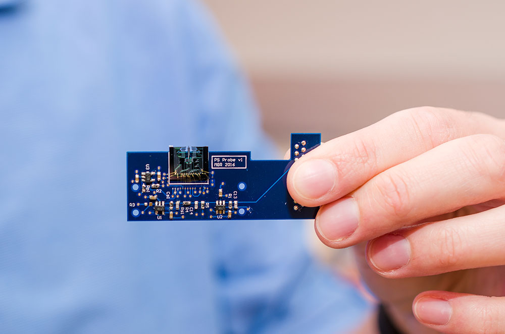
A MEMS-based atomic force microscope developed by engineers at UT Dallas is about 1 square centimeter in size (top center). Here it is attached to a small printed circuit board that contains circuitry, sensors and other miniaturized components that control the movement and other aspects of the device. Courtesy: University of Texas at Dallas

Of course, there’s still more work to be done as you’ll note when reading Dexter’s Feb. 21, 2017 posting where he features answers to questions he directed to the researchers.

Here’s a link to and a citation for the paper,

On-Chip Dynamic Mode Atomic Force Microscopy: A Silicon-on-Insulator MEMS Approach by  Michael G. Ruppert, Anthony G. Fowler, Mohammad Maroufi, S. O. Reza Moheimani. IEEE Journal of Microelectromechanical Systems Volume: 26 Issue: 1  Feb. 2017 DOI: 10.1109/JMEMS.2016.2628890 Date of Publication: 06 December 2016

This paper is behind a paywall.

Tiny sensors produced by nanoscale 3D printing could lead to new generation of atomic force microscopes

A Sept. 26, 2016 news item on Nanowerk features research into producing smaller sensors for atomic force microscopes (AFMs) to achieve greater sensitivity,

Tiny sensors made through nanoscale 3D printing may be the basis for the next generation of atomic force microscopes. These nanosensors can enhance the microscopes’ sensitivity and detection speed by miniaturizing their detection component up to 100 times. The sensors were used in a real-world application for the first time at EPFL, and the results are published in Nature Communications.

A Sept. 26, 2016 École Polytechnique Fédérale de Lausanne (EPFL; Switzerland) press release by Laure-Anne Pessina, which originated the news item, expands on the theme (Note: A link has been removed),

Atomic force microscopy is based on powerful technology that works a little like a miniature turntable. A tiny cantilever with a nanometric tip passes over a sample and traces its relief, atom by atom. The tip’s infinitesimal up-and-down movements are picked up by a sensor so that the sample’s topography can be determined. (…)

One way to improve atomic force microscopes is to miniaturize the cantilever, as this will reduce inertia, increase sensitivity, and speed up detection. Researchers at EPFL’s Laboratory for Bio- and Nano-Instrumentation achieved this by equipping the cantilever with a 5-nanometer thick sensor made with a nanoscale 3D-printing technique. “Using our method, the cantilever can be 100 times smaller,” says Georg Fantner, the lab’s director.

Electrons that jump over obstacles

The nanometric tip’s up-and-down movements can be measured through the deformation of the sensor placed at the fixed end of the cantilever. But because the researchers were dealing with minute movements – smaller than an atom – they had to pull a trick out of their hat.

Together with Michael Huth’s lab at Goethe Universität at Frankfurt am Main, they developed a sensor made up of highly conductive platinum nanoparticles surrounded by an insulating carbon matrix. Under normal conditions, the carbon isolates the electrons. But at the nano-scale, a quantum effect comes into play: some electrons jump through the insulating material and travel from one nanoparticle to the next. “It’s sort of like if people walking on a path came up against a wall and only the courageous few managed to climb over it,” said Fantner.

When the shape of the sensor changes, the nanoparticles move further away from each other and the electrons jump between them less frequently. Changes in the current thus reveal the deformation of the sensor and the composition of the sample.

Tailor-made sensors

The researchers’ real feat was in finding a way to produce these sensors in nanoscale dimensions while carefully controlling their structure and, by extension, their properties. “In a vacuum, we distribute a precursor gas containing platinum and carbon atoms over a substrate. Then we apply an electron beam. The platinum atoms gather and form nanoparticles, and the carbon atoms naturally form a matrix around them,” said Maja Dukic, the article’s lead author. “By repeating this process, we can build sensors with any thickness and shape we want. We have proven that we could build these sensors and that they work on existing infrastructures. Our technique can now be used for broader applications, ranging from biosensors, ABS sensors for cars, to touch sensors on flexible membranes in prosthetics and artificial skin.”

Here’s a link to and a citation for the paper,

Direct-write nanoscale printing of nanogranular tunnelling strain sensors for sub-micrometre cantilevers by Maja Dukic, Marcel Winhold, Christian H. Schwalb, Jonathan D. Adams, Vladimir Stavrov, Michael Huth, & Georg E. Fantner. Nature Communications 7, Article number: 12487 doi:10.1038/ncomms12487 Published  26 September 2016

This is an open access paper.

All about Atomic Force Microscopy (AFM) with Gerd Binnig and Christoph Gerber

Gerd Binnig, Christoph Gerber, and Calvin Quate invented the atomic force microscope in the 1980s and an Aug. 16, 2016 news item on Nanotechnology Now announces a discussion with two of the inventors, Binnig and Gerber (Note: Links have been removed),

The inventors of one of the most versatile tools in modern science – the atomic force microscope, or AFM – tell their story in an interview published online this week. The AFM was invented in the mid 1980s by Gerd Binnig, Christoph Gerber and Calvin Quate, three physicists who are sharing the 2016 Kavli Prize in Nanoscience.

Binnig and Gerber discuss their inspiration for the device, how they solved problems through sport, and why their invention continues to propel science at the nanoscale.

This charming Aug. 20, 2016 discussion for the Kavli Foundation focuses on more than the AFM although it is the main topic,

Our roundtable panelists were:

GERD BINNIG –is a physicist and Nobel Laureate for his invention (with Heinrich Rohrer and Christoph Gerber) of the scanning tunneling microscope while at IBM Zurich. He began development of the atomic force microscope in 1986 to overcome the limitations of his previous invention.
CHRISTOPH GERBER –is a physicist and director for scientific communication at the Swiss Nanoscience Institute at the University of Basel. While at IBM, Gerber worked closely with Binnig on bringing both the scanning tunneling microscope and atomic force microscope to fruition.

Calvin Quate was unable to participate in the roundtable. The transcript has been amended and edited by the laureates

THE KAVLI FOUNDATION [TKF]: You filed your first patent for the atomic force microscope (AFM) nearly 30 years ago. How has it changed the way we look at the world since then?

GERD BINNIG: It was like the first time people looked through an optical microscope and saw bacteria. That completely changed how we look at the world. Suddenly, we understood what was really going on in nature, and we used that knowledge to learn how diseases spread. The AFM is the next step. It lets us look at the molecules that make life possible in those bacteria – and everywhere else – and see things we could not see before. It teaches us how to make changes to surfaces or molecules that we attempted blindly in the past. And it has been used in so many different scientific studies, from looking at polymers and chemical reactions to modifying surfaces at the atomic level.

CHRISTOPH GERBER: As Gerd explained, seeing is believing, and now we can do that onthe atomic scale. AFM has turned into the most powerful and most versatile toolkit that we have for doing nanoscience. And it keeps evolving. In just the past few years, researchers have learned to pick up a molecule on the tip of an AFM, which we can think of as the needle on a record player, and reveal chemical bonds while imaging molecules on surfaces. Nobody thought that ever would be possible.

TKF: Has this changed how researchers think about the ways nanoscale interactions affect the things they study?

BINNIG: Very much so. Before AFM, people who wanted to model very small structures –molecules, cell walls, semiconductors – had to make indirect measurements of them. But those structures can be complex and disordered, and indirect measurements do not always capture that, so the models they came up with were often wrong. But now, we can look at those structures and adapt our models to match what we observe. We as scientists always have to connect our theories to reality. Atomic force microscopy lets us do this.

TKF: When you started thinking about the AFM, biology was one of the fields you had inmind. Yet even you must have been surprised at how it has revolutionized biology.

GERBER: Yes. AFM’s capabilities keep evolving, and researchers are always finding new ways to use it. For example, in recent years, researchers have made tremendous progress in taking AFM measurements in real time. It’s like watching a movie. They can now see biological interactions, such as how molecules degrade or how antimicrobials attack bacterial membranes as they occur – something nobody could have foreseen 20 years ago. It took 15 years to get there, but we can now see biology in action and compare that to our theories.

BINNIG: Exactly. In biology, the biggest and most important question is always whether a molecule will bind to another molecule, change it, and by changing it cause something important to happen. This is all about forces, and researchers can use AFM to bring two molecules or even two cells close together, or pull them apart, and measure those forces directly. We can learn how big those forces are and under what conditions they occur. We’re actually looking into the heart of biology when we do that.

GERBER: And atomic force microscopy can tell us about many different types of forces that determine the outcome of chemical reactions at the nanoscale. These range from chemical, mechanical and electrostatic through, most recently, to the very weak interactions between molecules.

BINNIG: A great example of this is how Hermann Gaub, a professor of biophysics at Ludwig Maximilians University of Munich, used AFM to unfold proteins. He actually attached one end of a protein to a surface and the other end to an AFM tip. When he pulled the tip up, the protein straightened out and he could create a fingerprint of the unfolding forces that he could compare with his model.

TKF: What about applications you could not have foreseen?

BINNIG: I could not have foreseen that we can image molecules with such a high resolution. It’s unbelievable. We can see the bonds between molecules. We can watch them change during a chemical reaction, and sometimes there are surprises. Some researchers have observed an intermediate state in a chemical reaction that should not have lasted long enough to see. So they have had to rethink their theories to take into account why this intermediate state lasted so long. That’s what happens when we can observe such high-resolution details.

GERBER: Another example is high-speed AFM, which biologists use to see the cellular machinery in action. No other technique can do that. It works by tapping a very, very thin cantilever up and down, taking one quick measurement after another.

BINNIG: It is amazing how many people use the AFM in so many different fields. We first thought, well, maybe biology or semiconductor research. But it was picked up everywhere, from studying friction to cosmetics.

GERBER: I recently looked it up, and AFM was mentioned in 353,000 peer-reviewed papers. Our original article was published in Physical Review Letters, the top journal in the field in which all the important theoretical work is published. Ours is the only experimental paper on its list of most-cited papers.

TKF: Amazing. And yet AFM was actually a follow-up to another technology you worked on, the scanning tunneling microscope, or STM. It was probably the first instrument to achieve nanoscale resolution without using electrons or other high-energy beams that can damage what you are observing, right?

BINNIG: Yes.

TKF: And where did that idea come from?

BINNIG: We were trying to solve a problem. IBM was working on a new type of semiconductor chip, and the insulator, which keeps the electric current from escaping the semiconductor, was leaking. But no one knew why. So Heinrich Rohrer, who was working at IBM Zurich, hired me. I looked to all the available instruments, and none of them could study materials on such a fine scale to find out.

So the two of us thought, well, okay, we’ll invent something. We thought we could take advantage of something called quantum tunneling. Quantum tunneling is when an electron tunnels through a conducting material and come out the other side. We developed STM to map the surface of the material by measuring where electrons emerged on the other side. Only later did we realize that we could move our probe from one spot to cover the entire surface.

TKF: Dr. Gerber, you quickly became part of the STM team. What convinced you to join?

GERBER: I felt this was such a crazy idea, and I’m always very fond of this sort of thing. I thought this was fantastic.

BINNIG: I can confirm this. Christoph always likes crazy things. That runs through his life.

GERBER: Actually, the development of STM was kind of an undercover project at the beginning, because Gerd and Heinrich were involved in other projects. I worked for a year or so on my own. When we started overcoming problems and we could see features on the surface of a material that were one-tenth of a nanometer, then it really took off.

I leave you to discover the discussion in its entirety: Aug. 20, 2016 discussion.

Barnacle footprints could be useful

An Aug. 18, 2016 news item on Nanowerk describes efforts by scientists at the University of Twente (The Netherlands) and A*STAR (Singapore) to trace a barnacle’s footprints (Note: A link has been removed),

Barnacle’s larvae leave behind tiny protein traces on a ship hull: but what is the type of protein and what is the protein-surface interaction? Conventional techniques can only identify dissolved proteins, and in large quantities. Using a modified type of an Atomic Force Microscope, scientists of the University of Twente in The Netherlands and A*STAR in Singapore, can now measure protein characteristics of even very small traces on a surface. They present the new technique in Nature Nanotechnology (“Measuring protein isoelectric points by AFM-based force spectroscopy using trace amounts of sample”).

An Aug. 16, 2016 University of Twente press release, which originated the news item, explains how the ‘footprints’ could lead to new applications for ships and boats and briefly describes the technical aspects of the research,

In infection diseases, membrane fouling, interaction with bacteria, as well as in rapid healing of wounds for example, the way proteins interact with a surface plays an important role. On a surface, they function in a different way than in solution. On a ship hull, the larvae of the barnacle will leave tiny traces of protein to test if the surface is attractive for long-term attachment. If we get to know more about this interaction, it will be possible to develop surface conditions that are less attractive for the barnacle. Large amounts of barnacles on a ship will have a destructive effect on flow resistance and will lead to more fuel consumption. The new measuring method makes use of a modified Atomic Force Microscope: a tiny ball glued to the cantilever of the microscope will attract protein molecules.

Modified AFM tip with a tiny ball that can attract protein molecules

FORCE MEASUREMENTS

An amount of just hundreds of protein molecules will be sufficient to determine a crucial value, called the iso-electric point (pI): this is the pH-value at which the protein has net zero electric charge. The pI value says a lot about the surroundings a protein will ‘feel comfortable’ in, and to which it preferably moves. Using the AFM microscope, of which the modified tip has collected protein molecules, it is possible to perform force measurements for different pH values. The tip will be attracted or repelled, or show no movement when the pI point is reached. For these measurement, the researchers made a special reference material consisting of several layers. Using this, the effect of a number of pH-values can be tested until the pI value is found.

The traces the larve leaves behind (left) and force measurements (right)

PAINT CHANGE

The tests have been successfully performed for a number of known proteins like fibrinogen, myoglobine and bovine albumin. And returning to the barnacle: the tiny protein footprint will contain enough molecules to determine the pI value. This quantifies the ideal surface conditions, and using this knowledge, new choices can be made for e.g. the paint that is used on a ship hull.

The research has been done within the group Materials Science and Technology of Polymers of Professor Julius Vancso, in close collaboration with colleagues of A*STAR in Singapore – Prof Vancso is a Visiting Professor there as well. His group is part of UT’s MESA+ Institute for Nanotechnology.

Here’s a link to and a citation for the paper,

Measuring protein isoelectric points by AFM-based force spectroscopy using trace amounts of sample by Shifeng Gu, Xiaoying Zhu, Dominik Jańczewski, Serina Siew Chen Lee, Tao He, Serena Lay Ming Teo, & G. Julius Vancso.  Nature Nanotechnology (2016) doi:10.1038/nnano.2016.118 Published online 25 July 2016

This paper is behind a paywall.
Graphene ribbons in solution bending and twisting like DNA

An Aug. 15, 2016 news item on ScienceDaily announces research into graphene nanoribbons and their DNA (deoxyribonucleic acid)-like properties,

Graphene nanoribbons (GNRs) bend and twist easily in solution, making them adaptable for biological uses like DNA analysis, drug delivery and biomimetic applications, according to scientists at Rice University.

Knowing the details of how GNRs behave in a solution will help make them suitable for wide use in biomimetics, according to Rice physicist Ching-Hwa Kiang, whose lab employed its unique capabilities to probe nanoscale materials like cells and proteins in wet environments. Biomimetic materials are those that imitate the forms and properties of natural materials.

An Aug. 15, 2016 Rice University (Texas, US) news release (also on EurekAlert), which originated the news item, describes the ribbons and the research in more detail,

Graphene nanoribbons can be thousands of times longer than they are wide. They can be produced in bulk by chemically “unzipping” carbon nanotubes, a process invented by Rice chemist and co-author James Tour and his lab.

Their size means they can operate on the scale of biological components like proteins and DNA, Kiang said. “We study the mechanical properties of all different kinds of materials, from proteins to cells, but a little different from the way other people do,” she said. “We like to see how materials behave in solution, because that’s where biological things are.” Kiang is a pioneer in developing methods to probe the energy states of proteins as they fold and unfold.

She said Tour suggested her lab have a look at the mechanical properties of GNRs. “It’s a little extra work to study these things in solution rather than dry, but that’s our specialty,” she said.

Nanoribbons are known for adding strength but not weight to solid-state composites, like bicycle frames and tennis rackets, and forming an electrically active matrix. A recent Rice project infused them into an efficient de-icer coating for aircraft.

But in a squishier environment, their ability to conform to surfaces, carry current and strengthen composites could also be valuable.

“It turns out that graphene behaves reasonably well, somewhat similar to other biological materials. But the interesting part is that it behaves differently in a solution than it does in air,” she said. The researchers found that like DNA and proteins, nanoribbons in solution naturally form folds and loops, but can also form helicoids, wrinkles and spirals.

Kiang, Wijeratne [Sithara Wijeratne, Rice graduate now a postdoctoral researcher at Harvard University] and Jingqiang Li, a co-author and student in the Kiang lab, used atomic force microscopy to test their properties. Atomic force microscopy can not only gather high-resolution images but also take sensitive force measurements of nanomaterials by pulling on them. The researchers probed GNRs and their precursors, graphene oxide nanoribbons.

The researchers discovered that all nanoribbons become rigid under stress, but their rigidity increases as oxide molecules are removed to turn graphene oxide nanoribbons into GNRs. They suggested this ability to tune their rigidity should help with the design and fabrication of GNR-biomimetic interfaces.

“Graphene and graphene oxide materials can be functionalized (or modified) to integrate with various biological systems, such as DNA, protein and even cells,” Kiang said. “These have been realized in biological devices, biomolecule detection and molecular medicine. The sensitivity of graphene bio-devices can be improved by using narrow graphene materials like nanoribbons.”

Wijeratne noted graphene nanoribbons are already being tested for use in DNA sequencing, in which strands of DNA are pulled through a nanopore in an electrified material. The base components of DNA affect the electric field, which can be read to identify the bases.

The researchers saw nanoribbons’ biocompatibility as potentially useful for sensors that could travel through the body and report on what they find, not unlike the Tour lab’s nanoreporters that retrieve information from oil wells.

Further studies will focus on the effect of the nanoribbons’ width, which range from 10 to 100 nanometers, on their properties.

Here’s a link to and a citation for the paper,

Detecting the Biopolymer Behavior of Graphene Nanoribbons in Aqueous Solution by Sithara S. Wijeratne, Evgeni S. Penev, Wei Lu, Jingqiang Li, Amanda L. Duque, Boris I. Yakobson, James M. Tour, & Ching-Hwa Kiang. Scientific Reports 6, Article number: 31174 (2016)  doi:10.1038/srep31174 Published online: 09 August 2016

This paper is open access.

Nature celebrates some nanotechnology anniversaries

An April 5, 2016 editorial in Nature magazine celebrates some nanotechnology milestones (Note: Links have been removed),

In March 1986, the atomic force microscope (AFM) was introduced by Gerd Binnig, Calvin Quate and Christoph Gerber with a paper in the journal Physical Review Letters titled simply ‘Atomic force microscope’1. This was 5 years (to the month) after the precursor to the AFM, the scanning tunnelling microscope (STM), had first been successfully tested at IBM’s Zurich Research Laboratory by Binnig and the late Heinrich Rohrer, and 7 months before Binnig and Rohrer were awarded a share of the Nobel Prize in Physics for the design of the STM (the prize was shared with Ernst Ruska, the inventor of the electron microscope). Achieving atomic resolution with the AFM proved more difficult than with the STM. It was, for example, only two years after its invention that the STM provided atomic-resolution images of an icon of surface science, the 7 × 7 surface reconstruction of Si(111) (ref. 2), whereas it took 8 years to achieve a similar feat with the AFM3, 4.

The editorial also provides an explanation of how the AFM works,

The AFM works by scanning a sharp tip attached to a flexible cantilever across a sample while measuring the interaction between the tip and the sample surface. The technique can operate in a range of environments, including in liquid and in air, and unlike the STM, it can be used with insulating materials; in their original paper, Binnig and colleagues used the instrument to analyse an aluminium oxide sample.

Then, the editorial touches on DNA (deoxyribonucleic acid) nanotechnology (Note: Links have been removed),

The history of structural DNA nanotechnology can, like the AFM, be traced back to the early 1980s, when Nadrian Seeman suggested that the exquisite base-pairing rules of DNA could be exploited to build artificial self-assembled structures11. But the founding experiment of the field came later. In April 1991, Seeman and Junghuei Chen reported building a cube-like molecular complex from DNA using a combination of branched junctions and single-stranded ‘sticky’ ends12. A range of significant advances soon followed, from 2D DNA arrays to DNA-based nanomechanical devices.

Then, in March 2006, the field of structural DNA nanotechnology experienced another decisive moment: Paul Rothemund reported the development of DNA origami13. This technique involves folding a long single strand of DNA into a predetermined shape with the help of short ‘staple’ strands. Used at first to create 2D structures, which were incidentally characterized using the AFM, the approach was quickly expanded to the building of intricate 3D structures and the organization of other species such as nanoparticles and proteins. …

Happy reading!

Developing optical microscopes that measure features down to 10 nanometer level on computer chips

The US National Institute of Standards and Technology (NIST) issued a Dec. 2, 2015 news release (also on EurekAlert) announcing a new kind of optical microscope and its possible impact on the semiconductor industry,

National Institute of Standards and Technology (NIST) researchers are seeing the light, but in an altogether different way. And how they are doing it just might be the semiconductor industry’s ticket for extending its use of optical microscopes to measure computer chip features that are approaching 10 nanometers, tiny fractions of the wavelength of light.

The news release goes on to provide details and an explanation of scatterfield imaging,

Using a novel microscope that combines standard through-the-lens viewing with a technique called scatterfield imaging, the NIST team accurately measured patterned features on a silicon wafer that were 30 times smaller than the wavelength of light (450 nanometers) used to examine them. They report* that measurements of the etched lines–as thin as 16 nanometers wide–on the SEMATECH-fabricated wafer were accurate to one nanometer. With the technique, they spotted variations in feature dimensions amounting to differences of a few atoms.

Measurements were confirmed by those made with an atomic force microscope, which achieves sub-nanometer resolution, but is considered too slow for online quality-control measurements. Combined with earlier results, the NIST researchers write, the new proof-of-concept study* suggests that the innovative optical approach could be a “realistic solution to a very challenging problem” facing chip makers and others aiming to harness advances in nanotechnology. All need the means for “nondestructive measurement of nanometer-scale structures with sub-nanometer sensitivity while still having high throughput.

“Light-based, or optical, microscopes can’t “see” features smaller than the wavelength of light, at least not in the crisp detail necessary for making accurate measurements. However, light does scatter when it strikes so-called subwavelength features and patterned arrangements of such features. “Historically, we would ignore this scattered light because it did not yield sufficient resolution,” explains Richard Silver, the physicist who initiated NIST’s scatterfield imaging effort. “Now we know it contains helpful information that provides signatures telling us something about where the light came from.”

With scatterfield imaging, Silver and colleagues methodically illuminate a sample with polarized light from different angles. From this collection of scattered light–nothing more than a sea of wiggly lines to the untrained eye–the NIST team can extract characteristics of the bounced lightwaves that, together, reveal the geometry of features on the specimen.

Light-scattering data are gathered in slices, which together image the volume of scattered light above and into the sample. These slices are analyzed and reconstructed to create a three-dimensional representation. The process is akin to a CT scan, except that the slices are collections of interfering waves, not cross-sectional pictures.

“It’s the ensemble of data that tells us what we’re after,” says project leader Bryan Barnes.” We may not be able see the lines on the wafer, but we can tell you what you need to know about them–their size, their shape, their spacing.”

Scatterfield imaging has critical prerequisites that must be met before it can yield useful data for high-accuracy measurements of exceedingly small features. Key steps entail detailed evaluation of the path light takes as it beams through lenses, apertures and other system elements before reaching the sample. The path traversed by light scattering from the specimen undergoes the same level of scrutiny. Fortunately, scatterfield imaging lends itself to thorough characterization of both sequences of optical devices, according to the researchers. These preliminary steps are akin to error mapping so that recognized sources of inaccuracy are factored out of the data.

The method also benefits from a little advance intelligence–the as-designed arrangement of circuit lines on a chip, down to the size of individual features. Knowing what is expected to be the result of the complex chip-making process sets up a classic matchup of theory vs. experiment.

The NIST researchers can use standard equations to simulate light scattering from an ideal, defect-free pattern and, in fact, any variation thereof. Using wave analysis software they developed, the team has assembled an indexed library of light-scattering reference models. So once a specimen is scanned, the team relies on computers to compare their real-world data to models and to find close matches.

From there, succeeding rounds of analysis homes in on the remaining differences, reducing them until the only ones that remain are due to variations in geometry such as irregularities in the height, width, or shape of a line.

Measurement results achieved with the NIST approach might be said to cast light itself in an entirely new light. Their new study, the researchers say, shows that once disregarded scattered light “contains a wealth of accessible optical information.”

Next steps include extending the technique to even shorter wavelengths of light, down to ultraviolet, or 193 nanometers. The aim is to accurately measure features as small as 5 nanometers.

This work is part of a larger NIST effort to supply measurement tools that enable the semiconductor industry to continue doubling the number of devices on a chip about every two years and to help other industries make products with nanoscale features. Recently, NIST and Intel researchers reported using an X-ray technique to accurately measure features on a silicon chip to within fractions of a nanometer.

Here’s a link to and a citation for a PDF of the paper,

Deep-subwavelength Nanometric Image Reconstruction using Fourier Domain Optical Normalization by Jing  Qin, Richard  M Silver, Bryan  M  Barnes, Hui Zhou, Ronald G Dixson, and Mark Alexander Hen. Light: Science & Applications accepted article preview 5 November 2015; e16038 doi: 10.1038/lsa.2016.38

[Note:] This is a PDF file of an unedited peer-reviewed manuscript that has been accepted for publication. NPG are providing this early version of the manuscript as a service to our customers. The manuscript will undergo copy editing, typesetting and a proof review before it is published in its final form. Please note that during the production process errors may be discovered which could affect the content, and all legal disclaimers apply.

This seems to be an open access paper but it is an early version.

Characterizing anatase titanium dixoide at the nanoscale

An international collaboration of researchers combined atomic force microscopy (AFM) and scanning tunneling microscopy (STM) to characterize anatase titanium dixoxide. From a Sept. 14, 2015 news item on Azonano,

A [Japan National Institute for Materials Science] NIMS research team successfully identified the atoms and common defects existing at the most stable surface of the anatase form of titanium dioxide by characterizing this material at the atomic scale with scanning probe microscopy. This work was published under open access policy in the online version of Nature Communications on June 29, 2015.

A June 29, 2015 NIMS press release, which originated the news item, includes the paper’s abstract in numbered point form,

  1. The research team consisting of Oscar Custance and Tomoko Shimizu, group leader and senior scientist, respectively, at the Atomic Force Probe Group, NIMS, Daisuke Fujita and Keisuke Sagisaka, group leader and senior researcher, respectively, at the Surface Characterization Group, NIMS, and scientists at Charles University in the Czech Republic, Autonomous University of Madrid in Spain, and other organizations combined simultaneous atomic force microscopy (AFM) and scanning tunneling microscopy (STM) measurements with first-principles calculations for the unambiguous identification of the atomic species at the most stable surface of the anatase form of titanium dioxide (hereinafter referred to as anatase) and its most common defects.
  2. In recent years, anatase has attracted considerable attention, because it has become a pivotal material in devices for photo-catalysis and for the conversion of solar energy to electricity. It is extremely challenging to grow large single crystals of anatase, and most of the applications of this material are in the form of nano crystals. To enhance the catalytic reactivity of anatase and the efficiency of devices for solar energy conversion based on anatase, it is critical to gain in-depth understanding and control of the reactions taking place at the surface of this material down to the atomic level. Only a few research groups worldwide possess the technology to create proper test samples and to make in-situ atomic-level observations of anatase surfaces.
  3. In this study, the research team used samples obtained from anatase natural single crystals extracted from naturally occurring anatase rocks. The team characterized the (101) surface of anatase at atomic level by means of simultaneous AFM and STM. Using single water molecules as atomic markers, the team successfully identified the atomic species of this surface; result that was additionally confirmed by the comparison of simultaneous AFM and STM measurements with the outcomes of first-principles calculations.
  4. In regular STM, in which an atomically sharp probe is scanned over the surface by keeping constant an electrical current flowing between them, it is difficult to stably image anatase surfaces as this material presents poor electrical conductivity over some of the atomic positions of the surface. However, simultaneous operation of AFM and STM allowed imaging the surface with atomic resolution even within the materials band gap (a region where the flow of current between the probe and the surface is, in principle, prohibited). Here, the detection of inter-atomic forces between the last atom of the atomically sharp probe and the atoms of the surface by AFM was of crucial importance. By regulating the probe-surface distance using AFM, it was possible to image the surface at atomic-scale while collecting STM data over both conductive and not conductive areas of the surface. By comparing simultaneous AFM and STM measurements with theoretical simulations, the team was not only able to discern which atomic species were contributing to the AFM and the STM images but also to identify the most common defects found at the surface.
  5. In the future, based on the information gained from this study, the NIMS research team will conduct research on molecules of technologically relevance that adsorb on anatase and characterize these hybrid systems by using simultaneous AFM and STM. Their ultimate goal is to formulate novel approaches for the development of photo-catalysts and solar cell materials and devices.

Here’s a link to and a citation for the paper,

Atomic species identification at the (101) anatase surface by simultaneous scanning tunnelling and atomic force microscopy by Oleksandr Stetsovych, Milica Todorović, Tomoko K. Shimizu, César Moreno, James William Ryan, Carmen Pérez León, Keisuke Sagisaka, Emilio Palomares, Vladimír Matolín, Daisuke Fujita, Ruben Perez, & Oscar Custance. Nature Communications 6, Article number: 7265 doi:10.1038/ncomms8265 Published 29 June 2015

This is an open access paper.

Corrections: Hybrid Photonic-Nanomechanical Force Microscopy uses vibration for better chemical analysis

*ETA  Nov. 4, 2015: I’m apologizing to anyone wishing to read this posting as it’s a bit of a mess. I deeply regret mishandling the situation. In future, I shall not be taking any corrections from individual researchers to materials such as news releases that have been issued by an institution. Whether or not the individual researchers are happy with how their contributions or how a colleague’s contributions or how their home institutions have been characterized is a matter for them and their home institutions.

The August 10, 2015 ORNL news release with all the correct details has been added to the end of this post.*

A researcher at the University of Central Florida (UCF) has developed a microscope that uses vibrations for better analysis of chemical composition. From an Aug. 10, 2015 news item on Nanowerk,

It’s a discovery that could have promising implications for fields as varied as biofuel production, solar energy, opto-electronic devices, pharmaceuticals and medical research.

“What we’re interested in is the tools that allow us to understand the world at a very small scale,” said UCF professor Laurene Tetard, formerly of the Oak Ridge National Laboratory. “Not just the shape of the object, but its mechanical properties, its composition and how it evolves in time.”

An Aug. 10, 2015 UCF news release (also on EurekAlert), which originated the news item, describes the limitations of atomic force microscopy and gives a few details about the hybrid microscope (Note: A link has been removed),

For more than two decades, scientists have used atomic force microscopy – a probe that acts like an ultra-sensitive needle on a record player – to determine the surface characteristics of samples at the microscopic scale. A “needle” that comes to an atoms-thin point traces a path over a sample, mapping the surface features at a sub-cellular level [nanoscale].

But that technology has its limits. It can determine the topographical characteristics of [a] sample, but it can’t identify its composition. And with the standard tools currently used for chemical mapping, anything smaller than roughly half a micron is going to look like a blurry blob, so researchers are out of luck if they want to study what’s happening at the molecular level.

A team led by Tetard has come up with a hybrid form of that technology that produces a much clearer chemical image. As described Aug. 10 in the journal Nature Nanotechnology, Hybrid Photonic-Nanomechanical Force Microscopy (HPFM) can discern a sample’s topographic characteristics together with the chemical properties at a much finer scale.

The HPFM method is able to identify materials based on differences in the vibration produced when they’re subjected to different wavelengths of light – essentially a material’s unique “fingerprint.”

“What we are developing is a completely new way of making that detection possible,” said Tetard, who has joint appointments to UCF’s Physics Department, Material Science and Engineering Department and the NanoScience Technology Center.

The researchers proved the effectiveness of HPFM while examining samples from an eastern cottonwood tree, a potential source of biofuel. By examining the plant samples at the nanoscale, the researchers for the first time were able to determine the molecular traits of both untreated and chemically processed cottonwood inside the plant cell walls.

The research team included Tetard; Ali Passian, R.H. Farahi and Brian Davison, all of Oak Ridge National Laboratory; and Thomas Thundat of the University of Alberta.

Long term, the results will help reveal better methods for producing the most biofuel from the cottonwood, a potential boon for industry. Likewise, the new method could be used to examine samples of myriad plants to determine whether they’re good candidates for biofuel production.

Potential uses of the technology go beyond the world of biofuel. Continued research may allow HPFM to be used as a probe so, for instance, it would be possible to study the effect of new treatments being developed to save plants such as citrus trees from bacterial diseases rapidly decimating the citrus industry, or study fundamental photonically-induced processes in complex systems such as in solar cell materials or opto-electronic devices.

Here’s a link to and a citation for the paper,

Opto-nanomechanical spectroscopic material characterization by L. Tetard, A. Passian, R. H. Farahi, T. Thundat, & B. H. Davison. Nature Nanotechnology (2015) doi:10.1038/nnano.2015.168 Published online 10 August 2015

This paper is behind a paywall.

*ETA August 27, 2015:

August 10, 2015 ORNL news release (Note: Funding information and a link to the paper [previously given] have been removed):

A microscope being developed at the Department of Energy’s Oak Ridge National Laboratory will allow scientists studying biological and synthetic materials to simultaneously observe chemical and physical properties on and beneath the surface.

The Hybrid Photonic Mode-Synthesizing Atomic Force Microscope is unique, according to principal investigator Ali Passian of ORNL’s Quantum Information System group. As a hybrid, the instrument, described in a paper published in Nature Nanotechnology, combines the disciplines of nanospectroscopy and nanomechanical microscopy.

“Our microscope offers a noninvasive rapid method to explore materials simultaneously for their chemical and physical properties,” Passian said. “It allows researchers to study the surface and subsurface of synthetic and biological samples, which is a capability that until now didn’t exist.”

ORNL’s instrument retains all of the advantages of an atomic force microscope while simultaneously offering the potential for discoveries through its high resolution and subsurface spectroscopic capabilities.

“The originality of the instrument and technique lies in its ability to provide information about a material’s chemical composition in the broad infrared spectrum of the chemical composition while showing the morphology of a material’s interior and exterior with nanoscale – a billionth of a meter – resolution,” Passian said.

Researchers will be able to study samples ranging from engineered nanoparticles and nanostructures to naturally occurring biological polymers, tissues and plant cells.

The first application as part of DOE’s BioEnergy Science Center was in the examination of plant cell walls under several treatments to provide submicron characterization. The plant cell wall is a layered nanostructure of biopolymers such as cellulose. Scientists want to convert such biopolymers to free the useful sugars and release energy.

An earlier instrument, also invented at ORNL, provided imaging of poplar cell wall structures that yielded unprecedented topological information, advancing fundamental research in sustainable biofuels.

Because of this new instrument’s impressive capabilities, the researcher team envisions broad applications.
“An urgent need exists for new platforms that can tackle the challenges of subsurface and chemical characterization at the nanometer scale,” said co-author Rubye Farahi. “Hybrid approaches such as ours bring together multiple capabilities, in this case, spectroscopy and high-resolution microscopy.”

Looking inside, the hybrid microscope consists of a photonic module that is incorporated into a mode-synthesizing atomic force microscope. The modular aspect of the system makes it possible to accommodate various radiation sources such as tunable lasers and non-coherent monochromatic or polychromatic sources.

ETA2 August 27, 2015: I’ve received an email from one of the paper’s authors (RH Farahi of the US Oak Ridge National Laboratory [ORNL]) who claims some inaccuracies in this piece.  The news release supplied by the University of Central Florida states that Dr. Tetard led the team and that is not so. According to Dr. Farahi, she had a postdoctoral position on the team which she left two years ago. You might also get the impression that some of the work was performed at the University of Central Florida. That is not so according to Dr. Farahi.  As a courtesy Dr. Tetard was retained as first author of the paper.

*Nov. 4, 2015: I suspect some of the misunderstanding was due to overeagerness and/or time pressures. Whoever wrote the news release may have made some assumptions. It’s very easy to make a mistake when talking to an ebullient scientist who can unintentionally lead you to believe something that’s not so. I worked in a high tech company and believed that there was some new software being developed which turned out to be a case of high hopes. Luckily, I said something that triggered a rapid rebuttal to the fantasies. Getting back to this situation, other contributing factors could include the writer not having time to get the news release reviewed the scientist or the scientist skimming the release and missing a few bits due to time pressure.*

Molecules (arynes) seen for first time in 113 years

Arynes were first theorized in 1902 and they’ve been used as building blocks to synthesize a variety of compounds but they’re existence hasn’t been confirmed until now.

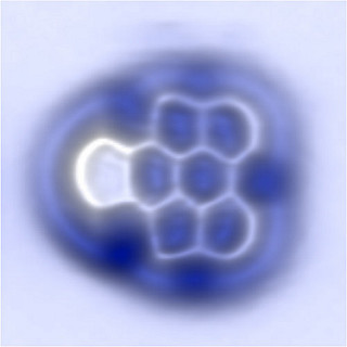
AFM image of an aryne molecule imaged with a CO tip. Courtesy: IBM

AFM image of an aryne molecule imaged with a CO tip. Courtesy: IBM

A July 13, 2015 news item in Nanowerk makes the announcement (Note: A link has been removed),

chemistry teachers and students can breath a sigh of relief. After teaching and learning about a particular family of molecules for decades, scientists have finally proven that they do in fact exist.

In a new paper published online today in Nature Chemistry (“On-surface generation and imaging of arynes by atomic force microscopy”), scientists from IBM Research and CIQUS at the University of Santiago de Compostela, Spain, have confirmed the existence and characterized the structure of arynes, a family of highly-reactive short-lived molecules which was first suggested 113 years ago. The technique has broad applications for on-surface chemistry and electronics, including the preparation of graphene nanoribbons and novel single-molecule devices.

A July 13, 2015 IBM news release by Chris Sciacca, which originated the news item, describes arynes and the imaging process used to capture them for the first time (Note: Links have been removed),

“Arynes are discussed in almost every undergraduate course on organic chemistry around the world. Therefore, it’s kind of a relief to find the final confirmation that these molecules truly exist,” said Prof. Diego Peña, a chemist at the University of Santiago de Compostela.

“I look forward to seeing new chemical challenges solved by the combination of organic synthesis and atomic force microscopy.”

There are trillions of molecules in the universe and some of them are stable enough to be isolated and characterized, but many others are so short-lived that they can only be proposed indirectly, via chemical reactions or spectroscopic methods.

One such species are arynes, which were first suggested in 1902, and since then have been used as intermediates or building blocks in the synthesis of a variety of compounds for applications including medicine, organic electronics and molecular materials. The challenge with these particular molecules is that they only exist for several milliseconds making them extremely challenging to image, until now.

The imaging was accomplished by means of atomic force microscopy (AFM), a scanning technique that can accomplish nanometer-level resolution. After the preparation of the key aryne precursor by CIQUS, IBM scientists used the sharp tip of a scanning tunneling microscope (STM) to generate individual aryne molecules from precursor molecules by atomic manipulation. The experiments were performed on films of sodium chloride, at temperatures near absolute zero, to stabilize the aryne.

Once the molecules were isolated, the team used AFM to measure the tiny forces between the STM tip, which is terminated with a single carbon monoxide molecule, and the sample to image the aryne’s molecular structure. The resulting image was so clear that the scientists could study their chemical nature based on the minute differences between individual bonds.

“Our team has developed several state-of-the-art techniques since 2009, which made this achievement possible,” said Dr. Niko Pavliček, a physicist at IBM Research – Zurich and lead author of the paper. “For this study, it was absolutely essential to pick an insulating film on which the molecules were adsorbed and to deliberately choose the atomic tip-terminations to probe them. We hope this technique will have profound effects on the future of chemistry and electronics.”

Prof. Peña, added that “These findings on arynes can be compared with the long-standing search for the giant squid. For centuries, fishermen had found clues of the existence of this legendary animal. But it was only very recently that scientists managed to film a giant squid alive. In both cases, state-of-the-art technologies were crucial to observe these elusive species alive: a low-noise submarine for the giant squid; a low-temperature AFM for the aryne.”

This research is part of IBM’s five-year, $3 billion investment to push the limits of chip technology and semiconductor innovations needed to meet the emerging demands of cloud computing and Big Data systems.

This work is a result of the large European project called (Planar Atomic and Molecular Scale Devices). PAMS’ main objective is to develop and investigate novel electronic devices of nanometric-scale size. Part of this research is also funded by a European Research Council Advanced Grant awarded to IBM scientist Gerhard Meyer, who is also a co-author of the paper.

Here’s a link to and a citation for the paper,

On-surface generation and imaging of arynes by atomic force microscopy by Niko Pavliček, Bruno Schuler, Sara Collazos, Nikolaj Moll, Dolores Pérez, Enrique Guitián, Gerhard Meyer, Diego Peña, & Leo Gross. Nature Chemistry (2015) doi:10.1038/nchem.2300 Published online 13 July 2015

This paper is behind a paywall.