Tag Archives: atomic force microscopy

Tiny sensors produced by nanoscale 3D printing could lead to new generation of atomic force microscopes

A Sept. 26, 2016 news item on Nanowerk features research into producing smaller sensors for atomic force microscopes (AFMs) to achieve greater sensitivity,

Tiny sensors made through nanoscale 3D printing may be the basis for the next generation of atomic force microscopes. These nanosensors can enhance the microscopes’ sensitivity and detection speed by miniaturizing their detection component up to 100 times. The sensors were used in a real-world application for the first time at EPFL, and the results are published in Nature Communications.

A Sept. 26, 2016 École Polytechnique Fédérale de Lausanne (EPFL; Switzerland) press release by Laure-Anne Pessina, which originated the news item, expands on the theme (Note: A link has been removed),

Atomic force microscopy is based on powerful technology that works a little like a miniature turntable. A tiny cantilever with a nanometric tip passes over a sample and traces its relief, atom by atom. The tip’s infinitesimal up-and-down movements are picked up by a sensor so that the sample’s topography can be determined. (…)

One way to improve atomic force microscopes is to miniaturize the cantilever, as this will reduce inertia, increase sensitivity, and speed up detection. Researchers at EPFL’s Laboratory for Bio- and Nano-Instrumentation achieved this by equipping the cantilever with a 5-nanometer thick sensor made with a nanoscale 3D-printing technique. “Using our method, the cantilever can be 100 times smaller,” says Georg Fantner, the lab’s director.

Electrons that jump over obstacles

The nanometric tip’s up-and-down movements can be measured through the deformation of the sensor placed at the fixed end of the cantilever. But because the researchers were dealing with minute movements – smaller than an atom – they had to pull a trick out of their hat.

Together with Michael Huth’s lab at Goethe Universität at Frankfurt am Main, they developed a sensor made up of highly conductive platinum nanoparticles surrounded by an insulating carbon matrix. Under normal conditions, the carbon isolates the electrons. But at the nano-scale, a quantum effect comes into play: some electrons jump through the insulating material and travel from one nanoparticle to the next. “It’s sort of like if people walking on a path came up against a wall and only the courageous few managed to climb over it,” said Fantner.

When the shape of the sensor changes, the nanoparticles move further away from each other and the electrons jump between them less frequently. Changes in the current thus reveal the deformation of the sensor and the composition of the sample.

Tailor-made sensors

The researchers’ real feat was in finding a way to produce these sensors in nanoscale dimensions while carefully controlling their structure and, by extension, their properties. “In a vacuum, we distribute a precursor gas containing platinum and carbon atoms over a substrate. Then we apply an electron beam. The platinum atoms gather and form nanoparticles, and the carbon atoms naturally form a matrix around them,” said Maja Dukic, the article’s lead author. “By repeating this process, we can build sensors with any thickness and shape we want. We have proven that we could build these sensors and that they work on existing infrastructures. Our technique can now be used for broader applications, ranging from biosensors, ABS sensors for cars, to touch sensors on flexible membranes in prosthetics and artificial skin.”

Here’s a link to and a citation for the paper,

Direct-write nanoscale printing of nanogranular tunnelling strain sensors for sub-micrometre cantilevers by Maja Dukic, Marcel Winhold, Christian H. Schwalb, Jonathan D. Adams, Vladimir Stavrov, Michael Huth, & Georg E. Fantner. Nature Communications 7, Article number: 12487 doi:10.1038/ncomms12487 Published  26 September 2016

This is an open access paper.

Graphene ribbons in solution bending and twisting like DNA

An Aug. 15, 2016 news item on ScienceDaily announces research into graphene nanoribbons and their DNA (deoxyribonucleic acid)-like properties,

Graphene nanoribbons (GNRs) bend and twist easily in solution, making them adaptable for biological uses like DNA analysis, drug delivery and biomimetic applications, according to scientists at Rice University.

Knowing the details of how GNRs behave in a solution will help make them suitable for wide use in biomimetics, according to Rice physicist Ching-Hwa Kiang, whose lab employed its unique capabilities to probe nanoscale materials like cells and proteins in wet environments. Biomimetic materials are those that imitate the forms and properties of natural materials.

An Aug. 15, 2016 Rice University (Texas, US) news release (also on EurekAlert), which originated the news item, describes the ribbons and the research in more detail,

Graphene nanoribbons can be thousands of times longer than they are wide. They can be produced in bulk by chemically “unzipping” carbon nanotubes, a process invented by Rice chemist and co-author James Tour and his lab.

Their size means they can operate on the scale of biological components like proteins and DNA, Kiang said. “We study the mechanical properties of all different kinds of materials, from proteins to cells, but a little different from the way other people do,” she said. “We like to see how materials behave in solution, because that’s where biological things are.” Kiang is a pioneer in developing methods to probe the energy states of proteins as they fold and unfold.

She said Tour suggested her lab have a look at the mechanical properties of GNRs. “It’s a little extra work to study these things in solution rather than dry, but that’s our specialty,” she said.

Nanoribbons are known for adding strength but not weight to solid-state composites, like bicycle frames and tennis rackets, and forming an electrically active matrix. A recent Rice project infused them into an efficient de-icer coating for aircraft.

But in a squishier environment, their ability to conform to surfaces, carry current and strengthen composites could also be valuable.

“It turns out that graphene behaves reasonably well, somewhat similar to other biological materials. But the interesting part is that it behaves differently in a solution than it does in air,” she said. The researchers found that like DNA and proteins, nanoribbons in solution naturally form folds and loops, but can also form helicoids, wrinkles and spirals.

Kiang, Wijeratne [Sithara Wijeratne, Rice graduate now a postdoctoral researcher at Harvard University] and Jingqiang Li, a co-author and student in the Kiang lab, used atomic force microscopy to test their properties. Atomic force microscopy can not only gather high-resolution images but also take sensitive force measurements of nanomaterials by pulling on them. The researchers probed GNRs and their precursors, graphene oxide nanoribbons.

The researchers discovered that all nanoribbons become rigid under stress, but their rigidity increases as oxide molecules are removed to turn graphene oxide nanoribbons into GNRs. They suggested this ability to tune their rigidity should help with the design and fabrication of GNR-biomimetic interfaces.

“Graphene and graphene oxide materials can be functionalized (or modified) to integrate with various biological systems, such as DNA, protein and even cells,” Kiang said. “These have been realized in biological devices, biomolecule detection and molecular medicine. The sensitivity of graphene bio-devices can be improved by using narrow graphene materials like nanoribbons.”

Wijeratne noted graphene nanoribbons are already being tested for use in DNA sequencing, in which strands of DNA are pulled through a nanopore in an electrified material. The base components of DNA affect the electric field, which can be read to identify the bases.

The researchers saw nanoribbons’ biocompatibility as potentially useful for sensors that could travel through the body and report on what they find, not unlike the Tour lab’s nanoreporters that retrieve information from oil wells.

Further studies will focus on the effect of the nanoribbons’ width, which range from 10 to 100 nanometers, on their properties.

Here’s a link to and a citation for the paper,

Detecting the Biopolymer Behavior of Graphene Nanoribbons in Aqueous Solution by Sithara S. Wijeratne, Evgeni S. Penev, Wei Lu, Jingqiang Li, Amanda L. Duque, Boris I. Yakobson, James M. Tour, & Ching-Hwa Kiang. Scientific Reports 6, Article number: 31174 (2016)  doi:10.1038/srep31174 Published online: 09 August 2016

This paper is open access.

Pushing efficiency of perovskite-based solar cells to 31%

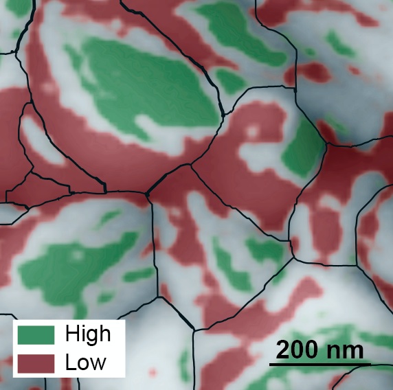
This atomic force microscopy image of the grainy surface of a perovskite solar cell reveals a new path to much greater efficiency. Individual grains are outlined in black, low-performing facets are red, and high-performing facets are green. A big jump in efficiency could possibly be obtained if the material can be grown so that more high-performing facets develop. (Credit: Berkeley Lab)

This atomic force microscopy image of the grainy surface of a perovskite solar cell reveals a new path to much greater efficiency. Individual grains are outlined in black, low-performing facets are red, and high-performing facets are green. A big jump in efficiency could possibly be obtained if the material can be grown so that more high-performing facets develop. (Credit: Berkeley Lab)

It’s always fascinating to observe a trend (or a craze) in science, an endeavour that outsiders (like me) tend to think of as impervious to such vagaries. Perovskite seems to be making its way past the trend/craze phase and moving into a more meaningful phase. From a July 4, 2016 news item on Nanowerk,

Scientists from the Department of Energy’s Lawrence Berkeley National Laboratory (Berkeley Lab) have discovered a possible secret to dramatically boosting the efficiency of perovskite solar cells hidden in the nanoscale peaks and valleys of the crystalline material.

Solar cells made from compounds that have the crystal structure of the mineral perovskite have captured scientists’ imaginations. They’re inexpensive and easy to fabricate, like organic solar cells. Even more intriguing, the efficiency at which perovskite solar cells convert photons to electricity has increased more rapidly than any other material to date, starting at three percent in 2009 — when researchers first began exploring the material’s photovoltaic capabilities — to 22 percent today. This is in the ballpark of the efficiency of silicon solar cells.

Now, as reported online July 4, 2016 in the journal Nature Energy (“Facet-dependent photovoltaic efficiency variations in single grains of hybrid halide perovskite”), a team of scientists from the Molecular Foundry and the Joint Center for Artificial Photosynthesis, both at Berkeley Lab, found a surprising characteristic of a perovskite solar cell that could be exploited for even higher efficiencies, possibly up to 31 percent.

A July 4, 2016 Berkeley Lab news release (also on EurekAlert), which originated the news item, details the research,

Using photoconductive atomic force microscopy, the scientists mapped two properties on the active layer of the solar cell that relate to its photovoltaic efficiency. The maps revealed a bumpy surface composed of grains about 200 nanometers in length, and each grain has multi-angled facets like the faces of a gemstone.

Unexpectedly, the scientists discovered a huge difference in energy conversion efficiency between facets on individual grains. They found poorly performing facets adjacent to highly efficient facets, with some facets approaching the material’s theoretical energy conversion limit of 31 percent.

The scientists say these top-performing facets could hold the secret to highly efficient solar cells, although more research is needed.

“If the material can be synthesized so that only very efficient facets develop, then we could see a big jump in the efficiency of perovskite solar cells, possibly approaching 31 percent,” says Sibel Leblebici, a postdoctoral researcher at the Molecular Foundry.

Leblebici works in the lab of Alexander Weber-Bargioni, who is a corresponding author of the paper that describes this research. Ian Sharp, also a corresponding author, is a Berkeley Lab scientist at the Joint Center for Artificial Photosynthesis. Other Berkeley Lab scientists who contributed include Linn Leppert, Francesca Toma, and Jeff Neaton, the director of the Molecular Foundry.

A team effort

The research started when Leblebici was searching for a new project. “I thought perovskites are the most exciting thing in solar right now, and I really wanted to see how they work at the nanoscale, which has not been widely studied,” she says.

She didn’t have to go far to find the material. For the past two years, scientists at the nearby Joint Center for Artificial Photosynthesis have been making thin films of perovskite-based compounds, and studying their ability to convert sunlight and CO2 into useful chemicals such as fuel. Switching gears, they created pervoskite solar cells composed of methylammonium lead iodide. They also analyzed the cells’ performance at the macroscale.

The scientists also made a second set of half cells that didn’t have an electrode layer. They packed eight of these cells on a thin film measuring one square centimeter. These films were analyzed at the Molecular Foundry, where researchers mapped the cells’ surface topography at a resolution of ten nanometers. They also mapped two properties that relate to the cells’ photovoltaic efficiency: photocurrent generation and open circuit voltage.

This was performed using a state-of-the-art atomic force microscopy technique, developed in collaboration with Park Systems, which utilizes a conductive tip to scan the material’s surface. The method also eliminates friction between the tip and the sample. This is important because the material is so rough and soft that friction can damage the tip and sample, and cause artifacts in the photocurrent.

Surprise discovery could lead to better solar cells

The resulting maps revealed an order of magnitude difference in photocurrent generation, and a 0.6-volt difference in open circuit voltage, between facets on the same grain. In addition, facets with high photocurrent generation had high open circuit voltage, and facets with low photocurrent generation had low open circuit voltage.

“This was a big surprise. It shows, for the first time, that perovskite solar cells exhibit facet-dependent photovoltaic efficiency,” says Weber-Bargioni.

Adds Toma, “These results open the door to exploring new ways to control the development of the material’s facets to dramatically increase efficiency.”

In practice, the facets behave like billions of tiny solar cells, all connected in parallel. As the scientists discovered, some cells operate extremely well and others very poorly. In this scenario, the current flows towards the bad cells, lowering the overall performance of the material. But if the material can be optimized so that only highly efficient facets interface with the electrode, the losses incurred by the poor facets would be eliminated.

“This means, at the macroscale, the material could possibly approach its theoretical energy conversion limit of 31 percent,” says Sharp.

A theoretical model that describes the experimental results predicts these facets should also impact the emission of light when used as an LED. …

The Molecular Foundry is a DOE Office of Science User Facility located at Berkeley Lab. The Joint Center for Artificial Photosynthesis is a DOE Energy Innovation Hub led by the California Institute of Technology in partnership with Berkeley Lab.

Here’s a link to and a citation for the paper,

Facet-dependent photovoltaic efficiency variations in single grains of hybrid halide perovskite by Sibel Y. Leblebici, Linn Leppert, Yanbo Li, Sebastian E. Reyes-Lillo, Sebastian Wickenburg, Ed Wong, Jiye Lee, Mauro Melli, Dominik Ziegler, Daniel K. Angell, D. Frank Ogletree, Paul D. Ashby, Francesca M. Toma, Jeffrey B. Neaton, Ian D. Sharp, & Alexander Weber-Bargioni. Nature Energy 1, Article number: 16093 (2016  doi:10.1038/nenergy.2016.93 Published online: 04 July 2016

This paper is behind a paywall.

Dexter Johnson’s July 6, 2016 posting on his Nanoclast blog (on the IEEE [Institute of Electrical and Electronics Engineers] website} presents his take on the impact that this new finding may have,

The rise of the crystal perovskite as a potential replacement for silicon in photovoltaics has been impressive over the last decade, with its conversion efficiency improving from 3.8 to 22.1 percent over that time period. Nonetheless, there has been a vague sense that this rise is beginning to peter out of late, largely because when a solar cell made from perovskite gets larger than 1 square centimeter the best conversion efficiency had been around 15.6 percent. …

Nano and a Unified Microbiome Initiative (UMI)

A Jan. 6, 2015 news item on Nanowerk features a proposal by US scientists for a Unified Microbiome Initiative (UMI),

In October [2015], an interdisciplinary group of scientists proposed forming a Unified Microbiome Initiative (UMI) to explore the world of microorganisms that are central to life on Earth and yet largely remain a mystery.

An article in the journal ACS Nano (“Tools for the Microbiome: Nano and Beyond”) describes the tools scientists will need to understand how microbes interact with each other and with us.

A Jan. 6, 2016 American Chemical Society (ACS) news release, which originated the news item, expands on the theme,

Microbes live just about everywhere: in the oceans, in the soil, in the atmosphere, in forests and in and on our bodies. Research has demonstrated that their influence ranges widely and profoundly, from affecting human health to the climate. But scientists don’t have the necessary tools to characterize communities of microbes, called microbiomes, and how they function. Rob Knight, Jeff F. Miller, Paul S. Weiss and colleagues detail what these technological needs are.

The researchers are seeking the development of advanced tools in bioinformatics, high-resolution imaging, and the sequencing of microbial macromolecules and metabolites. They say that such technology would enable scientists to gain a deeper understanding of microbiomes. Armed with new knowledge, they could then tackle related medical and other challenges with greater agility than what is possible today.

Here’s a link to and a citation for the paper,

Tools for the Microbiome: Nano and Beyond by Julie S. Biteen, Paul C. Blainey, Zoe G. Cardon, Miyoung Chun, George M. Church, Pieter C. Dorrestein, Scott E. Fraser, Jack A. Gilbert, Janet K. Jansson, Rob Knight, Jeff F. Miller, Aydogan Ozcan, Kimberly A. Prather, Stephen R. Quake, Edward G. Ruby, Pamela A. Silver, Sharif Taha, Ger van den Engh, Paul S. Weiss, Gerard C. L. Wong, Aaron T. Wright, and Thomas D. Young. ACS Nano, Article ASAP DOI: 10.1021/acsnano.5b07826 Publication Date (Web): December 22, 2015

Copyright © 2015 American Chemical Society

This is an open access paper.

I sped through very quickly and found a couple of references to ‘nano’,

Ocean Microbiomes and Nanobiomes

Life in the oceans is supported by a community of extremely small organisms that can be called a “nanobiome.” These nanoplankton particles, many of which measure less than 0.001× the volume of a white blood cell, harvest solar and chemical energy and channel essential elements into the food chain. A deep network of larger life forms (humans included) depends on these tiny microbes for its energy and chemical building blocks.

The importance of the oceanic nanobiome has only recently begun to be fully appreciated. Two dominant forms, Synechococcus and Prochlorococcus, were not discovered until the 1980s and 1990s.(32-34) Prochloroccus has now been demonstrated to be so abundant that it may account for as much as 10% of the world’s living organic carbon. The organism divides on a diel cycle while maintaining constant numbers, suggesting that about 5% of the world’s biomass flows through this species on a daily basis.(35-37)

Metagenomic studies show that many other less abundant life forms must exist but elude direct observation because they can neither be isolated nor grown in culture.

The small sizes of these organisms (and their genomes) indicate that they are highly specialized and optimized. Metagenome data indicate a large metabolic heterogeneity within the nanobiome. Rather than combining all life functions into a single organism, the nanobiome works as a network of specialists that can only exist as a community, therein explaining their resistance to being cultured. The detailed composition of the network is the result of interactions between the organisms themselves and the local physical and chemical environment. There is thus far little insight into how these networks are formed and how they maintain steady-state conditions in the turbulent natural ocean environment.

Rather than combining all life functions into a single organism, the nanobiome works as a network of specialists that can only exist as a community

The serendipitous discovery of Prochlorococcus happened by applying flow cytometry (developed as a medical technique for counting blood cells) to seawater.(34) With these medical instruments, the faint signals from nanoplankton can only be seen with great difficulty against noisy backgrounds. Currently, a small team is adapting flow cytometric technology to improve the capabilities for analyzing individual nanoplankton particles. The latest generation of flow cytometers enables researchers to count and to make quantitative observations of most of the small life forms (including some viruses) that comprise the nanobiome. To our knowledge, there are only two well-equipped mobile flow cytometry laboratories that are regularly taken to sea for real-time observations of the nanobiome. The laboratories include equipment for (meta)genome analysis and equipment to correlate the observations with the local physical parameters and (nutrient) chemistry in the ocean. Ultimately, integration of these measurements will be essential for understanding the complexity of the oceanic microbiome.

The ocean is tremendously undersampled. Ship time is costly and limited. Ultimately, inexpensive, automated, mobile biome observatories will require methods that integrate microbiome and nanobiome measurements, with (meta-) genomics analyses, with local geophysical and geochemical parameters.(38-42) To appreciate how the individual components of the ocean biome are related and work together, a more complete picture must be established.

The marine environment consists of stratified zones, each with a unique, characteristic biome.(43) The sunlit waters near the surface are mixed by wind action. Deeper waters may be mixed only occasionally by passing storms. The dark deepest layers are stabilized by temperature/salinity density gradients. Organic material from the photosynthetically active surface descends into the deep zone, where it decomposes into nutrients that are mixed with compounds that are released by volcanic and seismic action. These nutrients diffuse upward to replenish the depleted surface waters. The biome is stratified accordingly, sometimes with sudden transitions on small scales. Photo-autotrophs dominate near the surface. Chemo-heterotrophs populate the deep. The makeup of the microbial assemblages is dictated by the local nutrient and oxygen concentrations. The spatiotemporal interplay of these systems is highly relevant to such issues as the carbon budget of the planet but remains little understood.

And then, there was this,

Nanoscience and Nanotechnology Opportunities

The great advantage of nanoscience and nanotechnology in studying microbiomes is that the nanoscale is the scale of function in biology. It is this convergence of scales at which we can “see” and at which we can fabricate that heralds the contributions that can be made by developing new nanoscale analysis tools.(159-168) Microbiomes operate from the nanoscale up to much larger scales, even kilometers, so crossing these scales will pose significant challenges to the field, in terms of measurement, stimulation/response, informatics, and ultimately understanding.

Some progress has been made in creating model systems(143-145, 169-173) that can be used to develop tools and methods. In these cases, the tools can be brought to bear on more complex and real systems. Just as nanoscience began with the ability to image atoms and progressed to the ability to manipulate structures both directly and through guided interactions,(162, 163, 174-176) it has now become possible to control structure, materials, and chemical functionality from the submolecular to the centimeter scales simultaneously. Whereas substrates and surface functionalization have often been tailored to be resistant to bioadhesion, deliberate placement of chemical patterns can also be used for the growth and patterning of systems, such as biofilms, to be put into contact with nanoscale probes.(177-180) Such methods in combination with the tools of other fields (vide infra) will provide the means to probe and to understand microbiomes.

Key tools for the microbiome will need to be miniaturized and made parallel. These developments will leverage decades of work in nanotechnology in the areas of nanofabrication,(181) imaging systems,(182, 183) lab-on-a-chip systems,(184) control of biological interfaces,(185) and more. Commercialized and commoditized tools, such as smart phone cameras, can also be adapted for use (vide infra). By guiding the development and parallelization of these tools, increasingly complex microbiomes will be opened for study.(167)

Imaging and sensing, in general, have been enjoying a Renaissance over the past decades, and there are various powerful measurement techniques that are currently available, making the Microbiome Initiative timely and exciting from the broad perspective of advanced analysis techniques. Recent advances in various -omics technologies, electron microscopy, optical microscopy/nanoscopy and spectroscopy, cytometry, mass spectroscopy, atomic force microscopy, nuclear imaging, and other techniques, create unique opportunities for researchers to investigate a wide range of questions related to microbiome interactions, function, and diversity. We anticipate that some of these advanced imaging, spectroscopy, and sensing techniques, coupled with big data analytics, will be used to create multimodal and integrated smart systems that can shed light onto some of the most important needs in microbiome research, including (1) analyzing microbial interactions specifically and sensitively at the relevant spatial and temporal scales; (2) determining and analyzing the diversity covered by the microbial genome, transcriptome, proteome, and metabolome; (3) managing and manipulating microbiomes to probe their function, evaluating the impact of interventions and ultimately harnessing their activities; and (4) helping us identify and track microbial dark matter (referring to 99% of micro-organisms that cannot be cultured).

In this broad quest for creating next-generation imaging and sensing instrumentation to address the needs and challenges of microbiome-related research activities comprehensively, there are important issues that need to be considered, as discussed below.

The piece is extensive and quite interesting, if you have the time.
Characterizing anatase titanium dixoide at the nanoscale

An international collaboration of researchers combined atomic force microscopy (AFM) and scanning tunneling microscopy (STM) to characterize anatase titanium dixoxide. From a Sept. 14, 2015 news item on Azonano,

A [Japan National Institute for Materials Science] NIMS research team successfully identified the atoms and common defects existing at the most stable surface of the anatase form of titanium dioxide by characterizing this material at the atomic scale with scanning probe microscopy. This work was published under open access policy in the online version of Nature Communications on June 29, 2015.

A June 29, 2015 NIMS press release, which originated the news item, includes the paper’s abstract in numbered point form,

  1. The research team consisting of Oscar Custance and Tomoko Shimizu, group leader and senior scientist, respectively, at the Atomic Force Probe Group, NIMS, Daisuke Fujita and Keisuke Sagisaka, group leader and senior researcher, respectively, at the Surface Characterization Group, NIMS, and scientists at Charles University in the Czech Republic, Autonomous University of Madrid in Spain, and other organizations combined simultaneous atomic force microscopy (AFM) and scanning tunneling microscopy (STM) measurements with first-principles calculations for the unambiguous identification of the atomic species at the most stable surface of the anatase form of titanium dioxide (hereinafter referred to as anatase) and its most common defects.
  2. In recent years, anatase has attracted considerable attention, because it has become a pivotal material in devices for photo-catalysis and for the conversion of solar energy to electricity. It is extremely challenging to grow large single crystals of anatase, and most of the applications of this material are in the form of nano crystals. To enhance the catalytic reactivity of anatase and the efficiency of devices for solar energy conversion based on anatase, it is critical to gain in-depth understanding and control of the reactions taking place at the surface of this material down to the atomic level. Only a few research groups worldwide possess the technology to create proper test samples and to make in-situ atomic-level observations of anatase surfaces.
  3. In this study, the research team used samples obtained from anatase natural single crystals extracted from naturally occurring anatase rocks. The team characterized the (101) surface of anatase at atomic level by means of simultaneous AFM and STM. Using single water molecules as atomic markers, the team successfully identified the atomic species of this surface; result that was additionally confirmed by the comparison of simultaneous AFM and STM measurements with the outcomes of first-principles calculations.
  4. In regular STM, in which an atomically sharp probe is scanned over the surface by keeping constant an electrical current flowing between them, it is difficult to stably image anatase surfaces as this material presents poor electrical conductivity over some of the atomic positions of the surface. However, simultaneous operation of AFM and STM allowed imaging the surface with atomic resolution even within the materials band gap (a region where the flow of current between the probe and the surface is, in principle, prohibited). Here, the detection of inter-atomic forces between the last atom of the atomically sharp probe and the atoms of the surface by AFM was of crucial importance. By regulating the probe-surface distance using AFM, it was possible to image the surface at atomic-scale while collecting STM data over both conductive and not conductive areas of the surface. By comparing simultaneous AFM and STM measurements with theoretical simulations, the team was not only able to discern which atomic species were contributing to the AFM and the STM images but also to identify the most common defects found at the surface.
  5. In the future, based on the information gained from this study, the NIMS research team will conduct research on molecules of technologically relevance that adsorb on anatase and characterize these hybrid systems by using simultaneous AFM and STM. Their ultimate goal is to formulate novel approaches for the development of photo-catalysts and solar cell materials and devices.

Here’s a link to and a citation for the paper,

Atomic species identification at the (101) anatase surface by simultaneous scanning tunnelling and atomic force microscopy by Oleksandr Stetsovych, Milica Todorović, Tomoko K. Shimizu, César Moreno, James William Ryan, Carmen Pérez León, Keisuke Sagisaka, Emilio Palomares, Vladimír Matolín, Daisuke Fujita, Ruben Perez, & Oscar Custance. Nature Communications 6, Article number: 7265 doi:10.1038/ncomms8265 Published 29 June 2015

This is an open access paper.

Corrections: Hybrid Photonic-Nanomechanical Force Microscopy uses vibration for better chemical analysis

*ETA  Nov. 4, 2015: I’m apologizing to anyone wishing to read this posting as it’s a bit of a mess. I deeply regret mishandling the situation. In future, I shall not be taking any corrections from individual researchers to materials such as news releases that have been issued by an institution. Whether or not the individual researchers are happy with how their contributions or how a colleague’s contributions or how their home institutions have been characterized is a matter for them and their home institutions.

The August 10, 2015 ORNL news release with all the correct details has been added to the end of this post.*

A researcher at the University of Central Florida (UCF) has developed a microscope that uses vibrations for better analysis of chemical composition. From an Aug. 10, 2015 news item on Nanowerk,

It’s a discovery that could have promising implications for fields as varied as biofuel production, solar energy, opto-electronic devices, pharmaceuticals and medical research.

“What we’re interested in is the tools that allow us to understand the world at a very small scale,” said UCF professor Laurene Tetard, formerly of the Oak Ridge National Laboratory. “Not just the shape of the object, but its mechanical properties, its composition and how it evolves in time.”

An Aug. 10, 2015 UCF news release (also on EurekAlert), which originated the news item, describes the limitations of atomic force microscopy and gives a few details about the hybrid microscope (Note: A link has been removed),

For more than two decades, scientists have used atomic force microscopy – a probe that acts like an ultra-sensitive needle on a record player – to determine the surface characteristics of samples at the microscopic scale. A “needle” that comes to an atoms-thin point traces a path over a sample, mapping the surface features at a sub-cellular level [nanoscale].

But that technology has its limits. It can determine the topographical characteristics of [a] sample, but it can’t identify its composition. And with the standard tools currently used for chemical mapping, anything smaller than roughly half a micron is going to look like a blurry blob, so researchers are out of luck if they want to study what’s happening at the molecular level.

A team led by Tetard has come up with a hybrid form of that technology that produces a much clearer chemical image. As described Aug. 10 in the journal Nature Nanotechnology, Hybrid Photonic-Nanomechanical Force Microscopy (HPFM) can discern a sample’s topographic characteristics together with the chemical properties at a much finer scale.

The HPFM method is able to identify materials based on differences in the vibration produced when they’re subjected to different wavelengths of light – essentially a material’s unique “fingerprint.”

“What we are developing is a completely new way of making that detection possible,” said Tetard, who has joint appointments to UCF’s Physics Department, Material Science and Engineering Department and the NanoScience Technology Center.

The researchers proved the effectiveness of HPFM while examining samples from an eastern cottonwood tree, a potential source of biofuel. By examining the plant samples at the nanoscale, the researchers for the first time were able to determine the molecular traits of both untreated and chemically processed cottonwood inside the plant cell walls.

The research team included Tetard; Ali Passian, R.H. Farahi and Brian Davison, all of Oak Ridge National Laboratory; and Thomas Thundat of the University of Alberta.

Long term, the results will help reveal better methods for producing the most biofuel from the cottonwood, a potential boon for industry. Likewise, the new method could be used to examine samples of myriad plants to determine whether they’re good candidates for biofuel production.

Potential uses of the technology go beyond the world of biofuel. Continued research may allow HPFM to be used as a probe so, for instance, it would be possible to study the effect of new treatments being developed to save plants such as citrus trees from bacterial diseases rapidly decimating the citrus industry, or study fundamental photonically-induced processes in complex systems such as in solar cell materials or opto-electronic devices.

Here’s a link to and a citation for the paper,

Opto-nanomechanical spectroscopic material characterization by L. Tetard, A. Passian, R. H. Farahi, T. Thundat, & B. H. Davison. Nature Nanotechnology (2015) doi:10.1038/nnano.2015.168 Published online 10 August 2015

This paper is behind a paywall.

*ETA August 27, 2015:

August 10, 2015 ORNL news release (Note: Funding information and a link to the paper [previously given] have been removed):

A microscope being developed at the Department of Energy’s Oak Ridge National Laboratory will allow scientists studying biological and synthetic materials to simultaneously observe chemical and physical properties on and beneath the surface.

The Hybrid Photonic Mode-Synthesizing Atomic Force Microscope is unique, according to principal investigator Ali Passian of ORNL’s Quantum Information System group. As a hybrid, the instrument, described in a paper published in Nature Nanotechnology, combines the disciplines of nanospectroscopy and nanomechanical microscopy.

“Our microscope offers a noninvasive rapid method to explore materials simultaneously for their chemical and physical properties,” Passian said. “It allows researchers to study the surface and subsurface of synthetic and biological samples, which is a capability that until now didn’t exist.”

ORNL’s instrument retains all of the advantages of an atomic force microscope while simultaneously offering the potential for discoveries through its high resolution and subsurface spectroscopic capabilities.

“The originality of the instrument and technique lies in its ability to provide information about a material’s chemical composition in the broad infrared spectrum of the chemical composition while showing the morphology of a material’s interior and exterior with nanoscale – a billionth of a meter – resolution,” Passian said.

Researchers will be able to study samples ranging from engineered nanoparticles and nanostructures to naturally occurring biological polymers, tissues and plant cells.

The first application as part of DOE’s BioEnergy Science Center was in the examination of plant cell walls under several treatments to provide submicron characterization. The plant cell wall is a layered nanostructure of biopolymers such as cellulose. Scientists want to convert such biopolymers to free the useful sugars and release energy.

An earlier instrument, also invented at ORNL, provided imaging of poplar cell wall structures that yielded unprecedented topological information, advancing fundamental research in sustainable biofuels.

Because of this new instrument’s impressive capabilities, the researcher team envisions broad applications.
“An urgent need exists for new platforms that can tackle the challenges of subsurface and chemical characterization at the nanometer scale,” said co-author Rubye Farahi. “Hybrid approaches such as ours bring together multiple capabilities, in this case, spectroscopy and high-resolution microscopy.”

Looking inside, the hybrid microscope consists of a photonic module that is incorporated into a mode-synthesizing atomic force microscope. The modular aspect of the system makes it possible to accommodate various radiation sources such as tunable lasers and non-coherent monochromatic or polychromatic sources.

ETA2 August 27, 2015: I’ve received an email from one of the paper’s authors (RH Farahi of the US Oak Ridge National Laboratory [ORNL]) who claims some inaccuracies in this piece.  The news release supplied by the University of Central Florida states that Dr. Tetard led the team and that is not so. According to Dr. Farahi, she had a postdoctoral position on the team which she left two years ago. You might also get the impression that some of the work was performed at the University of Central Florida. That is not so according to Dr. Farahi.  As a courtesy Dr. Tetard was retained as first author of the paper.

*Nov. 4, 2015: I suspect some of the misunderstanding was due to overeagerness and/or time pressures. Whoever wrote the news release may have made some assumptions. It’s very easy to make a mistake when talking to an ebullient scientist who can unintentionally lead you to believe something that’s not so. I worked in a high tech company and believed that there was some new software being developed which turned out to be a case of high hopes. Luckily, I said something that triggered a rapid rebuttal to the fantasies. Getting back to this situation, other contributing factors could include the writer not having time to get the news release reviewed the scientist or the scientist skimming the release and missing a few bits due to time pressure.*

Park Nano Academy: How Graphene–based Nanomaterials and Films Revolutionize Science webinar

There’s another Park Systems webinar coming up on July 9, 2015 (the last one concerning Nanostructured Polymers and Nanomaterials for Oil & Gas was mentioned  in my June 9, 2015 posting).

This latest webinar series is focused on graphene, from a June 29, 2015 Park Systems news release,

Park Systems, world-leader in atomic force microscopy (AFM) is hosting a webinar to provide advanced scientific research into new classes of Nanoscale Graphene-based materials poised to revolutionize industries such as semiconductor, material science, bio science and energy.   Touted as ‘the wonder material of the 21st Century’ by the researchers who were awarded the 2010 Nobel Prize in physics for their graphene research,  this carbon-based lightweight material is 200 times stronger than steel and one of the most promising and versatile materials ever discovered.

The Park Systems Webinar titled Graphene Based Nanomaterials and Films will be given by Professor Rigoberto Advincula of Case Western Reserve University on July 9, 2015 at 9am PST.  Prof. Advincula is an eminent professor, researcher and expert in the area of polymers, smart coatings, nanomaterials, surface analytical methods for a variety of applications.

“The discovery of graphene is but a continuing evolution on how we analyze, treat, synthesize carbon based nanomaterials which includes the fullerenes, nanotubes, and now C polymorph platelets called graphene,” explains Dr. Advincula.  “Graphene is used in many areas of research and potential applications for electronics, solid-state devices, biosensors, coatings and much more for numerous industries where there are opportunities to make quantum improvements in methods and materials.”

Graphene is part of the C polymorph family of nanomaterials and because of the platy nature of the basal plane, it’s reactivity on the edges, and various redox forms, it is an excellent thin film additive and component that can be grown by vapor deposition methods as well as exfoliation. Current research into dispersion, preparations, and patterning of graphene using Park Systems AFM to identify nanoscale characteristics and surface properties as well as conductivity indicates that numerous breakthroughs in materials and chemicals are on the horizon.

“Park AFM is the natural tool to investigate Graphene’s adsorbed state on a flat substrate as well as characterize its surface properties and conductivity because of the reliability and accuracy of the equipment,” adds Dr. Advincula who will give the Webinar on July 9. “AFM is useful in understanding the surface properties of these products but is equally valuable in failure analysis because of the capability to do in-situ or real time measurements of failure with a temperature stage or a magnetic field.”

Graphene-based Nanomaterials offer many innovations in industries such as electronics, semiconductor, life science, material science and bio science. Some potential advancements already being researched include flexible electronics, anti bacterial paper, actuators, electrochoromic devices and transistors.

“Park Systems is presenting this webinar as part of Park Nano Academy, which will offer valuable education and shared knowledge across many Nano Science Disciplines and Industries as a way to further enable NanoScale advancements,” comments Keibock Lee, Park Systems President.  “We invite all curious Nano Researchers to join our webinars and educational forums to launch innovative ideas that propel us into future Nano Scientific Technologies.”

The webinar will highlight how the research into is conducted and present some of the findings by Professor Rigoberto Advincula of Case Western Reserve University.

This webinar is available at no cost and is part of Park Systems Nano Academy.

To register go to: http://www.parkafm.com/index.php/medias/nano-academy/webinars/115-webinars/486-nanomaterials-webinar-july-9-2015

Enjoy!

Nanotech and the oil and gas industry: a webinar

How serendipitous! I stumbled on an announcement from Park Systems for a webinar designed for the oil and gas industry after my June 8, 2015 post featuring Abakan and its new Alberta (Canada)-based cladding facility designed for oil and gas pipes in particular. From a June 8, 2015 news item on Nanowerk,

Park Systems, world-leader in atomic force microscopy (AFM) today announced a webinar to provide next generation technology to improve oil and gas production in both traditional drilling and hydraulic fracturing for oil & gas producers and equipment manufacturers as they continue to pursue the latest developments in production efficiencies.

A June 8, 2015 Park Systems news release, which originated the news item, expands on the theme,

The oil and gas industry is ripe for innovation and the cost of extracting oil can be reduced. Research at PETRO Case Consortium is uncovering new materials, chemicals and coatings that improves yield and reduce costs and with an eye towards diminishing the impact on our environment. This webinar is part of an ongoing series offered by Park System’s new Nano Academy, a platform for providing education and shared knowledge on the latest advancements across a wide spectrum of nanosciences.

This webinar titled Nanostructured Polymers and Nanomaterials for Oil & Gas will be given June 11 [2015] by Dr. Rigoberto Advincula, Director of the Petro Case Consortium and Professor with the Department of Macromolecular Science and Engineering at Case Western Reserve University and is designed to offer innovations in microscopy nanotechnology for oil & gas producers and suppliers.

“Our best in class AFM equipment registers nanoparticle observations and analysis not previously available that extends the ability to analyze chemicals and materials to develop the optimum efficiency,” said Keibock Lee, President of Park Systems. “We are proud to offer this webinar for the oil & gas industry, showcasing Dr. Advincula’s outstanding contribution towards cost reduction and sustainability for the current energy producers and paving the way for future innovations that can enable global energy solutions.”

PETRO Case Consortium at Case Western [Reserve] University, led by Dr. Advincula, is working hard to ensure that the industry can catch up with new technology and apply it to oil & gas production that improves productivity by creating longer lasting concrete, coatings and apply other methods to increase yield in production. This webinar is the first of a series that will cover multiple topics related to nano scale developments across a wide variety of research applications and bio scientific fields.
“Hydraulic fracturing and directional drilling has unlocked many resources,” states Dr. Advincula. “Revolutionary new microscopy technology provided thru Park Systems AFM (Atomic Force Microscopy) and new innovations in chemical and material research indicates that there is a defined opportunity to use the advances in chemistry, materials, and nanoscience to make valuable industry process updates.”

For the last 10 years there has been an increase in interest and research for new materials useful for upstream, midstream, and downstream processes to effectively find function in demanding environments including directional drilling and hydraulic fracturing. High temperature high pressure (HT/HP) and brine conditions pose a challenge for emulsification, demulsification, and viscosity of drilling fluids. Usually the “easy” oil or conventional oil has allowed technologies even dating back to the first oil well in Pennsylvania to become very profitable. But with high pressure high temperature (HPHT) conditions in the most challenging wells, many of the established technologies and materials do not suffice.

The discovery driven group, PETRO Case Consortium at Case Western University, a Park AFM user, investigates the area of molecular, macromolecular, and supramolecular synthesis and structure of polymers and nanomaterials capable of controlled-assembly to form ultrathin films and dispersions with the aim of finding new technologies and materials that improve and replace established oil and gas field formations.

For instance, the evaluation of chemicals and changing or altering the formulas can greatly improve production yields. Different chemicals used for the field include inhibitors for scaling, fouling, corrosion, asphaltene control, formation damage, differential pressures in multiphase environments which will be met by new synthesis methods including metathesis reactions, bio based feedstocks, new polymer surfactants, living polymers, and nanoparticle. Other uses of new chemical technologies include tracers and reporters for geomapping and well connectivity, as well as different types of fluid loss agents that prevent formation damage or keep well integrity, and smart and stimuli-responsive nanoparticles that can be used for improving gelation.

This webinar is available at no cost and is part of Park Systems Nano Academy which will offer valuable education and shared knowledge across many Nano Science Disciplines and Industries as a way to further enable NanoScale advancements. To register go to: http://bit.do/polyoilgas

Webinar logistics (from the Park Systems news release),

About Webinar
Title: Nanostructured Polymers and Nanomaterials for Oil & Gas
Date: June 11, 2015
Time: 9am PST
To Register, go to: http://bit.do/polyoilgas
Pre-requisite: Knowledge of oil field chemicals and rubber materials is preferred but not required.

Here’s more about the expert (from the news release),

About Prof. Rigoberto Advincula
Prof. Rigoberto Advincula, Director of the Petro Case Consortium, is recognized industry-wide as an expert regarding polymer and materials challenges of the oil-gas industry. He is currently a Professor with the Department of Macromolecular Science and Engineering at Case Western Reserve University and is the recipient of numerous awards including Fellow of the American Chemical Society, Herman Mark Scholar Award of the Polymer Division, and Humboldt Fellow.

The news release also included some information about Park Systems,

About Park Systems
Park Systems is a world-leading manufacturer of atomic force microscopy (AFM) systems with a complete range of products for researchers and industry engineers in chemistry, materials, physics, life sciences, semiconductor and data storage industries. Park’s products are used by over a thousand of institutions and corporations worldwide. Park’s AFM provides highest data accuracy at nanoscale resolution, superior productivity, and lowest operating cost thanks to its unique technology and innovative engineering. Park Systems, Inc. is headquartered in Santa Clara, California with its global manufacturing, and R&D headquarters in Korea. Park’s products are sold and supported worldwide with regional headquarters in the US, Korea, Japan, and Singapore, and distribution partners throughout Europe, Asia, and America. Please visit http://www.parkafm.com or call 408-986-1110 for more information.

So there you have it.

Nanorobotic approach to studying how skin falls apart

Scientists have combined robotic techniques with atomic force microscopy to achieve understanding of how skin falls apart at the nanoscale. From a Sept. 11, 2014 news item on Azonano,

University at Buffalo researchers and colleagues studying a rare, blistering disease have discovered new details of how autoantibodies destroy healthy cells in skin. This information provides new insights into autoimmune mechanisms in general and could help develop and screen treatments for patients suffering from all autoimmune diseases, estimated to affect 5-10 percent of the U.S. population.

“Our work represents a unique intersection between the fields of biology and engineering that allowed for entirely new investigational strategies applied to the study of clinical disease,” says Animesh A. Sinha, MD, PhD, Rita M. and Ralph T. Behling Professor and chair of the Department of Dermatology in the UB School of Medicine and Biomedical Sciences and senior author on the study.

A Sept. 9, 2014 University of Buffalo news release by Ellen Goldbaum (also on EurekAlert dated Sept. 10, 2014), which originated the news item, describes the condition and the research in more detail,

PV [Pemphigus Vulgaris] results in the often painful blistering of the skin and mucous membranes. Generally treated with corticosteroids and other immunosuppressive agents, the condition is life-threatening if untreated.

Sinha’s research team, in collaboration with scientists at Michigan State University, describe the use of atomic force microscopy (AFM), a technique originally developed to study nonbiological materials, to look at cell junctions and how they rupture, a process called acantholysis.

“It has been very difficult to study cell junctions, which maintain the skin’s barrier function by keeping cells attached to each other,” says Sinha. “These junctions, micron-sized spots on cell membranes, are very complex molecular structures. Their small size has made them resistant to detailed investigation.”

Sinha’s interest lies in determining what destroys those junctions in Pemphigus Vulgaris.

“We haven’t understood why some antibodies generated by the condition cause blisters and why other antibodies it generates do not,” says Sinha.

By studying the connections between skin cells using AFM and other techniques that probe cells at the nanoscale, Sinha and his colleagues report that pathogenic antibodies change structural and functional properties of skin cells in distinct ways.

“Our data suggest a new model for the action of autoantibodies in which there are two steps or ‘hits’ in the development of lesions,” says Sinha. “The first hit results in the initial separation of cells but only the pathogenic antibodies drive further intracellular changes that lead to the breaking of the cell junction and blistering.”

The researchers examined the cells using AFM, which requires minimal sample preparation and provides three-dimensional images of cell surfaces.

The AFM tip acts like a little probe, explains Sinha. When tapped against a cell, it sends back information regarding the cell’s mechanical properties, such as thickness, elasticity, viscosity and electrical potential.

“We combined existing and novel nanorobotic techniques with AFM, including a kind of nanodissection, where we physically detached cells from each other at certain points so that we could test what that did to their mechanical and biological functions,” Sinha adds.

Those data were then combined with information about functional changes in cell behavior to develop a nanomechanical profile, or phenotype, for specific cellular states.

He also envisions that this kind of nanomechanical phenotyping should allow for the development of predictive models for cellular behavior for any kind of cell.

“Ultimately, in the case of autoimmunity, we should be able to use these techniques as a high-throughput assay to screen hundreds or thousands of compounds that might block the effects of autoantibodies and identify novel agents with therapeutic potential in given individuals,” says Sinha.  “Such strategies aim to advance us toward a new era of personalized medicine”.

I found some more information about the nanorobotics technique, mentioned in the news release, in the researchers’ paper (Note: A link has been removed),

Nanorobotic surgery

AFM-based nanorobotics enables accurate and convenient sample manipulation and drug delivery. This capability was used in the current study to control the AFM tip position over the intercellular junction area, and apply vertical indentation forces, so that bundles of intercellular adhesion structures can be dissected precisely with an accuracy of less than 100 nm in height. We used a tip sharp enough (2 nm in tip apex diameter) to penetrate the cell membrane and the intermediate filaments. It has been shown that intermediate filaments have extremely high tensile strength by in vitro AFM stretching [19]. Thus, the vertical force and moving speed of the AFM cantilever (0.06 N/m in vertical spring constant) was controlled at a vertical force of 5 nN at an indentation speed of 0.1 µm/s to guarantee the rupture of the filament and to partially dissect cell adhesion structures between two neighboring cells.

For those who want to know more, here’s a link to and a citation for the paper,

Nanorobotic Investigation Identifies Novel Visual, Structural and Functional Correlates of Autoimmune Pathology in a Blistering Skin Disease Model by Kristina Seiffert-Sinha, Ruiguo Yang, Carmen K. Fung, King W. Lai, Kevin C. Patterson, Aimee S. Payne, Ning Xi, Animesh A. Sinha. PLOSONE Published: September 08, 2014 DOI: 10.1371/journal.pone.0106895

This is an open access paper.

Atomic force microscopes, images, and friction

To me, this looks like the ‘batman’ symbol but it’s not.

Nanofriction at the tip of the microscope. Courtesy SISSA [Scuola Internazionale Superiore di Studi Avanzat], Italy

Nanofriction at the tip of the microscope. Courtesy SISSA [Scuola Internazionale Superiore di Studi Avanzat], Italy

Here’s more about the work that produced this image from a Dec. 17, 2013 news item on Azonano,

Atomic force microscopes are able to reproduce spectacular images, at the scale of single atoms. This is made possible by the oscillation of a very sharp probe tip over the surface being observed. The tip never touches the surface but gets so close to it, at distances in the order of one billionth of a metre, that it “feels” the force due to the interaction with the atoms making up the material being observed.

These are tiny forces, in the order of nanonewtons (meaning one billion times smaller than the weight of an apple). By measuring these forces one can reproduce an image of the material. A research group, which brings together experimental physicists from the University of Basel and theoretical physicists from SISSA, has observed and explained a peculiar effect, a source of “friction” in this type of nanoscopic observations.

The Dec. 16,  2013 SISSA (Scuola Internazionale Superiore di Studi Avanzat) press release, which originated the news item, provides more specific detail,

 When the tip of the microscope oscillates over certain surfaces, in this case over NbSe2 (niobium selenide), peaks of “dissipation” (i.e., loss of energy) can be seen when the tip is at specific distances from the surface, as if it were held back, at certain locations, by some frictional force. This effect, which is related to a property of the surface known as charge density waves (CDW), was experimentally observed by the Basel physicists and first explained by Franco Pellegrini, Giuseppe Santoro and Erio Tosatti, of SISSA, by means of a theoretical model analysed with the use of numerical simulations.

“Our model describes in detail the interaction between the tip of the atomic force microscope and the CDW,” explains Pellegrini. “The model reproduces – and predicts – the data observed experimentally”.

“Knowledge of nanofriction is important today. Progressive miniaturization of electronic devices makes it crucial to understand the mechanisms underlying energy losses, continues Pelligrini.

Here’s a link to and a citation for the paper,

Giant frictional dissipation peaks and charge-density-wave slips at the NbSe2 surface by Markus Langer, Marcin Kisiel, Rémy Pawlak, Franco Pellegrini, Giuseppe E. Santoro, Renato Buzio, Andrea Gerbi, Geetha Balakrishnan, Alexis Baratoff, Erio Tosatti & Ernst Meyer. Nature Materials (2013) doi:10.1038/nmat3836 Published online 15 December 2013

This paper is behind a paywall although you can obtain a preview through ReadCube Access.