Tag Archives: University of California at Los Angeles

Making a graphene micro-supercapacitor with a home DVD burner

Not all science research and breakthroughs require massive investments of money, sometimes all you need is a home DVD burner as this Feb. 19, 2013 news release on EurekAlert notes,

While the demand for ever-smaller electronic devices has spurred the miniaturization of a variety of technologies, one area has lagged behind in this downsizing revolution: energy-storage units, such as batteries and capacitors.

Now, Richard Kaner, a member of the California NanoSystems Institute at UCLA and a professor of chemistry and biochemistry, and Maher El-Kady, a graduate student in Kaner’s laboratory, may have changed the game.

The UCLA researchers have developed a groundbreaking technique that uses a DVD burner to fabricate micro-scale graphene-based supercapacitors — devices that can charge and discharge a hundred to a thousand times faster than standard batteries. These micro-supercapacitors, made from a one-atom–thick layer of graphitic carbon, can be easily manufactured and readily integrated into small devices such as next-generation pacemakers.

The new cost-effective fabrication method, described in a study published this week in the journal Nature Communications, holds promise for the mass production of these supercapacitors, which have the potential to transform electronics and other fields.

“Traditional methods for the fabrication of micro-supercapacitors involve labor-intensive lithographic techniques that have proven difficult for building cost-effective devices, thus limiting their commercial application,” El-Kady said. “Instead, we used a consumer-grade LightScribe DVD burner to produce graphene micro-supercapacitors over large areas at a fraction of the cost of traditional devices. [emphasis mine] Using this technique, we have been able to produce more than 100 micro-supercapacitors on a single disc in less than 30 minutes, using inexpensive materials.”

The University of California at Los Angeles (UCLA) Feb. 19, 2013 news release written by David Malasarn, the origin of the EurekAlert news release, features more information about the process,

The process of miniaturization often relies on flattening technology, making devices thinner and more like a geometric plane that has only two dimensions. In developing their new micro-supercapacitor, Kaner and El-Kady used a two-dimensional sheet of carbon, known as graphene, which only has the thickness of a single atom in the third dimension.
Kaner and El-Kady took advantage of a new structural design during the fabrication. For any supercapacitor to be effective, two separated electrodes have to be positioned so that the available surface area between them is maximized. This allows the supercapacitor to store a greater charge. A previous design stacked the layers of graphene serving as electrodes, like the slices of bread on a sandwich. While this design was functional, however, it was not compatible with integrated circuits.
In their new design, the researchers placed the electrodes side by side using an interdigitated pattern, akin to interwoven fingers. This helped to maximize the accessible surface area available for each of the two electrodes while also reducing the path over which ions in the electrolyte would need to diffuse. As a result, the new supercapacitors have more charge capacity and rate capability than their stacked counterparts.
Interestingly, the researchers found that by placing more electrodes per unit area, they boosted the micro-supercapacitor’s ability to store even more charge.
Kaner and El-Kady were able to fabricate these intricate supercapacitors using an affordable and scalable technique that they had developed earlier. They glued a layer of plastic onto the surface of a DVD and then coated the plastic with a layer of graphite oxide. Then, they simply inserted the coated disc into a commercially available LightScribe optical drive — traditionally used to label DVDs — and took advantage of the drive’s own laser to create the interdigitated pattern. The laser scribing is so precise that none of the “interwoven fingers” touch each other, which would short-circuit the supercapacitor.
“To label discs using LightScribe, the surface of the disc is coated with a reactive dye that changes color on exposure to the laser light. Instead of printing on this specialized coating, our approach is to coat the disc with a film of graphite oxide, which then can be directly printed on,” Kaner said. “We previously found an unusual photo-thermal effect in which graphite oxide absorbs the laser light and is converted into graphene in a similar fashion to the commercial LightScribe process. With the precision of the laser, the drive renders the computer-designed pattern onto the graphite oxide film to produce the desired graphene circuits.”
“The process is straightforward, cost-effective and can be done at home,” El-Kady said. “One only needs a DVD burner and graphite oxide dispersion in water, which is commercially available at a moderate cost.”
The new micro-supercapacitors are also highly bendable and twistable, making them potentially useful as energy-storage devices in flexible electronics like roll-up displays and TVs, e-paper, and even wearable electronics.

The reference to e-paper and roll-up displays calls to mind work being done at Queen’s University (Kingston, Canada) and Roel Vertegaal’s work on bendable, flexible phones and computers (my Jan. 9, 2013 posting). Could this work on micro-supercapacitors have an impact on that work?

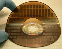
Here’s an image (supplied by UCLA) of the micro-supercapacitors ,

Kaner and El-Kady's micro-supercapacitors

Kaner and El-Kady’s micro-supercapacitors

UCLA has  also supplied a video of Kaner and El-Kady discussing their work,

Interestingly this video has been supported by GE (General Electric), a company which seems to be doing a great deal to be seen on the internet these days as per my Feb. 11, 2013 posting titled, Visualizing nanotechnology data with Seed Media Group and GE (General Electric).

Getting back to the researchers, they are looking for industry partners as per Malasarn’s news release.

‘Touching’ infrared light, if you’re a rat followed by announcement of US FDA approval of first commercial artificial retina (bionic eye)

Researcher Miguel Nicolelis and his colleagues at Duke University have implanted a neuroprosthetic device in the portion of a rat’s brain related to touch that allows the rats to see infrared light. From the Feb. 12, 2013 news release on EurekAlert,

Researchers have given rats the ability to “touch” infrared light, normally invisible to them, by fitting them with an infrared detector wired to microscopic electrodes implanted in the part of the mammalian brain that processes tactile information. The achievement represents the first time a brain-machine interface has augmented a sense in adult animals, said Duke University neurobiologist Miguel Nicolelis, who led the research team.

The experiment also demonstrated for the first time that a novel sensory input could be processed by a cortical region specialized in another sense without “hijacking” the function of this brain area said Nicolelis. This discovery suggests, for example, that a person whose visual cortex was damaged could regain sight through a neuroprosthesis implanted in another cortical region, he said.

Although the initial experiments tested only whether rats could detect infrared light, there seems no reason that these animals in the future could not be given full-fledged infrared vision, said Nicolelis. For that matter, cortical neuroprostheses could be developed to give animals or humans the ability to see in any region of the electromagnetic spectrum, or even magnetic fields. “We could create devices sensitive to any physical energy,” he said. “It could be magnetic fields, radio waves, or ultrasound. We chose infrared initially because it didn’t interfere with our electrophysiological recordings.”

Interestingly, the research was supported by the US National Institute of Mental Health (as per the news release).

The researchers have more to say about what they’re doing,

“The philosophy of the field of brain-machine interfaces has until now been to attempt to restore a motor function lost to lesion or damage of the central nervous system,” said Thomson, [Eric Thomson] first author of the study. “This is the first paper in which a neuroprosthetic device was used to augment function—literally enabling a normal animal to acquire a sixth sense.”

Here’s how they conducted the research,

The mammalian retina is blind to infrared light, and mammals cannot detect any heat generated by the weak infrared light used in the studies. In their experiments, the researchers used a test chamber that contained three light sources that could be switched on randomly. Using visible LED lights, they first taught each rat to choose the active light source by poking its nose into an attached port to receive a reward of a sip of water.

After training the rats, the researchers implanted in their brains an array of stimulating microelectrodes, each roughly a tenth the diameter of a human hair. The microelectrodes were implanted in the cortical region that processes touch information from the animals’ facial whiskers.

Attached to the microelectrodes was an infrared detector affixed to the animals’ foreheads. The system was programmed so that orientation toward an infrared light would trigger an electrical signal to the brain. The signal pulses increased in frequency with the intensity and proximity of the light.

The researchers returned the animals to the test chamber, gradually replacing the visible lights with infrared lights. At first in infrared trials, when a light was switched on the animals would tend to poke randomly at the reward ports and scratch at their faces, said Nicolelis. This indicated that they were initially interpreting the brain signals as touch. However, over about a month, the animals learned to associate the brain signal with the infrared source. They began to actively “forage” for the signal, sweeping their heads back and forth to guide themselves to the active light source. Ultimately, they achieved a near-perfect score in tracking and identifying the correct location of the infrared light source.

To ensure that the animals were really using the infrared detector and not their eyes to sense the infrared light, the researchers conducted trials in which the light switched on, but the detector sent no signal to the brain. In these trials, the rats did not react to the infrared light.

Their finding could have an impact on notions of mammalian brain plasticity,

A key finding, said Nicolelis, was that enlisting the touch cortex for light detection did not reduce its ability to process touch signals. “When we recorded signals from the touch cortex of these animals, we found that although the cells had begun responding to infrared light, they continued to respond to whisker touch. It was almost like the cortex was dividing itself evenly so that the neurons could process both types of information.

This finding of brain plasticity is in contrast with the “optogenetic” approach to brain stimulation, which holds that a particular neuronal cell type should be stimulated to generate a desired neurological function. Rather, said Nicolelis, the experiments demonstrate that a broad electrical stimulation, which recruits many distinct cell types, can drive a cortical region to adapt to a new source of sensory input.

All of this work is part of Nicolelis’ larger project ‘Walk Again’ which is mentioned in my March 16, 2012 posting and includes a reference to some ethical issues raised by the work. Briefly, Nicolelis and an international team of collaborators are developing a brain-machine interface that will enable full mobility for people who are severely paralyzed. From the news release,

The Walk Again Project has recently received a $20 million grant from FINEP, a Brazilian research funding agency to allow the development of the first brain-controlled whole body exoskeleton aimed at restoring mobility in severely paralyzed patients. A first demonstration of this technology is expected to happen in the opening game of the 2014 Soccer World Cup in Brazil.

Expanding sensory abilities could also enable a new type of feedback loop to improve the speed and accuracy of such exoskeletons, said Nicolelis. For example, while researchers are now seeking to use tactile feedback to allow patients to feel the movements produced by such “robotic vests,” the feedback could also be in the form of a radio signal or infrared light that would give the person information on the exoskeleton limb’s position and encounter with objects.

There’s more information including videos about the work with infrared light and rats at the Nicolelis Lab website.  Here’s a citation for and link to the team’s research paper,

Perceiving invisible light through a somatosensory cortical prosthesis by Eric E. Thomson, Rafael Carra, & Miguel A.L. Nicolelis. Nature Communications Published 12 Feb 2013 DOI: 10.1038/ncomms2497

Meanwhile, the US Food and Drug Administraton (FDA) has approved the first commercial artificial retina, from the Feb. 14, 2013 news release,

The U.S. Food and Drug Administration (FDA) granted market approval to an artificial retina technology today, the first bionic eye to be approved for patients in the United States. The prosthetic technology was developed in part with support from the National Science Foundation (NSF).

The device, called the Argus® II Retinal Prosthesis System, transmits images from a small, eye-glass-mounted camera wirelessly to a microelectrode array implanted on a patient’s damaged retina. The array sends electrical signals via the optic nerve, and the brain interprets a visual image.

The FDA approval currently applies to individuals who have lost sight as a result of severe to profound retinitis pigmentosa (RP), an ailment that affects one in every 4,000 Americans. The implant allows some individuals with RP, who are completely blind, to locate objects, detect movement, improve orientation and mobility skills and discern shapes such as large letters.

The Argus II is manufactured by, and will be distributed by, Second Sight Medical Products of Sylmar, Calif., which is part of the team of scientists and engineers from the university, federal and private sectors who spent nearly two decades developing the system with public and private investment.

Scientists are often compelled to do research in an area inspired by family,

“Seeing my grandmother go blind motivated me to pursue ophthalmology and biomedical engineering to develop a treatment for patients for whom there was no foreseeable cure,” says the technology’s co-developer, Mark Humayun, associate director of research at the Doheny Eye Institute at the University of Southern California and director of the NSF Engineering Research Center for Biomimetic MicroElectronic Systems (BMES). …”

There’s also been considerable government investment,

The effort by Humayun and his colleagues has received early and continuing support from NSF, the National Institutes of Health and the Department of Energy, with grants totaling more than $100 million. The private sector’s support nearly matched that of the federal government.

“The retinal implant exemplifies how NSF grants for high-risk, fundamental research can directly result in ground-breaking technologies decades later,” said Acting NSF Assistant Director for Engineering Kesh Narayanan. “In collaboration with the Second Sight team and the courageous patients who volunteered to have experimental surgery to implant the first-generation devices, the researchers of NSF’s Biomimetic MicroElectronic Systems Engineering Research Center are developing technologies that may ultimately have as profound an impact on blindness as the cochlear implant has had for hearing loss.”

Leaving aside controversies about cochlear implants and the possibility of such controversies with artificial retinas (bionic eyes), it’s interesting to note that this device is dependent on an external camera,

The researchers’ efforts have bridged cellular biology–necessary for understanding how to stimulate the retinal ganglion cells without permanent damage–with microelectronics, which led to the miniaturized, low-power integrated chip for performing signal conversion, conditioning and stimulation functions. The hardware was paired with software processing and tuning algorithms that convert visual imagery to stimulation signals, and the entire system had to be incorporated within hermetically sealed packaging that allowed the electronics to operate in the vitreous fluid of the eye indefinitely. Finally, the research team had to develop new surgical techniques in order to integrate the device with the body, ensuring accurate placement of the stimulation electrodes on the retina.

“The artificial retina is a great engineering challenge under the interdisciplinary constraint of biology, enabling technology, regulatory compliance, as well as sophisticated design science,” adds Liu.  [Wentai Liu of the University of California, Los Angeles] “The artificial retina provides an interface between biotic and abiotic systems. Its unique design characteristics rely on system-level optimization, rather than the more common practice of component optimization, to achieve miniaturization and integration. Using the most advanced semiconductor technology, the engine for the artificial retina is a ‘system on a chip’ of mixed voltages and mixed analog-digital design, which provides self-contained power and data management and other functionality. This design for the artificial retina facilitates both surgical procedures and regulatory compliance.”

The Argus II design consists of an external video camera system matched to the implanted retinal stimulator, which contains a microelectrode array that spans 20 degrees of visual field. [emphasis mine] …

“The external camera system-built into a pair of glasses-streams video to a belt-worn computer, which converts the video into stimulus commands for the implant,” says Weiland [USC researcher Jim Weiland], “The belt-worn computer encodes the commands into a wireless signal that is transmitted to the implant, which has the necessary electronics to receive and decode both wireless power and data. Based on those data, the implant stimulates the retina with small electrical pulses. The electronics are hermetically packaged and the electrical stimulus is delivered to the retina via a microelectrode array.”

You can see some footage of people using artificial retinas in the context of Grégoire Cosendai’s TEDx Vienna presentation. As I noted in my Aug. 18, 2011 posting where this talk and developments in human enhancement are mentioned, the relevant material can be seen at approximately 13 mins., 25 secs. in Cosendai’s talk.

Second Sight Medical Devices can be found here.

Turmeric, healing, and nanotechnology

Turmeric gives its distinctive yellow colour to the type of curry we always ate at home. All these years later, it’s a bit of a surprise to learn that turmeric has healing properties. From the Sept. 13, 2011 news item on MedicalXpress.com,

Curcumin, the main component in the spice turmeric, suppresses a cell signaling pathway that drives the growth of head and neck cancer, according to a pilot study using human saliva by researchers at UCLA’s Jonsson Comprehensive Cancer Center.

“This study shows that curcumin can work in the mouths of patients with head and neck malignancies and reduce activities that promote cancer growth,” Wang [Dr. Marilene Wang, senior author and professor of head and neck surgery] said. “And it not only affected the cancer by inhibiting a critical cell signaling pathway, it also affected the saliva itself by reducing pro-inflammatory cytokines within the saliva.”

Unfortunately, the amounts used in cooking are not sufficient for a cancer inhibiting effect,

To be effective in fighting cancer, the curcumin must be used in supplement form. Although turmeric is used in cooking, the amount of curcumin needed to produce a clinical response is much larger. Expecting a positive effect through eating foods spiced with turmeric is not realistic, Wang said.

There is a bit of a downside to the type of supplement they used in this study,

The curcumin was well tolerated by the patients and resulted in no toxic effects. The biggest problem was their mouths and teeth turned bright yellow.

As you might expect, the next study will be for a longer period,

The next step for Wang and her team is to treat patients with curcumin for longer periods of time to see if the inhibitory effects can be increased. They plan to treat cancer patients scheduled for surgery for a few weeks prior to their procedure. They’ll take a biopsy before the curcumin is started and then at the time of surgery and analyze the tissue to look for differences.

“There’s potential here for the development of curcumin as an adjuvant treatment for cancer,” Wang said. “It’s not toxic, well tolerated, cheap and easily obtained in any health food store. While this is a promising pilot study, it’s important to expand our work to more patients to confirm our findings.”

There have been two feature articles on Nanowerk about curcumin, its healing properties, which extend beyond treating head and neck cancer, and patents during fall 2011. From the Sept. 8, 2011 article, Nanotechnology-enhanced curcumin: Symbiosis of ancient wisdom of the East with modern medical science [Note: I have removed citation notes],

Turmeric (Curcuma longa L.) is the shining star among the cornucopia of traditional medicinal plants. It has a long history of usage in traditional medicine in India and China. Ancient Indians have known the medicinal properties of turmeric, thus curcumin, for several millennia.

The cultivation of turmeric plants began in Harappan civilization in 3000 BC and Susruta Samhita, dating back to 250 BC, highly recommends use of an ointment based on turmeric for relieving food poisoning effect. Turmeric was introduced to China from India by 700 A.D. and has been said to be long used as a medicinal herb. It has been used in Ayurvedic medicines internally as a stomach tonic and blood purifier, and topically in the prevention and treatment of skin diseases.

In the scientific literature there is a large body of evidence showing that curcuminoids exhibit a broad spectrum of biological and pharmacological activities including anti-oxidant, anti-inflammatory, anti-bacterial, anti-fungal, anti-parasitic, anti-mutagen, anti-cancer and detox properties. Curcumin’s unique ability to work through so many different pathways with its extraordinary antioxidant and anti-inflammatory attributes can have a positive influence in combating almost every known disease.

Extensive studies carried out by researchers around the globe have clearly demonstrated curcumin’s great potential as a thercurcuminapeutic agent, and have paved the way towards conducting clinical trials for a variety of diseases including cancer, cardiovascular, neurological and gastrointestinal disorders, multiple sclerosis, diabetes type II, skin diseases, cystic fibrosis, cataract etc.  [Note: There is also an extensive discussion of cancer treatment included in this article.]

Here are the active components (as understood by scientists currently),

The bio-active polyphenol component of turmeric is curcumin, also known as diferuloylmethane (C21H20O6), with an ability to prevent and cure diseases. Turmeric contains about 2-5% curcumin alone. Commercial curcumin contains three main types of curcuminoids, i.e., curcumin (diferuloylmethane or”Curcumin I” about 77%), demethoxy curcumin (“Curcumin II” ∼17%) and bis demethoxy curcumin (“Curcumin III” ∼3%)). Curcumin (diferuloylmethane renders its bright yellow color to turmeric. In addition to natural curcumin, several analogues of curcumin have been synthesized and studied. These include tetrahydrocurcumin (antioxidative), 4-hydroxy-3-methoxybenzoic acid methyl ester (HMBME), aromatic enone and dienone analogues, metal chelates of synthetic curcuminoids etc.

There has already been one court case regarding a curcumin patent,

Recently, turmeric came into the global limelight when the controversial patent “Use of Turmeric in Wound Healing” was awarded, in 1995, to the University of Mississippi Medical Center, USA. Indian Council of Scientific and Industrial Research (CSIR) aggressively contested this award of the patent. It was argued by them that turmeric has been an integral part of the traditional Indian medicinal system over several centuries, and therefore, is deemed to be ‘prior art’, hence is in the public domain. Subsequently, after protracted technical/legal battle USPTO decreed that turmeric is an Indian discovery and revoked the patent.

I wonder if this will set a precedent for other herbs and plants that are associated with specific cultures or indigenous groups as part of their healing tradition. Much of our modern pharamcopoeia is derived from traditional healing plants and the people who shared that knowledge have not shared in the benefits that large pharmaceutical companies have reaped.

Back to the curcumin and the issue of low bioavailability (in Wang’s study mentioned earlier, patients were given 2 tablets totaling 1000 miligrams of curcumin and it seems that was done once),

In practice, only very low or undetectable levels of curcumin can be achieved in blood by oral administration of curcumin. The low bioavailability of curcumin has been attributed to its very low aqueous solubility, tendency to degrade in the gastroinenstinal tract in the physiological environment, high rate of metabolism, and rapid systemic elimination. The low bioavailability of curcumin has so far limited its medical use. It has been suggested that a person is required to consume large doses (about 12-20g/day) of curcumin in order to achieve its therapeutic effects on the human body. That means one has to swallow 24 to 40 curcumin capsules of 500mg each. These doses are considered to be too high, and therefore, not feasible to be incorporated in clinical trials due to unbearable after-taste to the palate, possibility of giving rise to nauseatic feeling and perceived toxicity issues.

Therefore, to achieve the maximum response of this potentially useful chemopreventive agent, a number of approaches such as the use of adjuvants like piperine, synthetic analogues, chelating of curcumin with metals, combination with other dietary agents etc. have been investigated. Nanotechnology-based novel strategies are being aggressively explored worldwide to enhance curcumin’s bioavailability and reduce perceived toxicity as they offer several other additional benefits such as improved cellular uptake, enhanced dissolution rates, excellent blood stability, controlled release functions, multifunctional design, enhancement in its pharmacological activities (e.g. antioxidant and antihepatoma activities) etc.

This article and the Dec. 22, 2011 article, Nanotechnology-enhanced curcumin – literature and patent analysis, on Nanowerk were both written by Dr. Yashwant Mahajan (by himself for the Sept. 8  article and with Ratnesh Kumar Gaur for the Dec. 22 article), Centre for Knowledge Management of Nanoscience and Technology (CKMNT). There is more detail about the nanotechnology-based strategies to increase bioavailability in the Dec. 22, 1011 article,

These approaches include solid-lipid nanoparticles, nanosuspension, nanoemulsion, cyclodextrin curcumin self assembly, hydrogel nanoparticles, curcumin-phospholipid complex and curcumin incorporated within polymer nanoparticles. The figure below shows these various nano-based approaches for drug delivery of curcumin in the form of a pie chart and this survey is based on 124 relevant patents for the period from 2001 to 2010. As depicted in this pie chart [in the article on Nanowerk] polymer nanoparticles play a dominant role (34%) followed by curcumin nanoemulsion (20%), nanosuspension (13%), phospholipids complex (12%), cyclodextrin curcumin self-assembly, hydrogel NPs and SLNs in decreasing order. The polymer nanoparticles-incorporated drug delivery systems are further subdivided into various classes of polymers such as generic polymers, liposomal, PEG, micelle, PLGA, and as can be seen, generic polymers, liposomal, PEG and micelle play a dominant role in decreasing order.

Curcumin is still being patented but it seems the focus on delivering curcumin more efficiently for therapeutic use,

The analysis reveals that Laila Pharmaceuticals Private Ltd., Chennai, India is the world leader with 8 patent applications to their credit and their main focus is on nanoemulsification of curcumin and its derivatives. Second in the ranking are Johns Hopkins University, USA, and University of North Texas, USA with 7 patent applications each to their credit and their inventions are directed towards use of polymer nanoparticle encapsulated curcumin and curcumin loaded with PLGA-nanoparticles, respectively. The analysis has also revealed that R&D institutes, universities and only a few small bio and pharma companies such as Laila Pharmaceuticals, Magforce Nanotechnologies, Bioderm Research, Nano Cutting Edge Technologies etc. are involved in the patenting activity.

I did check out the Centre for Knowledge Management of Nanoscience and Technology (CKMNT) where Dr. Mahajan works and which was launched in 2009. From the About Us page,

CKMNT was launched on 1st April 2009 at Hyderabad by the International Advanced Research Centre for Powder Metallurgy and New Materials (ARCI) as one of its project centres. The centre has been set up to foster the exchange and dissemination of advanced technological knowledge and expertise to meet the needs of the nanoresearchers, industry, policy makers, financial institutions and venture capitalists. CKMNT has been partially funded by the Department of Science and Technology (DST), Govt. of India in a project mode and would help in fulfilling the objectives of the Nano Mission of DST.

The Centre’s team is made up of metallurgy and chemical engineering experts, none of whom seem to have worked in medicine or health care, which makes this interest in turmeric a little surprising.

Portable x-ray machine

It’s all about the adhesive tape according to the researchers at Tribogenics. Yes, they can create x-rays by unrolling scotch tape in a vacuum. Neal Ungerleider’s Dec. 8, 2011 article for Fast Company,

Tribogenics’ products rely on a counterintuitive discovery: X-rays are generated when unrolling Scotch tape in a vacuum. In a Nature article, UCLA researchers Carlos Camara, Juan Escobar, Jonathan Hird, and Seth Putterman detailed how Scotch tape can generate surprisingly large amounts of X-rays thanks to visible radiation generated by static electricity between two contacting surfaces. The research encountered challenges thanks to the fact that Scotch tape and generic brand adhesive tapes generated slightly different energy signatures; the composition of Scotch tape adhesive is a closely guarded 3M trade secret. …

Fox [Dale Fox, Tribogenics’ Chief Scientist] told Fast Company that “every other X-ray source in the world uses a high-voltage transformer connected to a vacuum tube. In contrast, we’ve harnessed the power of the immense voltages in static electricity to create tiny, low-cost, battery-operated X-ray sources for the first time in history. It’s like the jump the electronics industry took when it moved from vacuum tubes to transistors.” According to Fox, Tribogenics has already developed X-ray energy sources the size of a USB memory stick. While Tribogenics representatives declined to discuss pricing for upcoming products, the firm “very comfortably” promised that the cost would be less than 10% than that of any existing X-ray technology.

This technology can be traced back to DARPA (Defense Advanced Research Projects Agency) in 2007 when the agency funded the company’s first research, according to the company website. There have been other military funds as well, the US Army Telemedicine and Advanced Research Center in 2010.

The company describes itself this way (from the home page),

Tribogenics patented technology enables portable, compact x-ray solutions for applications in precious metal, mining, military, medical imaging, security and other industries. By miniaturizing X-ray sources and eliminating the need for high voltage, we can create products and solutions unattainable using existing X-ray technology. Tribogenics revolutionary X-ray solution emerged from DARPA and TATRC-funded initiatives at UCLA and was developed by prominent scientists.

Ungerleider notes that the company has not launched any commercial products yet but this one sure looks interesting,

… ultra-portable X-ray machines show the greatest potential for becoming a disruptive medical technology. Tribogenics’ methods have revolutionary ramifications for catheterized radiation therapy, which currently poses significant radiation risks for patients, doctors, and nurses. According to Fox, the company’s products eliminate the need for radioactive isotopes.

If you are interested in this technology, I would suggest reading Ungerleider’s article for additional details.

Misunderstanding the data or a failure to research? Georgia Straight article about nanoparticles

It’s good to see articles about nanotechnology. The recent, Tiny nanoparticles could be a big problem, article written by Alex Roslin for the Georgia Straight (July 21, 2011 online or July 21-28, 2011 paper edition) is the first I’ve seen on that topic in that particular newspaper. Unfortunately, there are  some curious bits of information included in the article, which render it, in my opinion, difficult to trust.

I do agree with Roslin that nanoparticles/nanomaterials could constitute a danger and there are a number of studies which indicate that, at the least, extreme caution in a number of cases should be taken if we choose to proceed with developing nanotechnology-enabled products.

One of my difficulties with the article is the information that has been left out. (Perhaps Roslin didn’t have time to properly research?) At the time (2009) I did read with much concern the reports Roslin mentions about the Chinese workers who were injured and/or died after working with nanomaterials. As Roslin points out,

Nanotech already appears to be affecting people’s health. In 2009, two Chinese factory workers died and another five were seriously injured in a plant that made paint containing nanoparticles.

The seven young female workers developed lung disease and rashes on their face and arms. Nanoparticles were found deep in the workers’ lungs.

“These cases arouse concern that long-term exposure to some nanoparticles without protective measures may be related to serious damage to human lungs,” wrote Chinese medical researchers in a 2009 study on the incident in the European Respiratory Journal.

Left undescribed by Roslin are the working conditions; the affected people were working in an unventilated room. From the European Respiratory Journal article (ERJ September 1, 2009 vol. 34 no. 3 559-567, free access), Exposure to nanoparticles is related to pleural effusion, pulmonary fibrosis and granuloma,

A survey of the patients’ workplace was conducted. It measures ∼70 m2, has one door, no windows and one machine which is used to air spray materials, heat and dry boards. This machine has three atomising spray nozzles and one gas exhauster (a ventilation unit), which broke 5 months before the occurrence of the disease. The paste material used is an ivory white soft coating mixture of polyacrylic ester.

Eight workers (seven female and one male) were divided into two equal groups each working 8–12 h shifts. Using a spoon, the workers took the above coating material (room temperature) to the open-bottom pan of the machine, which automatically air-sprayed the coating material at the pressure of 100–120 Kpa onto polystyrene (PS) boards (organic glass), which can then be used in the printing and decorating industry. The PS board was heated and dried at 75–100°C, and the smoke produced in the process was cleared by the gas exhauster. In total, 6 kg of coating material was typically used each day. The PS board sizes varied from 0.5–1 m2 and ∼5,000 m2 were handled each workday. The workers had several tasks in the process including loading the soft coating material in the machine, as well as clipping, heating and handling the PS board. Each worker participated in all parts of this process.

Accumulated dust particles were found at the intake of the gas exhauster. During the 5 months preceding illness the door of the workspace was kept closed due to cold outdoor temperatures. The workers were all peasants near the factory, and had no knowledge of industrial hygiene and possible toxicity from the materials they worked with. The only personal protective equipment used on an occasional basis was cotton gauze masks. According to the patients, there were often some flocculi produced during air spraying, which caused itching on their faces and arms. It is estimated that the airflow or turnover rates of indoor air would be very slow, or quiescent due to the lack of windows and the closed door. [emphases mine]

Here’s the full text from the researchers’ conclusion,

In conclusion, these cases arouse concern that long-term exposure to some nanoparticles without protective measures may be related to serious damage to human lungs. It is impossible to remove nanoparticles that have penetrated the cell and lodged in the cytoplasm and caryoplasm of pulmonary epithelial cells, or that have aggregated around the red blood cell membrane. Effective protective methods appear to be extremely important in terms of protecting exposed workers from illness caused by nanoparticles.

There is no question that serious issues about occupational health and safety with regards to nanomaterials were raised. But, we work with dangerous and hazardous materials all the time; precautions are necessary whether you’re working with hydrochloric acid or engineered nanoparticles. (There are naturally occurring nanoparticles too.)

Dr. Andrew Maynard (at the time he was the Chief Science Advisor for the Project on Emerging Nanotechnologies, today he is the Director of the University of Michigan’s Risk Science Center) on his 2020 Science blog wrote a number of posts dated Aug. 18, 2009 about this tragic industrial incident, including this one where he culled comments from six other researchers noting some of the difficulties the Chinese researchers experienced running a clinical study after the fact.

The material on silver nanoparticles and concerns about their use in consumer products and possible toxic consequences with their eventual appearance in the water supply seem unexceptionable to me. (Note:  I haven’t drilled down into the material and the writer cites studies unknown to me but they parallel information I’ve seen elsewhere).

The material on titanium dioxide as being asbestos-like was new to me, the only nanomaterial I’d previously heard described as being similar to asbestos is the long carbon nanotube. I am surprised Roslin didn’t mention that occupational health and safety research which is also quite disturbing, it’s especially surprising since Roslin does mention carbon nanotubes later in the article.

There is a Canadian expert, Dr. Claude Ostiguy, who consults internationally on the topic of nanotechnology and occupational health and safety. I wonder why he wasn’t consulted. (Note: He testified before Canada’s House of Commons Standing Committee on Health meeting in June 2010 on this topic. You can find more about this in my June 23, 2011 posting, Nanomaterials, toxicity, and Canada’s House of Commons Standing Committee on Health.)

Quoted quite liberally throughout the article is researcher, Dr.Robert Schiestl (professor of pathology and radiation oncology at the University of California at Los Angeles [UCLA]). This particular passage referencing Schiestl is a little disconcerting,

Schiestl said nanoparticles could also be helping to fuel a rise in the rates of some cancers. He wouldn’t make a link with any specific kind of cancer, but data from the U.S. National Cancer Institute show that kidney and renal-pelvis cancer rates rose 24 percent between 2000 and 2007 in the U.S., while the rates for melanoma of the skin went up 29 percent and thyroid cancer rose 54 percent.

Since Schiestl isn’t linking the nanoparticles to any specific cancers, why mention those statistics? Using that kind of logic I could theorize that the increase in the number and use of cell phones (mobiles) may have something to do with these cancers. Perhaps organic food has caused this increase? You see the problem?

As for the number of nanotechnology-enabled products in use, I’m not sure why Roslin chose to cite the Project on Emerging Nanotechnologies’ inventory which is not scrutinized, i. e., anyone can register any product as nanotechnology-enabled. The writer also mentioned a Canadian inventory listing over 1600 products  cited in an ETC Group report, The Big Downturn? Nanogeopolitics,

Has anyone ever seen this inventory? I’ve been chasing it for years and the only time the Canadian government reports on this inventory is in the Organization for Economic Cooperation and Development (OECD) report (cited by the ETC Group [no. 79 in their list of references] and noted in both my Feb. 1, 2011 posting and my April 12, 2010 posting). Here’s the OECD report, if you’d like to see it for yourself. The top three questions I keep asking myself is where is the report/inventory, how did they determine their terms of reference, and why don’t Canadian taxpayers have easy access to it? I’d best return to my main topic.

As for the material Roslin offers about nanosunscreens I was surprised given the tenor of the article to see that the Environmental Working Group (EWG) was listed as an information source since they recommend mineral sunscreens containing nanoscale ingredients such as titanium dioxide and/or zinc oxide as preferable to sunscreens containing hormone disruptors.  From the EWG page on sunscreens and nanomaterials,

Sunscreen makers offer mineral and non-mineral formulations, as well as products that combine both mineral and non-mineral active ingredients. Mineral formulations incorporate zinc oxide or titanium dioxide in nano- and micro-sized particles that can be toxic if they penetrate the skin. Most studies show that these ingredients do not penetrate through skin to the bloodstream, but research continues. These constitute one in five sunscreens on the market in 2010 and offer strong UVA protection that is rare in non-mineral sunscreens.

The most common ingredients in non-mineral sunscreens are oxybenzone, octisalate, octinoxate, and avobenzone found in 65, 58, 57, and 56 percent of all non-mineral sunscreens on the market, respectively. The most common, oxybenzone, can trigger allergic reactions, is a potential hormone disruptor and penetrates the skin in relatively large amounts. Some experts caution that it should not be used on children. Three of every five sunscreens rated by EWG are non-mineral, and one in five sunscreens combines both mineral and non-mineral active ingredients.

EWG reviewed the scientific literature on hazards and efficacy (UVB and UVA protection) for all active ingredients approved in the U.S. Though no ingredient is without hazard or perfectly effective, on balance our ratings tend to favor mineral sunscreens because of their low capacity to penetrate the skin and the superior UVA protection they offer. [emphasis mine]

(I did find some information (very little) about Health Canada and sunscreens which I discuss in June 3, 2011 posting [if you’re impatient, scroll down about 1/2 way].)

There was some mention of Health Canada in Roslin’s article but no mention of last year’s public consultation, although to be fair, it seemed a clandestine operation. (My latest update on the Health Canada public consultation about a definition for nanomaterials is May 27, 2011.)

I find some aspects of the article puzzling as Roslin is an award-winning investigative reporter. From the kitco bio page,

Alex Roslin is a leading Canadian investigative journalist and active trader based in Montreal. He has won a Canadian Association of Journalists award for investigative reporting and is a five-time nominee for investigative and writing prizes from the CAJ and the National Magazine Awards. He has worked on major investigations for Canada’s premier investigative television program, the fifth estate, and the CBC’s Disclosure program. His writing has appeared in Technical Analysis of Stocks & Commodities, The Financial Post, Toronto Star and Montreal Gazette. He regularly writes about investing for The Montreal Gazette.

I notice there’s no mention of writing in either science or health matters so I imagine this is an early stage piece in this aspect of Roslin’s career, which may explain some of the leaps in logic and misleading information. Happily, I did learn a few things from reading the article and while I don’t trust much of the information in it, I will investigate further as time permits.

In general, I found the tenor of the article more alarmist than informational and I’m sorry about that as I would like to see more information being shared and, ultimately, public discussion in Canada about nanotechnology and other emerging technologies.
Cell phone microscopy

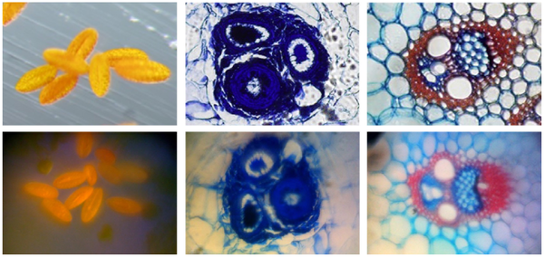
You can make a microscope or a spectrometer out of your cell phone for about $20, say researchers at the University of California at Los Angeles and the University of California at Davis. Here’s an image contrasting standard microscopy with cell phone microscopy,

Images of several commercially prepared microscope slides featuring stained samples. Top row, commercial microscope. Bottom row, cell phone microscope. Left column, pollen grains. Right two columns, plant stems. (copied from PLoS article: DOI: 10.1371/journal.pone.0017150

There’s a serious note to this activity (from the March 11, 2011 news item on Nanowerk),

With health care costs increasing throughout the world, there is a pressing need for reducing the cost and complexity of biomedical devices. Additionally, with growing demand for high-quality health care in regions of the world where medical infrastructure is below levels found in developed countries, portable devices that can transmit relevant data to remote experts are likely to have a large impact on quantity and quality of care. To this end, several groups have focused on the development of low-cost and rapidly deployable technologies that address common diseases afflicting the third world and common tests performed in both hospital and field environments.

Researchers at UCLA have constructed a modified lensless cell phone that enables holography-based digital microscopy, while researchers at UC Berkeley have constructed a complex objective attachment that also transforms a cell phone into a microscope. Additionally, a patent was recently awarded for the use of a cell phone as a spectrometer. However, there is still a need for more research directed towards utilizing cell-phone cameras to record images or spectra of biological samples.

Dave Mosher’s March 11, 2011 article for Wired magazine offers instructions on how to create the cell phone microscope,

Using tape, rubber and a tiny glass ball, researchers transformed an iPhone into a cheap, yet powerful microscope able to image tiny blood cells. They’ve also added a clinical-grade cellphone spectroscope that might be able to measure some vital signs.

And with a few dollars and some patience, you can do the same to your own phone. (See instructions below.)

“It still amazes me how you can build near-research-grade instruments with cheap consumer electronics,” said physicist Sebastian Wachsmann-Hogiu of the University of California at Davis, leader of a study March 2 in PLoS ONE. “And with cellphones, you can record and transmit data anywhere. In rural or remote areas, you could get a diagnosis from a professional pathologist halfway around the world.”

Cellphone Microscope – Step 1

Grab any cellphone with a camera, but note models that use touchscreen focusing and/or have manual focus options are best.

Find some thin, dark, rubbery material and poke a small hole in it (less than 1 millimeter in diameter). This can be done using a pin or needle.

Cellphone Microscope – Step 2

Order a 1-millimeter-diameter ball or half-ball lens. One from Edmund Optics costs between $15 and $25.

Note that lenses with larger diameters can be used, but they will provide a smaller magnification.

Cellphone Microscope – Step 3

Carefully mount it to the iris, covering as little of the lens as possible.

Cellphone Microscope – Step 4

Center the iris with the ball lens tucked in the middle over the camera of the cellphone (above).

From black electrical tape, cut out a hole larger than the diameter of the ball lens, but smaller than the diameter of the iris (below [image omitted, see Wired article]).

Cellphone Microscope – Step 5

Attach the iris to the camera body using the electrical tape mask. You may need to adjust the position of the iris to ensure the microscope images are centered in the camera’s field of view.

As with a standard microscope, use plenty of light to illuminate your sample. Liquid samples should be placed between a glass slide and coverslip.

Mosher’s article also provides instructions on how create a cell phone spectrometer. Or you can read the research article on the Public Library of Science website (open access):

Cell-Phone-Based Platform for Biomedical Device Development and Education Applications

Zachary J. Smith, Kaiqin Chu, Alyssa R. Espenson, Mehdi Rahimzadeh, Amy Gryshuk, Marco Molinaro, Denis M. Dwyre, Stephen Lane, Dennis Matthews,  and Sebastian Wachsmann-Hogiu

PLoS ONE, Vol. 6, Issue 3. March 2, 2011. DOI: 10.1371/journal.pone.0017150

Sometimes I find it all kind of amazing. I mean, whodathunk you could create a microscope with a phone?

California’s call for information about nanomaterials

A little late but better than never, the US state of California has issued a call for information focused on analytical test methods, i.e., lab procedures for testing, nano silver, nano zero valent iron, nano titanium dioxide, nano zinc oxide, nano cerium oxide, and quantum dots. The deadline for a response is Dec. 21, 2011, one year from the date of the request. From the Dec. 27, 2010 news item on Nanowerk,

DTSC [Department of Toxic Substances Control] has conducted a search of known public sources for analytical test methods for these six nanomaterials. We have compiled our research in this bibliography. DTSC has also contacted and consulted with manufacturers, researchers, environmental laboratory experts, other governments, and stakeholders regarding analytical test methods for these nanomaterials in these matrices. We convened public workshops and symposia on nanotechnology and, in particular, these six nanomaterials.

From our research, consultations, and workshops, we have determined that little or no information on analytical test methods for these nanomaterials in the human body or the environment now exists. To better understand the behavior, fate and transport of the se six nanomaterials, appropriate analytical test methods are needed for manufacturers, for contract and reference laboratories, and for regulatory agencies.

You can get more information about the call from the DTSC site including a list of companies that received the ‘call for information’ letter.

UK strategy for investing in nanotech; new insight into titanium dioxide toxicology; creative nonfiction writing for scientists

It sounds promising. UK strategy for nanotechnology business investment is the title of a news item on Nanowerk which outlines the UK Technology Board’s investment strategy. From the news item,

The UK’s Technology Strategy Board has developed a nanotechnology strategy document (pdf download) that sets out the processes the Technology Strategy Board will use to determine how it will invest in the nanotechnology space in a way that helps UK businesses to succeed on a global scale. It is based on the fundamental premise that the technologies likely to see the most success will be those that result in developing materials and devices with new functionality that address markets driven by society’s greatest challenges.

The item goes on to outline the specific areas (environment/energy; aging population, media) where investments will be made but gives no details about the amount of funding available or the source for funds. Curious, I checked out the UK Technology’s Board’s site.  No details to be found on the About Us pages although there is a link to a  Dept. of Business Innovation and Skills (presumably a government department). My guess is that these are government funds and the board has decided to be discreet about the connection. I’m not ready to draw any conclusions; I’m just noticing.

I’ve been following  (somewhat lazily) discussions around titanium dioxide particles (widely used in sunscreens) and their possible toxicology. Nanoparticles used in common household goods caused genetic damage in mice on Nanowerk sheds some new light on the subject. From the news item,

In the past, these TiO2 [titanium dioxide] nanoparticles have been considered non-toxic in that they do not incite a chemical reaction. Instead, it is surface interactions that the nanoparticles have within their environment- in this case inside a mouse – that is causing the genetic damage, [Robert] Schiestl [professor of pathology, radiation oncology and environmental health sciences at Jonsson Cancer Center at the University of California, Los Angeles] said. They wander throughout the body causing oxidative stress, which can lead to cell death. It is a novel mechanism of toxicity, a physicochemical reaction, these particles cause in comparison to regular chemical toxins, which are the usual subjects of toxicological research, Schiestl said. “The novel principle is that titanium by itself is chemically inert. However, when the particles become progressively smaller, their surface, in turn, becomes progressively bigger and in the interaction of this surface with the environment oxidative stress is induced,” he said.

I have posted about titanium dioxide in the past, this posting is the most relevant to this discussion as it contains a reference to some work by Japanese researchers who demonstrated that titanium dioxide cause genetic damage in mice. Presumably building on this work, the researchers at Jonsson Cancer Center have determined a possible mechanism for how the damage is caused.

This is the first time I’ve seen a study that doesn’t ‘shrink’ standard toxicology to the nanoscale. For example, “carbon nanotubes look like asbestos fibres so we should test to find out if they have the same effect on lungs. ” This makes sense and it should be done. At the same time, I’m glad to see that researchers are taking into account the fact that materials at the nanoscale behave in novel ways leading to novel forms of toxicology.

I was intrigued to read Dr. Kristen Kulinowki’s opinion piece in  Azonano’s Nanotechnology Thought Leaders Series … insights from the world’s leading players.  Her piece titled, Temptation, Temptation, Temptation: Why Easy Answers About Nanomaterial Risk are Probably Wrong, provided some valuable insights for me about the work that has been done to collect information about nanomaterials and their potentials risks while citing some useful resources.

Before you go to read the article there are a few things you might want to keep in mind. There are a couple themes that are not followed through so the piece jumps around, the tone is problematic, and the academic style is sometimes inserted into a more chatty blog style. All of which made reading the opinion piece a little more work for me.

I got the impression that Kulinowski did not put much effort into writing this piece, i.e. she tossed it off. The chatty, casual style (a creative writing technique) takes a lot more effort and practice and is much more difficult to pull off  than most people realize, especially when you’re writing nonfiction. (Yes, some people are naturals but even they need to work at it if they plan to continue long term.)