Tag Archives: Brown University

‘Smart’ fabric that’s bony

Researchers at Australia’s University of New South of Wales (UNSW) have devised a means of ‘weaving’ a material that mimics *bone tissue, periosteum according to a Jan. 11, 2017 news item on ScienceDaily,

For the first time, UNSW [University of New South Wales] biomedical engineers have woven a ‘smart’ fabric that mimics the sophisticated and complex properties of one nature’s ingenious materials, the bone tissue periosteum.

Having achieved proof of concept, the researchers are now ready to produce fabric prototypes for a range of advanced functional materials that could transform the medical, safety and transport sectors. Patents for the innovation are pending in Australia, the United States and Europe.

Potential future applications range from protective suits that stiffen under high impact for skiers, racing-car drivers and astronauts, through to ‘intelligent’ compression bandages for deep-vein thrombosis that respond to the wearer’s movement and safer steel-belt radial tyres.

A Jan. 11, 2017 UNSW press release on EurekAlert, which originated the news item, expands on the theme,

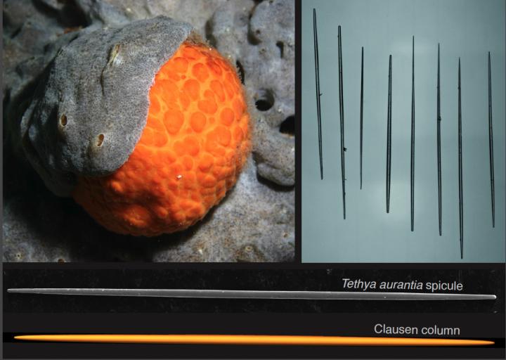
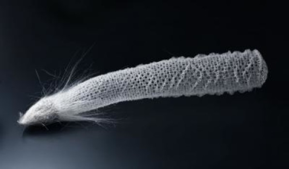
Many animal and plant tissues exhibit ‘smart’ and adaptive properties. One such material is the periosteum, a soft tissue sleeve that envelops most bony surfaces in the body. The complex arrangement of collagen, elastin and other structural proteins gives periosteum amazing resilience and provides bones with added strength under high impact loads.

Until now, a lack of scalable ‘bottom-up’ approaches by researchers has stymied their ability to use smart tissues to create advanced functional materials.

UNSW’s Paul Trainor Chair of Biomedical Engineering, Professor Melissa Knothe Tate, said her team had for the first time mapped the complex tissue architectures of the periosteum, visualised them in 3D on a computer, scaled up the key components and produced prototypes using weaving loom technology.

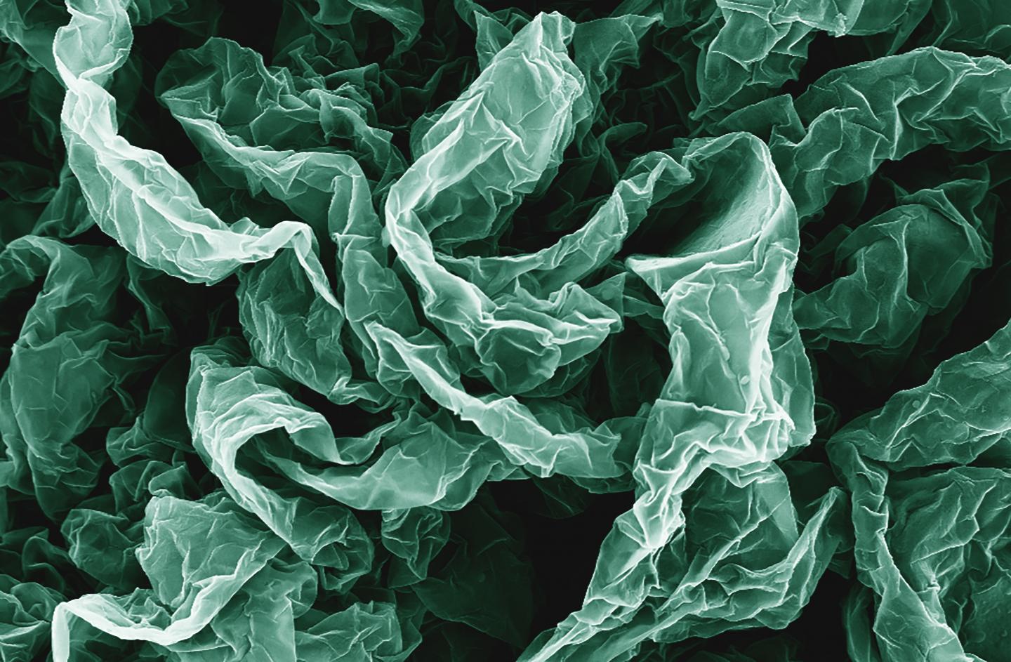
“The result is a series of textile swatch prototypes that mimic periosteum’s smart stress-strain properties. We have also demonstrated the feasibility of using this technique to test other fibres to produce a whole range of new textiles,” Professor Knothe Tate said.

In order to understand the functional capacity of the periosteum, the team used an incredibly high fidelity imaging system to investigate and map its architecture.

“We then tested the feasibility of rendering periosteum’s natural tissue weaves using computer-aided design software,” Professor Knothe Tate said.

The computer modelling allowed the researchers to scale up nature’s architectural patterns to weave periosteum-inspired, multidimensional fabrics using a state-of-the-art computer-controlled jacquard loom. The loom is known as the original rudimentary computer, first unveiled in 1801.

“The challenge with using collagen and elastin is their fibres, that are too small to fit into the loom. So we used elastic material that mimics elastin and silk that mimics collagen,” Professor Knothe Tate said.

In a first test of the scaled-up tissue weaving concept, a series of textile swatch prototypes were woven, using specific combinations of collagen and elastin in a twill pattern designed to mirror periosteum’s weave. Mechanical testing of the swatches showed they exhibited similar properties found in periosteum’s natural collagen and elastin weave.

First author and biomedical engineering PhD candidate, Joanna Ng, said the technique had significant implications for the development of next-generation advanced materials and mechanically functional textiles.

While the materials produced by the jacquard loom have potential manufacturing applications – one tyremaker believes a titanium weave could spawn a new generation of thinner, stronger and safer steel-belt radials – the UNSW team is ultimately focused on the machine’s human potential.

“Our longer term goal is to weave biological tissues – essentially human body parts – in the lab to replace and repair our failing joints that reflect the biology, architecture and mechanical properties of the periosteum,” Ms Ng said.

An NHMRC development grant received in November [2016] will allow the team to take its research to the next phase. The researchers will work with the Cleveland Clinic and the University of Sydney’s Professor Tony Weiss to develop and commercialise prototype bone implants for pre-clinical research, using the ‘smart’ technology, within three years.

In searching for more information about this work, I found a Winter 2015 article (PDF; pp. 8-11) by Amy Coopes and Steve Offner for UNSW Magazine about Knothe Tate and her work (Note: In Australia, winter would be what we in the Northern Hemisphere consider summer),

Tucked away in a small room in UNSW’s Graduate School of Biomedical Engineering sits a 19th century–era weaver’s wooden loom. Operated by punch cards and hooks, the machine was the first rudimentary computer when it was unveiled in 1801. While on the surface it looks like a standard Jacquard loom, it has been enhanced with motherboards integrated into each of the loom’s five hook modules and connected to a computer. This state-of-the-art technology means complex algorithms control each of the 5,000 feed-in fibres with incredible precision.

That capacity means the loom can weave with an extraordinary variety of substances, from glass and titanium to rayon and silk, a development that has attracted industry attention around the world.

The interest lies in the natural advantage woven materials have over other manufactured substances. Instead of manipulating material to create new shades or hues as in traditional weaving, the fabrics’ mechanical properties can be modulated, to be stiff at one end, for example, and more flexible at the other.

“Instead of a pattern of colours we get a pattern of mechanical properties,” says Melissa Knothe Tate, UNSW’s Paul Trainor Chair of Biomedical Engineering. “Think of a rope; it’s uniquely good in tension and in bending. Weaving is naturally strong in that way.”


The interface of mechanics and physiology is the focus of Knothe Tate’s work. In March [2015], she travelled to the United States to present another aspect of her work at a meeting of the international Orthopedic Research Society in Las Vegas. That project – which has been dubbed “Google Maps for the body” – explores the interaction between cells and their environment in osteoporosis and other degenerative musculoskeletal conditions such as osteoarthritis.

Using previously top-secret semiconductor technology developed by optics giant Zeiss, and the same approach used by Google Maps to locate users with pinpoint accuracy, Knothe Tate and her team have created “zoomable” anatomical maps from the scale of a human joint down to a single cell.

She has also spearheaded a groundbreaking partnership that includes the Cleveland Clinic, and Brown and Stanford universities to help crunch terabytes of data gathered from human hip studies – all processed with the Google technology. Analysis that once took 25 years can now be done in a matter of weeks, bringing researchers ever closer to a set of laws that govern biological behaviour. [p. 9]

I gather she was recruited from the US to work at the University of New South Wales and this article was to highlight why they recruited her and to promote the university’s biomedical engineering department, which she chairs.

Getting back to 2017, here’s a link to and citation for the paper,

Scale-up of nature’s tissue weaving algorithms to engineer advanced functional materials by Joanna L. Ng, Lillian E. Knothe, Renee M. Whan, Ulf Knothe & Melissa L. Knothe Tate. Scientific Reports 7, Article number: 40396 (2017) doi:10.1038/srep40396 Published online: 11 January 2017

This paper is open access.

One final comment, that’s a lot of people (three out of five) with the last name Knothe in the author’s list for the paper.

*’the bone tissue’ changed to ‘bone tissue’ on July 17,2017.

Sea sponges don’t buckle under pressure

You wouldn’t think a sponge (the sea creature) was particularly tough but it is according to a Jan. 4, 2017 news item on Nanowerk,

Judging by their name alone, orange puffball sea sponges might seem unlikely paragons of structural strength. But maintaining their shape at the bottom of the churning ocean is critical to the creatures’ survival, and new research shows that tiny structural rods in their bodies have evolved the optimal shape to avoid buckling under pressure.

The rods, called strongyloxea spicules, measure about 2 millimeters long and are thinner than a human hair. Hundreds of them are bundled together, forming stiff rib-like structures inside the orange puffball’s spongy body. It was the odd and remarkably consistent shape of each spicule that caught the eye of Brown University engineers Haneesh Kesari and Michael Monn. Each one is symmetrically tapered along its length — going gradually from fatter in the middle to thinner at the ends.

Caption: Tiny rods found inside the bodies of orange puffball sea sponges have an interesting tapered shape. That shape, new research shows, turns out to be a match for the Clausen profile, a column shape shown to be optimal for resistance to buckling failure. Credit: Michael Monn, Haneesh Kesari / Brown University

A Jan. 4, 2017 Brown University news release on EurekAlert, which originated the news item, describes the research in more detail,

Using structural mechanics models and a bit of digging in obscure mathematics journals, Monn and Kesari showed the peculiar shape of the spicules to be optimal for resistance to buckling, the primary mode of failure for slender structures. This natural shape could provide a blueprint for increasing the buckling resistance in all kinds of slender human-made structures, from building columns to bicycle spokes to arterial stents, the researchers say.

“This is one of the rare examples that we’re aware of where a natural structure is not just well-suited for a given function, but actually approaches a theoretical optimum,” said Kesari, an assistant professor of engineering at Brown. “There’s no engineering analog for this shape — we don’t see any columns or other slender structures that are tapered in this way. So in this case, nature has shown us something quite new that we think could be useful in engineering.”

The findings are published in the journal Scientific Reports.

Function and form

Orange puffball sponges (Tethya aurantia) are native to the Mediterranean Sea. They live mainly in rocky coastal environments, where they’re subject to the constant stress of underwater waves and tidal forces. Sponges are filter feeders — they pump water through their bodies to extract nutrients and oxygen. To do this, their bodies need to be porous and compliant, but they also need enough stiffness to avoid being deformed too much.

“If you compress them too much, you’re essentially choking them,” Kesari said. “So maintaining their stiffness is critical to their survival.”

And that means the spicules, which make up the rib-like structures that give sponges their stiffness, are critical components. When Monn and Kesari saw the shapes of the spicules under a microscope, the consistency of the tapered shape from spicule to spicule was hard to miss.

“We saw the shape and wondered if there might be an engineering principle at work here,” Kesari said.

To figure that out, the researchers first needed to understand what forces were acting on each individual spicule. So Monn and Kesari developed a structural mechanics model of spicules bundled within a sponge’s ribs. The model showed that the mismatch in stiffness between the bulk of the sponge’s soft body and the more rigid spicules causes each spicule to experience primarily one type of mechanical loading — a compression load on each of its ends.

“You can imagine taking a toothpick and trying to squeeze it longways between your fingers,” Monn said. “That’s how these spicules see the world.”

The primary mode of failure for a structure with this mechanical load is through buckling. At a certain critical load, the structure starts to bend somewhere along its length. Once the bending starts, the force transferred by the load is amplified at the bending point, which causes the structure to break or collapse.

Once Kesari and Monn knew what forces were acting on the spicules and how they would fail, the next step was looking to see if there was anything special about them that helped them resist buckling. Scanning electron microscope images of the inside of a spicule and other tests showed that they were monolithic silica — essentially glass.

“We could see that there was no funny business going on with the material properties,” Monn said. “If there was anything contributing to its mechanical performance, it would have to be the shape.”

Optimal shape

Kesari and Monn combed the literature to see if they could find anything on tapering in slender structures. They came up empty in the modern engineering literature. But they found something interesting published more than 150 years ago by a German scientist named Thomas Clausen.

In 1851, Clausen proposed that columns that are tapered toward their ends should have more buckling resistance than plain cylinders, which had been and still are the primary design for architectural columns. In the 1960s, mathematician Joseph Keller published an ironclad mathematical proof that the Clausen column was indeed optimal for resistance to buckling — having 33 percent better resistance than a cylinder. Even compared to a very similar shape — an ellipse, which is slightly fatter in the middle and pointier at the ends — the Clausen column had 18 percent better buckling resistance.

Knowing what the optimal column shape is, Monn and Kesari started making precise dimensional measurements of dozens of spicules. They showed that their shapes were remarkably consistent and nearly identical to that of the Clausen column.

“The spicules were a match for the best shape of all possible column shapes,” Monn said.

It seems in this case, natural selection figured out something that engineers have not. Despite the fact that it’s been mathematically shown to be the optimal column shape, the Clausen profile isn’t widely known in the engineering community. Kesari and Monn hope this work might bring it out of the shadows.

“We see this as an addition to our library of structural designs,” Monn said. “We’re not just talking about an improvement of a few percent. This shape is 33 percent better than the cylinder, which is quite an improvement.”

In particular, the shape would be particularly useful in a new generation of materials made from nanoscale truss structures. “It would be easy to 3-D print the Clausen profile into these materials, and you’d get a tremendous increase in buckling resistance, which is often how these materials fail.”

Lessons from nature

The field of bio-inspired engineering began at a time when many people viewed adaptive evolution as an unceasing march toward perfection. If that were true, scientists should find untold numbers of optimal structures in nature.

But the modern understanding of evolution is a bit different. It’s now understood that in order for a trait to be conserved by natural selection, it doesn’t need to be optimal. It just needs to be good enough to work. That has put a bit of a damper on the enthusiasm for bio-inspired engineering, Kesari and Monn say.

However, they say, this work shows that nearly optimal structures are out there if researchers look in the right places. In this case, they looked at creatures from a very old phylum — sea sponges are among the very first animals on Earth — with plenty of time to evolve under consistent selection pressures.

Sponges are also fairly simple creatures, so understanding the function of a given trait is relatively straightforward. In this case, the spicule appears to have one and only one job to do — provide stiffness. Compare that to, for example, human bone, which not only provides support but must also accommodate arteries, provide attachment points for muscles and house bone marrow. Those other functions may cause tradeoffs in adaptations for strength or stiffness.

“With the sponges, you have lots of evolutionary pressure, lots of time and opportunity to respond to that pressure, and functional elements that can be easily identified,” Kesari said.

With those as guiding principles, there may well be more ideal structures out there waiting to be found.

“This work shows that nature can hit an optimum,” Kesari said, “and the biological world can still be hiding completely new designs of considerable technological significance in plain sight.”

Here’s a link to and a citation for the paper,

A new structure-property connection in the skeletal elements of the marine sponge Tethya aurantia that guards against buckling instability by Michael A. Monn & Haneesh Kesari. Scientific Reports 7, Article number: 39547 (2017) doi:10.1038/srep39547 Published online: 04 January 2017

This paper is open access.

Kesari and Monn have researched sea sponges previously as can be seen in my April 7, 2015 posting, which highlights their work on strength and Venus’ flower basket sea sponge.

A new Shrinky Dinks story: super-wrinkled and super-crumpled graphene for self-cleaning surfaces and other applications

Caption: Wrinkles and crumples, introduced by placing graphene on shrinky polymers, can enhance graphene's properties. Credit: Hurt and Wong Labs / Brown University

Caption: Wrinkles and crumples, introduced by placing graphene on shrinky polymers, can enhance graphene’s properties. Credit: Hurt and Wong Labs / Brown University

A March 21, 2016 news item on ScienceDaily describes how Brown University (US) researchers developed super-wrinkled and super-crumpled graphene,

Crumple a piece of paper and it’s probably destined for the trash can, but new research shows that repeatedly crumpling sheets of the nanomaterial graphene can actually enhance some of its properties. In some cases, the more crumpled the better.

The research by engineers from Brown University shows that graphene, wrinkled and crumpled in a multi-step process, becomes significantly better at repelling water–a property that could be useful in making self-cleaning surfaces. Crumpled graphene also has enhanced electrochemical properties, which could make it more useful as electrodes in batteries and fuel cells.

A March 21, 2016 Brown University news release (also on EurekAlert), which originated the news item, provides more detail about the current and previous research,

This new research builds on previous work done by Robert Hurt and Ian Wong, from Brown’s School of Engineering. The team had previously showed that by introducing wrinkles into graphene, they could make substrates for culturing cells that were more similar to the complex environments in which cells grow in the body. For this latest work, the researchers led by Po-Yen Chen, a Hibbit postdoctoral fellow, wanted to build more complex architectures incorporating both wrinkles and crumples. “I wanted to see if there was a way to create higher-generational structures,” Chen said.

To do that, the researchers deposited layers of graphene oxide onto shrink films–polymer membranes that shrink when heated (kids may know these as Shrinky Dinks [emphasis mine]). As the films shrink, the graphene on top is compressed, causing it to wrinkle and crumple. To see what kind of structures they could create, the researchers compressed same graphene sheets multiple times. After the first shrink, the film was dissolved away, and the graphene was placed in a new film to be shrunk again.

The researchers experimented with different configurations in the successive generations of shrinking. For example, sometimes they clamped opposite ends of the films, which caused them to shrink only along one axis. Clamped films yielded graphene sheets with periodic, basically parallel wrinkles across its surface. Unclamped films shrank in two dimensions, both length- and width-wise, creating a graphene surface that was crumpled in random shapes.

The team experimented with those different modes of shrinking over three successive generations. For example, they might shrink the same graphene sheet on a clamped film, then an unclamped film, then clamped again; or unclamped, clamped, unclamped. They also rotated the graphene in different configurations between shrinkings, sometimes placing the sheet perpendicular to its original orientation.

The team found that the multi-generational approach could substantially compress the graphene sheets, making them as small as one-fortieth their original size. They also showed that successive generations could create interesting patterns along the surface–wrinkles and crumples that were superimposed onto each other, for example.

“As you go deeper into the generations you tend to get larger wavelength structures with the original, smaller wavelength structure from earlier generations built into them,” said Robert Hurt, a professor of engineering at Brown and one of the paper’s corresponding authors.

A sheet that was shrunk clamped, unclamped, and then clamped looked different from ones that were unclamped, clamped, unclamped, for example.

“The sequence matters,” said Wong, also a corresponding author on the paper. “It’s not like multiplication where 2 times 3 is the same as 3 times 2. The material has a ‘memory’ and we get different results when we wrinkle or crumple in a different order.”

The researchers generated a kind of taxonomy of structures born from different shrinking configurations. They then tested several of those structures to see how they altered the properties of the graphene sheets.

Enhanced properties

They showed that a highly crumpled graphene surface becomes superhydrophobic–able to resist wetting by water. When water touches a hydrophobic surface, it beads up and rolls off. When the contact angle of those water beads with an underlying surface exceeds 160 degrees–meaning very little of the water bead’s surface touches the material–the material is said to be superhydrophobic. The researchers showed that they could make superhydrophobic graphene with three unclamped shrinks.

The team also showed that crumpling could enhance the electrochemical behaviors of graphene, which could be useful in next-generation energy storage and generation. The research showed that crumpled graphene used as a battery electrode had as much as a 400 percent increase in electrochemical current density over flat graphene sheets. That increase in current density could make for vastly more efficient batteries.

“You don’t need a new material to do it,” Chen said. “You just need to crumple the graphene.”

In additional to batteries and water resistant coatings, graphene compressed in this manner might also be useful in stretchable electronics–a wearable sensor, for example.

The group plans to continue experimenting with different ways of generating structures on graphene and other nanomaterials.

“There are many new two-dimensional nanomaterials that have interesting properties, not just graphene,” Wong said. “So other materials or combinations of materials may also organize into interesting structures with unexpected functionalities.”

Here’s a link to and a citation for the paper,

Multiscale Graphene Topographies Programmed by Sequential Mechanical Deformation by Po-Yen Chen, Jaskiranjeet Sodhi, Yang Qiu, Thomas M. Valentin, Ruben Spitz Steinberg, Zhongying Wang, Robert H. Hurt, and Ian Y. Wong. Advanced Materials DOI: 10.1002/adma.201506194 Article first published online: 21 MAR 2016

© 2016 WILEY-VCH Verlag GmbH & Co. KGaA, Weinheim

This paper is behind a paywall.

As for Shrinky Dinks, I first featured this material and its use in science research in an Aug. 16, 2010 posting about Shrinky Dinks and nanopatterning. It was originally developed by Betty J. Morris as craft material for children. Both she and the scientist kindly answered some followup questions inspired by the original news release and published in the 2010 post.

Plasmonic interferometry without coherent light

There are already a number of biosensors based on plasmonic interferometry in use but this latest breakthrough from Brown University (US) could make them cheaper and more accessible. A Feb. 16, 2016 Brown University news release (also on EurekAlert), announces the new technique,

Imagine a hand-held environmental sensor that can instantly test water for lead, E. coli, and pesticides all at the same time, or a biosensor that can perform a complete blood workup from just a single drop. That’s the promise of nanoscale plasmonic interferometry, a technique that combines nanotechnology with plasmonics–the interaction between electrons in a metal and light.

Now researchers from Brown University’s School of Engineering have made an important fundamental advance that could make such devices more practical. The research team has developed a technique that eliminates the need for highly specialized external light sources that deliver coherent light, which the technique normally requires. The advance could enable more versatile and more compact devices.

“It has always been assumed that coherent light was necessary for plasmonic interferometry,” said Domenico Pacifici, a professor of engineering who oversaw the work with his postdoctoral researcher Dongfang Li, and graduate student Jing Feng. “But we were able to disprove that assumption.”

The research is described in Nature Scientific Reports.

Plasmonic interferometers make use of the interaction between light and surface plasmon polaritons, density waves created when light energy rattles free electrons in a metal. One type of interferometer looks like a bull’s-eye structure etched into a thin layer of metal. In the center is a hole poked through the metal layer with a diameter of about 300 nanometers–about 1,000 times smaller than the diameter of a human hair. The hole is encircled by a series of etched grooves, with diameters of a few micrometers. Thousands of these bulls-eyes can be placed on a chip the size of a fingernail.

When light from an external source is shown onto the surface of an interferometer, some of the photons go through the central hole, while others are scattered by the grooves. Those scattered photons generate surface plasmons that propagate through the metal inward toward the hole, where they interact with photons passing through the hole. That creates an interference pattern in the light emitted from the hole, which can be recorded by a detector beneath the metal surface.

When a liquid is deposited on top of an interferometer, the light and the surface plasmons propagate through that liquid before they interfere with each other. That alters the interference patterns picked up by the detector depending on the chemical makeup of the liquid or compounds present in it. By using different sizes of groove rings around the hole, the interferometers can be tuned to detect the signature of specific compounds or molecules. With the ability to put many differently tuned interferometers on one chip, engineers can hypothetically make a versatile detector.

Up to now, all plasmonic interferometers have required the use of highly specialized external light sources that can deliver coherent light–beams in which light waves are parallel, have the same wavelength, and travel in-phase (meaning the peaks and valleys of the waves are aligned). Without coherent light sources, the interferometers cannot produce usable interference patterns. Those kinds of light sources, however, tend to be bulky, expensive, and require careful alignment and periodic recalibration to obtain a reliable optical response.

But Pacifici and his group have come up with a way to eliminate the need for external coherent light. In the new method, fluorescent light-emitting atoms are integrated directly within the tiny hole in the center of the interferometer. An external light source is still necessary to excite the internal emitters, but it need not be a specialized coherent source.

“This is a whole new concept for optical interferometry,” Pacifici said, “an entirely new device.”

In this new device, incoherent light shown on the interferometer causes the fluorescent atoms inside the center hole to generate surface plasmons. Those plasmons propagate outward from the hole, bounce off the groove rings, and propagate back toward the hole after. Once a plasmon propagates back, it interacts with the atom that released it, causing an interference with the directly transmitted photon. Because the emission of a photon and the generation of a plasmon are indistinguishable, alternative paths originating from the same emitter, the process is naturally coherent and interference can therefore occur even though the emitters are excited incoherently.

“The important thing here is that this is a self-interference process,” Pacifici said. “It doesn’t matter that you’re using incoherent light to excite the emitters, you still get a coherent process.”

In addition to eliminating the need for specialized external light sources, the approach has several advantages, Pacifici said. Because the surface plasmons travel out from the hole and back again, they probe the sample on top of the interferometer surface twice. That makes the device more sensitive.

But that’s not the only advantage. In the new device, external light can be projected from underneath the metal surface containing the interferometers instead of from above. That eliminates the need for complex illumination architectures on top of the sensing surface, which could make for easier integration into compact devices.

The embedded light emitters also eliminate the need to control the amount of sample liquid deposited on the interferometer’s surface. Large droplets of liquid can cause lensing effects, a bending of light that can scramble the results from the interferometer. Most plasmonic sensors make use of tiny microfluidic channels to deliver a thin film of liquid to avoid lensing problems. But with internal light emitters excited from the bottom surface, the external light never comes in contact with the sample, so lensing effects are negated, as is the need for microfluidics.

Finally, the internal emitters produce a low intensity light. That’s good for probing delicate samples, such as proteins, than can be damaged by high-intensity light.

More work is required to get the system out of the lab and into devices, and Pacifici and his team plan to continue to refine the idea. The next step will be to try eliminating the external light source altogether. It might be possible, the researchers say, to eventually excite the internal emitters using tiny fiber optic lines, or perhaps electric current.

Still, this initial proof-of-concept is promising, Pacifici said.

“From a fundamental standpoint, we think this new device represents a significant step forward,” he said, “a first demonstration of plasmonic interferometry with incoherent light”.

Here’s a link to and a citation for the paper,

Nanoscale optical interferometry with incoherent light by Dongfang Li, Jing Feng, & Domenico Pacifici. Scientific Reports 6, Article number: 20836 (2016) doi:10.1038/srep20836 Published online: 16 February 2016

This paper is open access.

One final comment, Dexter Johnson has a Feb. 18, 2016 posting about this interferometer where he references Pacifici’s past work in this area, as well as, this latest breakthrough. Dexter’s posting can be found on his Nanoclast blog which is on the IEEE (Institute of Electrical and Electronics Engineers) website.

Brown University (US) gets big bucks to study effect on nanomaterials on human health

In over seven years of blogging about nanotechnology, this is the most active funding period for health and environmental effects impacts I’ve seen yet. A Sept. 26, 2015 news item on Azonano features another such grant,

With a new federal grant of nearly $10.8 million over the next five years, Brown University researchers and students in the Superfund Research Program (SRP) will be able to advance their work studying how toxicant exposures affect health, how such exposures occur, how nanotechnologies could contain contamination, and how to make sure those technologies are safe.

A Sept. 24, 2015 Brown University news release, which originated the news item, describes of Brown’s SRP work already underway and how this new grant will support it,

“There is more research to be performed,” said Kim Boekelheide, program director, professor of pathology and laboratory medicine, and fellow of the Institute at Brown for Environment and Society (IBES). “Our scientific theme is integrated biomedical and engineering solutions to regulatory uncertainty, using interdisciplinary approaches to attack the really difficult contamination problems that matter.”

The program is pursuing four integrated projects. In one led by Boekelheide, a team is looking at the physiological effects of exposure to toxicants like trichloroethylene on the male reproductive system. In particular he hopes to find the subtle differences in biomolecular markers in sperm that could allow for very early detection of exposure. Meanwhile in another line of research, Eric Suuberg, professor of engineering, is studying how vapors from toxic material releases can re-emerge from the soil entering into buildings built at or near the polluted sites — and why it is hard to predict the level of exposure that inhabitants of these buildings may suffer.

In another project, Robert Hurt, an IBES fellow, SRP co-primary investigator and professor of engineering, is studying how graphene, an atomically thin carbon material, can be used to block the release and transport of toxicants to prevent human exposures. Hurt is also collaborating with Agnes Kane, an IBES fellow and chair and professor of pathology and laboratory medicine, who is leading a study of nanomaterial effects on human health, so they can be designed and used safely in environmental and other applications.

The program will also continue the program’s community outreach efforts in which they work and share information with communities near the state’s Superfund-designated and Brownfield contaminated sites. Scott Frickel, an IBES fellow and associate professor of sociology, is the new leader of community engagement. The program also includes a research translation core in which researchers share their findings and expertise with the U.S. Environmental Protection Agency, state agencies, and professionals involved in contamination management and cleanup. A training core provides opportunities for interdisciplinary research, field work, and industry “externships” for graduate students in engineering, pathobiology, and social sciences at Brown.

It’s good to see they are integrating social sciences into this project although I hope they aren’t attempting this move as a means to coopt and/or stifle genuine dissent and disagreement by giving a superficial nod to the social sciences and public engagement  while wending on their merry way.

Venus’* flower basket sea sponge has strength

Despite being made essentially of glass, the skeleton of the sea sponge known as Venus' flower basket is remarkably strong -- right down to the tiny, hair-like fibers that hold the creatures to the sea floor. Researchers from Brown University have shown that those fibers, called spicules, have an intricate internal structure that is fine-tuned to boost strength. The findings could inform the engineering of human-made materials. Credit: Kesari Lab / Brown University

Despite being made essentially of glass, the skeleton of the sea sponge known as Venus’ flower basket is remarkably strong — right down to the tiny, hair-like fibers that hold the creatures to the sea floor. Researchers from Brown University have shown that those fibers, called spicules, have an intricate internal structure that is fine-tuned to boost strength. The findings could inform the engineering of human-made materials.
Credit: Kesari Lab / Brown University

I’m not sure how anyone saw a flower basket in that sponge but I bow to a more poetic soul. In any event, scientists at Brown University (US) have shown that this sponge has unexpected strength according to an April 6, 2015 news item on ScienceDaily,

Life may seem precarious for the sea sponge known as Venus’ flower basket. Tiny, hair-like appendages made essentially of glass are all that hold the creatures to their seafloor homes. But fear not for these creatures of the deep. Those tiny lifelines, called basalia spicules, are fine-tuned for strength, according to new research led by Brown University engineers.

In a paper published in the Proceedings of the National Academy of Sciences, the researchers show that the secret to spicules’ strength lies in their remarkable internal structure. The spicules, each only 50 microns in diameter, are made of a silica (glass) core surrounded by 10 to 50 concentric cylinders of glass, each separated by an ultra-thin layer of an organic material. The walls of each cylinder gradually decrease in thickness moving from the core toward the outside edge of the spicule.

An April 6, 2015 Brown University news release (also on EurekAlert), which originated the news item, describes the research in more detail,

When Haneesh Kesari, assistant professor of engineering at Brown, first saw this structure, he wasn’t sure what to make of it. But the pattern of decreasing thickness caught his eye.

“It was not at all clear to me what this pattern was for, but it looked like a figure from a math book,” Kesari said. “It had such mathematical regularity to it that I thought it had to be for something useful and important to the animal.”

The lives of these sponges depend on their ability to stay fixed to the sea floor. They sustain themselves by filtering nutrients out of the water, which they cannot do if they’re being cast about with the flow. So it would make sense, Kesari thought, that natural selection may have molded the creatures’ spicule anchors into models of strength — and the thickness pattern could be a contributing factor.

“If it can’t anchor, it can’t survive,” Kesari said. “So we thought this internal structure must be contributing to these spicules being a better anchor.”

To find out, Kesari worked with graduate student Michael Monn to build a mathematical model of the spicules’ structure. Among the model’s assumptions was that the organic layers between the glass cylinders allowed the cylinders to slide against each other.

“We prepared a mechanical model of this system and asked the question: Of all possible ways the thicknesses of the layers can vary, how should they vary so that the spicule’s anchoring ability is maximized?” Kesari said.

The model predicted that the structure’s load capacity would be greatest when the layers decrease in thickness toward the outside, just as was initially observed in actual spicules. Kesari and Monn then worked with James Weaver and Joanna Aizenberg of Harvard’s Wyss Institute for Biologically Inspired Engineering, who have worked with this sponge species for years. The team carefully compared the layer thicknesses predicted by the mechanics model to the actual layer thicknesses in more than a hundred spicule samples from sponges.

The work showed that the predictions made by the model matched very closely with the observed layer thicknesses in the samples. “It appears that the arrangement and thicknesses of these layers does indeed contribute to the spicules’ strength, which helps make them better anchors,” Kesari said.

The researchers say this is the first time to their knowledge that anyone has evaluated the mechanical advantage of this particular arrangement of layers. It could add to the list of useful engineered structures inspired by nature.

“In the engineered world, you see all kinds of instances where the external geometry of a structure is modified to enhance its specific strength — I-beams are one example,” Monn said. “But you don’t see a huge effort focused toward the internal mechanical design of these structures.”

This study, however, suggests that sponge spicules could provide a blueprint for load-bearing beams made stronger from the inside out.

Here’s a link to and a citation for the paper,

New functional insights into the internal architecture of the laminated anchor spicules of Euplectella aspergillum by Michael A. Monn, James C. Weaver, Tianyang Zhang, Joanna Aizenberg, and Haneesh Kesari. Published online before print April 6, 2015, doi: 10.1073/pnas.1415502112 PNAS April 6, 2015

This paper is behind a paywall.

*’A Venus flower basket sea sponge’ corrected to ‘Venus’ flower basket sea sponge’ on Jan. 5, 2017

High-order Brownian motion observed

A Nov. 17, 2014 news item on ScienceDaily highlights a new technique for observing Brownian motion,

For the first time, scientists have vividly mapped the shapes and textures of high-order modes of Brownian motions–in this case, the collective macroscopic movement of molecules in microdisk resonators–researchers at Case Western Reserve University report.

To do this, they used a record-setting scanning optical interferometry technique, described in a study published today in the journal Nature Communications.

The new technology holds promise for multimodal sensing and signal processing, and to develop optical coding for computing and other information-processing functions by exploiting the spatially resolved multimode Brownian resonances and their splitting pairs of modes.

A Nov. 17, 2014 Case Western Reserve University news release on EurekAlert, which originated the news item, provides more information about the technique and the research,

Interferometry uses the interference of light waves reflected off a surface to measure distances, a technique invented by Case School of Applied Science physicist Albert A. Michelson (who won the Nobel prize in science in 1907). Michelson and Western Reserve University chemist Edward Morley used the instrument to famously disprove that light traveled through “luminous ether” in 1887, setting the groundwork for Albert Einstein’s theory of relativity.

The technology has evolved since then. The keys to Feng’s new interferometry technique are focusing a tighter-than-standard laser spot on the surface of novel silicon carbide microdisks.

The microdisks, which sit atop pedestals of silicon oxide like cymbals on stands, are extremely sensitive to the smallest fluctuations arising from Brownian motions, even at thermodynamic equilibrium. Hence, they exhibit very small oscillations without external driving forces. These oscillations include fundamental and higher modes, called thermomechanical resonances.

Some of the light from the laser reflects back to a sensor after striking the top surface of the silicon dioxide film. And some of the light is refracted through the film and reflected back on a different path, causing interference in the light waves.

The narrow laser spot scans the disk surface and measures movement, or displacement, of the disk with a sensitivity of about 7 femtometers per square-root of a hertz at room temperature, which researchers believe is a record for interferometric systems. To put that in perspective, the width of a hair is about 40 microns, and a femtometer is 100 million times smaller than a micron.

Although higher frequency modes have small motion amplitudes, the technology enabled the group to spatially map and clearly visualize the first through ninth Brownian modes in the high frequency band, ranging from 5.78 to 26.41 megahertz.

In addition to detecting the shapes and textures of Brownian motions, multimode mapping identified subtle structural imperfections and defects, which are ubiquitous but otherwise invisible, or can’t be quantified most of the time. This capability may be useful for probing the dynamics and propagation of defects and defect arrays in nanodevices, as well as for future engineering of controllable defects to manipulate information in silicon carbide nanostructures

The high sensitivity and spatial resolution also enabled them to identify mode splitting, crossing and degeneracy, spatial asymmetry and other effects that may be used to encode information with increasing complexity. The researchers are continuing to explore the capabilities of the technology.

Here’s a link to and a citation for the paper,

Spatial mapping of multimode Brownian motions in high-frequency ​silicon carbide microdisk resonators by Zenghui Wang, Jaesung Lee & Philip X. -L. Feng. Nature Communications 5, Article number: 5158 doi:10.1038/ncomms6158 Published 17 November 2014

This paper is behind a paywall.

For those who would like a little more information about Brownian motion, there’s this from its Wikipedia entry,

Brownian motion or pedesis (from Greek: πήδησις /pɛ̌ːdɛːsis/ “leaping”) is the random motion of particles suspended in a fluid (a liquid or a gas) resulting from their collision with the quick atoms or molecules in the gas or liquid. The term “Brownian motion” can also refer to the mathematical model used to describe such random movements, which is often called a particle theory.

The Wikipedia entry also includes this gif

This is a simulation of Brownian motion of a big particle (dust particle) that collides with a large set of smaller particles (molecules of a gas) which move with different velocities in different random directions. http://weelookang.blogspot.com/2010/06/ejs-open-source-brownian-motion-gas.html Lookang Author of computer model: Francisco Esquembre, Fu-Kwun and lookang - Own work

This is a simulation of Brownian motion of a big particle (dust particle) that collides with a large set of smaller particles (molecules of a gas) which move with different velocities in different random directions. http://weelookang.blogspot.com/2010/06/ejs-open-source-brownian-motion-gas.html
Lookang Author of computer model: Francisco Esquembre, Fu-Kwun and lookang – Own work

On a tangential and amusing note, Brown University celebrating its 250th anniversary this year (2014) commissioned a Brownian Motion composition as part of its commemoration activities (from a Feb. 21, 2014 Brown University news release),

While Brown University and its neighbors celebrate the University’s first 250 years during the Opening Celebration Friday and Saturday, March 7-8, 2014, some new history will be made as well. On Friday night, the Brown University Wind Symphony will present the world premier of Brownian Motion, a piece commissioned for the semiquincentenary.

Written by the composer and saxophonist Patrick Zimmerli, the commission was funded by Edward Guiliano, a 1972 Brown graduate who was president of the Brown Band and founded the Brown Wind Ensemble during his time on College Hill.

Zimmerli admits to feeling excitement when approached with the commission. “I didn’t go to Brown but I have many connections to people who did, and I was really looking forward to the challenge of writing for an undergraduate wind ensemble, something I’d never done before.”

McGarrell [Matthew McGarrell, director of bands at Brown] and Zimmerli met last summer to talk about the commission for the first time. Aside from sending Zimmerli a few pieces to use as models, McGarrell gave the composer free reign over over everything from the feel to the length of the piece.

The resulting composition, which Zimmerli presented to McGarrell at the beginning of January, is dominated by jazz rhythms, with some nods to vernacular musics, including Caribbean and calypso, mixed in.

“The piece has several different moods but overall it is celebratory,” Zimmerli said. “After all it’s a birthday piece. It’s meant to be challenging but fun for the players.”

Listeners with a link to Brown may also find parts of the work familiar. Zimmerli subtly weaves an early melody known as “Araby’s Daughter” — Brown’s Alma Mater — throughout the piece, building on it until it’s played in its full glory by the French horns toward the end.

For inspiration, Zimmerli did extensive research on Brown’s early history and was intrigued to learn that Brown’s founding was initially opposed by a group of preachers who had a mistrust for those who had been formally educated. The result is a theme — “learning is evil,” a nod to those early roots — that winds its way throughout the song.

“Brown is an amazing example of an institution that has been able to evolve and transform itself from within, and I thought that fact should be celebrated,” said Zimmerli.

Other parts of the song inspired the Brownian Motion name.

“There’s a jagged theme toward the beginning of the piece that is a bit cheeky, even subversive. The way it moves and darts around through the instruments unexpectedly is what eventually led me to the actual title of the piece,” Zimmerli said.

“We knew we wanted to make it special concert,” said McGarrell of the program selections. “We wanted to reach both the Brown community in history, through the alumni, through musical representation, and we wanted to reach out to the extended Brown community in Rhode Island and southeastern New England, through history and intercultural outreach.”

The Brown musicians have been hard at work since the end of January learning Brownian Motion. While technically challenging, McGarrell said the students have been appreciating the skill level required and that “morale has remained high within the group.” Zimmerli arrives on campus on Wednesday, March 5, to help put the finishing touches on the performance.

There is a youtube video (over 60 mins.) of the Brownian Motion March 2014 performance.

Boron as a ‘buckyball’ or borospherene

First there was the borophene (like graphene but using boron rather than carbon) announcement from Brown University in my Jan. 28, 214 posting and now US (Brown University again) and Chinese researchers have developed a boron ‘buckyball’. Coincidentally, this announcement comes just after the 2014 World Cup final (July 13, 2014). Representations of buckyballs always resemble soccer balls. (Note: Germany won.)

From a July 14, 2014 news item on Azonano,

The discovery 30 years ago of soccer-ball-shaped carbon molecules called buckyballs helped to spur an explosion of nanotechnology research. Now, there appears to be a new ball on the pitch.

Researchers from Brown University, Shanxi University and Tsinghua University in China have shown that a cluster of 40 boron atoms forms a hollow molecular cage similar to a carbon buckyball. It’s the first experimental evidence that a boron cage structure—previously only a matter of speculation—does indeed exist.

“This is the first time that a boron cage has been observed experimentally,” said Lai-Sheng Wang, a professor of chemistry at Brown who led the team that made the discovery. “As a chemist, finding new molecules and structures is always exciting. The fact that boron has the capacity to form this kind of structure is very interesting.”

The researchers have provided an illustration of their borospherene,

The carbon buckyball has a boron cousin. A cluster for 40 boron atoms forms a hollow cage-like molecule. Courtesy Brown University

The carbon buckyball has a boron cousin. A cluster for 40 boron atoms forms a hollow cage-like molecule. Courtesy Brown University

A July 9, 2104 Brown University news release (also on EurekAlert), which originated the news item, describes the borosphene’s predecessor, the carbon buckyball, and provides more details about this new molecule,

Carbon buckyballs are made of 60 carbon atoms arranged in pentagons and hexagons to form a sphere — like a soccer ball. Their discovery in 1985 was soon followed by discoveries of other hollow carbon structures including carbon nanotubes. Another famous carbon nanomaterial — a one-atom-thick sheet called graphene — followed shortly after.

After buckyballs, scientists wondered if other elements might form these odd hollow structures. One candidate was boron, carbon’s neighbor on the periodic table. But because boron has one less electron than carbon, it can’t form the same 60-atom structure found in the buckyball. The missing electrons would cause the cluster to collapse on itself. If a boron cage existed, it would have to have a different number of atoms.

Wang and his research group have been studying boron chemistry for years. In a paper published earlier this year, Wang and his colleagues showed that clusters of 36 boron atoms form one-atom-thick disks, which might be stitched together to form an analog to graphene, dubbed borophene. Wang’s preliminary work suggested that there was also something special about boron clusters with 40 atoms. They seemed to be abnormally stable compared to other boron clusters.

Figuring out what that 40-atom cluster actually looks like required a combination of experimental work and modeling using high-powered supercomputers.

On the computer, Wang’s colleagues modeled over 10,000 possible arrangements of 40 boron atoms bonded to each other. The computer simulations estimate not only the shapes of the structures, but also estimate the electron binding energy for each structure — a measure of how tightly a molecule holds its electrons. The spectrum of binding energies serves as a unique fingerprint of each potential structure.

The next step is to test the actual binding energies of boron clusters in the lab to see if they match any of the theoretical structures generated by the computer. To do that, Wang and his colleagues used a technique called photoelectron spectroscopy.

Chunks of bulk boron are zapped with a laser to create vapor of boron atoms. A jet of helium then freezes the vapor into tiny clusters of atoms. The clusters of 40 atoms were isolated by weight then zapped with a second laser, which knocks an electron out of the cluster. The ejected electron flies down a long tube Wang calls his “electron racetrack.” The speed at which the electrons fly down the racetrack is used to determine the cluster’s electron binding energy spectrum — its structural fingerprint.

The experiments showed that 40-atom-clusters form two structures with distinct binding spectra. Those spectra turned out to be a dead-on match with the spectra for two structures generated by the computer models. One was a semi-flat molecule and the other was the buckyball-like spherical cage.

“The experimental sighting of a binding spectrum that matched our models was of paramount importance,” Wang said. “The experiment gives us these very specific signatures, and those signatures fit our models.”

The borospherene molecule isn’t quite as spherical as its carbon cousin. Rather than a series of five- and six-membered rings formed by carbon, borospherene consists of 48 triangles, four seven-sided rings and two six-membered rings. Several atoms stick out a bit from the others, making the surface of borospherene somewhat less smooth than a buckyball.

As for possible uses for borospherene, it’s a little too early to tell, Wang says. One possibility, he points out, could be hydrogen storage. Because of the electron deficiency of boron, borospherene would likely bond well with hydrogen. So tiny boron cages could serve as safe houses for hydrogen molecules.

But for now, Wang is enjoying the discovery.

“For us, just to be the first to have observed this, that’s a pretty big deal,” Wang said. “Of course if it turns out to be useful that would be great, but we don’t know yet. Hopefully this initial finding will stimulate further interest in boron clusters and new ideas to synthesize them in bulk quantities.”

The theoretical modeling was done with a group led by Prof. Si-Dian Li from Shanxi University and a group led by Prof. Jun Li from Tsinghua University. The work was supported by the U.S. National Science Foundation (CHE-1263745) and the National Natural Science Foundation of China.

Here’s a link to and a citation for the paper,

Observation of an all-boron fullerene by Hua-Jin Zhai, Ya-Fan Zhao, Wei-Li Li, Qiang Chen, Hui Bai, Han-Shi Hu, Zachary A. Piazza, Wen-Juan Tian, Hai-Gang Lu, Yan-Bo Wu, Yue-Wen Mu, Guang-Feng Wei, Zhi-Pan Liu, Jun Li, Si-Dian Li, & Lai-Sheng Wang. Nature Chemistry (2014) doi:10.1038/nchem.1999 Published online 13 July 2014

This paper is behind a paywall.

Physics, nanopores, viruses, and DNA

A June 17, 2014 news item on Azonano describes a project which could help scientists decode strands of DNA at top speeds,

Nanopores may one day lead a revolution in DNA sequencing. By sliding DNA molecules one at a time through tiny holes in a thin membrane, it may be possible to decode long stretches of DNA at lightning speeds. Scientists, however, haven’t quite figured out the physics of how polymer strands like DNA interact with nanopores. Now, with the help of a particular type of virus, researchers from Brown University have shed new light on this nanoscale physics.

“What got us interested in this was that everybody in the field studied DNA and developed models for how they interact with nanopores,” said Derek Stein, associate professor of physics and engineering at Brown [Brown University, US] who directed the research. “But even the most basic things you would hope models would predict starting from the basic properties of DNA — you couldn’t do it. The only way to break out of that rut was to study something different.”

A June 16, 2014 Brown University news release (also on EurekAlert), which originated the news item, describes the problems with nanopores,

The concept behind nanopore sequencing is fairly simple. A hole just a few billionths of a meter wide is poked in a membrane separating two pools of salty water. An electric current is applied to the system, which occasionally snares a charged DNA strand and whips it through the pore — a phenomenon called translocation. When a molecule translocates, it causes detectable variations in the electric current across the pore. By looking carefully at those variations in current, scientists may be able to distinguish individual nucleotides — the A’s, C’s, G’s and T’s coded in DNA molecules.

The first commercially available nanopore sequencers may only be a few years away, but despite advances in the field, surprisingly little is known about the basic physics involved when polymers interact with nanopores. That’s partly because of the complexities involved in studying DNA. In solution, DNA molecules form balls of random squiggles, which make understanding their physical behavior extremely difficult.

For example, the factors governing the speed of DNA translocation aren’t well understood. Sometimes molecules zip through a pore quickly; other times they slither more slowly, and nobody completely understands why.

One possible explanation is that the squiggly configuration of DNA causes each molecule to experience differences in drag as they’re pulled through the water toward the pore. “If a molecule is crumpled up next to the pore, it has a shorter distance to travel and experiences less drag,” said Angus McMullen, a physics graduate student at Brown and the study’s lead author. “But if it’s stretched out then it would feel drag along the whole length and that would cause it to go slower.”

The news release then goes on to detail a possible solution to the problem of why DNA translocation varies in speed. Answering this question about DNA translocation could lead to faster and more accurate nanopore sequencing,

The drag effect is impossible to isolate experimentally using DNA, but the virus McMullen and his colleagues studied offered a solution.

The researchers looked at fd, a harmless virus that infects e. coli bacteria. Two things make the virus an ideal candidate for study with nanpores. First, fd viruses are all identical clones of each other. Second, unlike squiggly DNA, fd virus is a stiff, rod-like molecule. Because the virus doesn’t curl up like DNA does, the effect of drag on each one should be essentially the same every time.

With drag eliminated as a source of variation in translocation speed, the researchers expected that the only source of variation would be the effect of thermal motion. The tiny virus molecules constantly bump up against the water molecules in which they are immersed. A few random thermal kicks from the rear would speed the virus up as it goes through the pore. A few kicks from the front would slow it down.

The experiments showed that while thermal motion explained much of the variation in translocation speed, it didn’t explain it all. Much to the researchers’ surprise, they found another source of variation that increased when the voltage across the pore was increased.

“We thought that the physics would be crystal clear,” said Jay Tang, associate professor of physics and engineering at Brown and one of the study’s co-authors. “You have this stiff [virus] with well-defined diameter and size and you would expect a very clear-cut signal. As it turns out, we found some puzzling physics we can only partially explain ourselves.”

The researchers can’t say for sure what’s causing the variation they observed, but they have a few ideas.

“It’s been predicted that depending on where [an object] is inside the pore, it might be pulled harder or weaker,” McMullen said. “If it’s in the center of the pore, it pulls a little bit weaker than if it’s right on the edge. That’s been predicted, but never experimentally verified. This could be evidence of that happening, but we’re still doing follow up work.

The new approach using a virus answered questions while leading to new insights and possibilities (from the news release),

A better understanding of translocation speed could improve the accuracy of nanopore sequencing, McMullen says. It would also be helpful in the crucial task of measuring the length of DNA strands. “If you can predict the translocation speed,” McMullen said, “then you can easily get the length of the DNA from how long its translocation was.”

The research also helped to reveal other aspects of the translocation process that could be useful in designing future devices. The study showed that the electrical current tends to align the viruses head first to the pore, but on occasions when they’re not lined up, they tend to bounce around on the edge of the pore until thermal motion aligns them to go through. However, when the voltage was turned too high, the thermal effects were suppressed and the virus became stuck to the membrane. That suggests a sweet spot in voltage where headfirst translocation is most likely.

None of this is observable directly — the system is simply too small to be seen in action. But the researchers could infer what was happening by looking at slight changes in the current across the pore.

“When the viruses miss, they rattle around and we see these little bumps in the current,” Stein said. “So with these little bumps, we’re starting to get an idea of what the molecule is doing before it slides through. Normally these sensors are blind to anything that’s going on until the molecule slides through.”

That would have been impossible to observe using DNA. The floppiness of the DNA molecule allows it to go through a pore in a folded configuration even if it’s not aligned head-on. But because the virus is stiff, it can’t fold to go through. That enabled the researchers to isolate and observe those contact dynamics.

“These viruses are unique,” Stein said. “They’re like perfect little yardsticks.”

In addition to shedding light on basic physics, the work might also have another application. While the fd virus itself is harmless, the bacteria it infects — e. coli — is not. Based on this work, it might be possible to build a nanopore device for detecting the presence of fd, and by proxy, e. coli. Other dangerous viruses — Ebola and Marburg among them — share the same rod-like structure as fd.

“This might be an easy way to detect these viruses,” Tang said. “So that’s another potential application for this.”

Here’s a link to and a citation for the paper,

Stiff filamentous virus translocations through solid-state nanopores by Angus McMullen, Hendrick W. de Haan, Jay X. Tang, & Derek Stein. Nature Communications 5, Article number: 4171 doi:10.1038/ncomms5171 Published 16 June 2014

This paper is behind a paywall.

Bioceramic armour: tough and clear

This story about a mollusk and its armour eventually led me back to one of my favourite science writers, David L. Chandler at the Massachusetts Institute of Technology (MIT). First, here’s an excerpt from a March 30, 2014 news item on ScienceDaily,

The shells of a sea creature, the mollusk Placuna placenta, are not only exceptionally tough, but also clear enough to read through. Now, researchers at MIT have analyzed these shells to determine exactly why they are so resistant to penetration and damage — even though they are 99 percent calcite, a weak, brittle mineral.

The shells’ unique properties emerge from a specialized nanostructure that allows optical clarity, as well as efficient energy dissipation and the ability to localize deformation, the researchers found. The results are published this week in the journal Nature Materials, in a paper co-authored by MIT graduate student Ling Li and professor Christine Ortiz.

A March 30, 2014 MIT press release (I’m not positive Chandler wrote this but he is the press contact) describes both the engineered bioceramic armour and the mollusk’s naturally occurring armour,

Engineered ceramic-based armor, while designed to resist penetration, often lacks the ability to withstand multiple blows, due to large-scale deformation and fracture that can compromise its structural integrity, Ortiz says. In transparent armor systems, such deformation can also obscure visibility.

Creatures that have evolved natural exoskeletons — many of them ceramic-based — have developed ingenious designs that can withstand multiple penetrating attacks from predators. The shells of a few species, such as Placuna placenta, are also optically clear.

To test exactly how the shells — which combine calcite with about 1 percent organic material — respond to penetration, the researchers subjected samples to indentation tests, using a sharp diamond tip in an experimental setup that could measure loads precisely. They then used high-resolution analysis methods, such as electron microscopy and diffraction, to examine the resulting damage.

The material initially isolates damage through an atomic-level process called “twinning” within the individual ceramic building blocks: A crystal breaks up into a pair of mirror-image regions that share a common boundary, rather like a butterfly’s wings. This twinning process occurs all around the stressed region, helping to form a kind of boundary that keeps the damage from spreading outward.

The MIT researchers found that twinning then activates “a series of additional energy-dissipation mechanisms … which preserve the mechanical and optical integrity of the surrounding material,” Li says. This produces a material that is 10 times more efficient in dissipating energy than the pure mineral, Li adds.

The properties of this natural armor make it a promising template for the development of bio-inspired synthetic materials for both commercial and military applications — such as eye and face protection for soldiers, windows and windshields, and blast shields, Ortiz says.

Huajian Gao, a professor of engineering at Brown University who was not involved in this research, calls it “an excellent and elegant piece of work.” He says it “successfully demonstrates the effectiveness of nanoscale deformation twins in energy dissipation in bioceramics, and should be able to inspire and guide the development of manmade ceramic materials.” He adds, “As a first-of-its-kind [demonstration of] the effectiveness of deformation twins in natural materials, this work should have huge practical impact.”

The work was supported by the National Science Foundation; the U.S. Army Research Office through the MIT Institute for Soldier Nanotechnologies; the National Security Science and Engineering Faculty Fellowships Program; and the Office of the Assistant Secretary of Defense for Research and Engineering.

The researchers have produced an image showing how the mollusk shell reacts to being damaged,

A Scanning Electron Microscope (SEM) image of the region surrounding an indentation the researchers made in a piece of shell from Placuna placenta. The image shows the localization of damage to the area immediately surrounding the stress. Image: Ling Li and James C. Weaver. Courtesy: MIT

A Scanning Electron Microscope (SEM) image of the region surrounding an indentation the researchers made in a piece of shell from Placuna placenta. The image shows the localization of damage to the area immediately surrounding the stress.
Image: Ling Li and James C. Weaver. Courtesy: MIT

Here’s a link to and a citation for the paper,

Pervasive nanoscale deformation twinning as a catalyst for efficient energy dissipation in a bioceramic armour by Ling Li & Christine Ortiz. Nature Materials (2014) doi:10.1038/nmat3920 Published online 30 March 2014

This paper is behind a paywall.