Tag Archives: Kansas State University

US Dept. of Agriculture announces its nanotechnology research grants

I don’t always stumble across the US Department of Agriculture’s nanotechnology research grant announcements but I’m always grateful when I do as it’s good to find out about  nanotechnology research taking place in the agricultural sector. From a July 21, 2017 news item on Nanowerk,,

The U.S. Department of Agriculture’s (USDA) National Institute of Food and Agriculture (NIFA) today announced 13 grants totaling $4.6 million for research on the next generation of agricultural technologies and systems to meet the growing demand for food, fuel, and fiber. The grants are funded through NIFA’s Agriculture and Food Research Initiative (AFRI), authorized by the 2014 Farm Bill.

“Nanotechnology is being rapidly implemented in medicine, electronics, energy, and biotechnology, and it has huge potential to enhance the agricultural sector,” said NIFA Director Sonny Ramaswamy. “NIFA research investments can help spur nanotechnology-based improvements to ensure global nutritional security and prosperity in rural communities.”

A July 20, 2017 USDA news release, which originated the news item, lists this year’s grants and provides a brief description of a few of the newly and previously funded projects,

Fiscal year 2016 grants being announced include:

Nanotechnology for Agricultural and Food Systems

  • Kansas State University, Manhattan, Kansas, $450,200
  • Wichita State University, Wichita, Kansas, $340,000
  • University of Massachusetts, Amherst, Massachusetts, $444,550
  • University of Nevada, Las Vegas, Nevada,$150,000
  • North Dakota State University, Fargo, North Dakota, $149,000
  • Cornell University, Ithaca, New York, $455,000
  • Cornell University, Ithaca, New York, $450,200
  • Oregon State University, Corvallis, Oregon, $402,550
  • University of Pennsylvania, Philadelphia, Pennsylvania, $405,055
  • Gordon Research Conferences, West Kingston, Rhode Island, $45,000
  • The University of Tennessee,  Knoxville, Tennessee, $450,200
  • Utah State University, Logan, Utah, $450,200
  • The George Washington University, Washington, D.C., $450,200

Project details can be found at the NIFA website (link is external).

Among the grants, a University of Pennsylvania project will engineer cellulose nanomaterials [emphasis mine] with high toughness for potential use in building materials, automotive components, and consumer products. A University of Nevada-Las Vegas project will develop a rapid, sensitive test to detect Salmonella typhimurium to enhance food supply safety.

Previously funded grants include an Iowa State University project in which a low-cost and disposable biosensor made out of nanoparticle graphene that can detect pesticides in soil was developed. The biosensor also has the potential for use in the biomedical, environmental, and food safety fields. University of Minnesota (link is external) researchers created a sponge that uses nanotechnology to quickly absorb mercury, as well as bacterial and fungal microbes from polluted water. The sponge can be used on tap water, industrial wastewater, and in lakes. It converts contaminants into nontoxic waste that can be disposed in a landfill.

NIFA invests in and advances agricultural research, education, and extension and promotes transformative discoveries that solve societal challenges. NIFA support for the best and brightest scientists and extension personnel has resulted in user-inspired, groundbreaking discoveries that combat childhood obesity, improve and sustain rural economic growth, address water availability issues, increase food production, find new sources of energy, mitigate climate variability and ensure food safety. To learn more about NIFA’s impact on agricultural science, visit www.nifa.usda.gov/impacts, sign up for email updates (link is external) or follow us on Twitter @USDA_NIFA (link is external), #NIFAImpacts (link is external).

Given my interest in nanocellulose materials (Canada was/is a leader in the production of cellulose nanocrystals [CNC] but there has been little news about Canadian research into CNC applications), I used the NIFA link to access the table listing the grants and clicked on ‘brief’ in the View column in the University of Pennsylania row to find this description of the project,

ENGINEERING CELLULOSE NANOMATERIALS WITH HIGH TOUGHNESS

NON-TECHNICAL SUMMARY: Cellulose nanofibrils (CNFs) are natural materials with exceptional mechanical properties that can be obtained from renewable plant-based resources. CNFs are stiff, strong, and lightweight, thus they are ideal for use in structural materials. In particular, there is a significant opportunity to use CNFs to realize polymer composites with improved toughness and resistance to fracture. The overall goal of this project is to establish an understanding of fracture toughness enhancement in polymer composites reinforced with CNFs. A key outcome of this work will be process – structure – fracture property relationships for CNF-reinforced composites. The knowledge developed in this project will enable a new class of tough CNF-reinforced composite materials with applications in areas such as building materials, automotive components, and consumer products.The composite materials that will be investigated are at the convergence of nanotechnology and bio-sourced material trends. Emerging nanocellulose technologies have the potential to move biomass materials into high value-added applications and entirely new markets.

It’s not the only nanocellulose material project being funded in this round, there’s this at North Dakota State University, from the NIFA ‘brief’ project description page,

NOVEL NANOCELLULOSE BASED FIRE RETARDANT FOR POLYMER COMPOSITES

NON-TECHNICAL SUMMARY: Synthetic polymers are quite vulnerable to fire.There are 2.4 million reported fires, resulting in 7.8 billion dollars of direct property loss, an estimated 30 billion dollars of indirect loss, 29,000 civilian injuries, 101,000 firefighter injuries and 6000 civilian fatalities annually in the U.S. There is an urgent need for a safe, potent, and reliable fire retardant (FR) system that can be used in commodity polymers to reduce their flammability and protect lives and properties. The goal of this project is to develop a novel, safe and biobased FR system using agricultural and woody biomass. The project is divided into three major tasks. The first is to manufacture zinc oxide (ZnO) coated cellulose nanoparticles and evaluate their morphological, chemical, structural and thermal characteristics. The second task will be to design and manufacture polymer composites containing nano sized zinc oxide and cellulose crystals. Finally the third task will be to test the fire retardancy and mechanical properties of the composites. Wbelieve that presence of zinc oxide and cellulose nanocrystals in polymers will limit the oxygen supply by charring, shielding the surface and cellulose nanocrystals will make composites strong. The outcome of this project will help in developing a safe, reliable and biobased fire retardant for consumer goods, automotive, building products and will help in saving human lives and property damage due to fire.

One day, I hope to hear about Canadian research into applications for nanocellulose materials. (fingers crossed for good luck)
Detonating (exploding) your way to graphene

Physicists at Kansas State University use controlled detonation to make graphene according to a Jan. 25, 2017 news item on Nanowerk (Note: A link has been removed),

Forget chemicals, catalysts and expensive machinery — a Kansas State University team of physicists has discovered a way to mass-produce graphene with three ingredients: hydrocarbon gas, oxygen and a spark plug.

Their method is simple: Fill a chamber with acetylene or ethylene gas and oxygen. Use a vehicle spark plug to create a contained detonation. Collect the graphene that forms afterward.

Chris Sorensen, Cortelyou-Rust university distinguished professor of physics, is the lead inventor of the recently issued patent, “Process for high-yield production of graphene via detonation of carbon-containing material”. Other Kansas State University researchers involved include Arjun Nepal, postdoctoral researcher and instructor of physics, and Gajendra Prasad Singh, former visiting scientist.

For further reading here’s the Jan. 25, 2017 Kansas State University news release, which originated the news item,

“We have discovered a viable process to make graphene,” Sorensen said. “Our process has many positive properties, from the economic feasibility, the possibility for large-scale production and the lack of nasty chemicals. What might be the best property of all is that the energy required to make a gram of graphene through our process is much less than other processes because all it takes is a single spark.”

Graphene is a single atom-thick sheet of hexagonally coordinated carbon atoms, which makes it the world’s thinnest material. Since graphene was isolated in 2004, scientists have found it has valuable physical and electronic properties with many possible applications, such as more efficient rechargeable batteries or better electronics.

For Sorensen’s research team, the serendipitous path to creating graphene started when they were developing and patenting carbon soot aerosol gels. They created the gels by filling a 17-liter aluminum chamber with acetylene gas and oxygen. Using a spark plug, they created a detonation in the chamber. The soot from the detonation formed aerosol gels that looked like “black angel food cake,” Sorensen said.

But after further analysis, the researchers found that the aerosol gel was more than lookalike dark angel food cake — it was graphene.

“We made graphene by serendipity,” Sorensen said. “We didn’t plan on making graphene. We planned on making the aerosol gel and we got lucky.”

But unlike other methods of creating graphene, Sorensen’s method is simple, efficient, low-cost and scalable for industry.

Other methods of creating graphene involve “cooking” the mineral graphite with chemicals — such as sulfuric acid, sodium nitrate, potassium permanganate or hydrazine — for a long time at precisely prescribed temperatures. Additional methods involve heating hydrocarbons to 1,000 degrees Celsius in the presence of catalysts.

Such methods are energy intensive — and even dangerous — and have low yield, while Sorensen and his team’s method makes larger quantities with minimal energy and no dangerous chemicals.

“The real charm of our experiment is that we can produce graphene in the quantity of grams rather than milligrams,” Nepal said.

Now the research team — including Justin Wright, doctoral student in physics, Camp Hill, Pennsylvania — is working to improve the quality of the graphene and scale the laboratory process to an industrial level. They are upgrading some of the equipment to make it easier to get graphene from the chamber seconds — rather than minutes — after the detonation. Accessing the graphene more quickly could improve the quality of the material, Sorensen said.

The patent was issued to the Kansas State University Research Foundation, a nonprofit corporation responsible for managing technology transfer activities at the university.

I wish they’d filmed one of their graphene explosions even if it meant that all we’d get is the sight of a canister and the sound of a boom. Still, they did show a brief spark from the spark plug.

Growing and sharpening gold

An Oct. 19, 2016 news item on phys.org compares nanogold to a snowflake,

Grown like a snowflake and sharpened with a sewing machine, a novel device by Kansas State University researchers may benefit biomedical professionals and the patients they serve during electrode and organ transplant procedures.

The device uses gold nanowires and was developed by Bret Flanders, associate professor of physics, and Govind Paneru, former graduate research assistant in physics, to manipulate and sense characteristics of individual cells in medical procedures. The gold nanowires are 1,000 times smaller than a human hair.

An Oct. 19, 2016 Kansas State University news release (also on EurekAlert) by Tiffany Roney, which originated the news item, expands on the theme,

“Conventional surgical tools, including electrodes that are implanted in people’s tissue, are unfavorably large on the cellular level,” Flanders said. “Working at the individual cellular level is of increasing importance in areas such as neurosurgery. Potentially, this sleek device, made from gold nanowires, could get in close and do the job.”

Flanders said the size of the nanowires is what makes their device so unique.

Each wire is less than 100 nanometers in diameter. Cells in skin and hair are about 10-20 micrometers in diameter, while red blood cells measure about 7 micrometers. Because the wire is so small, it can pierce a biological cell to stimulate the cell membrane and investigate its interior.

The nanowires are electrochemically grown, meaning they do not grow by a lengthening or enlarging an existing wire, but rather by accumulating particles from solution into a new wire.

In heavily zoomed video footage the nanowire appears to grow out of the micrometer-thick electrode. Actually, the nanowire forms similarly to how a snowflake is assembled in the sky when water vapor molecules in the air condense onto the surface of pollen or dust and grow non-uniformly until they become a recognizable snowflake.

“We start with a sharp microelectrode on a microscope stage,” Flanders said. “Similar to snowflake formation, the gold atoms condense onto its sharp tip. Like the water condensing onto the snowflake seed, the golden solution condenses onto the gold ‘seed,’ or the microelectrode.”

The researchers developed sharp electrodes with an unconventional tool not found in many laboratories: a sewing machine.

“It’s like putting the wire in a pencil sharpener, where you turn the crank to sharpen it, except we don’t do it mechanically with a pencil sharpener — we do it with a common salt solution and a sewing machine,” Flanders said. “This turned out to be the approach that worked the best, and the sewing machine cost only $10 at the Salvation Army.”

The sewing machine oscillates the microelectrode up and down in a beaker of potassium chloride solution. Application of a voltage dissolves the tip of the microelectrode.

“The process sharpens the electrode because the tip is in the solution longer than any other point,” Flanders said. “If we did not oscillate the wire, the whole wire would dissolve. Instead, dipping the tip in and out causes the tip to dissolve the most, thereby sharpening it.”

The sharpened electrode allows the nanowire to grow. The researchers then dismount the nanowire from the electrode and ship it to collaborators across the country, including a nanofabrication company that may incorporate the invention into a pre-existing device to provide it with greater power.

There are two published pieces associated with the research but they are older. Here’s a link to and a citation for each,

Single-step growth and low resistance interconnecting of gold nanowires by Birol Ozturk, Bret N Flanders, Daniel R Grischkowsky, and Tetsuya D Mishima. Nanotechnology, Volume 18, Number 17 doi:10.1088/0957-4484/18/17/175707 Published 2 April 2007
Directed growth of single-crystal indium wires by Ishan Talukdar, Birol Ozturk, Bret N. Flanders, and Tetsuya D. Mishima. Appl. Phys. Lett. 88, 221907 (2006); http://dx.doi.org/10.1063/1.2208431 Published online 31 May 2006

Both papers are behind paywalls.

Killing mosquitos and other pests with genetics-based technology

Having supplied more than one tasty meal for mosquitos (or, as some prefer, mosquitoes), I am not their friend but couldn’t help but wonder about unintended consequences (as per Max Weber) on reading about a new patent awarded to Kansas State University (from a Nov. 12, 2014 news item on Nanowerk),

Kansas State University researchers have developed a patented method of keeping mosquitoes and other insect pests at bay.

U.S. Patent 8,841,272, “Double-Stranded RNA-Based Nanoparticles for Insect Gene Silencing,” was recently awarded to the Kansas State University Research Foundation, a nonprofit corporation responsible for managing technology transfer activities at the university. The patent covers microscopic, genetics-based technology that can help safely kill mosquitos and other insect pests.

A Nov. 12, 2014 Kansas State University news release, which originated the news item, provides more detail about the research,

Kun Yan Zhu, professor of entomology; Xin Zhang, research associate in the Division of Biology; and Jianzhen Zhang, visiting scientist from Shanxi University in China, developed the technology: nanoparticles comprised of a nontoxic, biodegradable polymer matrix and insect derived double-stranded ribonucleic acid, or dsRNA. Double-stranded RNA is a synthesized molecule that can trigger a biological process known as RNA interference, or RNAi, to destroy the genetic code of an insect in a specific DNA sequence.

The technology is expected to have great potential for safe and effective control of insect pests, Zhu said.

“For example, we can buy cockroach bait that contains a toxic substance to kill cockroaches. However, the bait could potentially harm whatever else ingests it,” Zhu said. “If we can incorporate dsRNA specifically targeting a cockroach gene in the bait rather than a toxic substance, the bait would not harm other organisms, such as pets, because the dsRNA is designed to specifically disable the function of the cockroach gene.”

Researchers developed the technology while looking at how to disable gene functions in mosquito larvae. After testing a series of unsuccessful genetic techniques, the team turned to a nanoparticle-based approach.

Once ingested, the nanoparticles act as a Trojan horse, releasing the loosely bound dsRNA into the insect gut. The dsRNA then triggers a genetic chain reaction that destroys specific messenger RNA, or mRNA, in the developing insects. Messenger RNA carries important genetic information.

In the studies on mosquito larvae, researchers designed dsRNA to target the mRNA encoding the enzymes that help mosquitoes produce chitin, the main component in the hard exoskeleton of insects, crustaceans and arachnids.

Researchers found that the developing mosquitoes produced less chitin. As a result, the mosquitoes were more prone to insecticides as they no longer had a sufficient amount of chitin for a normal functioning protective shell. If the production of chitin can be further reduced, the insects can be killed without using any toxic insecticides.

While mosquitos were the primary insect for which the nanoparticle-based method was developed, the technology can be applied to other insect pests, Zhu said.

“Our dsRNA molecules were designed based on specific gene sequences of the mosquito,” Zhu said. “You can design species-specific dsRNA for the same or different genes for other insect pests. When you make baits containing gene-specific nanoparticles, you may be able to kill the insects through the RNAi pathway. We see this having really broad applications for insect pest management.”

The patent is currently available to license through the Kansas State University Institute for Commercialization, which licenses the university’s intellectual property. The Institute for Commercialization can be contacted at 785-532-3900 and ic@k-state.edu.

Eight U.S. patents have been awarded to the Kansas State University Research Foundation in 2014 for inventions by Kansas State University researchers.

Here’s an image of the ‘Trojan horse’ nanoparticles,

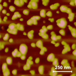
The nanoparticles, pictured as gold colored, are less than 100 nanometers in diameter. photo credit: bogdog Dan via photopincc

The nanoparticles, pictured as gold colored, are less than 100 nanometers in diameter. photo credit: bogdog Dan via photopincc

My guess is that the photographer has added some colour such as the gold and the pink to enhance the image as otherwise this would be a symphony of grey tones.

So, if this material will lead to weakened chitin such that pesticides and insecticides are more effective, does this mean that something else in the food chain will suffer because it no longer has mosquitos and other pests to munch on?

One last note, usually my ‘mosquito’ pieces concern malaria and the most recent of those was a Sept. 4, 2014 posting about a possible malaria vaccine being developed at the University of Connecticut.

Storing isotopes in nanocontainers for safer radiation therapy

While it can be effective, radiation therapy is known to be destructive  for cancerous cells and healthy cells. Researchers at Kansas State University and their colleagues in other institutions have devised a new technique that contains the isotopes so they reach the cancerous cells only. From an April 2, 2014 news item in ScienceDaily,

Researchers have discovered that microscopic “bubbles” developed at Kansas State University are safe and effective storage lockers for harmful isotopes that emit ionizing radiation for treating tumors.

The findings can benefit patient health and advance radiation therapy used to treat cancer and other diseases, said John M. Tomich, a professor of biochemistry and molecular biophysics who is affiliated with the university’s Johnson Cancer Research Center.

Tomich conducted the study with Ekaterina Dadachova, a radiochemistry specialist at Albert Einstein College of Medicine in New York, along with researchers from his group at Kansas State University, the University of Kansas, Jikei University School of Medicine in Japan and the Institute for Transuranium Elements in Germany. They recently published their findings in the study “Branched Amphiphilic Peptide Capsules: Cellular Uptake and Retention of Encapsulated Solutes,” which appears in the scientific journal Biochimica et Biophysica Acta.

The study looks at the ability of nontoxic molecules to store and deliver potentially harmful alpha emitting radioisotopes — one of the most effective forms of radiation therapy.

An April 2, 2014 Kansas State University news release (also on EurekAlert), which originated the news item, provides more details about this research that in some ways dates from 2012,

The study looks at the ability of nontoxic molecules to store and deliver potentially harmful alpha emitting radioisotopes — one of the most effective forms of radiation therapy.

In 2012, Tomich and his research lab team combined two related sequences of amino acids to form a very small, hollow nanocapsule similar to a bubble.

“We found that the two sequences come together to form a thin membrane that assembled into little spheres, which we call capsules,” Tomich said. “While other vesicles have been created from lipids, most are much less stable and break down. Ours are like stones, though. They’re incredibly stable and are not destroyed by cells in the body.”

The ability of the capsules to stay intact with the isotope inside and remain undetected by the body’s clearance systems prompted Tomich to investigate using the capsules as unbreakable storage containers that can be used for biomedical research, particularly in radiation therapies.

“The problem with current alpha-particle radiation therapies used to treat cancer is that they lead to the release of nontargeted radioactive daughter ions into the body,” Tomich said. “Radioactive atoms break down to form new atoms, called daughter ions, with the release of some form of energy or energetic particles. Alpha emitters give off an energetic particle that comes off at nearly the speed of light.”

These particles are like a car careening on ice, Tomich said. They are very powerful but can only travel a short distance. On collision, the alpha particle destroys DNA and whatever vital cellular components are in its path. Similarly, the daughter ions recoil with high energy on ejection of the alpha particle — similar to how a gun recoils as it is fired. The daughter ions have enough energy to escape the targeting and containment molecules that currently are in use.

“Once freed, the daughter isotopes can end up in places you don’t want them, like bone marrow, which can then lead to leukemia and new challenges,” Tomich said. “We don’t want any stray isotopes because they can harm the body. The trick is to get the radioactive isotopes into and contained in just diseases cells where they can work their magic.”

The radioactive compound that the team works with is 225Actinium, which on decay releases four alpha particles and numerous daughter ions.

Tomich and Dadachova tested the retention and biodistribution of alpha-emitting particles trapped inside the peptide capsules in cells. The capsules readily enter cells. Once inside, they migrate to a position alongside the nucleus, where the DNA is.

Tomich and Dadachova found that as the alpha particle-emitting isotopes decayed, the recoiled daughter ion collides with the capsule walls and essentially bounces off them and remains trapped inside the capsule. This completely blocked the release of the daughter ions, which prevented uptake in certain nontarget tissues and protected the subject from harmful radiation that would have otherwise have been releases into the body.

Tomich said that more studies are needed to add target molecules to the surface of the capsules. He anticipates that this new approach will provide a safer option for treating tumors with radiation therapy by reducing the amount of radioisotope required for killing the cancer cells and reducing the side effects caused by off-target accumulation of the radioisotopes.

“These capsules are easy to make and easy to work with,” Tomich said. “I think we’re just scratching the surface of what we can do with them to improve human health and nanomaterials.”

I hope this new technique proves effective and travels soon from the laboratory to clinical practice in the foreseeable future.

In the meantime, here’s a link to and a citation for the paper,

Branched amphiphilic peptide capsules: Cellular uptake and retention of encapsulated solutes by Pinakin Sukthankar, L. Adriana Avila, Susan K. Whitaker, Takeo Iwamoto, Alfred Morgenstern, Christos Apostolidis, Ke Liu, Robert P. Hanzlik, Ekaterina Dadachova, and John M. Tomich. Biochimica et Biophysica Acta (BBA) – Biomembranes (Biochim Biophys Acta) 2014 Feb 22. pii: S0005-2736(14)00069-8. doi: 10.1016/j.bbamem.2014.02.005. Available online 22 February 2014

This paper is behind a paywall.

Quantum dots and graphene; a mini roundup

I’ve done very little writing about quantum dots (so much nano, so little time) but there’s been a fair amount of activity lately which has piqued my interest. In the last few days researchers at Kansas State University have been getting noticed for being able to control the size and shape of the graphene quantum dots they produce.  This one has gotten extensive coverage online including this May 17, 2012 news item on physorg.com,

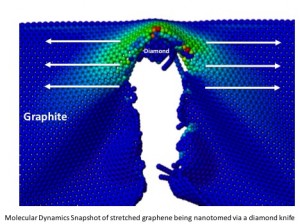
Vikas Berry, William H. Honstead professor of chemical engineering, has developed a novel process that uses a diamond knife to cleave graphite into graphite nanoblocks, which are precursors for graphene quantum dots. These nanoblocks are then exfoliated to produce ultrasmall sheets of carbon atoms of controlled shape and size.

By controlling the size and shape, the researchers can control graphene’s properties over a wide range for varied applications, such as solar cells, electronics, optical dyes, biomarkers, composites and particulate systems. Their work has been published in Nature Communications and supports the university’s vision to become a top 50 public research university by 2025. The article is available online.

Here’s an image of graphene being cut by a diamond knife from the May 16, 2012 posting by jtorline on the K-State News blog,

Molecular dynamics snapshot of stretched graphene being nanotomed via a diamond knife.

Here’s why standardizing the size is so important,

While other researchers have been able to make quantum dots, Berry’s research team can make quantum dots with a controlled structure in large quantities, which may allow these optically active quantum dots to be used in solar cell and other optoelectronic applications. [emphasis mine]

While all this is happening in Kansas, the Econ0mist magazine published a May 12, 2012 article about some important quantum dot optoelectronic developments in Spain (an excellent description for relative beginners is given and, if this area interests you, I’d suggest reading it in full),

Actually converting the wonders of graphene into products has been tough. But Frank Koppens and his colleagues at the Institute of Photonic Sciences in Barcelona think they have found a way to do so. As they describe in Nature Nanotechnology, they believe graphene can be used to make ultra-sensitive, low-cost photodetectors.

A typical photodetector is made of a silicon chip a few millimetres across onto which light is focused by a small lens. Light striking the chip knocks electrons free from some of the silicon atoms, producing a signal that the chip’s electronics convert into a picture or other useful information. …

Silicon photodetectors suffer, though, from a handicap: they are inflexible. Nor are they particularly cheap. And they are not that sensitive. They absorb only 10-20% of the light that falls on to them. For years, therefore, engineers have been on the lookout for a cheap, bendable, sensitive photodetector. …

By itself, graphene is worse than silicon at absorbing light. According to Dr Koppens only 2.7% of the photons falling on it are captured. But he and his colleague Gerasimos Konstantatos have managed to increase this to more than 50% by spraying tiny crystals of lead sulphide onto the surface of the material.

So combining the ability to size quantum dots uniformly with this discovery on how to make graphene more sensitive (and more useful in potential products) with quantum dots suggests some very exciting possibilities including this one mentioned by Dexter Johnson (who’s living in Spain these days) in his May 16, 2012 posting on Nanoclast (on the Institute of Electrical and Electronics Engineers [IEEE] website),

The researchers offer a range of applications for the graphene-and-quantum-dot combination, including digital cameras and sensors.  [emphasis mine] But it seems the researchers seem particularly excited about one application in particular. They expect the material will be used for night-vision technologies in automobiles—an application I have never heard trotted out before in relation to nanotech.

You can get more insights, more precise descriptions if you want to follow up from the Econ0mist article,  and Dexter’s links to more information about the research in his posting.

In my final roundup piece, I received a news release (dated April 24, 2012) about a quantum dot commercialization project at the University of Utah,

One of the biggest challenges for advancing quantum dots is the manufacturing process. Conventional processes are expensive, require high temperatures and produce low yields. However, researchers at the University of Utah believe they have a solution. They recently formed a startup company called Navillum Nanotechnologies, and their efforts are gaining national attention with help from a team of M.B.A. students from the David Eccles School of Business.
The students recently won first place and $100,000 at the regional CU Cleantech New Venture Challenge. The student competition concluded at the University of Colorado in Boulder on Friday, April 20. The student team advances to the national championship, which will be held in June in Washington, D.C. Student teams from six regions will compete for additional prizes and recognition at the prestigious event. Other regional competitions were held at MIT, Cal Tech, the University of Maryland, Clean Energy Trust (Chicago) and Rice University. All the competitions are financed by the U.S. Department of Energy.

The students will be competing in the national Clean Energy Business Plan Competition taking place June 12-13, 2012 in Washington, D.C.  Here are a few more details from the national competition webpage,

Winners of the six regional competitions will represent their home universities and regions as they vie for the honor of presenting the best clean energy business plan before a distinguished panel of expert judges and invited guests from federal agencies, industry, national labs and the venture capital community.

Confirmed Attendees include:

The Honorable Steven Chu
Energy Secretary [US federal government]

Dr. David Danielson
Assistant Secretary, EERE  [US Dept. of Energy, energy efficiency and renewable energy technologies)

Dr. Karina Edmonds
Technology Transfer Coordinator [US Dept. of Energy]

Mr. Todd Park
Chief Technology Officer, White House

Good luck to the students!