Tag Archives: US Department of Energy

Commercializing nanotechnology: Peter Thiel’s Breakout Labs and Argonne National Laboratories

Breakout Labs

I last wrote about entrepreneur Peter Thiel’s Breakout Labs project in an Oct. 26, 2011 posting announcing its inception. An Oct. 6, 2015 Breakout Labs news release (received in my email) highlights a funding announcement for four startups of which at least three are nanotechnology-enabled,

Breakout Labs, a program of Peter Thiel’s philanthropic organization, the Thiel Foundation, announced today that four new companies advancing scientific discoveries in biomedical, chemical engineering, and nanotechnology have been selected for funding.

“We’re always hearing about bold new scientific research that promises to transform the world, but far too often the latest discoveries are left withering in a lab,” said Lindy Fishburne, Executive Director of Breakout Labs. “Our mission is to help a new type of scientist-entrepreneur navigate the startup ecosystem and build lasting companies that can make audacious scientific discoveries meaningful to everyday life. The four new companies joining the Breakout Labs portfolio – nanoGriptech, Maxterial, C2Sense, and CyteGen – embody that spirit and we’re excited to be working with them to help make their vision a reality.”

The future of adhesives: inspired by geckos

Inspired by the gecko’s ability to scuttle up walls and across ceilings due to their millions of micro/nano foot-hairs,nanoGriptech (http://nanogriptech.com/), based in Pittsburgh, Pa., is developing a new kind of microfiber adhesive material that is strong, lightweight, and reusable without requiring glues or producing harmful residues. Currently being tested by the U.S. military, NASA, and top global brands, nanoGriptech’s flagship product Setex™ is the first adhesive product of its kind that is not only strong and durable, but can also be manufactured at low cost, and at scale.

“We envision a future filled with no-leak biohazard enclosures, ergonomic and inexpensive car seats, extremely durable aerospace adhesives, comfortable prosthetic liners, high performance athletic wear, and widely available nanotechnology-enabled products manufactured less expensively — all thanks to the grippy little gecko,” said Roi Ben-Itzhak, CFO and VP of Business Development for nanoGriptech.

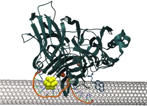
A sense of smell for the digital world

Despite the U.S. Department of Agriculture’s recent goals to drastically reduce food waste, most consumers don’t realize the global problem created by 1.3 billion metric tons of food wasted each year — clogging landfills and releasing unsustainable levels of methane gas into the atmosphere. Using technology developed at MIT’s Swager lab, Cambridge, Ma.-based C2Sense(http://www.c2sense.com/) is developing inexpensive, lightweight hand-held sensors based on carbon nanotubes which can detect fruit ripeness and meat, fish and poultry freshness. Smaller than a half of a business card, these sensors can be developed at very low cost, require very little power to operate, and can be easily integrated into most agricultural supply chains, including food storage packaging, to ensure that food is picked, stored, shipped, and sold at optimal freshness.

“Our mission is to bring a sense of smell to the digital world. With our technology, that package of steaks in your refrigerator will tell you when it’s about to go bad, recommend some recipe options and help build out your shopping list,” said Jan Schnorr, Chief Technology Officer of C2Sense.

Amazing metals that completely repel water

MaxterialTM, Inc. develops amazing materials that resist a variety of detrimental environmental effects through technology that emulates similar strategies found in nature, such as the self-cleaning lotus leaf and antifouling properties of crabs. By modifying the surface shape or texture of a metal, through a method that is very affordable and easy to introduce into the existing manufacturing process, Maxterial introduces a microlayer of air pockets that reduce contact surface area. The underlying material can be chemically the same as ever, retaining inherent properties like thermal and electrical conductivity. But through Maxterial’s technology, the metallic surface also becomes inherently water repellant. This property introduces the superhydrophobic maxterial as a potential solution to a myriad of problems, such as corrosion, biofouling, and ice formation. Maxterial is currently focused on developing durable hygienic and eco-friendly anti-corrosion coatings for metallic surfaces.

“Our process has the potential to create metallic objects that retain their amazing properties for the lifetime of the object – this isn’t an aftermarket coating that can wear or chip off,” said Mehdi Kargar, Co-founder and CEO of Maxterial, Inc. “We are working towards a day when shipping equipment can withstand harsh arctic environments, offshore structures can resist corrosion, and electronics can be fully submersible and continue working as good as new.”

New approaches to combat aging

CyteGen (http://cytegen.com/) wants to dramatically increase the human healthspan, tackle neurodegenerative diseases, and reverse age-related decline. What makes this possible now is new discovery tools backed by the dream team of interdisciplinary experts the company has assembled. CyteGen’s approach is unusually collaborative, tapping into the resources and expertise of world-renowned researchers across eight major universities to focus different strengths and perspectives to achieve the company’s goals. By approaching aging from a holistic, systematic point of view, rather than focusing solely on discrete definitions of disease, they have developed a new way to think about aging, and to develop treatments that can help people live longer, healthier lives.

“There is an assumption that aging necessarily brings the kind of physical and mental decline that results in Parkinson’s, Alzheimer’s, and other diseases. Evidence indicates otherwise, which is what spurred us to launch CyteGen,” said George Ugras, Co-Founder and President of CyteGen.

To date, Breakout Labs has invested in more than two dozen companies at the forefront of science, helping radical technologies get beyond common hurdles faced by early stage companies, and advance research and development to market much more quickly. Portfolio companies have raised more than six times the amount of capital invested in the program by the Thiel Foundation, and represent six Series A valuations ranging from $10 million to $60 million as well as one acquisition.

You can see the original Oct. 6, 2015 Breakout Labs news release here or in this Oct. 7, 2015 news item on Azonano.

Argonne National Labs and Nano Design Works (NDW) and the Argonne Collaborative Center for Energy Storage Science (ACCESS)

The US Department of Energy’s Argonne National Laboratory’s Oct. 6, 2015 press release by Greg Cunningham announced two initiatives meant to speed commercialization of nanotechnology-enabled products for the energy storage and other sectors,

Few technologies hold more potential to positively transform our society than energy storage and nanotechnology. Advances in energy storage research will revolutionize the way the world generates and stores energy, democratizing the delivery of electricity. Grid-level storage can help reduce carbon emissions through the increased adoption of renewable energy and use of electric vehicles while helping bring electricity to developing parts of the world. Nanotechnology has already transformed the electronics industry and is bringing a new set of powerful tools and materials to developers who are changing everything from the way energy is generated, stored and transported to how medicines are delivered and the way chemicals are produced through novel catalytic nanomaterials.

Recognizing the power of these technologies and seeking to accelerate their impact, the U.S. Department of Energy’s Argonne National Laboratory has created two new collaborative centers that provide an innovative pathway for business and industry to access Argonne’s unparalleled scientific resources to address the nation’s energy and national security needs. These centers will help speed discoveries to market to ensure U.S. industry maintains a lead in this global technology race.

“This is an exciting time for us, because we believe this new approach to interacting with business can be a real game changer in two areas of research that are of great importance to Argonne and the world,” said Argonne Director Peter B. Littlewood. “We recognize that delivering to market our breakthrough science in energy storage and nanotechnology can help ensure our work brings the maximum benefit to society.”

Nano Design Works (NDW) and the Argonne Collaborative Center for Energy Storage Science (ACCESS) will provide central points of contact for companies — ranging from large industrial entities to smaller businesses and startups, as well as government agencies — to benefit from Argonne’s world-class expertise, scientific tools and facilities.

NDW and ACCESS represent a new way to collaborate at Argonne, providing a single point of contact for businesses to assemble tailored interdisciplinary teams to address their most challenging R&D questions. The centers will also provide a pathway to Argonne’s fundamental research that is poised for development into practical products. The chance to build on existing scientific discovery is a unique opportunity for businesses in the nano and energy storage fields.

The center directors, Andreas Roelofs of NDW and Jeff Chamberlain of ACCESS, have both created startups in their careers and understand the value that collaboration with a national laboratory can bring to a company trying to innovate in technologically challenging fields of science. While the new centers will work with all sizes of companies, a strong emphasis will be placed on helping small businesses and startups, which are drivers of job creation and receive a large portion of the risk capital in this country.

“For a startup like mine to have the ability to tap the resources of a place like Argonne would have been immensely helpful,” said Roelofs. “We”ve seen the power of that sort of access, and we want to make it available to the companies that need it to drive truly transformative technologies to market.”

Chamberlain said his experience as an energy storage researcher and entrepreneur led him to look for innovative approaches to leveraging the best aspects of private industry and public science. The national laboratory system has a long history of breakthrough science that has worked its way to market, but shortening that journey from basic research to product has become a growing point of emphasis for the national laboratories over the past couple of decades. The idea behind ACCESS and NDW is to make that collaboration even easier and more powerful.

“Where ACCESS and NDW will differ from the conventional approach is through creating an efficient way for a business to build a customized, multi-disciplinary team that can address anything from small technical questions to broad challenges that require massive resources,” Chamberlain said. “That might mean assembling a team with chemists, physicists, computer scientists, materials engineers, imaging experts, or mechanical and electrical engineers; the list goes on and on. It’s that ability to tap the full spectrum of cross-cutting expertise at Argonne that will really make the difference.”

Chamberlain is deeply familiar with the potential of energy storage as a transformational technology, having led the formation of Argonne’s Joint Center for Energy Storage Research (JCESR). The center’s years-long quest to discover technologies beyond lithium-ion batteries has solidified the laboratory’s reputation as one of the key global players in battery research. ACCESS will tap Argonne’s full battery expertise, which extends well beyond JCESR and is dedicated to fulfilling the promise of energy storage.

Energy storage research has profound implications for energy security and national security. Chamberlain points out that approximately 1.3 billion people across the globe do not have access to electricity, with another billion having only sporadic access. Energy storage, coupled with renewable generation like solar, could solve that problem and eliminate the need to build out massive power grids. Batteries also have the potential to create a more secure, stable grid for countries with existing power systems and help fight global climate disruption through adoption of renewable energy and electric vehicles.

Argonne researchers are pursuing hundreds of projects in nanoscience, but some of the more notable include research into targeted drugs that affect only cancerous cells; magnetic nanofibers that can be used to create more powerful and efficient electric motors and generators; and highly efficient water filtration systems that can dramatically reduce the energy requirements for desalination or cleanup of oil spills. Other researchers are working with nanoparticles that create a super-lubricated state and other very-low friction coatings.

“When you think that 30 percent of a car engine’s power is sacrificed to frictional loss, you start to get an idea of the potential of these technologies,” Roelofs said. “But it’s not just about the ideas already at Argonne that can be brought to market, it’s also about the challenges for businesses that need Argonne-level resources. I”m convinced there are many startups out there working on transformational ideas that can greatly benefit from the help of a place Argonne to bring those ideas to fruition. That is what has me excited about ACCESS and NDW.”

For more information on ACCESS, see: access.anl.gov

For more information on NDW, see: nanoworks.anl.gov

You can read more about the announcement in an Oct. 6, 2015 article by Greg Watry for R&D magazine featuring an interview with Andreas Roelofs.

Enzymatic fuel cells with ultrasmall gold nanocluster

Scientists at the US Department of Energy’s Los Alamos National Laboratory have developed a DNA-templated gold nanocluster (AuNC) for more efficient biofuel cell design (Note: A link has been removed). From a Sept. 24, 2015 news item on ScienceDaily,

With fossil-fuel sources dwindling, better biofuel cell design is a strong candidate in the energy field. In research published in the Journal of the American Chemical Society (“A Hybrid DNA-Templated Gold Nanocluster For Enhanced Enzymatic Reduction of Oxygen”), Los Alamos researchers and external collaborators synthesized and characterized a new DNA-templated gold nanocluster (AuNC) that could resolve a critical methodological barrier for efficient biofuel cell design.

Here’s an image illustrating the DNA-templated gold nanoclusters,

Caption: Gold nanoclusters (~1 nm) are efficient mediators of electron transfer between co-self-assembled enzymes and carbon nanotubes in an enzyme fuel cell. The efficient electron transfer from this quantized nano material minimizes the energy waste and improves the kinetics of the oxygen reduction reaction, toward a more efficient fuel cell cycle. Credit: Los Alamos National Laboratory

Caption: Gold nanoclusters (~1 nm) are efficient mediators of electron transfer between co-self-assembled enzymes and carbon nanotubes in an enzyme fuel cell. The efficient electron transfer from this quantized nano material minimizes the energy waste and improves the kinetics of the oxygen reduction reaction, toward a more efficient fuel cell cycle.
Credit: Los Alamos National Laboratory

A Sept. 24, 2015 Los Alamos National Laboratory news release, which originated the news item, provides more details,

“Enzymatic fuel cells and nanomaterials show great promise and as they can operate under environmentally benign neutral pH conditions, they are a greener alternative to existing alkaline or acidic fuel cells, making them the subject of worldwide research endeavors,” said Saumen Chakraborty, a scientist on the project. “Our work seeks to boost electron transfer efficiency, creating a potential candidate for the development of cathodes in enzymatic fuel cells.”

Ligands, molecules that bind to a central metal atom, are necessary to form stable nanoclusters. For this study, the researchers chose single-stranded DNA as the ligand, as DNA is a natural nanoscale material having high affinity for metal cations and can be used to assembly the cluster to other nanoscale material such as carbon nanotubes.

In enzymatic fuel cells, fuel is oxidized on the anode, while oxygen reduction reactions take place on the cathode, often using multi copper oxidases. Enzymatic fuel cell performance depends critically on how effectively the enzyme active sites can accept and donate electrons from the electrode by direct electron transfer (ET). However, the lack of effective ET between the enzyme active sites, which are usually buried ~10Å from their surface, and the electrode is a major barrier to their development. Therefore, effective mediators of this electron transfer are needed.

The team developed a new DNA-templated gold nanocluster (AuNC) that enhanced electron transfer. This novel role of the AuNC as enhancer of electron transfer at the enzyme-electrode interface could be effective for cathodes in enzymatic fuel cells, thus removing a critical methodological barrier for efficient biofuel cell design.

Possessing many unique properties due to their discrete electron state distributions, metal nanoclusters (<1.5 nm diameter; ~2-144 atoms of gold, silver, platinum, or copper) show application in many fields.

Hypothesizing that due to the ultra-small size (the clusters are ~7 atoms, ~0.9 nm in diameter), and unique electrochemical properties, the AuNC can facilitate electron transfer to an oxygen-reduction reaction enzyme-active site and therefore, lower the overpotential of the oxygen reaction. Overpotential is the extra amount of energy required to drive an electrochemical reaction.

Ideally, it is desirable that all electrochemical reactions have minimal to no overpotential, but in reality they all have some. Therefore, to design an efficient electrocatalyst (for reduction or oxidation) we want to design it so that the reaction can proceed with a minimal amount of extra, applied energy.

When self assembled with bilirubin oxidase and carbon nanotubes, the AuNC acts to enhance the electron transfer, and it lowers the overpotential of oxygen reduction by a significant ~15 mV (as opposed to ~1-2 mV observed using other types of mediators) compared to the enzyme alone. The AuNC also causes significant enhancement of electrocatalytic current densities. Proteins are electronically insulating (they are complex, greasy and large), so the use of carbon nanotubes helps the enzyme stick to the electrode as well as to facilitate electron transfer.

Although gold nanoclusters have been used in chemical catalysis, this is the first time that we demonstrate they can also act as electron relaying agents to enzymatic oxygen reduction reaction monitored by electrochemistry.

Finally, the presence of AuNC does not perturb the mechanism of enzymatic O2 reduction. Such unique application of AuNC as facilitator of ET by improving thermodynamics and kinetics of O2 reduction is unprecedented.

Here’s a link to and a citation for the paper,

A Hybrid DNA-Templated Gold Nanocluster For Enhanced Enzymatic Reduction of Oxygen by Saumen Chakraborty, Sofia Babanova, Reginaldo C. Rocha, Anil Desireddy, Kateryna Artyushkova, Amy E. Boncella, Plamen Atanassov, and Jennifer S. Martinez. J. Am. Chem. Soc., 2015, 137 (36), pp 11678–11687 DOI: 10.1021/jacs.5b05338 Publication Date (Web): August 19, 2015

Copyright © 2015 American Chemical Society

This paper is behind a paywall.

Carbyne: 40x stiffer than diamond

A material that’s tougher than diamond is the object of interest for researchers at the US Department of Energy’s Lawrence Livermore National Laboratory (LLNL) according to a Sept. 18, 2015 news item by Beth Ellison on Azonano (Note: A link has been removed),

Researchers at Lawrence Livermore National Laboratory (LLNL) have explored a method that uses laser-melted graphite to develop linear chains of carbon atoms.

This material, referred to as carbyne, could possess numerous unique properties, such as modification of the quantity of electrical current passing through a circuit according to the needs of a user. This research could probably lead to the creation of tiny electronics capable of turning on and off at an atomic scale.

A Sept. 17, 2015 LLNL news release (also on EurekAlert) details the research (Note: A link has been removed),

Carbyne is the subject of intense research because of its presence in astrophysical bodies, as well as its potential use in nanoelectronic devices and superhard materials. Its linear shape gives it unique electrical properties that are sensitive to stretching and bending, and it is 40 times stiffer than diamond. It also was found in the Murchison and Allende meteorites and could be an ingredient of interstellar dust.

Using computer simulations, LLNL scientist Nir Goldman and colleague Christopher Cannella, an undergraduate summer researcher from Caltech, initially intended to study the properties of liquid carbon as it evaporates, after being formed by shining a laser beam on the surface of graphite. The laser can heat the graphite surface to a few thousands of degrees, which then forms a fairly volatile droplet. To their surprise, as the liquid droplet evaporated and cooled in their simulations, it formed bundles of linear chains of carbon atoms.

“There’s been a lot of speculation about how to make carbyne and how stable it is,” Goldman said. “We showed that laser melting of graphite is one viable avenue for its synthesis. If you regulate carbyne synthesis in a controlled way, it could have applications as a new material for a number of different research areas, including as a tunable semiconductor or even for hydrogen storage.

“Our method shows that carbyne can be formed easily in the laboratory or otherwise. The process also could occur in astrophysical bodies or in the interstellar medium, where carbon-containing material can be exposed to relatively high temperatures and carbon can liquefy.”

Goldman’s study and computational models allow for direct comparison with experiments and can help determine parameters for synthesis of carbon-based materials with potentially exotic properties.

“Our simulations indicate a possible mechanism for carbyne fiber synthesis that confirms previous experimental observation of its formation,” Goldman said. “These results help determine one set of thermodynamic conditions for its synthesis and could account for its detection in meteorites resulting from high-pressure conditions due to impact.”

Here’s a link to and a citation for the paper,

Carbyne Fiber Synthesis within Evaporating Metallic Liquid Carbon by Christopher B. Cannella and Nir Goldman. J. Phys. Chem. C, 2015, 119 (37), pp 21605–21611 DOI: 10.1021/acs.jpcc.5b03781 Publication Date (Web): July 9, 2015 (print): Sept. 17, 2015

Copyright © 2015 American Chemical Society

This paper is behind a paywall.

Dexter Johnson in a Sept. 18, 2015 posting about the latest carbyne developments on his Nanoclast blog (on the IEEE [Institute for Electrical and Electronics Engineers] website) provides a little history (Note: Links have been removed),

A couple of years ago, a material dubbed carbyne—which is a chain of carbon atoms held together by either double or alternating single and triple atomic bonds—was awarded the title of the world’s strongest material. Later, scientists also demonstrated that it has the unusual property of being able to change from being a conductor to an insulator when it’s stretched by as little as 3 percent.

Here’s an image illustrating the process,

A carbyne strand forms in laser-melted graphite. Carbyne is found in astrophysical bodies and has the potential to be used in nanoelectronic devices and superhard materials. Image by Liam Krauss/LLNL

A carbyne strand forms in laser-melted graphite. Carbyne is found in astrophysical bodies and has the potential to be used in nanoelectronic devices and superhard materials. Image by Liam Krauss/LLNL

Windows as solar panels

Thanks to Dexter Johnson’s Aug. 27, 2015 posting, I’ve found another type of ‘smart’ window (I have written many postings about nanotechnology-enabled windows, especially self-cleaning ones); this window is a solar panel (Note: Links have been removed),

In joint research between the Department of Energy’s Los Alamos National Laboratory (LANL) and the University of Milan-Bicocca (UNIMIB) in Italy, researchers have spent the last 16 months perfecting a technique that makes it possible to embed quantum dots into windows so that the window itself becomes a solar panel.

Of course, this is not the first time someone thought that it would be a good idea to make windows into solar collectors. But this latest iteration marks a significant development in the evolution of the technology. Previous technologies used organic emitters that limited the size of the concentrators to just a few centimeters.

The energy conversion efficiency the researchers were able to acheive with the solar windows was around 3.2 percent, which stands up pretty well when compared with state-of-the-art quantum dot-based solar cells that have reached 9 percent conversion efficiency.

An August 24, 2015 US Los Alamos National Laboratory news release, which inspired Dexter’s posting, describes the research and the US-Italian collaboration in more detail,

A luminescent solar concentrator [LSC] is an emerging sunlight harvesting technology that has the potential to disrupt the way we think about energy; It could turn any window into a daytime power source.

“In these devices, a fraction of light transmitted through the window is absorbed by nanosized particles (semiconductor quantum dots) dispersed in a glass window, re-emitted at the infrared wavelength invisible to the human eye, and wave-guided to a solar cell at the edge of the window,” said Victor Klimov, lead researcher on the project at the Department of Energy’s Los Alamos National Laboratory. “Using this design, a nearly transparent window becomes an electrical generator, one that can power your room’s air conditioner on a hot day or a heater on a cold one.”

… The work was performed by researchers at the Center for Advanced Solar Photophysics (CASP) of Los Alamos, led by Klimov and the research team coordinated by Sergio Brovelli and Francesco Meinardi of the Department of Materials Science of the University of Milan-Bicocca (UNIMIB) in Italy.

The news release goes on to describe the precursor work which made this latest step forward possible,

In April 2014, using special composite quantum dots, the American-Italian collaboration demonstrated the first example of large-area luminescent solar concentrators free from reabsorption losses of the guided light by the nanoparticles. This represented a fundamental advancement with respect to the earlier technology, which was based on organic emitters that allowed for the realization of concentrators of only a few centimeters in size.

However, the quantum dots used in previous proof-of-principle devices were still unsuitable for real-world applications, as they were based on the toxic heavy metal cadmium and were capable of absorbing only a small portion of the solar light. This resulted in limited light-harvesting efficiency and strong yellow/red coloring of the concentrators, which complicated their application in residential environments.

Here’s how they solved the problem (from the news release),

Klimov, CASP’s director, explained how the updated approach solves the coloring problem: “Our new devices use quantum dots of a complex composition which includes copper (Cu), indium (In), selenium (Se) and sulfur (S). This composition is often abbreviated as CISeS. Importantly, these particles do not contain any toxic metals that are typically present in previously demonstrated LSCs.”

“Furthermore,” Klimov noted, “the CISeS quantum dots provide a uniform coverage of the solar spectrum, thus adding only a neutral tint to a window without introducing any distortion to perceived colors. In addition, their near-infrared emission is invisible to a human eye, but at the same time is ideally suited for most common solar cells based on silicon.”

Francesco Meinardi, professor of Physics at UNIMIB, described the emerging work, noting, “In order for this technology to leave the research laboratories and reach its full potential in sustainable architecture, it is necessary to realize non-toxic concentrators capable of harvesting the whole solar spectrum.”

“We must still preserve the key ability to transmit the guided luminescence without reabsorption losses, though, so as to complement high photovoltaic efficiency with dimensions compatible with real windows. The aesthetic factor is also of critical importance for the desirability of an emerging technology,” Meinardi said. [emphasis mine]

I couldn’t agree more with Professor Meinardi. You’re much more likely to adopt something that’s good for you and the planet if you like the look. Following on that thought, you’re much more likely to adopt solar panel windows if they’re aesthetically pleasing.

However, there is still a problem to be solved,

Hunter McDaniel, formerly a Los Alamos CASP postdoctoral fellow and presently a quantum dot entrepreneur (UbiQD founder and president), added, “with a new class of low-cost, low-hazard quantum dots composed of CISeS, we have overcome some of the biggest roadblocks to commercial deployment of this technology.”

“One of the remaining problems to tackle is reducing cost, but already this material is significantly less expensive to manufacture than alternative quantum dots used in previous LSC demonstrations,” McDaniel said.

Nonetheless, they have high hopes the technology can be commercialized (although as Dexter notes, it’s probably not going to be in the near future), from the news release,

A key element of this work is a procedure comparable to the cell casting industrial method used for fabricating high optical quality polymer windows. It involves a new UNIMIB protocol for encapsulating quantum dots into a high-optical quality transparent polymer matrix. The polymer used in this study is a cross-linked polylaurylmethacrylate, which belongs to the family of acrylate polymers. Its long side-chains prevent agglomeration of the quantum dots and provide them with the “friendly” local environment, which is similar to that of the original colloidal suspension. This allows one to preserve light emission properties of the quantum dots upon encapsulation into the polymer.

Sergio Brovelli, the lead researcher on the Italian team, concluded: “Quantum dot solar window technology, of which we had demonstrated the feasibility just one year ago, now becomes a reality that can be transferred to the industry in the short to medium term, allowing us to convert not only rooftops, as we do now, but the whole body of urban buildings, including windows, into solar energy generators.”

“This is especially important in densely populated urban area where the rooftop surfaces are too small for collecting all the energy required for the building operations,” he said. He proposes that the team’s estimations indicate that by replacing the passive glazing of a skyscraper such as the One World Trade Center in NYC (72,000 square meters divided into 12,000 windows) with our technology, it would be possible to generate the equivalent of the energy need of over 350 apartments.

“Add to these remarkable figures, the energy that would be saved by the reduced need for air conditioning thanks to the filtering effect by the LSC, which lowers the heating of indoor spaces by sunlight, and you have a potentially game-changing technology towards “net-zero” energy cities,” Brovelli said.

For anyone interested in this latest work on energy harvesting and windows, here’s a link to and a citation for the paper,

Highly efficient large-area colourless luminescent solar concentrators using heavy-metal-free colloidal quantum dots by Francesco Meinardi, Hunter McDaniel, Francesco Carulli, Annalisa Colombo, Kirill A. Velizhanin, Nikolay S. Makarov, Roberto Simonutti, Victor I. Klimov, & Sergio Brovelli. Nature Nanotechnology (2015) doi:10.1038/nnano.2015.178 Published online 24 August 2015

This paper is behind a paywall.

Corrections: Hybrid Photonic-Nanomechanical Force Microscopy uses vibration for better chemical analysis

*ETA  Nov. 4, 2015: I’m apologizing to anyone wishing to read this posting as it’s a bit of a mess. I deeply regret mishandling the situation. In future, I shall not be taking any corrections from individual researchers to materials such as news releases that have been issued by an institution. Whether or not the individual researchers are happy with how their contributions or how a colleague’s contributions or how their home institutions have been characterized is a matter for them and their home institutions.

The August 10, 2015 ORNL news release with all the correct details has been added to the end of this post.*

A researcher at the University of Central Florida (UCF) has developed a microscope that uses vibrations for better analysis of chemical composition. From an Aug. 10, 2015 news item on Nanowerk,

It’s a discovery that could have promising implications for fields as varied as biofuel production, solar energy, opto-electronic devices, pharmaceuticals and medical research.

“What we’re interested in is the tools that allow us to understand the world at a very small scale,” said UCF professor Laurene Tetard, formerly of the Oak Ridge National Laboratory. “Not just the shape of the object, but its mechanical properties, its composition and how it evolves in time.”

An Aug. 10, 2015 UCF news release (also on EurekAlert), which originated the news item, describes the limitations of atomic force microscopy and gives a few details about the hybrid microscope (Note: A link has been removed),

For more than two decades, scientists have used atomic force microscopy – a probe that acts like an ultra-sensitive needle on a record player – to determine the surface characteristics of samples at the microscopic scale. A “needle” that comes to an atoms-thin point traces a path over a sample, mapping the surface features at a sub-cellular level [nanoscale].

But that technology has its limits. It can determine the topographical characteristics of [a] sample, but it can’t identify its composition. And with the standard tools currently used for chemical mapping, anything smaller than roughly half a micron is going to look like a blurry blob, so researchers are out of luck if they want to study what’s happening at the molecular level.

A team led by Tetard has come up with a hybrid form of that technology that produces a much clearer chemical image. As described Aug. 10 in the journal Nature Nanotechnology, Hybrid Photonic-Nanomechanical Force Microscopy (HPFM) can discern a sample’s topographic characteristics together with the chemical properties at a much finer scale.

The HPFM method is able to identify materials based on differences in the vibration produced when they’re subjected to different wavelengths of light – essentially a material’s unique “fingerprint.”

“What we are developing is a completely new way of making that detection possible,” said Tetard, who has joint appointments to UCF’s Physics Department, Material Science and Engineering Department and the NanoScience Technology Center.

The researchers proved the effectiveness of HPFM while examining samples from an eastern cottonwood tree, a potential source of biofuel. By examining the plant samples at the nanoscale, the researchers for the first time were able to determine the molecular traits of both untreated and chemically processed cottonwood inside the plant cell walls.

The research team included Tetard; Ali Passian, R.H. Farahi and Brian Davison, all of Oak Ridge National Laboratory; and Thomas Thundat of the University of Alberta.

Long term, the results will help reveal better methods for producing the most biofuel from the cottonwood, a potential boon for industry. Likewise, the new method could be used to examine samples of myriad plants to determine whether they’re good candidates for biofuel production.

Potential uses of the technology go beyond the world of biofuel. Continued research may allow HPFM to be used as a probe so, for instance, it would be possible to study the effect of new treatments being developed to save plants such as citrus trees from bacterial diseases rapidly decimating the citrus industry, or study fundamental photonically-induced processes in complex systems such as in solar cell materials or opto-electronic devices.

Here’s a link to and a citation for the paper,

Opto-nanomechanical spectroscopic material characterization by L. Tetard, A. Passian, R. H. Farahi, T. Thundat, & B. H. Davison. Nature Nanotechnology (2015) doi:10.1038/nnano.2015.168 Published online 10 August 2015

This paper is behind a paywall.

*ETA August 27, 2015:

August 10, 2015 ORNL news release (Note: Funding information and a link to the paper [previously given] have been removed):

A microscope being developed at the Department of Energy’s Oak Ridge National Laboratory will allow scientists studying biological and synthetic materials to simultaneously observe chemical and physical properties on and beneath the surface.

The Hybrid Photonic Mode-Synthesizing Atomic Force Microscope is unique, according to principal investigator Ali Passian of ORNL’s Quantum Information System group. As a hybrid, the instrument, described in a paper published in Nature Nanotechnology, combines the disciplines of nanospectroscopy and nanomechanical microscopy.

“Our microscope offers a noninvasive rapid method to explore materials simultaneously for their chemical and physical properties,” Passian said. “It allows researchers to study the surface and subsurface of synthetic and biological samples, which is a capability that until now didn’t exist.”

ORNL’s instrument retains all of the advantages of an atomic force microscope while simultaneously offering the potential for discoveries through its high resolution and subsurface spectroscopic capabilities.

“The originality of the instrument and technique lies in its ability to provide information about a material’s chemical composition in the broad infrared spectrum of the chemical composition while showing the morphology of a material’s interior and exterior with nanoscale – a billionth of a meter – resolution,” Passian said.

Researchers will be able to study samples ranging from engineered nanoparticles and nanostructures to naturally occurring biological polymers, tissues and plant cells.

The first application as part of DOE’s BioEnergy Science Center was in the examination of plant cell walls under several treatments to provide submicron characterization. The plant cell wall is a layered nanostructure of biopolymers such as cellulose. Scientists want to convert such biopolymers to free the useful sugars and release energy.

An earlier instrument, also invented at ORNL, provided imaging of poplar cell wall structures that yielded unprecedented topological information, advancing fundamental research in sustainable biofuels.

Because of this new instrument’s impressive capabilities, the researcher team envisions broad applications.
“An urgent need exists for new platforms that can tackle the challenges of subsurface and chemical characterization at the nanometer scale,” said co-author Rubye Farahi. “Hybrid approaches such as ours bring together multiple capabilities, in this case, spectroscopy and high-resolution microscopy.”

Looking inside, the hybrid microscope consists of a photonic module that is incorporated into a mode-synthesizing atomic force microscope. The modular aspect of the system makes it possible to accommodate various radiation sources such as tunable lasers and non-coherent monochromatic or polychromatic sources.

ETA2 August 27, 2015: I’ve received an email from one of the paper’s authors (RH Farahi of the US Oak Ridge National Laboratory [ORNL]) who claims some inaccuracies in this piece.  The news release supplied by the University of Central Florida states that Dr. Tetard led the team and that is not so. According to Dr. Farahi, she had a postdoctoral position on the team which she left two years ago. You might also get the impression that some of the work was performed at the University of Central Florida. That is not so according to Dr. Farahi.  As a courtesy Dr. Tetard was retained as first author of the paper.

*Nov. 4, 2015: I suspect some of the misunderstanding was due to overeagerness and/or time pressures. Whoever wrote the news release may have made some assumptions. It’s very easy to make a mistake when talking to an ebullient scientist who can unintentionally lead you to believe something that’s not so. I worked in a high tech company and believed that there was some new software being developed which turned out to be a case of high hopes. Luckily, I said something that triggered a rapid rebuttal to the fantasies. Getting back to this situation, other contributing factors could include the writer not having time to get the news release reviewed the scientist or the scientist skimming the release and missing a few bits due to time pressure.*

MOFs (metal-organic frameworks) to clean up nuclear waste?

There’s a possibility that metal-organic frameworks could be used to clean up nuclear waste according to an Aug. 5, 2015 news item on phys.org,

One of the most versatile and widely applicable classes of materials being studied today are the metal-organic frameworks. These materials, known as MOFs, are characterized by metal ions or metal-ion clusters that are linked together with organic molecules, forming ordered crystal structures that contain tiny cage-like pores with diameters of two nanometers or less.

MOFs can be thought of as highly specialized and customizable sieves. By designing them with pores of a certain size, shape, and chemical composition, researchers can tailor them for specific purposes. A few of the many, many possible applications for MOFs are storing hydrogen in fuel cells, capturing environmental contaminants, or temporarily housing catalytic agents for chemical reactions.

At [US Department of Energy] Brookhaven National Laboratory, physicist Sanjit Ghose and his collaborators have been studying MOFs designed for use in the separation of waste from nuclear reactors, which results from the reprocessing of nuclear fuel rods. He is targeting two waste products in particular: the noble gases xenon (Xe) and krypton (Kr).

An Aug. 4, 2015 Brookhaven National Laboratory news release, which originated the news item, describes not only the research and the reasons for it but also the institutional collaborations necessary to conduct the research,

There are compelling economic and environmental reasons to separate Xe and Kr from the nuclear waste stream. For one, because they have very different half-lives – about 36 days for Xe and nearly 11 years for Kr – pulling out the Xe greatly reduces the amount of waste that needs to be stored long-term before it is safe to handle. Additionally, the extracted Xe can be used for industrial applications, such as in commercial lighting and as an anesthetic. This research may also help scientists determine how to create MOFs that can remove other materials from the nuclear waste stream and expose the remaining unreacted nuclear fuel for further re-use. This could lead to much less overall waste that must be stored long-term and a more efficient system for producing nuclear energy, which is the source of about 20 percent of the electricity in the U.S.

Because Xe and Kr are noble gases, meaning their outer electron orbitals are filled and they don’t tend to bind to other atoms, they are difficult to manipulate. The current method for extracting them from the nuclear waste stream is cryogenic distillation, a process that is energy-intensive and expensive. The MOFs studied here use a very different approach: polarizing the gas atoms dynamically, just enough to draw them in using the van der Waals force. The mechanism works at room temperature, but also at hotter temperatures, which is key if the MOFs are to be used in a nuclear environment.

Recently, Ghose co-authored two papers that describe MOFs capable of adsorbing Xe and Kr, and excel at separating the Xe from the Kr. The papers are published in the May 22 online edition of the Journal of the American Chemical Society and the April 16 online edition of the Journal of Physical Chemistry Letters.

“Only a handful of noble-gas-specific MOFs have been studied so far, and we felt there was certainly scope for improvement through the discovery of more selective materials,” said Ghose.

Both MOF studies were carried out by large multi-institution collaborations, using a combination of X-ray diffraction, theoretical modeling, and other methods. The X-ray work was performed at Brookhaven’s former National Synchrotron Light Source (permanently closed and replaced by its successor, NSLS-II) and the Advanced Photon Source at Argonne National Laboratory (ANL), both DOE Office of Science User Facilities.

The JACS paper was co-authored by researchers from Brookhaven Lab, Stony Brook University (SBU), Pacific Northwest National Laboratory (PNNL), and the University of Amsterdam. Authors on the JPCL paper include scientists from Brookhaven, SBU, PNNL, ANL, the Deutsches Elektronen-Synchrotron (DESY) in Germany, and DM Strachan, LLC.

Here’s more about the first published paper in the Journal of Physical Chemistry Letters (JCPL) (from the news release)

A nickel-based MOF

The MOF studied in the JCPL paper consists of nickel (Ni) and the organic compound dioxido-benzene-dicarboxylate (DOBC), and is thus referred to as Ni-DOBDC. Ni-DOBDC can adsorb both Xe and Kr at room temperature but is highly selective toward Xe. In fact, it boasts what may be the highest Xe adsorption capacity of a MOF discovered to date.

The group studied Ni-DOBC using two main techniques: X-ray diffraction and first-principles density functional theory (DFT). The paper is the first published report to detail the adsorption mechanism by which the MOF takes in these noble gases at room temperature and pressure.

“Our results provide a fundamental understanding of the adsorption structure and the interactions between the MOF and the gas by combining direct structural analyses from experimental X-ray diffraction data and DFT calculations,” said Ghose.

The group was also able to discover the existence of a secondary site at the pore center in addition to the six-fold primary site. The seven-atom loading scheme was initially proposed by theorist Yan Li, an co-author of the JCPL paper and formerly on staff at Brookhaven (she is now an editor at Physical Review B), which was confirmed experimentally and theoretically. Data also indicate that Xe are adsorbed more strongly than Kr, due to its higher atomic polarizability. They also discovered a temperature-dependence of the adsorption that furthers this MOF’s selectivity for Xe over Kr. As the temperature was increased above room temperature, the Kr adsorption drops more drastically than for Xe. Over the entire temperature range tested, Xe adsorption always dominates that of Kr.

“The high separation capacity of Ni-DOBDC suggests that it has great potential for removing Xe from Kr in the off-gas streams in nuclear spent fuel reprocessing, as well as filtering Xe at low concentration from other gas mixtures,” said Ghose.

Ghose and Li are now preparing a manuscript that will discuss a more in-depth investigation into the possibility of packing in even more Xe atoms.

“Because of the confinement offered by each pore, we want to see if it’s possible to fit enough Xe in each chamber to form a solid,” said Li.

Ghose and Li hope to experimentally test this idea at NSLS-II in the future, at the facility’s X-ray Powder Diffraction (XPD) beamline, which Ghose has helped develop and build. Additional future studies of these and other MOFs will also take place at XPD. For example, they want to see what happens when other gases are present, such as nitrogen oxides, to mimic what happens in an actual nuclear reactor.

Then, there was the second paper published in the Journal of the American Chemical Society (JACS),

Another MOF, Another Promising Result

In the JACS paper, Ghose and researchers from Brookhaven, SBU, PNNL, and the University of Amsterdam describe a second MOF, dubbed Stony Brook MOF-2 (SBMOF-2). It also captures both Xe and Kr at room temperature and pressure, although is about ten times as effective at taking in Xe, with Xe taking up as much as 27 percent of its weight. SBMOF-2 had been theoretically predicted to be an efficient adsorbent for Xe and Kr, but until this research there had been no experimental results to back up the prediction.

“Our study is different than MOF research done by other groups,” said chemist John Parise, a coauthor of the JACS paper who holds a joint position with Brookhaven and SBU. “We did a lot of testing and investigated the capture mechanism very closely to get clues that would help us understand why the MOF worked, and how to tailor the structure to have even better properties.”

SBMOF-2 contains calcium (Ca) ions and an organic compound with the chemical formula C34H22O8. X-ray data show that its structure is unusual among microporous MOFs. It has fewer calcium sites than expected and an excess of oxygen over calcium. The calcium and oxgyen form CaO6, which takes the form of a three-dimensional octahedron. Notably, none of the six oxygen atoms bound to the calcium ion are shared with any other nearby calcium ions. The authors believe that SBMOF-2 is the first microporous MOF with these isolated CaO6 octahedra, which are connected by organic linker molecules.

The group discovered that the preference of SBMOF-2 for Xe over Kr is due to both the geometry and chemistry of its pores. All the pores have diamond-shaped cross sections, but they come in two sizes, designated type-1 and type-2. Both sizes are a better fit for the Xe molecule. The interiors of the pores have walls made of phenyl groups – ring-shaped C6H5 molecules – along with delocalized electron clouds and H atoms pointing into the pore. The type-2 pores also have hydroxyl anions (OH-) available. All of these features provide are potential sites for adsorbed Xe and Kr atoms.

In follow-up studies, Ghose and his colleagues will use these results to guide them as they determine what changes can be made to these MOFs to improve adsorption, as well as to determine what existing MOFs may yield similar or better performance.

Here are links to and citations for both papers,

Understanding the Adsorption Mechanism of Xe and Kr in a Metal–Organic Framework from X-ray Structural Analysis and First-Principles Calculations by Sanjit K. Ghose, Yan Li, Andrey Yakovenko, Eric Dooryhee, Lars Ehm, Lynne E. Ecker, Ann-Christin Dippel, Gregory J. Halder, Denis M. Strachan, and Praveen K. Thallapally. J. Phys. Chem. Lett., 2015, 6 (10), pp 1790–1794 DOI: 10.1021/acs.jpclett.5b00440 Publication Date (Web): April 16, 2015

Copyright © 2015 American Chemical Society

Direct Observation of Xe and Kr Adsorption in a Xe-Selective Microporous Metal–Organic Framework by Xianyin Chen, Anna M. Plonka, Debasis Banerjee, Rajamani Krishna, Herbert T. Schaef, Sanjit Ghose, Praveen K. Thallapally, and John B. Parise. J. Am. Chem. Soc., 2015, 137 (22), pp 7007–7010 DOI: 10.1021/jacs.5b02556 Publication Date (Web): May 22, 2015
Copyright © 2015 American Chemical Society

Both papers are behind a paywall.

SINGLE (3D Structure Identification of Nanoparticles by Graphene Liquid Cell Electron Microscopy) and the 3D structures of two individual platinum nanoparticles in solution

It seems to me there’s been an explosion of new imaging techniques lately. This one from the Lawrence Berkelely National Laboratory is all about imaging colloidal nanoparticles (nanoparticles in solution), from a July 20, 2015 news item on Azonano,

Just as proteins are one of the basic building blocks of biology, nanoparticles can serve as the basic building blocks for next generation materials. In keeping with this parallel between biology and nanotechnology, a proven technique for determining the three dimensional structures of individual proteins has been adapted to determine the 3D structures of individual nanoparticles in solution.

A multi-institutional team of researchers led by the U.S. Department of Energy (DOE)’s Lawrence Berkeley National Laboratory (Berkeley Lab), has developed a new technique called “SINGLE” that provides the first atomic-scale images of colloidal nanoparticles. SINGLE, which stands for 3D Structure Identification of Nanoparticles by Graphene Liquid Cell Electron Microscopy, has been used to separately reconstruct the 3D structures of two individual platinum nanoparticles in solution.

A July 16, 2015 Berkeley Lab news release, which originated the news item, reveals more details about the reason for the research and the research itself,

“Understanding structural details of colloidal nanoparticles is required to bridge our knowledge about their synthesis, growth mechanisms, and physical properties to facilitate their application to renewable energy, catalysis and a great many other fields,” says Berkeley Lab director and renowned nanoscience authority Paul Alivisatos, who led this research. “Whereas most structural studies of colloidal nanoparticles are performed in a vacuum after crystal growth is complete, our SINGLE method allows us to determine their 3D structure in a solution, an important step to improving the design of nanoparticles for catalysis and energy research applications.”

Alivisatos, who also holds the Samsung Distinguished Chair in Nanoscience and Nanotechnology at the University of California Berkeley, and directs the Kavli Energy NanoScience Institute at Berkeley (Kavli ENSI), is the corresponding author of a paper detailing this research in the journal Science. The paper is titled “3D Structure of Individual Nanocrystals in Solution by Electron Microscopy.” The lead co-authors are Jungwon Park of Harvard University, Hans Elmlund of Australia’s Monash University, and Peter Ercius of Berkeley Lab. Other co-authors are Jong Min Yuk, David Limmer, Qian Chen, Kwanpyo Kim, Sang Hoon Han, David Weitz and Alex Zettl.

Colloidal nanoparticles are clusters of hundreds to thousands of atoms suspended in a solution whose collective chemical and physical properties are determined by the size and shape of the individual nanoparticles. Imaging techniques that are routinely used to analyze the 3D structure of individual crystals in a material can’t be applied to suspended nanomaterials because individual particles in a solution are not static. The functionality of proteins are also determined by their size and shape, and scientists who wanted to image 3D protein structures faced a similar problem. The protein imaging problem was solved by a technique called “single-particle cryo-electron microscopy,” in which tens of thousands of 2D transmission electron microscope (TEM) images of identical copies of an individual protein or protein complex frozen in random orientations are recorded then computationally combined into high-resolution 3D reconstructions. Alivisatos and his colleagues utilized this concept to create their SINGLE technique.

“In materials science, we cannot assume the nanoparticles in a solution are all identical so we needed to develop a hybrid approach for reconstructing the 3D structures of individual nanoparticles,” says co-lead author of the Science paper Peter Ercius, a staff scientist with the National Center for Electron Microscopy (NCEM) at the Molecular Foundry, a DOE Office of Science User Facility.

“SINGLE represents a combination of three technological advancements from TEM imaging in biological and materials science,” Ercius says. “These three advancements are the development of a graphene liquid cell that allows TEM imaging of nanoparticles rotating freely in solution, direct electron detectors that can produce movies with millisecond frame-to-frame time resolution of the rotating nanocrystals, and a theory for ab initio single particle 3D reconstruction.”

The graphene liquid cell (GLC) that helped make this study possible was also developed at Berkeley Lab under the leadership of Alivisatos and co-author Zettl, a physicist who also holds joint appointments with Berkeley Lab, UC Berkeley and Kavli ENSI. TEM imaging uses a beam of electrons rather than light for illumination and magnification but can only be used in a high vacuum because molecules in the air disrupt the electron beam. Since liquids evaporate in high vacuum, samples in solutions must be hermetically sealed in special solid containers – called cells – with a very thin viewing window before being imaged with TEM. In the past, liquid cells featured silicon-based viewing windows whose thickness limited resolution and perturbed the natural state of the sample materials. The GLC developed at Berkeley lab features a viewing window made from a graphene sheet that is only a single atom thick.

“The GLC provides us with an ultra-thin covering of our nanoparticles while maintaining liquid conditions in the TEM vacuum,” Ercius says. “Since the graphene surface of the GLC is inert, it does not adsorb or otherwise perturb the natural state of our nanoparticles.”

Working at NCEM’s TEAM I, the world’s most powerful electron microscope, Ercius, Alivisatos and their colleagues were able to image in situ the translational and rotational motions of individual nanoparticles of platinum that were less than two nanometers in diameter. Platinum nanoparticles were chosen because of their high electron scattering strength and because their detailed atomic structure is important for catalysis.

“Our earlier GLC studies of platinum nanocrystals showed that they grow by aggregation, resulting in complex structures that are not possible to determine by any previously developed method,” Ercius says. “Since SINGLE derives its 3D structures from images of individual nanoparticles rotating freely in solution, it enables the analysis of heterogeneous populations of potentially unordered nanoparticles that are synthesized in solution, thereby providing a means to understand the structure and stability of defects at the nanoscale.”

The next step for SINGLE is to recover a full 3D atomic resolution density map of colloidal nanoparticles using a more advanced camera installed on TEAM I that can provide 400 frames-per-second and better image quality.

“We plan to image defects in nanoparticles made from different materials, core shell particles, and also alloys made of two different atomic species,” Ercius says. [emphasis mine]

“Two different atomic species?”, they really are pushing that biology analogy.

Here’s a link to and a citation for the paper,

3D structure of individual nanocrystals in solution by electron microscopy by Jungwon Park, Hans Elmlund, Peter Ercius, Jong Min Yuk, David T. Limme, Qian Chen, Kwanpyo Kim, Sang Hoon Han, David A. Weitz, A. Zettl, A. Paul Alivisatos. Science 17 July 2015: Vol. 349 no. 6245 pp. 290-295 DOI: 10.1126/science.aab1343

This paper is behind a paywall.

Extending catalyst life for oil and gas

A July 6, 2015 news item on Nanowerk describes the progress on determining exactly how catalysis is achieved when using zeolite (Note: A link has been removed),

Despite decades of industrial use, the exact chemical transformations occurring within zeolites, a common material used in the conversion of oil to gasoline, remain poorly understood. Now scientists have found a way to locate—with atomic precision—spots within the material where chemical reactions take place, and how these spots shut down.

Called active sites, the spots help rip apart and rearrange molecules as they pass through nanometer-sized channels, like an assembly line in a factory. A process called steaming causes these active sites to cluster, effectively shutting down the factory, the scientists reported in Nature Communications (“Determining the location and nearest neighbours of aluminium in zeolites with atom probe tomography”). This knowledge could help devise how to keep the factory running longer, so to speak, and improve catalysts that help produce fuel, biofuel and other chemicals.

A July 6, 2015 Pacific Northwest National Laboratories (PNNL) news release (also on EurekAlert), which originated the news item, describes the collaboration and the research in more detail (Note: Links have been removed),

The team included scientists from the Department of Energy’s Pacific Northwest National Laboratory, petroleum refining technology company UOP LLC and Utrecht University. To make this discovery, they reconstructed the first 3-D atomic map of an industrially relevant zeolite material to track down its key element, aluminum.

When things get steamy, structure changes

Zeolites are minerals made up of aluminum, silicon and oxygen atoms arranged in a three-dimensional crystalline structure. Though they look like white powder to the naked eye, zeolites have a sponge-like network of molecule-size pores. Aluminum atoms along these pores act like workers on an assembly line-they create active sites that give zeolites their catalytic properties.

Industry uses about a dozen synthetic zeolites as catalysts to process petroleum and chemicals. One major conversion process, called fluid catalytic cracking, depends on zeolites to produce the majority of the world’s gasoline. [emphasis mine]

To awaken active sites within zeolites, industry pretreats the material with heat and water, a process called steaming. But too much steaming somehow switches the sites off. Changing the conditions of steaming could extend the catalyst’s life, thus producing fuel more efficiently.

Scientists have long suspected that steaming causes aluminum to move around within the material, thus changing its properties. But until now aluminum has evaded detailed analysis.

Strip away the atoms

Most studies of zeolite structure rely on electron microscopy, which can’t easily distinguish aluminum from silicon because of their similar masses. Worse, the instrument’s intense electron beam tends to damage the material, changing its inherent structure before it’s seen.

Instead, the team of scientists turned to a characterization technique that had never before been successfully applied to zeolites. Called atom probe tomography, it works by zapping a sample with a pulsing laser, providing just enough energy to knock off one atom at a time. Time-of-flight mass spectrometers analyze each atom-at a rate of about 1,000 atoms per second. Unlike an electron microscope, this technique can distinguish aluminum from silicon.

Though atom probe tomography has been around for 50 years, it was originally designed to look at conductive materials, such as metals. Less conductive zeolites presented a problem.

PNNL materials scientist Danny Perea and his colleagues overcame this hurdle by adapting a Local Electrode Atom Probe at EMSL, the Environmental Molecular Sciences Laboratory, a DOE Office of Science User Facility accessible to scientists around the world. Most attempts to image the material ended prematurely, when electromagnetic forces within the instrument vaporized the entire sample. The key to success was to find the right conditions to prepare a sample and then to coat it with a layer of metal to help provide conductivity and strength to withstand analysis.

After hours of blasting tens-of-millions of atoms, the scientists could reconstruct an atomic map of a sample about a thousand times smaller than the width of a human hair. These maps hold clues as to why the catalyst fails.

The news release reveals what the scientists were able to see for the first time,

The images confirmed what scientists have long suspected: Steaming causes aluminum atoms to cluster. Like workers crowded around one spot on the assembly line, this clustering effectively shuts down the catalytic factory.

The scientists even pinpointed the place where aluminum likes to cluster. Zeolite crystals often grow in overlapping sub-units, forming something like a 3-D Venn diagram. Scientists call the edge between two sub-units a grain boundary, and that’s where the aluminum clustered. The scientists suspect that open space along grain boundaries attracted the aluminum.

With the guidance of these atomic maps, industry could one day modify how it steams zeolites to produce a more efficient, longer lasting catalyst. The research team will next examine other industrially important zeolites at different stages of steaming to provide a more detailed map of this transformation.

Here’s a link to and a citation for the paper,

Determining the location and nearest neighbours of aluminium in zeolites with atom probe tomography by Daniel E. Perea, Ilke Arslan, Jia Liu, Zoran Ristanović, Libor Kovarik, Bruce W. Arey, Johannes A. Lercher, Simon R. Bare, & Bert M. Weckhuysen.  Nature Communications 6, Article number: 7589 doi:10.1038/ncomms8589 Published 02 July 2015

This is an open access paper.

Saharan silver ants: the nano of it all (science and technology)

Researchers at Columbia University (US) are on quite a publishing binge lately. The latest is a biomimicry story where researchers (from Columbia amongst other universities and including Brookhaven National Laboratory, which has issued its own news release) have taken a very close look at Saharan silver ants to determine how they stay cool in one of the hottest climates in the world. From a June 18, 2015 Columbia University news release (also on EurekAlert), Note: Links have been removed,

Nanfang Yu, assistant professor of applied physics at Columbia Engineering, and colleagues from the University of Zürich and the University of Washington, have discovered two key strategies that enable Saharan silver ants to stay cool in one of the hottest terrestrial environments on Earth. Yu’s team is the first to demonstrate that the ants use a coat of uniquely shaped hairs to control electromagnetic waves over an extremely broad range from the solar spectrum (visible and near-infrared) to the thermal radiation spectrum (mid-infrared), and that different physical mechanisms are used in different spectral bands to realize the same biological function of reducing body temperature. Their research, “Saharan silver ants keep cool by combining enhanced optical reflection and radiative heat dissipation,” is published June 18 [2015] in Science magazine.

The Columbia University news release expands on the theme,

“This is a telling example of how evolution has triggered the adaptation of physical attributes to accomplish a physiological task and ensure survival, in this case to prevent Saharan silver ants from getting overheated,” Yu says. “While there have been many studies of the physical optics of living systems in the ultraviolet and visible range of the spectrum, our understanding of the role of infrared light in their lives is much less advanced. Our study shows that light invisible to the human eye does not necessarily mean that it does not play a crucial role for living organisms.”

The project was initially triggered by wondering whether the ants’ conspicuous silvery coats were important in keeping them cool in blistering heat. Yu’s team found that the answer to this question was much broader once they realized the important role of infrared light. Their discovery that there is a biological solution to a thermoregulatory problem could lead to the development of novel flat optical components that exhibit optimal cooling properties.

“Such biologically inspired cooling surfaces will have high reflectivity in the solar spectrum and high radiative efficiency in the thermal radiation spectrum,” Yu explains. “So this may generate useful applications such as a cooling surface for vehicles, buildings, instruments, and even clothing.”

Saharan silver ants (Cataglyphis bombycina) forage in the Saharan Desert in the full midday sun when surface temperatures reach up to 70°C (158°F), and they must keep their body temperature below their critical thermal maximum of 53.6°C (128.48°F) most of the time. In their wide-ranging foraging journeys, the ants search for corpses of insects and other arthropods that have succumbed to the thermally harsh desert conditions, which they are able to endure more successfully. Being most active during the hottest moment of the day also allows these ants to avoid predatory desert lizards. Researchers have long wondered how these tiny insects (about 10 mm, or 3/8” long) can survive under such thermally extreme and stressful conditions.

Using electron microscopy and ion beam milling, Yu’s group discovered that the ants are covered on the top and sides of their bodies with a coating of uniquely shaped hairs with triangular cross-sections that keep them cool in two ways. These hairs are highly reflective under the visible and near-infrared light, i.e., in the region of maximal solar radiation (the ants run at a speed of up to 0.7 meters per second and look like droplets of mercury on the desert surface). The hairs are also highly emissive in the mid-infrared portion of the electromagnetic spectrum, where they serve as an antireflection layer that enhances the ants’ ability to offload excess heat via thermal radiation, which is emitted from the hot body of the ants to the cold sky. This passive cooling effect works under the full sun whenever the insects are exposed to the clear sky.

“To appreciate the effect of thermal radiation, think of the chilly feeling when you get out of bed in the morning,” says Yu. “Half of the energy loss at that moment is due to thermal radiation since your skin temperature is temporarily much higher than that of the surrounding environment.”

The researchers found that the enhanced reflectivity in the solar spectrum and enhanced thermal radiative efficiency have comparable contributions to reducing the body temperature of silver ants by 5 to 10 degrees compared to if the ants were without the hair cover. “The fact that these silver ants can manipulate electromagnetic waves over such a broad range of spectrum shows us just how complex the function of these seemingly simple biological organs of an insect can be,” observes Norman Nan Shi, lead author of the study and PhD student who works with Yu at Columbia Engineering.

Yu and Shi collaborated on the project with Rüdiger Wehner, professor at the Brain Research Institute, University of Zürich, Switzerland, and Gary Bernard, electrical engineering professor at the University of Washington, Seattle, who are renowned experts in the study of insect physiology and ecology. The Columbia Engineering team designed and conducted all experimental work, including optical and infrared microscopy and spectroscopy experiments, thermodynamic experiments, and computer simulation and modeling. They are currently working on adapting the engineering lessons learned from the study of Saharan silver ants to create flat optical components, or “metasurfaces,” that consist of a planar array of nanophotonic elements and provide designer optical and thermal radiative properties.

Yu and his team plan next to extend their research to other animals and organisms living in extreme environments, trying to learn the strategies these creatures have developed to cope with harsh environmental conditions.

“Animals have evolved diverse strategies to perceive and utilize electromagnetic waves: deep sea fish have eyes that enable them to maneuver and prey in dark waters, butterflies create colors from nanostructures in their wings, honey bees can see and respond to ultraviolet signals, and fireflies use flash communication systems,” Yu adds. “Organs evolved for perceiving or controlling electromagnetic waves often surpass analogous man-made devices in both sophistication and efficiency. Understanding and harnessing natural design concepts deepens our knowledge of complex biological systems and inspires ideas for creating novel technologies.”

Next, there’s the perspective provided by Brookhaven National Laboratory in a June 18, 2015 news item on Nanowerk (Note: It is very similar to the Columbia University news release but it takes a turn towards the technical challenges as you’ll see if you keep reading),

The paper, published by Columbia Engineering researchers and collaborators—including researchers from the U.S. Department of Energy’s (DOE) Brookhaven National Laboratory—describes how the nanoscale structure of the hairs helps increase the reflectivity of the ant’s body in both visible and near-infrared wavelengths, allowing the insects to deflect solar radiation their bodies would otherwise absorb. The hairs also enhance emissivity in the mid-infrared spectrum, allowing heat to dissipate efficiently from the hot body of the ants to the cool, clear sky.

A June 18, 2015 BNL news release by Alasdair Wilkins, which originated the Nanowerk news item, describes the collaboration between the researchers and the special adjustments made to the equipment in service of this project (Note: A link has been removed),

In a typical experiment involving biological material such as nanoscale hairs, it would usually be sufficient to use an electron microscope to create an image of the surface of the specimen. This research, however, required Yu’s group to look inside the ant hairs and produce a cross-section of the structure’s interior. The relatively weak beam of electrons from a standard electron microscope would not be able to penetrate the surface of the sample.

The CFN’s dual beam system solves the problem by combining the imaging of an electron microscope with a much more powerful beam of gallium ions.  With 31 protons and 38 neutrons, each gallium ion is about 125,000 times more massive than an electron, and massive enough to create dents in the nanoscale structure – like throwing a stone against a wall. The researchers used these powerful beams to drill precise cuts into the hairs, revealing the crucial information hidden beneath the surface. Indeed, this particular application, in which the system was used to investigate a biological problem, was new for the team at CFN.

“Conventionally, this tool is used to produce cross-sections of microelectronic circuits,” said Camino. “The focused ion beam is like an etching tool. You can think of it like a milling tool in a machine shop, but at the nanoscale. It can remove material at specific places because you can see these locations with the SEM. So locally you remove material and you look at the under layers, because the cuts give you access to the cross section of whatever you want to look at.”

The ant hair research challenged the CFN team to come up with novel solutions to investigate the internal structures without damaging the more delicate biological samples.

“These hairs are very soft compared to, say, semiconductors or crystalline materials. And there’s a lot of local heat that can damage biological samples. So the parameters have to be carefully tuned not to do much damage to it,” he said. “We had to adapt our technique to find the right conditions.”

Another challenge lay in dealing with the so-called charging effect. When the dual beam system is trained on a non-conducting material, electrons can build up at the point where the beams hit the specimen, distorting the resulting image. The team at CFN was able to solve this problem by placing thin layers of gold over the biological material, making the sample just conductive enough to avoid the charging effect.

Revealing Reflectivity

While Camino’s team focused on helping Yu’s group investigate the structure of the ant hairs, Matthew Sfeir’s work with high-brightness Fourier transform optical spectroscopy helped to reveal how the reflectivity of the hairs helped Saharan silver ants regulate temperature. Sfeir’s spectrometer revealed precisely how much those biological structures reflect light across multiple wavelengths, including both visible and near-infrared light.

“It’s a multiplexed measurement,” Sfeir said, explaining his team’s spectrometer. “Instead of tuning through this wavelength and this wavelength, that wavelength, you do them all in one swoop to get all the spectral information in one shot. It gives you very fast measurements and very good resolution spectrally. Then we optimize it for very small samples. It’s a rather unique capability of CFN.”

Sfeir’s spectroscopy work draws on knowledge gained from his work at another key Brookhaven facility: the original National Synchrotron Light Source, where he did much of his postdoc work. His experience was particularly useful in analyzing the reflectivity of the biological structures across many different wavelengths of the electromagnetic spectrum.

“This technique was developed from my experience working with the infrared synchrotron beamlines,” said Sfeir. “Synchrotron beamlines are optimized for exactly this kind of thing. I thought, ‘Hey, wouldn’t it be great if we could develop a similar measurement for the type of solar devices we make at CFN?’ So we built a bench-top version to use here.”

Fascinating, non? At last, here’s a link to and a citation for the paper,

Keeping cool: Enhanced optical reflection and heat dissipation in silver ants by Norman Nan Shi, Cheng-Chia Tsai, Fernando Camino, Gary D. Bernard, Nanfang Yu, and Rüdiger Wehner. Science DOI: 10.1126/science.aab3564 Published online June 18, 2015

This paper is behind a paywall.

Platinum catalysts and their shortcomings

The problem boils down to the fact that platinum isn’t cheap and so US Dept. of Energy research laboratories are looking for alternatives to or ways of making more efficient use of platinum according to a June 16, 2015 news item on Nanowerk,

Visions of dazzling engagement rings may pop to mind when platinum is mentioned, but a significant share of the nearly half a million pounds of the rare metalExternal link [sic] mined each year ends up in vehicle emission systems and chemical manufacturing plants. The silvery white metal speeds up or enhances reactions, a role scientists call serving as a catalyst, and platinum is fast and efficient performing this function.

Because of its outstanding performance as a catalyst, platinum plays a major role in fuel cells. Inside a fuel cell, tiny platinum particles break apart hydrogen fuel to create electricity. Leftover protons are combined with oxygen ions to create pure water.

Fuel cells could let scientists turn wind into fuel. Right now, electricity generated by wind turbines is not stored. If that energy could be converted into hydrogen to power fuel cells, it would turn a sporadic source into a continuous one.

The problem is the platinum – a scarce and costly metal. Scientists funded by the U.S. Department of Energy’s Office of Science are seeing if something more readily available, such as iron or nickel, could catalyze the reaction.

But, earth-abundant metals cannot simply be used in place of platinum and other rare metals. Each metal works differently at the atomic level. It takes basic research to understand the interactions and use that knowledge to create the right catalysts.

A June 15, 2015 US Department of Energy Office of Science news release, which originated the news item, describes various efforts,

At the Center for Molecular Electrocatalysis, an Energy Frontier Research Center, scientists are gaining new understanding of catalysts based on common metals and how they move protons, the positively charged, oft-ignored counterpart to the electron.

Center Director Morris Bullock and his colleagues showed that protons’ ability to move through the catalyst greatly influences the catalyst’s speed and efficiency. Protons move via relays — clusters of atoms that convey protons to or from the active site of catalysts, where the reaction of interest occurs. The constitution, placement, and number of relays can let a reaction zip along or grind to a halt. Bullock and his colleagues are creating “design guidelines” for building relays.

Further, the team is expanding the guidelines to examine proton movement related to the solutions and surfaces where the catalyst resides. For example, matching the proton-donating abilityExternal link [sic] of a nickel-based catalyst to that of the surrounding liquid, much like matching your clothing choice with the event you’re attending, eases protons’ travels. The benefit? Speed. A coordinated catalyst pumped out 96,000 hydrogen molecules a second — compared to just 27,000 molecules a second without the adjustment.

This and other research at the Energy Frontier Research Center is funded by the DOE Office of Science’s Office of Basic Energy Sciences. The Center is led by Pacific Northwest National Laboratory.

At two other labs, research shows how changing the catalyst’s superstructure, which contains the proton relays and wraps around the active site, can also increase the speed of the reaction. Led by Argonne National Lab’s Vojislav Stamenkovic and Berkeley Lab’s Peidong Yang, researchers created hollow platinum and nickel nanoparticles, a thousand times smaller in diameter than a human hair. The 12-sided particles split oxygen molecules into charged oxygen ions, a reaction that’s needed in fuel cells. The new catalyst is far more active and uses far less platinum than conventional platinum-carbon catalysts.

Building the catalysts begins with tiny structures made of platinum and nickel held in solution. Oxygen from the air dissolves into the liquid and selectively etches away some of the nickel atoms. The result is a hollow framework with a highly active platinum skin over the surface. The open design of the catalyst allows the oxygen to easily access the platinum. The new catalyst has a 36-fold increase in activity compared to traditional platinum–carbon catalysts. Further, the new hollow structure continues to work far longer in operating fuel cells than traditional catalysts.

I think we’re entering the ‘slow’ season newswise so there are likely to be more of these ’roundup’ pieces being circulated in the online nanosciencesphere and, consequently, here. too.