Tag Archives: deoxyribonucleic acid

Viewing RNA (ribonucleic acid) more closely at the nanoscale with expansion microscopy (EXM) and off-the-shelf parts

A close cousin to DNA (deoxyribonucleic acid), RNA (ribonucleic acid) is a communicator according to a July 4, 2016 news item on ScienceDaily describing how a team at the Massachusetts Institute of Technology (MIT) managed to image RNA more precisely,

Cells contain thousands of messenger RNA molecules, which carry copies of DNA’s genetic instructions to the rest of the cell. MIT engineers have now developed a way to visualize these molecules in higher resolution than previously possible in intact tissues, allowing researchers to precisely map the location of RNA throughout cells.

Key to the new technique is expanding the tissue before imaging it. By making the sample physically larger, it can be imaged with very high resolution using ordinary microscopes commonly found in research labs.

“Now we can image RNA with great spatial precision, thanks to the expansion process, and we also can do it more easily in large intact tissues,” says Ed Boyden, an associate professor of biological engineering and brain and cognitive sciences at MIT, a member of MIT’s Media Lab and McGovern Institute for Brain Research, and the senior author of a paper describing the technique in the July 4, 2016 issue of Nature Methods.

A July 4, 2016 MIT news release (also on EurekAlert), which originated the news item, explains why scientists want a better look at RNA and how the MIT team accomplished the task,

Studying the distribution of RNA inside cells could help scientists learn more about how cells control their gene expression and could also allow them to investigate diseases thought to be caused by failure of RNA to move to the correct location.

Boyden and colleagues first described the underlying technique, known as expansion microscopy (ExM), last year, when they used it to image proteins inside large samples of brain tissue. In a paper appearing in Nature Biotechnology on July 4, the MIT team has now presented a new version of the technology that employs off-the-shelf chemicals, making it easier for researchers to use.

MIT graduate students Fei Chen and Asmamaw Wassie are the lead authors of the Nature Methods paper, and Chen and graduate student Paul Tillberg are the lead authors of the Nature Biotechnology paper.

A simpler process

The original expansion microscopy technique is based on embedding tissue samples in a polymer that swells when water is added. This tissue enlargement allows researchers to obtain images with a resolution of around 70 nanometers, which was previously possible only with very specialized and expensive microscopes. However, that method posed some challenges because it requires generating a complicated chemical tag consisting of an antibody that targets a specific protein, linked to both a fluorescent dye and a chemical anchor that attaches the whole complex to a highly absorbent polymer known as polyacrylate. Once the targets are labeled, the researchers break down the proteins that hold the tissue sample together, allowing it to expand uniformly as the polyacrylate gel swells.

In their new studies, to eliminate the need for custom-designed labels, the researchers used a different molecule to anchor the targets to the gel before digestion. This molecule, which the researchers dubbed AcX, is commercially available and therefore makes the process much simpler.

AcX can be modified to anchor either proteins or RNA to the gel. In the Nature Biotechnology study, the researchers used it to anchor proteins, and they also showed that the technique works on tissue that has been previously labeled with either fluorescent antibodies or proteins such as green fluorescent protein (GFP).

“This lets you use completely off-the-shelf parts, which means that it can integrate very easily into existing workflows,” Tillberg says. “We think that it’s going to lower the barrier significantly for people to use the technique compared to the original ExM.”

Using this approach, it takes about an hour to scan a piece of tissue 500 by 500 by 200 microns, using a light sheet fluorescence microscope. The researchers showed that this technique works for many types of tissues, including brain, pancreas, lung, and spleen.

Imaging RNA

In the Nature Methods paper, the researchers used the same kind of anchoring molecule but modified it to target RNA instead. All of the RNAs in the sample are anchored to the gel, so they stay in their original locations throughout the digestion and expansion process.

After the tissue is expanded, the researchers label specific RNA molecules using a process known as fluorescence in situ hybridization (FISH), which was originally developed in the early 1980s and is widely used. This allows researchers to visualize the location of specific RNA molecules at high resolution, in three dimensions, in large tissue samples.

This enhanced spatial precision could allow scientists to explore many questions about how RNA contributes to cellular function. For example, a longstanding question in neuroscience is how neurons rapidly change the strength of their connections to store new memories or skills. One hypothesis is that RNA molecules encoding proteins necessary for plasticity are stored in cell compartments close to the synapses, poised to be translated into proteins when needed.

With the new system, it should be possible to determine exactly which RNA molecules are located near the synapses, waiting to be translated.

“People have found hundreds of these locally translated RNAs, but it’s hard to know where exactly they are and what they’re doing,” Chen says. “This technique would be useful to study that.”

Boyden’s lab is also interested in using this technology to trace the connections between neurons and to classify different subtypes of neurons based on which genes they are expressing.

There’s a brief (30 secs.), silent video illustrating the work (something about a ‘Brainbow Hippocampus’) made available by MIT,


Here’s a link to and a citation for the paper,

Nanoscale imaging of RNA with expansion microscopy by Fei Chen, Asmamaw T Wassie, Allison J Cote, Anubhav Sinha, Shahar Alon, Shoh Asano, Evan R Daugharthy, Jae-Byum Chang, Adam Marblestone, George M Church, Arjun Raj, & Edward S Boyden.     Nature Methods (2016)  doi:10.1038/nmeth.3899 Published online 04 July 2016

This paper is behind a paywall.

DNA as a framework for rationally designed nanostructures

After publishing a June 15, 2016 post about taking DNA (deoxyribonucleic acid) beyond genetics, it seemed like a good to publish a companion piece featuring a more technical explanation of at least one way DNA might provide the base for living computers and robots. From a June 13, 2016 BrookHaven National Laboratory news release (also on EurekAlert),

A cube, an octahedron, a prism–these are among the polyhedral structures, or frames, made of DNA that scientists at the U.S. Department of Energy’s (DOE) Brookhaven National Laboratory have designed to connect nanoparticles into a variety of precisely structured three-dimensional (3D) lattices. The scientists also developed a method to integrate nanoparticles and DNA frames into interconnecting modules, expanding the diversity of possible structures.

These achievements, described in papers published in Nature Materials and Nature Chemistry, could enable the rational design of nanomaterials with enhanced or combined optical, electric, and magnetic properties to achieve desired functions.

“We are aiming to create self-assembled nanostructures from blueprints,” said physicist Oleg Gang, who led this research at the Center for Functional Nanomaterials (CFN), a DOE Office of Science User Facility at Brookhaven. “The structure of our nanoparticle assemblies is mostly controlled by the shape and binding properties of precisely designed DNA frames, not by the nanoparticles themselves. By enabling us to engineer different lattices and architectures without having to manipulate the particles, our method opens up great opportunities for designing nanomaterials with properties that can be enhanced by precisely organizing functional components. For example, we could create targeted light-absorbing materials that harness solar energy, or magnetic materials that increase information-storage capacity.”

The news release goes on to describe the frames,

Gang’s team has previously exploited DNA’s complementary base pairing–the highly specific binding of bases known by the letters A, T, G, and C that make up the rungs of the DNA double-helix “ladder”–to bring particles together in a precise way. Particles coated with single strands of DNA link to particles coated with complementary strands (A binds with T and G binds with C) while repelling particles coated with non-complementary strands.

They have also designed 3D DNA frames whose corners have single-stranded DNA tethers to which nanoparticles coated with complementary strands can bind. When the scientists mix these nanoparticles and frames, the components self-assemble into lattices that are mainly defined by the shape of the designed frame. The Nature Materials paper describes the most recent structures achieved using this strategy.

“In our approach, we use DNA frames to promote the directional interactions between nanoparticles such that the particles connect into specific configurations that achieve the desired 3D arrays,” said Ye Tian, lead author on the Nature Materials paper and a member of Gang’s research team. “The geometry of each particle-linking frame is directly related to the lattice type, though the exact nature of this relationship is still being explored.”

So far, the team has designed five polyhedral frame shapes–a cube, an octahedron, an elongated square bipyramid, a prism, and a triangular bypyramid–but a variety of other shapes could be created.

“The idea is to construct different 3D structures (buildings) from the same nanoparticle (brick),” said Gang. “Usually, the particles need to be modified to produce the desired structures. Our approach significantly reduces the structure’s dependence on the nature of the particle, which can be gold, silver, iron, or any other inorganic material.”

Nanoparticles (yellow balls) capped with short single-stranded DNA (blue squiggly lines) are mixed with polyhedral DNA frames (from top to bottom): cube, octahedron, elongated square bipyramid, prism, and triangular bipyramid. The frames' vertices are encoded with complementary DNA strands for nanoparticle binding. When the corresponding frames and particles mix, they form a framework. Courtesy of Brookhaven National Laboratory

Nanoparticles (yellow balls) capped with short single-stranded DNA (blue squiggly lines) are mixed with polyhedral DNA frames (from top to bottom): cube, octahedron, elongated square bipyramid, prism, and triangular bipyramid. The frames’ vertices are encoded with complementary DNA strands for nanoparticle binding. When the corresponding frames and particles mix, they form a framework. Courtesy of Brookhaven National Laboratory

There’s also a discussion about how DNA origami was used to design the frames,

To design the frames, the team used DNA origami, a self-assembly technique in which short synthetic strands of DNA (staple strands) are mixed with a longer single strand of biologically derived DNA (scaffold strand). When the scientists heat and cool this mixture, the staple strands selectively bind with or “staple” the scaffold strand, causing the scaffold strand to repeatedly fold over onto itself. Computer software helps them determine the specific sequences for folding the DNA into desired shapes.

The folding of the single-stranded DNA scaffold introduces anchoring points that contain free “sticky” ends–unpaired strings of DNA bases–where nanoparticles coated with complementary single-strand tethers can attach. These sticky ends can be positioned anywhere on the DNA frame, but Gang’s team chose the corners so that multiple frames could be connected.

For each frame shape, the number of DNA strands linking a frame corner to an individual nanoparticle is equivalent to the number of edges converging at that corner. The cube and prism frames have three strands at each corner, for example. By making these corner tethers with varying numbers of bases, the scientists can tune the flexibility and length of the particle-frame linkages.

The interparticle distances are determined by the lengths of the frame edges, which are tens of nanometers in the frames designed to date, but the scientists say it should be possible to tailor the frames to achieve any desired dimensions.

The scientists verified the frame structures and nanoparticle arrangements through cryo-electron microscopy (a type of microscopy conducted at very low temperatures) at the CFN and Brookhaven’s Biology Department, and x-ray scattering at the National Synchrotron Light Source II (NSLS-II), a DOE Office of Science User Facility at Brookhaven.

The team started with a relatively simple form (from the news release),

In the Nature Chemistry paper, Gang’s team described how they used a similar DNA-based approach to create programmable two-dimensional (2D), square-like DNA frames around single nanoparticles.

DNA strands inside the frames provide coupling to complementary DNA on the nanoparticles, essentially holding the particle inside the frame. Each exterior side of the frame can be individually encoded with different DNA sequences. These outer DNA strands guide frame-frame recognition and connection.

Gang likens these DNA-framed nanoparticle modules to Legos whose interactions are programmed: “Each module can hold a different kind of nanoparticle and interlock to other modules in different but specific ways, fully determined by the complementary pairing of the DNA bases on the sides of the frame.”

In other words, the frames not only determine if the nanoparticles will connect but also how they will connect. Programming the frame sides with specific DNA sequences means only frames with complementary sequences can link up.

Mixing different types of modules together can yield a variety of structures, similar to the constructs that can be generated from Lego pieces. By creating a library of the modules, the scientists hope to be able to assemble structures on demand.

Finally, the discussion turns to the assembly of multifuctional nanomaterials (from the news release),

The selectivity of the connections enables different types and sizes of nanoparticles to be combined into single structures.

The geometry of the connections, or how the particles are oriented in space, is very important to designing structures with desired functions. For example, optically active nanoparticles can be arranged in a particular geometry to rotate, filter, absorb, and emit light–capabilities that are relevant for energy-harvesting applications, such as display screens and solar panels.

By using different modules from the “library,” Gang’s team demonstrated the self-assembly of one-dimensional linear arrays, “zigzag” chains, square-shaped and cross-shaped clusters, and 2D square lattices. The scientists even generated a simplistic nanoscale model of Leonardo da Vinci’s Vitruvian Man.

“We wanted to demonstrate that complex nanoparticle architectures can be self-assembled using our approach,” said Gang.

Again, the scientists used sophisticated imaging techniques–electron and atomic force microscopy at the CFN and x-ray scattering at NSLS-II–to verify that their structures were consistent with the prescribed designs and to study the assembly process in detail.

“Although many additional studies are required, our results show that we are making advances toward our goal of creating designed matter via self-assembly, including periodic particle arrays and complex nanoarchitectures with freeform shapes,” said Gang. “Our approach is exciting because it is a new platform for nanoscale manufacturing, one that can lead to a variety of rationally designed functional materials.”

Here’s an image illustrating among other things da Vinci’s Vitruvian Man,

A schematic diagram (left) showing how a nanoparticle (yellow ball) is incorporated within a square-like DNA frame. The DNA strands inside the frame (blue squiggly lines) are complementary to the DNA strands on the nanoparticle; the colored strands on the outer edges of the frame have different DNA sequences that determine how the DNA-framed nanoparticle modules can connect. The architecture shown (middle) is a simplistic nanoscale representation of Leonardo da Vinci's Vitruvian Man, assembled from several module types. The scientists used atomic force microscopy to generate the high-magnification image of this assembly (right). Courtesy Brookhaven National Laboratory

A schematic diagram (left) showing how a nanoparticle (yellow ball) is incorporated within a square-like DNA frame. The DNA strands inside the frame (blue squiggly lines) are complementary to the DNA strands on the nanoparticle; the colored strands on the outer edges of the frame have different DNA sequences that determine how the DNA-framed nanoparticle modules can connect. The architecture shown (middle) is a simplistic nanoscale representation of Leonardo da Vinci’s Vitruvian Man, assembled from several module types. The scientists used atomic force microscopy to generate the high-magnification image of this assembly (right). Courtesy Brookhaven National Laboratory

I enjoy the overviews provided by various writers and thinkers in the field but it’s details such as these that are often most compelling to me.

Taking DNA beyond genetics with living computers and nanobots

You might want to keep a salt shaker with you while reading a June 7, 2016 essay by Matteo Palma (Queen Mary’s University of London) about nanotechnology and DNA on The Conversation website (h/t June 7, 2016 news item on Nanowerk).

This is not a ‘hype’ piece as Palma backs every claim with links to the research while providing a good overview of some very exciting work but the mood is a bit euphoric so you may want to keep the earlier mentioned salt shaker nearby.

Palma offers a very nice beginner introduction especially helpful for someone who only half-remembers their high school biology (from the June 7, 2016 essay)

DNA is one of the most amazing molecules in nature, providing a way to carry the instructions needed to create almost any lifeform on Earth in a microscopic package. Now scientists are finding ways to push DNA even further, using it not just to store information but to create physical components in a range of biological machines.

Deoxyribonucleic acid or “DNA” carries the genetic information that we, and all living organisms, use to function. It typically comes in the form of the famous double-helix shape, made up of two single-stranded DNA molecules folded into a spiral. Each of these is made up of a series of four different types of molecular component: adenine (A), guanine (G), thymine (T), and cytosine (C).

Genes are made up from different sequences of these building block components, and the order in which they appear in a strand of DNA is what encodes genetic information. But by precisely designing different A,G,T and C sequences, scientists have recently been able to develop new ways of folding DNA into different origami shapes, beyond the conventional double helix.

This approach has opened up new possibilities of using DNA beyond its genetic and biological purpose, turning it into a Lego-like material for building objects that are just a few billionths of a metre in diameter (nanoscale). DNA-based materials are now being used for a variety of applications, ranging from templates for electronic nano-devices, to ways of precisely carrying drugs to diseased cells.

He highlights some Canadian work,

Designing electronic devices that are just nanometres in size opens up all sorts of possible applications but makes it harder to spot defects. As a way of dealing with this, researchers at the University of Montreal have used DNA to create ultrasensitive nanoscale thermometers that could help find minuscule hotspots in nanodevices (which would indicate a defect). They could also be used to monitor the temperature inside living cells.

The nanothermometers are made using loops of DNA that act as switches, folding or unfolding in response to temperature changes. This movement can be detected by attaching optical probes to the DNA. The researchers now want to build these nanothermometers into larger DNA devices that can work inside the human body.

He also mentions the nanobots that will heal your body (according to many works of fiction),

Researchers at Harvard Medical School have used DNA to design and build a nanosized robot that acts as a drug delivery vehicle to target specific cells. The nanorobot comes in the form of an open barrel made of DNA, whose two halves are connected by a hinge held shut by special DNA handles. These handles can recognise combinations of specific proteins present on the surface of cells, including ones associated with diseases.

When the robot comes into contact with the right cells, it opens the container and delivers its cargo. When applied to a mixture of healthy and cancerous human blood cells, these robots showed the ability to target and kill half of the cancer cells, while the healthy cells were left unharmed.

Palma is describing a very exciting development and there are many teams worldwide working on ways to make drugs more effective and less side effect-ridden. However there does seem to be a bit of a problem with targeted drug delivery as noted in my April 27, 2016 posting,

According to an April 27, 2016 news item on Nanowerk researchers at the University of Toronto (Canada) along with their collaborators in the US (Harvard Medical School) and Japan (University of Tokyo) have determined that less than 1% of nanoparticle-based drugs reach their intended destination …

Less than 1%? Admittedly, nanoparticles are not the same as nanobots but the problem is in the delivery, from my April 27, 2016 posting,

… the authors argue that, in order to increase nanoparticle delivery efficiency, a systematic and coordinated long-term strategy is necessary. To build a strong foundation for the field of cancer nanomedicine, researchers will need to understand a lot more about the interactions between nanoparticles and the body’s various organs than they do today. …

I imagine nanobots will suffer a similar fate since the actual delivery mechanism to a targeted cell is still a mystery.

I quite enjoyed Palma’s essay and appreciated the links he provided. My only proviso, keep a salt shaker nearby. That rosy future is going take a while to get here.

Two May 31, 2016 talks (Why nuclear power is necessary and DNA is not destiny) in Vancouver, Canada

Both the upcoming science talks in Vancouver are scheduled for May 31, 2016. Isn’t that always the way?

Why nuclear power is necessary

This talk is being held by ARPICO (Society of Italian Researchers & Professionals in Western Canada). From the ARPICO event page,

Why Nuclear Power is Necessary

Presenter

Patrick Walden graduated with a B.Sc. in Physics from UBC and a Ph.D in Particle Physics from Caltech. His Post Doctoral research was done at the Stanford University Linear Accelerator (SLAC), and since 1974 he has been at TRIUMF here in Vancouver. Patrick has been active in the fields of pion photo-production, meson spectroscopy, the dynamics of pion production from nuclei, and nuclear astrophysics.

Abstract

Nuclear power is the second largest source of greenhouse gas emissions-free energy in the world. It supplies approximately 5% of the world’s total energy demand. Presently, human activity is on the brink of initiating a global greenhouse climate catastrophe unless we can limit our greenhouse gas emissions.

In this talk, Dr. Patrick Walden will examine the concerns about nuclear power and the reasons why, contrary to public perception, nuclear power is one of the safest, most economical, plentiful, and greenest sources of energy available.

Logistics

  • May 31, 2016 – 7:00pm
  • Roundhouse Community Centre – Room B – (181 Roundhouse Mews, Vancouver BC V6Z2W3)
  • Underground pay parking is available, access off Drake St. south of Pacific Blvd.
    Admission by donation. Q&A and complimentary refreshments follow. Registration is highly recommended as seating is limited. RSVP at info@arpico.ca or at EventBrite by May 28th, 2016.

A map for the location can be found here.

There is a Skytrain station nearbyYaletown-Roundhouse Canada Line Station

DNA is not destiny

This month’s Café Scientifique talk is being held in downtown Vancouver at Yaggers (433 W. Pender St.). Details of the talk are (from the May 13, 2016 email announcement,

… Our speaker for the evening will be Dr. Steven Heine, a Professor in the Department of Psychology at UBC [University of British Columbia]. The title of his talk is:

DNA is Not Destiny: How Essences Distort how we Think about Genes

People the world over are essentialist thinkers – they are attracted to the idea that hidden essences make things as they are. And because genetic concepts remind people of essences, they tend to think of genes in ways similar to essences. That is, people tend to think about genetic causes as immutable, deterministic, homogenous, discrete, and natural.  Dr. Heine will discuss how our essentialist biases lead people to think differently about sex, race, crime, eugenics, and disease whenever these are described in genetic terms. Moreover, Dr. Heine will discuss how our essentialistic biases make people vulnerable to the sensationalist hype that has emerged with the genomic revolution and access to direct-to-consumer genotyping services.

Logistics

Tuesday May 31st, 7:30pm at Yagger’s Downtown (433 W Pender).

I have found a little more information about Dr. Steven Heine and his work (from his University of British Columbia webpage),

Our lab is currently working on three distinct research programs, which we refer to as Cultural Psychology, Meaning Maintenance, and Genetic Essentialism.

Our third research program on genetic esssentialism considers how people understand essences and genetic foundations for human behavior. We propose that encounters with genetic explanations for human outcomes prompts people to think of those outcomes in essentiialized ways, by viewing those outcomes as more deterministic, immutable, and fatalistic. For example, we find that women are more vulnerable to stereotype threat when they hear of genetic reasons for why men outperform women in math than when they hear of environmental reasons for this difference. We also find that men are more tolerant of sex crimes when they learn of genetic basis for sexual motivations than when they hear of social-constructivist accounts. We are conducting several studies to explore the ways that people respond to genetic accounts for human conditions.

Have fun whichever one you choose to attend.

The Leonardo Project and the master’s DNA (deoxyribonucleic acid)

I’ve never really understood the mania for digging up bodies of famous people in history and trying to ascertain how the person really died or what kind of diseases they may have had but the practice fascinates me. The latest famous person to be subjected to a forensic inquiry centuries after death is Leonardo da Vinci. A May 5, 2016 Human Evolution (journal) news release on EurekAlert provides details,

A team of eminent specialists from a variety of academic disciplines has coalesced around a goal of creating new insight into the life and genius of Leonardo da Vinci by means of authoritative new research and modern detective technologies, including DNA science.

The Leonardo Project is in pursuit of several possible physical connections to Leonardo, beaming radar, for example, at an ancient Italian church floor to help corroborate extensive research to pinpoint the likely location of the tomb of his father and other relatives. A collaborating scholar also recently announced the successful tracing of several likely DNA relatives of Leonardo living today in Italy (see endnotes).

If granted the necessary approvals, the Project will compare DNA from Leonardo’s relatives past and present with physical remnants — hair, bones, fingerprints and skin cells — associated with the Renaissance figure whose life marked the rebirth of Western civilization.

The Project’s objectives, motives, methods, and work to date are detailed in a special issue of the journal Human Evolution, published coincident with a meeting of the group hosted in Florence this week under the patronage of Eugenio Giani, President of the Tuscan Regional Council (Consiglio Regionale della Toscana).

The news release goes on to provide some context for the work,

Born in Vinci, Italy, Leonardo died in 1519, age 67, and was buried in Amboise, southwest of Paris. His creative imagination foresaw and described innovations hundreds of years before their invention, such as the helicopter and armored tank. His artistic legacy includes the iconic Mona Lisa and The Last Supper.

The idea behind the Project, founded in 2014, has inspired and united anthropologists, art historians, genealogists, microbiologists, and other experts from leading universities and institutes in France, Italy, Spain, Canada and the USA, including specialists from the J. Craig Venter Institute of California, which pioneered the sequencing of the human genome.

The work underway resembles in complexity recent projects such as the successful search for the tomb of historic author Miguel de Cervantes and, in March 2015, the identification of England’s King Richard III from remains exhumed from beneath a UK parking lot, fittingly re-interred 500 years after his death.

Like Richard, Leonardo was born in 1452, and was buried in a setting that underwent changes in subsequent years such that the exact location of the grave was lost.

If DNA and other analyses yield a definitive identification, conventional and computerized techniques might reconstruct the face of Leonardo from models of the skull.”

In addition to Leonardo’s physical appearance, information potentially revealed from the work includes his ancestry and additional insight into his diet, state of health, personal habits, and places of residence.

According to the news release, the researchers have an agenda that goes beyond facial reconstruction and clues about  ancestry and diet,

Beyond those questions, and the verification of Leonardo’s “presumed remains” in the chapel of Saint-Hubert at the Château d’Amboise, the Project aims to develop a genetic profile extensive enough to understand better his abilities and visual acuity, which could provide insights into other individuals with remarkable qualities.

It may also make a lasting contribution to the art world, within which forgery is a multi-billion dollar industry, by advancing a technique for extracting and sequencing DNA from other centuries-old works of art, and associated methods of attribution.

Says Jesse Ausubel, Vice Chairman of the Richard Lounsbery Foundation, sponsor of the Project’s meetings in 2015 and 2016: “I think everyone in the group believes that Leonardo, who devoted himself to advancing art and science, who delighted in puzzles, and whose diverse talents and insights continue to enrich society five centuries after his passing, would welcome the initiative of this team — indeed would likely wish to lead it were he alive today.”

The researchers aim to have the work complete by 2019,

In the journal, group members underline the highly conservative, precautionary approach required at every phase of the Project, which they aim to conclude in 2019 to mark the 500th anniversary of Leonardo’s death.

For example, one objective is to verify whether fingerprints on Leonardo’s paintings, drawings, and notebooks can yield DNA consistent with that extracted from identified remains.

Early last year, Project collaborators from the International Institute for Humankind Studies in Florence opened discussions with the laboratory in that city where Leonardo’s Adoration of the Magi has been undergoing restoration for nearly two years, to explore the possibility of analyzing dust from the painting for possible DNA traces. A crucial question is whether traces of DNA remain or whether restoration measures and the passage of time have obliterated all evidence of Leonardo’s touch.

In preparation for such analysis, a team from the J. Craig Venter Institute and the University of Florence is examining privately owned paintings believed to be of comparable age to develop and calibrate techniques for DNA extraction and analysis. At this year’s meeting in Florence, the researchers also described a pioneering effort to analyze the microbiome of a painting thought to be about five centuries old.

If human DNA can one day be obtained from Leonardo’s work and sequenced, the genetic material could then be compared with genetic information from skeletal or other remains that may be exhumed in the future.

Here’s a list of the participating organizations (from the news release),

  • The Institut de Paléontologie Humaine, Paris
  • The International Institute for Humankind Studies, Florence
  • The Laboratory of Molecular Anthropology and Paleogenetics, Biology Department, University of Florence
  • Museo Ideale Leonardo da Vinci, in Vinci, Italy
  • J. Craig Venter Institute, La Jolla, California
  • Laboratory of Genetic Identification, University of Granada, Spain
  • The Rockefeller University, New York City

You can find the special issue of Human Evolution (HE Vol. 31, 2016 no. 3) here. The introductory essay is open access but the other articles are behind a paywall.

Disinfectant for backyard pools could be key to new nanomaterials

Research from McGill University (Québec, Canada) focuses on cyanuric acid, one of the chemicals used to disinfect backyard pools. according to a March 1, 2016 McGill University news release (received by email; it can also be found in a March 1, 2016 news item on Nanowerk *and on EurekAlert*),

Cyanuric acid is commonly used to stabilize chlorine in backyard pools; it binds to free chlorine and releases it slowly in the water. But researchers at McGill University have now discovered that this same small, inexpensive molecule can also be used to coax DNA into forming a brand new structure: instead of forming the familiar double helix, DNA’s nucleobases — which normally form rungs in the DNA ladder — associate with cyanuric acid molecules to form a triple helix.

The discovery “demonstrates a fundamentally new way to make DNA assemblies,” says Hanadi Sleiman, Canada Research Chair in DNA Nanoscience at McGill and senior author of the study, published in Nature Chemistry. “This concept may apply to many other molecules, and the resulting DNA assemblies could have applications in a range of technologies.”

The DNA alphabet, composed of the four letters A, T, G and C, is the underlying code that gives rise to the double helix famously discovered by Watson and Crick more than 60 years ago. The letters, or bases, of DNA can also interact in other ways to form a variety of DNA structures used by scientists in nanotechnology applications – quite apart from DNA’s biological role in living cells.

For years, scientists have sought to develop a larger, designer alphabet of DNA bases that would enable the creation of more DNA structures with unique, new properties. For the most part, however, devising these new molecules has involved costly and complex procedures.

The road to the McGill team’s discovery began some eight years ago, when Sleiman mentioned to others in her lab that cyanuric acid might be worth experimenting with because of its properties. The molecule has three faces with the same binding features as thymine (T in the DNA alphabet), the natural complement to adenine (A).  “One of my grad students tried it,” she recalls, “and came back and said he saw fibres” through an atomic force microscope.

The researchers later discovered that these fibres have a unique underlying structure. Cyanuric acid is able to coax strands composed of adenine bases into forming a novel motif in DNA assembly. The adenine and cyanuric acid units associate into flower-like rosettes; these form the cross-section of a triple helix.  The strands then combine to form long fibres.

“The nanofibre material formed in this way is easy to access, abundant and highly structured,” says Nicole Avakyan, a PhD student in Sleiman’s lab and first author of the study. “With further development, we can envisage a variety of applications of this material, from medicinal chemistry to tissue engineering and materials science.”

Here’s a link to and a citation for the paper,

Reprogramming the assembly of unmodified DNA with a small molecule by Nicole Avakyan, Andrea A. Greschner, Faisal Aldaye, Christopher J. Serpell, Violeta Toader,    Anne Petitjean, & Hanadi F. Sleiman. Nature Chemistry (2016) doi:10.1038/nchem.2451 Published online 22 February 2016

This paper is behind a paywall.

*’also on EurekAlert’ added on March 2, 2016.

A nanoparticle ‘printing press’

This research comes from Montréal, Canada via a Jan. 7, 2016 McGill University news release (also on EurekAlert*),

Gold nanoparticles have unusual optical, electronic and chemical properties, which scientists are seeking to put to use in a range of new technologies, from nanoelectronics to cancer treatments.

Some of the most interesting properties of nanoparticles emerge when they are brought close together – either in clusters of just a few particles or in crystals made up of millions of them. Yet particles that are just millionths of an inch in size are too small to be manipulated by conventional lab tools, so a major challenge has been finding ways to assemble these bits of gold while controlling the three-dimensional shape of their arrangement.

One approach that researchers have developed has been to use tiny structures made from synthetic strands of DNA to help organize nanoparticles. Since DNA strands are programmed to pair with other strands in certain patterns, scientists have attached individual strands of DNA to gold particle surfaces to create a variety of assemblies. But these hybrid gold-DNA nanostructures are intricate and expensive to generate, limiting their potential for use in practical materials. The process is similar, in a sense, to producing books by hand.

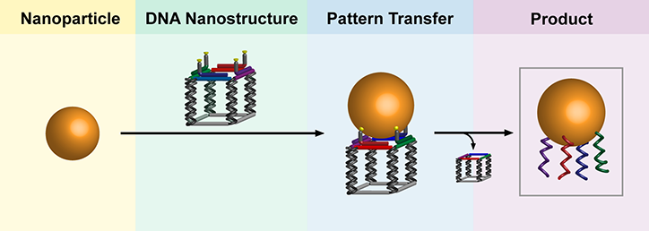
Enter the nanoparticle equivalent of the printing press. It’s efficient, re-usable and carries more information than previously possible. In results reported online in Nature Chemistry, researchers from McGill’s Department of Chemistry outline a procedure for making a DNA [deoxyribonucleic acid] structure with a specific pattern of strands coming out of it; at the end of each strand is a chemical “sticky patch.”  When a gold nanoparticle is brought into contact to the DNA nanostructure, it sticks to the patches. The scientists then dissolve the assembly in distilled water, separating the DNA nanostructure into its component strands and leaving behind the DNA imprint on the gold nanoparticle. …

The researchers have made an illustration of their concept available,

Credit: Thomas Edwardson

Credit: Thomas Edwardson

“These encoded gold nanoparticles are unprecedented in their information content,” says senior author Hanadi Sleiman, who holds the Canada Research Chair in DNA Nanoscience. “The DNA nanostructures, for their part, can be re-used, much like stamps in an old printing press.”

The news release includes suggestions for possible future applications,

From stained glass to optoelectronics

Some of the properties of gold nanoparticles have been recognized for centuries.  Medieval artisans added gold chloride to molten glass to create the ruby-red colour in stained-glass windows – the result, as chemists figured out much later, of the light-scattering properties of tiny gold particles.

Now, the McGill researchers hope their new production technique will help pave the way for use of DNA-encoded nanoparticles in a range of cutting-edge technologies. First author Thomas Edwardson says the next step for the lab will be to investigate the properties of structures made from these new building blocks. “In much the same way that atoms combine to form complex molecules, patterned DNA gold particles can connect to neighbouring particles to form well-defined nanoparticle assemblies.”

These could be put to use in areas including optoelectronic nanodevices and biomedical sciences, the researchers say. The patterns of DNA strands could, for example, be engineered to target specific proteins on cancer cells, and thus serve to detect cancer or to selectively destroy cancer cells.

Here’s a link to and a citation for the paper,

Transfer of molecular recognition information from DNA nanostructures to gold nanoparticles by Thomas G. W. Edwardson, Kai Lin Lau, Danny Bousmail, Christopher J. Serpell, & Hanadi F. Sleiman. Nature Chemistry (2016)  doi:10.1038/nchem.2420 Published online 04 January 2016

This paper is behind a paywall.
*’also on EurekAlert’ added on Jan. 8, 2016.

A bioelectronic future made possible with DNA-based electromechanical switch

DNA-based electronics are discussed in the context of a Dec. 14, 2015 news item by Beth Ellison for Azonano about research into electromechanical switches at the University of California at Davis,

Researchers from the University of California, Davis (UC Davis) and the University of Washington have shown the possibility of using DNA-based electromechanical switches for nanoscale computing.

DNA is considered to be the molecule of life, and researchers have shown considerable interest in utilizing DNA as a nanoscale material in various applications.

A Dec. 14, 2015 UC Davis news release on EurekAlert, which originated the news item, provides more detail,

In their paper published in Nature Communications, the team demonstrated that changing the structure of the DNA double helix by modifying its environment allows the conductance (the ease with which an electric current passes) to be reversibly controlled. This ability to structurally modulate the charge transport properties may enable the design of unique nanodevices based on DNA. These devices would operate using a completely different paradigm than today’s conventional electronics.

“As electronics get smaller they are becoming more difficult and expensive to manufacture, but DNA-based devices could be designed from the bottom-up using directed self-assembly techniques such as ‘DNA origami’,” said Josh Hihath, assistant professor of electrical and computer engineering at UC Davis and senior author on the paper. DNA origami is the folding of DNA to create two- and three-dimensional shapes at the nanoscale level.

“Considerable progress has been made in understanding DNA’s mechanical, structural, and self-assembly properties and the use of these properties to design structures at the nanoscale. The electrical properties, however, have generally been difficult to control,” said Hihath.

New Twist on DNA? Possible Paradigms for Computing

In addition to potential advantages in fabrication at the nanoscale level, such DNA-based devices may also improve the energy efficiency of electronic circuits. The size of devices has been significantly reduced over the last 40 years, but as the size has decreased, the power density on-chip has increased. Scientists and engineers have been exploring novel solutions to improve the efficiency.

“There’s no reason that computation must be done with traditional transistors. Early computers were fully mechanical and later worked on relays and vacuum tubes,” said Hihath. “Moving to an electromechanical platform may eventually allow us to improve the energy efficiency of electronic devices at the nanoscale.”

This work demonstrates that DNA is capable of operating as an electromechanical switch and could lead to new paradigms for computing.

To develop DNA into a reversible switch, the scientists focused on switching between two stable conformations of DNA, known as the A-form and the B-form. In DNA, the B-form is the conventional DNA duplex that is commonly associated with these molecules. The A-form is a more compact version with different spacing and tilting between the base pairs. Exposure to ethanol forces the DNA into the A-form conformation resulting in an increased conductance. Similarly, by removing the ethanol, the DNA can switch back to the B-form and return to its original reduced conductance value.

One Step Toward Molecular Computing

In order to develop this finding into a technologically viable platform for electronics, the authors also noted that there is still a great deal of work to be done. Although this discovery provides a proof-of-principle demonstration of electromechanical switching in DNA, there are generally two major hurdles yet to be overcome in the field of molecular electronics. First, billions of active molecular devices must be integrated into the same circuit as is done currently in conventional electronics. Next, scientists must be able to gate specific devices individually in such a large system.

“Eventually, the environmental gating aspect of this work will have to be replaced with a mechanical or electrical signal in order to locally address a single device,” noted Hihath.

Here’s a link to and a citation for the paper,

Conformational gating of DNA conductance by Juan Manuel Artés, Yuanhui Li, Jianqing Qi, M. P. Anantram, & Joshua Hihath. Nature Communications 6, Article number: 8870 doi:10.1038/ncomms9870 Published 09 December 2015

This paper is open access.

Enzymatic fuel cells with ultrasmall gold nanocluster

Scientists at the US Department of Energy’s Los Alamos National Laboratory have developed a DNA-templated gold nanocluster (AuNC) for more efficient biofuel cell design (Note: A link has been removed). From a Sept. 24, 2015 news item on ScienceDaily,

With fossil-fuel sources dwindling, better biofuel cell design is a strong candidate in the energy field. In research published in the Journal of the American Chemical Society (“A Hybrid DNA-Templated Gold Nanocluster For Enhanced Enzymatic Reduction of Oxygen”), Los Alamos researchers and external collaborators synthesized and characterized a new DNA-templated gold nanocluster (AuNC) that could resolve a critical methodological barrier for efficient biofuel cell design.

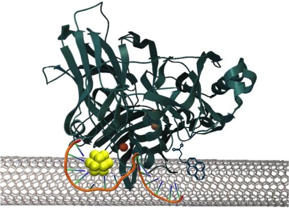
Here’s an image illustrating the DNA-templated gold nanoclusters,

Caption: Gold nanoclusters (~1 nm) are efficient mediators of electron transfer between co-self-assembled enzymes and carbon nanotubes in an enzyme fuel cell. The efficient electron transfer from this quantized nano material minimizes the energy waste and improves the kinetics of the oxygen reduction reaction, toward a more efficient fuel cell cycle. Credit: Los Alamos National Laboratory

Caption: Gold nanoclusters (~1 nm) are efficient mediators of electron transfer between co-self-assembled enzymes and carbon nanotubes in an enzyme fuel cell. The efficient electron transfer from this quantized nano material minimizes the energy waste and improves the kinetics of the oxygen reduction reaction, toward a more efficient fuel cell cycle.
Credit: Los Alamos National Laboratory

A Sept. 24, 2015 Los Alamos National Laboratory news release, which originated the news item, provides more details,

“Enzymatic fuel cells and nanomaterials show great promise and as they can operate under environmentally benign neutral pH conditions, they are a greener alternative to existing alkaline or acidic fuel cells, making them the subject of worldwide research endeavors,” said Saumen Chakraborty, a scientist on the project. “Our work seeks to boost electron transfer efficiency, creating a potential candidate for the development of cathodes in enzymatic fuel cells.”

Ligands, molecules that bind to a central metal atom, are necessary to form stable nanoclusters. For this study, the researchers chose single-stranded DNA as the ligand, as DNA is a natural nanoscale material having high affinity for metal cations and can be used to assembly the cluster to other nanoscale material such as carbon nanotubes.

In enzymatic fuel cells, fuel is oxidized on the anode, while oxygen reduction reactions take place on the cathode, often using multi copper oxidases. Enzymatic fuel cell performance depends critically on how effectively the enzyme active sites can accept and donate electrons from the electrode by direct electron transfer (ET). However, the lack of effective ET between the enzyme active sites, which are usually buried ~10Å from their surface, and the electrode is a major barrier to their development. Therefore, effective mediators of this electron transfer are needed.

The team developed a new DNA-templated gold nanocluster (AuNC) that enhanced electron transfer. This novel role of the AuNC as enhancer of electron transfer at the enzyme-electrode interface could be effective for cathodes in enzymatic fuel cells, thus removing a critical methodological barrier for efficient biofuel cell design.

Possessing many unique properties due to their discrete electron state distributions, metal nanoclusters (<1.5 nm diameter; ~2-144 atoms of gold, silver, platinum, or copper) show application in many fields.

Hypothesizing that due to the ultra-small size (the clusters are ~7 atoms, ~0.9 nm in diameter), and unique electrochemical properties, the AuNC can facilitate electron transfer to an oxygen-reduction reaction enzyme-active site and therefore, lower the overpotential of the oxygen reaction. Overpotential is the extra amount of energy required to drive an electrochemical reaction.

Ideally, it is desirable that all electrochemical reactions have minimal to no overpotential, but in reality they all have some. Therefore, to design an efficient electrocatalyst (for reduction or oxidation) we want to design it so that the reaction can proceed with a minimal amount of extra, applied energy.

When self assembled with bilirubin oxidase and carbon nanotubes, the AuNC acts to enhance the electron transfer, and it lowers the overpotential of oxygen reduction by a significant ~15 mV (as opposed to ~1-2 mV observed using other types of mediators) compared to the enzyme alone. The AuNC also causes significant enhancement of electrocatalytic current densities. Proteins are electronically insulating (they are complex, greasy and large), so the use of carbon nanotubes helps the enzyme stick to the electrode as well as to facilitate electron transfer.

Although gold nanoclusters have been used in chemical catalysis, this is the first time that we demonstrate they can also act as electron relaying agents to enzymatic oxygen reduction reaction monitored by electrochemistry.

Finally, the presence of AuNC does not perturb the mechanism of enzymatic O2 reduction. Such unique application of AuNC as facilitator of ET by improving thermodynamics and kinetics of O2 reduction is unprecedented.

Here’s a link to and a citation for the paper,

A Hybrid DNA-Templated Gold Nanocluster For Enhanced Enzymatic Reduction of Oxygen by Saumen Chakraborty, Sofia Babanova, Reginaldo C. Rocha, Anil Desireddy, Kateryna Artyushkova, Amy E. Boncella, Plamen Atanassov, and Jennifer S. Martinez. J. Am. Chem. Soc., 2015, 137 (36), pp 11678–11687 DOI: 10.1021/jacs.5b05338 Publication Date (Web): August 19, 2015

Copyright © 2015 American Chemical Society

This paper is behind a paywall.

What’s in your DNA (deoxyribonucleic acid)? an art auction at Christies

For this item, I have David Bruggeman’s Sept. 24, 2015 posting on his Pasco Phronesis blog to thank,

As part of a fundraising project for a building at the Francis Crick Institute, Christie’s will hold an auction for 30 double-helix sculptures on September 30 (H/T ScienceInsider).

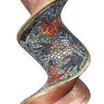
David has embedded a video featuring some of the artists and their works in his posting. By contrast, here are a few pictures of the DNA (deoxyribonucleic acid) art objects from the Cancer Research UK’s DNA Trail page,

For our London Art trail, which ran from 29 June – 6 September 2015, we asked internationally renowned artists to design a beautiful double helix sculpture inspired by the question: What’s in your DNA? Take a look at their sculptures and find out more about the artists’ inspirations.

This one is called The Journey and is by Gary Portell,

DNA_The Journey

His inspiration is: “My design is based on two symbols, the swallow who shares my journey from Africa to England and the hand print. The hand print as a symbol of creation and the swallow reflects the traveller.

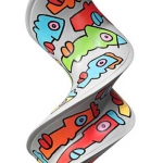
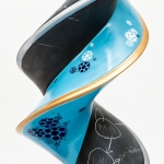
This one by Thiery Noir is titled Double Helix Noir.

DNA_DoubleHelixNoir

The inspiration is: For this sculpture, Noir wanted to pay tribute to the memory of his former assistant, Lisa Brown, who was affected by breast cancer and who passed away in July 2001, at the young age of 31 years old.

Growing Stem is by Orla Kiely,

CNA_GrowingStem

The inspiration is: I find inspiration in many things, but especially love nature with the abundance of colourful flowers, leaves, and stems. Applying our multi stem onto the DNA spiral seemed a natural choice as it represents positivity and growth: qualities that are so relevant for cancer research.

Double Dutch Delftblue DNA is by twins, Chris and Xand van Tulleken.

DNA_DoubleDutchDelftblue

The inspiration is: The recurrent motifs of Delft tiles reference those of DNA. Our inspiration was the combination of our family’s DNA, drawing on Dutch and Canadian origins, and the fact that twins have shared genomes.  (With thanks to Anthony van Tulleken)

Ted Baker’s Ted’s Helix of Haberdashery,

DNA_TedsHelixOfHaberdashery

Inspiration is: Always a fan of spinning a yarn, Ted Baker’s Helix of Haberdashery sculpture unravels the tale of his evolution from shirt specialist to global lifestyle brand. Ted’s DNA is represented as a cascading double helix of pearlescent buttons, finished with a typically playful story-telling flourish.

Finally, What Mad Pursuit is by Kindra Crick,

DNA_WhatMadPursuit

Inspiration is: What Mad Pursuit explores the creative possibilities achievable through the intermingling of art, science and imagination in the quest for knowledge. The piece is inspired by my family’s contribution to the discovery of the structure of DNA.

Aparna Vidyasagar interviewed Kindra Crick in a Sept. 24, 2015 Q&A for ScienceInsider (Note: Links have been removed),

Kindra Crick, granddaughter of Francis Crick, the co-discoverer of DNA’s structure, is one of more than 20 artists contributing sculptures to an auction fundraiser for a building at the new Francis Crick Institute. The auction is being organized by Cancer Research UK and will be held at Christie’s in London on 30 September. The auction will continue online until 13 October.

The new biomedical research institute, named for the Nobel laureate who died in 2004, aims to develop prevention strategies and treatments for diseases including cancer. It is a consortium of six partners, including Cancer Research UK.

Earlier this year, Cancer Research UK asked about two dozen artists—including Chinese superstar Ai Weiwei—to answer the question “What’s in your DNA?” through a sculpture based on DNA’s double helix structure. …

Q: “What’s in your DNA?” How did you build your sculpture around that question?

A: When I was given the theme, I thought this was a wonderful project for me, considering my family history. Also, in my own art practice I try to express the wonder and the process of scientific inquiry. This draws on my backgrounds; in molecular biology from when I was at Princeton [University], and in art while going to the School of the Art Institute of Chicago.

I was influenced by my grandparents, Francis Crick and Odile Crick. He was the scientist and she was the artist. My grandfather worked on elucidating the structure of DNA, and my grandmother, Odile, was the one to draw the first image of DNA. The illustration was used for the 1953 paper that my grandfather wrote with James Watson. So, there’s a rich history there that I can draw from, in terms of what’s in my DNA.

Should you be interested in bidding on one of the pieces, you can go to Christie’s What’s in your DNA webpage,

ONLINE AUCTION IS LIVE: 30 September – 13 October 2015

Good luck!

David Bruggeman has put in a request (from his Sept. 24, 2015 posting),

… if you become aware of human trials for 3D bioprinting, please give a holler.  I may now qualify.

Good luck David!