Tag Archives: sensing

Two-dimensional arsenic (arsenene) for electronics

Another day, another ‘ene’ (e.g., graphene, borene, germanene, etc.). This ‘ene’ is arsenene, from an October 15, 2018 Wiley (Publications) news release (also on EurekAlert),

The discovery of graphene, a material made of one or very few atomic layers of carbon, started a boom. Today, such two-dimensional materials are no longer limited to carbon and are hot prospects for many applications, especially in microelectronics. In the journal Angewandte Chemie, scientists have now introduced a new 2D material: they successfully modified arsenene (arsenic in a graphene-like structure) with chloromethylene groups.

Two-dimensional materials are crystalline materials made of just a single or very few layers of atoms that often display unusual properties. However, the use of graphene for applications such as transistors is limited because it behaves more like a conductor than a semiconductor. Modified graphene and 2D materials based on other chemical elements with semiconducting properties have now been developed. One such material is β-arsenene, a two-dimensional arsenic in a buckled honeycomb structure derived from gray arsenic. Researchers hope that modification of this previously seldom-studied material could improve its semiconducting properties and lead the way to new applications in fields such as sensing, catalysis, optoelectronics, and other semiconductor technologies.

A team at the University of Chemistry and Technology Prague (Czech Republic) and Nanyang Technical University (Singapore), led by Zdenek Sofer and Martin Pumera has now successfully produced a highly promising covalent modification of β-arsenene.

The arsenene was produced by milling gray arsenic in tetrahydrofuran. The shear forces cause two-dimensional layers to split off and disperse into the solvent. The researchers then introduce dichloromethane and add an organic lithium compound (butyllithium). These two reagents form an intermediate called chlorocarbene, a molecule made of one carbon atom, one hydrogen atom, and one chlorine atom. The carbon atom is short two bonding partners, a state that makes the whole class of carbene molecules highly reactive. Arsenene contains free electron pairs that “stick out” from the surface and can easily enter into bonds to chlorocarbene.

This approach leads to high coverage of the arsenene surface with chloromethylene groups, as confirmed by a variety of analysis methods (X-ray photoelectron spectroscopy, FT-IR spectroscopy, elemental analysis by transmission electron microscopy). The modified arsenene is more stable than pure arsenene and exhibits strong luminescence and electronic properties that make it attractive for optoelectronic applications. In addition, the chloromethylene units could serve as a starting point for further interesting modifications.

As always with an ‘ene’, the major focus is on electronics. Here’s a link to and a citation for the paper,

Covalent Functionalization of Exfoliated Arsenic with Chlorocarbene by Jiri Sturala, Adriano Ambrosi, Zdeněk Sofer, Martin Pumera. Angewandte Chimie International Edition Volume 57, Issue 45 November 5, 2018 Pages 14837-14840 DOI: https://doi.org/10.1002/anie.201809341 First published: 31 August 2018

This paper is behind a paywall.
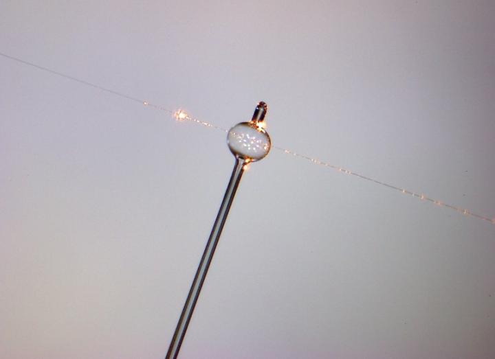
Nanoparticle detection with whispers and bubbles

Caption: A magnified photograph of a glass Whispering Gallery Resonator. The bubble is extremely small, less than the width of a human hair. Credit: OIST (Okinawa Institute of Science and Technology Graduate University)

It was the reference to a whispering gallery which attracted my attention; a July 11, 2018 news item on Nanowerk is where I found it,

Technology created by researchers at the Okinawa Institute of Science and Technology Graduate University (OIST) [Japan] is literally shedding light on some of the smallest particles to detect their presence – and it’s made from tiny glass bubbles.

The technology has its roots in a peculiar physical phenomenon known as the “whispering gallery,” described by physicist Lord Rayleigh (John William Strutt) in 1878 and named after an acoustic effect inside the dome of St Paul’s Cathedral in London. Whispers made at one side of the circular gallery could be heard clearly at the opposite side. It happens because sound waves travel along the walls of the dome to the other side, and this effect can be replicated by light in a tiny glass sphere just a hair’s breadth wide called a Whispering Gallery Resonator (WGR).

A July 11, 2018 OIST press release by Andrew Scott (also on EurekAlert), provides more details,

When light is shined into the sphere, it bounces around and around the inner surface, creating an optical carousel. Photons bouncing along the interior of the tiny sphere can end up travelling for long distances, sometimes as far as 100 meters. But each time a photon bounces off the sphere’s surface, a small amount of light escapes. This leaking light creates a sort of aura around the sphere, known as an evanescent light field. When nanoparticles come within range of this field, they distort its wavelength, effectively changing its color. Monitoring these color changes allows scientists to use the WGRs as a sensor; previous research groups have used them to detect individual virus particles in solution, for example. But at OIST’s Light-Matter Interactions Unit, scientists saw they could improve on previous work and create even more sensitive designs. The study is published in Optica.

Today, Dr. Jonathan Ward is using WGRs to detect minute particles more efficiently than ever before. The WGRs they have made are hollow glass bubbles rather than balls, explains Dr. Ward. “We heated a small glass tube with a laser and had air blown down it – it’s a lot like traditional glass blowing”. Blowing the air down the heated glass tube creates a spherical chamber that can support the sensitive light field. The most noticeable difference between a blown glass ornament and these precision instruments is the scale: the glass bubbles can be as small as 100 microns- a fraction of a millimeter in width. Their size makes them fragile to handle, but also malleable.

Working from theoretical models, Dr. Ward showed that they could increase the size of the light field by using a thin spherical shell (a bubble, in other words) instead of a solid sphere. A bigger field would increase the range in which particles can be detected, increasing the efficacy of the sensor. “We knew we had the techniques and the materials to fabricate the resonator”, said Dr. Ward. “Next we had to demonstrate that it could outperform the current types used for particle detection”.

To prove their concept, the team came up with a relatively simple test. The new bubble design was filled with a liquid solution containing tiny particles of polystyrene, and light was shined along a glass filament to generate a light field in its liquid interior. As particles passed within range of the light field, they produced noticeable shifts in the wavelength that were much more pronounced than those seen with a standard spherical WGR.

With a more effective tool now at their disposal, the next challenge for the team is to find applications for it. Learning what changes different materials make to the light field would allow Dr Ward to identify and target them, and even control their activity.

Despite their fragility, these new versions of WGRs are easy to manufacture and can be safely transported in custom made cases. That means these sensors could be used in a wide verity of fields, such as testing for toxic molecules in water to detect pollution, or detecting blood borne viruses in extremely rural areas where healthcare may be limited.

For Dr. Ward however, there’s always room from improvement: “We’re always pushing to get even more sensitivity and find the smallest particle this sensor can detect. We want to push our detection to the physical limits.”

Here’s a link to and a citation for the paper,

Nanoparticle sensing beyond evanescent field interaction with a quasi-droplet microcavity by Jonathan M. Ward, Yong Yang, Fuchuan Lei, Xiao-Chong Yu, Yun-Feng Xiao, and Síle Nic Chormaic. Optica Vol. 5, Issue 6, pp. 674-677 (2018) https://doi.org/10.1364/OPTICA.5.000674

This paper is open access.

Wearable microscopes

It never occurred to me that someone might want a wearable microscope but, apparently, there is a need. A Sept. 27, 2016 news item on phys.org,

UCLA [University of California at Los Angeles] researchers working with a team at Verily Life Sciences have designed a mobile microscope that can detect and monitor fluorescent biomarkers inside the skin with a high level of sensitivity, an important tool in tracking various biochemical reactions for medical diagnostics and therapy.

A Sept. 26, 2016 UCLA news release by Meghan Steele Horan, which originated the news item, describes the work in more detail,

This new system weighs less than a one-tenth of a pound, making it small and light enough for a person to wear around their bicep, among other parts of their body. In the future, technology like this could be used for continuous patient monitoring at home or at point-of-care settings.

The research, which was published in the journal ACS Nano, was led by Aydogan Ozcan, UCLA’s Chancellor’s Professor of Electrical Engineering and Bioengineering and associate director of the California NanoSystems Institute and Vasiliki Demas of Verily Life Sciences (formerly Google Life Sciences).

Fluorescent biomarkers are routinely used for cancer detection and drug delivery and release among other medical therapies. Recently, biocompatible fluorescent dyes have emerged, creating new opportunities for noninvasive sensing and measuring of biomarkers through the skin.

However, detecting artificially added fluorescent objects under the skin is challenging. Collagen, melanin and other biological structures emit natural light in a process called autofluorescence. Various methods have been tried to investigate this problem using different sensing systems. Most are quite expensive and difficult to make small and cost-effective enough to be used in a wearable imaging system.

To test the mobile microscope, researchers first designed a tissue phantom — an artificially created material that mimics human skin optical properties, such as autofluorescence, absorption and scattering. The target fluorescent dye solution was injected into a micro-well with a volume of about one-hundredth of a microliter, thinner than a human hair, and subsequently implanted into the tissue phantom half a millimeter to 2 millimeters from the surface — which would be deep enough to reach blood and other tissue fluids in practice.

To measure the fluorescent dye, the wearable microscope created by Ozcan and his team used a laser to hit the skin at an angle. The fluorescent image at the surface of the skin was captured via the wearable microscope. The image was then uploaded to a computer where it was processed using a custom-designed algorithm, digitally separating the target fluorescent signal from the autofluorescence of the skin, at a very sensitive parts-per-billion level of detection.

“We can place various tiny bio-sensors inside the skin next to each other, and through our imaging system, we can tell them apart,” Ozcan said. “We can monitor all these embedded sensors inside the skin in parallel, even understand potential misalignments of the wearable imager and correct it to continuously quantify a panel of biomarkers.”

This computational imaging framework might also be used in the future to continuously monitor various chronic diseases through the skin using an implantable or injectable fluorescent dye.

Here’s a link to and a citation for the paper,

Quantitative Fluorescence Sensing Through Highly Autofluorescent, Scattering, and Absorbing Media Using Mobile Microscopy by Zoltán Göröcs, Yair Rivenson, Hatice Ceylan Koydemir, Derek Tseng, Tamara L. Troy, Vasiliki Demas, and Aydogan Ozcan. ACS Nano, 2016, 10 (9), pp 8989–8999 DOI: 10.1021/acsnano.6b05129 Publication Date (Web): September 13, 2016

Copyright © 2016 American Chemical Society

This paper is behind a paywall.

DNA as a sensor

McMaster University (Ontario, Canada) researchers have developed a technique for using DNA (deoxyribonucleic acid) as a sensor according to a July 7, 2016 news item on ScienceDaily,

Researchers at McMaster University have established a way to harness DNA as the engine of a microscopic “machine” they can turn on to detect trace amounts of substances that range from viruses and bacteria to cocaine and metals.

“It’s a completely new platform that can be adapted to many kinds of uses,” says John Brennan, director of McMaster’s Biointerfaces Insitute and co-author of a paper in the journal Nature Communications that describes the technology. “These DNA nano-architectures are adaptable, so that any target should be detectable.”

A July 7, 2016 McMaster University news release (also on EurekAlert), which originated the news item, expands on the theme,

DNA is best known as a genetic material, but is also a very programmable molecule that lends itself to engineering for synthetic applications.

The new method shapes separately programmed pieces of DNA material into pairs of interlocking circles.

The first remains inactive until it is released by the second, like a bicycle wheel in a lock. When the second circle, acting as the lock, is exposed to even a trace of the target substance, it opens, freeing the first circle of DNA, which replicates quickly and creates a signal, such as a colour change.

“The key is that it’s selectively triggered by whatever we want to detect,” says Brennan, who holds the Canada Research Chair in Bioanalytical Chemistry and Biointerfaces. “We have essentially taken a piece of DNA and forced it to do something it was never designed to do. We can design the lock to be specific to a certain key. All the parts are made of DNA, and ultimately that key is defined by how we build it.”

The idea for the “DNA nanomachine” comes from nature itself, explains co-author Yingfu Li, who holds the Canada Research Chair in Nucleic Acids Research.

“Biology uses all kinds of nanoscale molecular machines to achieve important functions in cells,” Li says. “For the first time, we have designed a DNA-based nano-machine that is capable of achieving ultra-sensitive detection of a bacterial pathogen.”

The DNA-based nanomachine is being further developed into a user-friendly detection kit that will enable rapid testing of a variety of substances, and could move to clinical testing within a year.

Here’s a link to and a citation for the paper,

Programming a topologically constrained DNA nanostructure into a sensor by Meng Liu, Qiang Zhang, Zhongping Li, Jimmy Gu, John D. Brennan, & Yingfu Li. Nature Communications 7, Article number: 12074  doi:10.1038/ncomms12074 Published 23 June 2016

This paper is open access.

Nanotechnology Molecular Tagging for sniffing out explosives

A nifty technology for sniffing out explosives is described in a June 22, 2016 news item in Government Security News magazine. I do think they might have eased up on the Egypt Air disaster reference and the implication that it might have been avoided with the use of this technology,

The crash of an Egypt Air Flight 804 recently again raised concerns over whether a vulnerability in pre-flight security has led to another deadly terrorist attacks. Officials haven’t found a cause for the crash yet, but news reports indicate that officials believe either a bomb or fire are what brought the plane down [link included from press release].

Regardless of the cause, the Chief Executive Officer of British-based Ancon Technologies said that the incident shows the compelling need for more versatile and affordable explosive detection technology.

“There are still too many vulnerabilities in transportation systems around the world,” said CEO Dr. Robert Muir. “That’s why our focus has been on developing explosive detection technology that is highly efficient, easily deployable and economically priced.”

A June 21, 2015 Ancon Technologies press release on PR Web, which originated the news item, describes the technology in a little more detail,

Using nanotechnology to scan sensitive vapour readings, Ancon Technologies has developed unique security devices with exception sensitivity to detect explosive chemicals and materials. Called Nanotechnology Molecular Tagging, the technology is used to look for specific molecular markers that are emitted from the chemicals used in explosive compounds. An NMT device can then be programmed to look for these compounds and gauge concentrations.

“The result is unprecedented sensitivity for a device that is portable and versatile,” Dr. Muir said. “The technology is also highly selective, meaning it can distinguish the molecules is testing for against the backdrop of other chemicals and readings in the air.”

If terrorism is responsible for the crash of the Egypt Air flight on route to Cairo from Paris’ Charles de Gaulle Airport, the incident further shows the need for heightened screening processes, Muir said. Concerns about air travel’s vulnerabilities to terrorism were further raised in October when a Russian plane flying out of Egypt crashed in what several officials believe was a terrorist bombing.

Both cases show the need for improved security measures in airports around the world, especially those related to early explosive detection, Muir said. CNN reported that the Egypt Air crash would likely generate even more attention to airport security while Egypt has already been investing in new security measures following the October attack.

“An NMT device can bring laboratory-level sensitivity to the airport screening procedure, adding another level of safety in places where it’s needed most,” Muir said. “By being able to detect a compound at concentrations as small as a single molecule, NMT can pinpoint a threat and provide security teams with the early warning they need.”

The NMT device’s sensitivity and accuracy can also help balance another concern with airport security: long waits. Already, the Transportation Security Agency is coming under fire this summer for extended airport security screening lines, reports USA Today.

“An NMT device can produce results from test samples in minutes, meaning screenings can proceed at a reasonable pace without jeopardizing security,” Muir said.

Ancon Technologies has working arrangements with military and security agencies in both the United Kingdom and the United States, Muir said, following a recent round of investments. The company is headquartered in Canterbury, Kent and has an office in the U.S. in Bloomington, Minnesota.

So this is a sensing device and I believe this particular type can also be described as an artificial nose.

Using quantum dots to detect and identify explosives

This research is courtesy of the University College London (UCL) according to a Dec. 9, 2015 news item on Nanowerk,

A new test for detecting multiple explosives simultaneously has been developed by UCL scientists. The proof-of-concept sensor is designed to quickly identify and quantify five commonly used explosives in solution to help track toxic contamination in waste water and improve the safety of public spaces.

Lead researcher, Dr William Peveler (UCL Chemistry), said: “This is the first time multiple explosives have been detected using a single sensor before, demonstrating proof-of-concept for this approach. Our sensor changes colour within 10 seconds to give information about how much and what explosives are present in a sample. Following further development, we hope it will be used to quickly analyse the nature of threats and inform tailored responses.”

A Dec. 9, 2015 UCL press release (also on EurekAlert), which originated the news item, expands on the theme,

Dr Peveler, added: “We analysed explosives which are commonly used for industrial and military purposes to create a useful tool for environmental and security monitoring. For example, DNT is a breakdown product from landmines, and RDX and PETN have been used in terror plots in recent years as they can be hard to detect using sniffer dogs. Our test can quickly identify these compounds so we see it having a variety of applications from monitoring the waste water of munitions factories and military ranges to finding evidence of illicit activities.”

The sensor is made of quantum dots, which are tiny light-emitting particles or nanomaterials, to which explosive targeting receptors are attached. As each explosive binds to the quantum dot, it quenches the light being emitted to a different degree. The distinct changes in colour are analysed computationally in a variety of conditions to give a unique fingerprint for each compound, allowing multiple explosives to be detected with a single test.

Senior author, Professor Ivan Parkin (UCL Chemistry), said: “Our sensor is a significant step forward for multiple explosives detection. Current methods can be laborious and require expensive equipment but our test is designed to be inexpensive, fast and use a much smaller volume of sample than previously possible. Although all of these factors are important, speed and accuracy are crucial when identifying explosive compounds.”

The team plan to take it from the laboratory into the field by blind testing it with contaminated waste water samples. They also hope to improve the sensitivity of the test by tailoring the surfaces of the quantum dots. Currently, its limit is less than one part per million which the team hope to increase into the part per billion range.

Here’s a link to and a citation for the paper,

Multichannel Detection and Differentiation of Explosives with a Quantum Dot Array by William J. Peveler, Alberto Roldan, Nathan Hollingsworth, Michael J. Porte‡, and Ivan P. Parkin. ACS Nano, Article ASAP DOI: 10.1021/acsnano.5b06433 Publication Date (Web): November 18, 2015

Copyright © 2015 American Chemical Society

This paper is behind a paywall.

Herbicide nanometric sensor could help diagnose multiple sclerosis

This research into nanometric sensors and multiple sclerosis comes from Brazil. According to a June 23, 2015 news item on Nanowerk (Note: A link has been removed),

The early diagnosis of certain types of cancer, as well as nervous system diseases such as multiple sclerosis and neuromyelitis optica, may soon be facilitated by the use of a nanosensor capable of identifying biomarkers of these pathological conditions (“A Nanobiosensor Based on 4-Hydroxyphenylpyruvate Dioxygenase Enzyme for Mesotrione Detection”).

The nanobiosensor was developed at the Federal University of São Carlos (UFSCar), Sorocaba, in partnership with the São Paulo Federal Institute of Education, Science & Technology (IFSP), Itapetininga, São Paulo State, Brazil. It was originally designed to detect herbicides, heavy metals and other pollutants.

A June 23, 2015 Fundação de Amparo à Pesquisa do Estado de São Paulo news release on EurekAlert, which originated the news item, describes the sensor as it was originally used and explains its new function as a diagnostic tool for multiple sclerosis and other diseases,

“It’s a highly sensitive device, which we developed in collaboration with Alberto Luís Dario Moreau, a professor at IFSP. “We were able to increase sensitivity dramatically by going down to the nanometric scale,” said physicist Fábio de Lima Leite, a professor at UFSCar and the coordinator of the research group.

The nanobiosensor consists of a silicon nitride (Si3N4) or silicon (Si) nanoprobe with a molecular-scale elastic constant and a nanotip coupled to an enzyme, protein or other molecule.

When this molecule touches a target of interest, such as an antibody or antigen, the probe bends as the two molecules adhere. The deflection is detected and measured by the device, enabling scientists to identify the target.

“We started by detecting herbicides and heavy metals. Now we’re testing the device for use in detecting target molecules typical of nervous system diseases, in partnership with colleagues at leading centers of research on demyelinating diseases of the central nervous system”

The migration from herbicide detection to antibody detection was motivated mainly by the difficulty of diagnosing demyelinating diseases, cancer and other chronic diseases before they have advanced beyond an initial stage.

The criteria for establishing a diagnosis of multiple sclerosis or neuromyelitis optica are clinical (supplemented by MRI scans), and patients do not always present with a characteristic clinical picture. More precise diagnosis entails ruling out several other diseases.

The development of nanodevices will be of assistance in identifying these diseases and reducing the chances of false diagnosis.

The procedure can be as simple as placing a drop of the patient’s cerebrospinal fluid on a glass slide and observing its interaction with the nanobiosensor.

“If the interaction is low, we’ll be able to rule out multiple sclerosis with great confidence,” Leite said. “High interaction will indicate that the person is very likely to have the disease.” In this case, further testing would be required to exclude the possibility of a false positive.

“Different nervous system diseases have highly similar symptoms. Multiple sclerosis and neuromyelitis optica are just two examples. Even specialists experience difficulties or take a long time to diagnose them. Our technique would provide a differential diagnostic tool,” Leite said.

The next step for the group is to research biomarkers for these diseases that have not been completely mapped, including antibodies and antigens, among others. The group has begun tests for the detection of head and neck cancer.

Here’s a link to and a citation for the paper,

A Nanobiosensor Based on 4-Hydroxyphenylpyruvate Dioxygenase Enzyme for Mesotrione Detection by P. Soto Garcia, A.L.D Moreau, J.C. Magalhaes Ierich,  A.C Araujo Vig, A.M. Higa, G.S. Oliveira, F. Camargo Abdalla, M. Hausen, & F.L. Leite. Sensors Journal, IEEE  (Volume:15 ,  Issue: 4) pp. 2106 – 2113 Date of Publication: 20 November 2014 Date of Current Version: 27 January 2015 Issue Date: April 2015  DOI 10.1109/JSEN.2014.2371773

This paper is behind a paywall.

A ‘sweat’mometer—sensing your health through your sweat

At this point, it’s more fitness monitor than diagnostic tool, so, you’ll still need to submit blood, stool, and urine samples when the doctor requests it but the device does offer some tantalizing possibilities according to a May 15, 2015 news item on phys.org,

Made from state-of-the-art silicon transistors, an ultra-low power sensor enables real-time scanning of the contents of liquids such as perspiration. Compatible with advanced electronics, this technology boasts exceptional accuracy – enough to manufacture mobile sensors that monitor health.

Imagine that it is possible, through a tiny adhesive electronic stamp attached to the arm, to know in real time one’s level of hydration, stress or fatigue while jogging. A new sensor developed at the Nanoelectronic Devices Laboratory (Nanolab) at EPFL [École Polytechnique Fédérale de Lausanne in Switzerland] is the first step toward this application. “The ionic equilibrium in a person’s sweat could provide significant information on the state of his health,” says Adrian Ionescu, director of Nanolab. “Our technology detects the presence of elementary charged particles in ultra-small concentrations such as ions and protons, which reflects not only the pH balance of sweat but also more complex hydration of fatigues states. By an adapted functionalization I can also track different kinds of proteins.”

A May 15, 2015 EPFL press release by Laure-Anne Pessina, which originated the news item, includes a good technical explanation of the device for non-experts in the field,

Published in the journal ACS Nano, the device is based on transistors that are comparable to those used by the company Intel in advanced microprocessors. On the state-of-the-art “FinFET” transistor, researchers fixed a microfluidic channel through which the fluid to be analyzed flows. When the molecules pass, their electrical charge disturbs the sensor, which makes it possible to deduce the fluid’s composition.

The new device doesn’t host only sensors, but also transistors and circuits enabling the amplification of the signals – a significant innovation. The feat relies on a layered design that isolates the electronic part from the liquid substance. “Usually it is necessary to use separately a sensor for detection and a circuit for computing and signal amplification,” says Sara Rigante, lead author of the publication. “In our chip, sensors and circuits are in the same device – making it a ‘Sensing integrated circuit’. This proximity ensures that the signal is not disturbed or altered. We can thereby obtain extremely stable and accurate measurements.”

But that’s not all. Due to the size of the transistors – 20 nanometers, which is one hundred to one thousand times smaller than the thickness of a hair – it is possible to place a whole network of sensors on one chip, with each sensor locating a different particle. “We could also detect calcium, sodium or potassium in sweat,” the researcher elaborates.

As to what makes the device special (from the press release),

The technology developed at EPFL stands out from its competitors because it is extremely stable, compatible with existing electronics (CMOS), ultra-low power and easy to reproduce in large arrays of sensors. “In the field of biosensors, research around nanotechnology is intense, particularly regarding silicon nanowires and nanotubes. But these technologies are frequently unstable and therefore unusable for now in industrial applications,” says Ionescu. “In the case of our sensor, we started from extremely powerful, advanced technology and adapted it for sensing need in a liquid-gate FinFET configurations. The precision of the electronics is such that it is easy to clone our device in millions with identical characteristics.”

In addition, the technology is not energy intensive. “We could feed 10,000 sensors with a single solar cell,” Professor Ionescu asserts.

Of course, there does seem to be one shortcoming (from the press release),

Thus far, the tests have been carried out by circulating the liquid with a tiny pump. Researchers are currently working on a means of sucking the sweat into the microfluidic tube via wicking. This would rid the small analyzing “band-aid” of the need for an attached pump.

While they work on eliminating the pump part of the device, here’s  a link to and a citation for the paper,

Sensing with Advanced Computing Technology: Fin Field-Effect Transistors with High-k Gate Stack on Bulk Silicon by Sara Rigante, Paolo Scarbolo, Mathias Wipf, Ralph L. Stoop, Kristine Bedner, Elizabeth Buitrago, Antonios Bazigos, Didier Bouvet, Michel Calame, Christian Schönenberger, and Adrian M. Ionescu. ACS Nano, Article ASAP DOI: 10.1021/nn5064216 Publication Date (Web): March 27, 2015

Copyright © 2015 American Chemical Society

This paper is behind a paywall.

As for the ‘sweat’mometer in the headline, I was combining sweat with thermometer.

Changing the vibration of gold nanodisks (acoustic tuning) with light

A May 7, 2015 news item on phys.org describes research that could have a major impact on photonics applications,

In a study that could open doors for new applications of photonics from molecular sensing to wireless communications, Rice University [Texas, US] scientists have discovered a new method to tune the light-induced vibrations of nanoparticles through slight alterations to the surface to which the particles are attached.

n a study published online this week in Nature Communications, researchers at Rice’s Laboratory for Nanophotonics (LANP) used ultrafast laser pulses to induce the atoms in gold nanodisks to vibrate. These vibrational patterns, known as acoustic phonons, have a characteristic frequency that relates directly to the size of the nanoparticle. The researchers found they could fine-tune the acoustic response of the particle by varying the thickness of the material to which the nanodisks were attached.

A May 7, 2015 Rice University news release (also on EurekAlert), which originated the news item, expands on the theme (Note: A link has been removed),

Our results point toward a straightforward method for tuning the acoustic phonon frequency of a nanostructure in the gigahertz range by controlling the thickness of its adhesion layer,” said lead researcher Stephan Link, associate professor of chemistry and in electrical and computer engineering.

Light has no mass, but each photon that strikes an object imparts a miniscule amount of mechanical motion, thanks to a phenomenon known as radiation pressure. A branch of physics known as optomechanics has developed over the past decade to study and exploit radiation pressure for applications like gravity wave detection and low-temperature generation.

Link and colleagues at LANP specialize in another branch of science called plasmonics that is devoted to the study of light-activated nanostructures. Plasmons are waves of electrons that flow like a fluid across a metallic surface.

When a light pulse of a specific wavelength strikes a metal particle like the puck-shaped gold nanodisks in the LANP experiments, the light energy is converted into plasmons. These plasmons slosh across the surface of the particle with a characteristic frequency, in much the same way that each phonon has a characteristic vibrational frequency.

The study’s first author, Wei-Shun Chang, a postdoctoral researcher in Link’s lab, and graduate students Fangfang Wen and Man-Nung Su conducted a series of experiments that revealed a direct connection between the resonant frequencies of the plasmons and phonons in nanodisks that had been exposed to laser pulses.

“Heating nanostructures with a short light pulse launches acoustic phonons that depend sensitively on the structure’s dimensions,” Link said. “Thanks to advanced lithographic techniques, experimentalists can engineer plasmonic nanostructures with great precision. Based on our results, it appears that plasmonic nanostructures may present an interesting alternative to conventional optomechanical oscillators.”

Chang said plasmonics experts often rely on substrates when using electron-beam lithography to pattern plasmonic structures. For example, gold nanodisks like those used in the experiments will not stick to glass slides. But if a thin substrate of titanium or chromium is added to the glass, the disks will adhere and stay where they are placed.

“The substrate layer affects the mechanical properties of the nanostructure, but many questions remain as to how it does this,” Chang said. “Our experiments explored how the thickness of the substrate impacted properties like adhesion and phononic frequency.”

Link said the research was a collaborative effort involving research groups at Rice and the University of Melbourne in Victoria, Australia.

“Wei-Shun and Man-Nung from my lab did the ultrafast spectroscopy,” Link said. “Fangfang, who is in Naomi Halas’ group here at Rice, made the nanodisks. John Sader at the University of Melbourne, and his postdoc Debadi Chakraborty calculated the acoustic modes, and Yue Zhang, a Rice graduate student from Peter Nordlander’s group at Rice simulated the optical/plasmonic properties. Bo Shuang of the Landes’ research group at Rice contributed to the analysis of the experimental data.”

Here’s a link to and a citation for the paper,

Tuning the acoustic frequency of a gold nanodisk through its adhesion layer by Wei-Shun Chang, Fangfang Wen, Debadi Chakraborty, Man-Nung Su, Yue Zhang, Bo Shuang, Peter Nordlander, John E. Sader, Naomi J. Halas, & Stephan Link. Nature Communications 6, Article number: 7022 doi:10.1038/ncomms8022 Published 05 May 2015

This paper is behind a paywall but a free preview is available vie ReadCube Access.

Gold detection down to the nanoparticle?

It appears that detecting gold, presumably for mining purposes, isn’t as easy as one might think especially at the nanoscale. Researchers at Australia’s University of Adelaide have devised a new method according to an April 29, 2015 news item on Nanowerk (Note: A link has been removed),

University of Adelaide researchers are developing a portable, highly sensitive method for gold detection that would allow mineral exploration companies to test for gold on-site at the drilling rig.

Using light in two different processes (fluorescence and absorption), the researchers from the University’s Institute for Photonics and Advanced Sensing (IPAS), have been able to detect gold nanoparticles at detection limits 100 times lower than achievable under current methods.

An April 29, 2015 University of Adelaide news release details Australia’s interest in gold and offers a high level explanation of the need for better gold detection (Note: Links have been removed),

Australia is the world’s second largest gold producer, worth $13 billion in export earnings.

“Gold is not just used for jewellery, it is in high demand for electronics and medical applications around the world, but exploration for gold is extremely challenging with a desire to detect very low concentrations of gold in host rocks,” says postdoctoral researcher Dr Agnieszka Zuber, working on the project with Associate Professor Heike Ebendorff-Heidepriem.

“The presence of gold deep underground is estimated by analysis of rock particles coming out of the drilling holes. But current portable methods for detection are not sensitive enough, and the more sensitive methods require some weeks before results are available.

“This easy-to-use sensor will allow fast detection right at the drill rig with the amount of gold determined within an hour, at much lower cost.”

The researchers have been able to detect less than 100 parts per billion of gold in water. They are now testing using samples of real rock with initial promising results. The work is funded by the Deep Exploration Technologies Cooperative Research Centre.

The gold detection project is one of a series of projects which will be presented at the IPAS Minerals and Energy Sector Workshop today [April 29, 2015], aimed at linking resources specific research to local companies.

You can find out more about the University of Adelaide’s Institute of Photonics and Advanced Sensing here.