Half a century ago, a scientist won a Nobel Prize for Chemistry for work he’d done at the University of Saskatchewan and, later, at a National Research Council of Canada laboratory. The Nobel Prize was an unlikely event for more than one reason.
The history description I like the best is also the clunkiest (due to links and citations). From the essay by Denisa Popa for the Defining Moments Canada website (Note 1: I have removed the links; Note 2: NSERC is the Natural Sciences and Engineering Research Council of Canada),
…
Gerhard Herzberg was born in Hamburg, Germany on December 25th, 1904. From an early age Herzberg developed a keen interest in the sciences, particularly astronomy, physics and chemistry (Stoicheff, 2002). … Herzberg initially considered a career in astronomy, but lacked the funds to pursue it any further (NSERC). In 1924, he ultimately decided to pursue engineering physics and enrolled in the Technical University at Darmstadt (NSERC). By the time he was 24 years old, he was well established in his field, publishing a number of academic papers on the topics of atomic and molecular physics, as well as obtaining a Doctorate in Engineering Physics in 1928 (NSERC).
Following his graduation, he entered a postdoctoral fellowship at the University of Göttingen (University of Saskatchewan). Following that, Herzberg returned to Darmstadt where he spent five years conducting research on spectroscopy (University of Saskatchewan). Spectroscopy is used to analyze the ability of molecules and compounds to emit and absorb different wavelengths of light and electromagnetic radiation (Herschbach, 1999). Through understanding the properties of the light/radiation that is emitted (or absorbed) scientists can learn more about the characteristics of molecules and compounds, including their structure and the types of chemical bonds they contain (Herschbach, 1999).
While completing his postdoctoral fellowship, Herzberg met Luise Hedwig Oettinger, a university student also focusing on spectroscopic research (Stoicheff, 2002). The pair grew close and eventually married on December 30th, 1929 (Stoicheff, 2002). Over the years Luise, who received her Ph.D from the University of Frankfurt in 1933, co-authored a number of scientific papers with her husband (Stoicheff, 2002). The Herzbergs’ academic life in Germany would soon end in 1934 when the Nazi regime rose to power and began implementing new restrictions against Jewish scholars in academic institutions (Stoicheff, 2002). Herzberg received notice that he would no longer be permitted to teach at Darmstadt because of Luise’s Jewish heritage (Stoicheff, 2002; University of Saskatchewan). With the help of John W. T. Spinks (a chemist who visited and became closely acquainted with Herzberg in Darmstadt) and Walter C. Murray at the University of Saskatchewan, as well as funding from the Carnegie Foundation (as the university’s budget was limited during the depression era), the Herzbergs moved to Saskatoon that following year (NSERC).
From 1935 to 1945 Herzberg established himself at the University of Saskatchewan, where he continued his research on molecular and atomic spectroscopy, publishing three new books (NSERC). He then spent the following three years at the University of Chicago’s Yerkes Observatory investigating “the absorption spectra of many molecules of astrophysical interest.” (NSERC) In 1948, the Herzbergs relocated back to Canada when Herzberg was invited to “establish a laboratory for fundamental research in spectroscopy” at the National Research Council (NRC) of Canada. (NSERC) It was during his time at the NRC that one of his key discoveries was made–the observation of the spectra of methylene radical (CH2) (Stoicheff, 2002). Scientists describe free radicals as chemical species that have an unpaired electron in the outer valence shell (Winnewisser, 2004). Free radicals can be found as intermediates in a variety of chemical reactions (Herschbach, 1999). It was Herzberg’s contribution to the understanding of free radicals that contributed to his Nobel Prize win in 1971 (NSERC). Dr. Gerhard Herzberg had two children and passed away on March 3rd, 1999 at the age of 94 (Herschbach, 1999).
…
Kathryn Warden’s Saskatechwan-forward article was first published in August 2021 in the University of Saskatchewan’s Green & White magazine (Note: A link has been removed),
When Gerhard Herzberg was awarded the Nobel Prize in chemistry 50 years ago for ground-breaking discoveries in a lifelong exploration of the structure of matter, he publicly thanked the University of Saskatchewan.
“It is obvious that the work that has earned me the Nobel Prize was not done without a great deal of help,” Herzberg said in his acceptance speech, acknowledging “the full and understanding support” of successive USask presidents and faculty who “did their utmost to make it possible for me to proceed with my scientific work.”
Herzberg’s brilliance in studying the spectra of atoms and molecules to understand their physical properties significantly advanced astronomy, chemistry and physics—enhancing knowledge of the atmospheres of stars and planets and determining the existence of some molecules never before imagined.
“He was certainly a pioneer,” said USask PhD student Natasha Vetter, winner of both the 2014 Herzberg Scholarship and the 2018 Herzberg Fellowship. “Without his work, the fundamental tools we use as chemists and biochemists wouldn’t exist. I feel pretty honoured to be part of that legacy and to have received those awards.”
While at USask from 1935 to 1945, Herzberg made discoveries that laid the groundwork for his work at Chicago’s Yerkes Observatory and then at the National Research Council (NRC), culminating in his celebrated work on free radicals—highly unstable, short-lived molecules that are everywhere: in our bodies, in materials and in space. They help important reactions take place but an imbalance can cause damage such as cancer or age-related illness. Knowledge of their structure is now used to make pharmaceuticals, medical radiation tests, light sensors, and a wide range of innovative materials.
“This was the beginning of molecular spectroscopy, and it was an exciting time because it was all so new,” said Alexander Moewes, Canada Research Chair in Materials Science with Synchrotron Radiation.
“Herzberg was unravelling the structure of molecules, specifically free radicals. Many of today’s drugs and human biochemistry processes are governed by these molecules. So much that we have developed today would not have been discovered if Herzberg hadn’t done this fundamental research. This can’t be overstated.”
In honour of Herzberg, the University of Saskatchewan is naming both a hall and a lecture theatre at the Canadian Light Source (CLS), Canada’s synchrotron facility, after Herzberg, from a November 10, 2021 University of Saskatchewan news release,
As part of a national initiative to mark the 50th anniversary of Gerhard Herzberg’s Nobel Prize, the University of Saskatchewan (USask) is naming the main experimental hall of the Canadian Light Source (CLS) and a prominent physics lecture theatre on campus after the renowned scientist.
…
“Canada and the University of Saskatchewan welcomed Herzberg and his wife when no other country or university did,” said Stoicheff [USask President Peter Stoicheff]. “His legacy is evident today in so many ways, including at our Canadian Light Source where scientists from across Canada and around the world continue to unravel the mysteries of atomic structure.”
The Herzberg Experimental Hall is at the heart of the CLS, “the brightest light in Canada.” The enormous hall the size of a football field houses the synchrotron which supplies light to the many beamlines where wide-ranging experiments are conducted. The naming was endorsed by both the CLS board of directors and the CLS Users’ Executive Committee, and subsequently approved by the President’s Advisory Committee on Naming University Assets.
“As the father of modern spectroscopy, Herzberg conducted experiments that fundamentally changed scientific understanding of how molecules absorb and emit light,” said CLS board chair Pierre Lapointe.
“So it is very fitting that we honour his legacy at the Canadian Light Source where scientists from across Canada and around the world carry on the important work of using light to investigate the structure of matter—work that is leading to discoveries in fields as diverse as health, environment and new materials.”
In his 2020 co-authored book on the history of the CLS, former CLS director Michael Bancroft said Herzberg’s fundamental research program in spectroscopy at USask in the 1930s paved the way for Canada’s only synchrotron. He states that the close friendship that developed between USask chemistry researcher John Spinks and Herzberg in 1933 and 1934 in Germany, along with Herzberg’s subsequent hiring by USask President Walter Murray in 1935, “were the most important events in eventually landing the Canadian Light Source over 60 years later.”
As Herzberg was a member of the USask physics department for a decade, the Physics 107 Lecture Theatre, across from a display dedicated to Herzberg, will be named the Dr. Gerhard Herzberg Lecture Theatre.
…
Chris Putnam’s December 10, 2021 article for the University of Saskatchewan highlights Herzberg’s other interests such as music and humanitarian work.
Finally, Herzberg gave an interview to Mary Christine King on May 5, 1986 (audio file and text) for the Science History Institute. Here’s a little more about Ms. King who died months after the interview,
“… born in China and educated in Ireland. She obtained a BSc degree in chemistry from the University of London in 1968, which was followed by an MSc in polymer and fiber science (1970) and a PhD for a thesis on the hydrodynamic properties of paraffins in solution (1973), both from the University of Manchester Institute of Science and Technology. After working with Joseph Needham at Cambridge, she received a PhD in the history and philosophy of science from the Open University (1980) and thereafter worked at the University of California, Berkeley, and at the University of Ottawa, … King died in an automobile accident in late 1987 …”
The interview is an oral history as recounted by Herzberg.
![Fig. 1. Dress 1 circa 1865, dress 2 circa 1898, dress 3 circa 1878 and dress 4 circa 1885 (clock-wise from left top). Details of these dresses are presented in the Experimental section. [downloaded from http://www.sciencedirect.com/science/article/pii/S1386142515302742]](http://www.frogheart.ca/wp-content/uploads/2015/12/19thCenturyPurpleDresses.jpg)
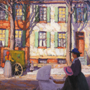
!["Autumn Harbour" [downloaded from http://metronews.ca/news/vancouver/1051693/chemists-in-vancouver-to-use-lasers-to-verify-group-of-seven-painting/]](http://www.frogheart.ca/wp-content/uploads/2014/06/autumn-harbour.jpg)