Monthly Archives: August 2014

I know something about your mummy or an ion beam microscope analyses a sarcophagus scrap

“Bearded Man, 170-180 A.D.” from the Walters Art Museum collection, object #32.6

“Bearded Man, 170-180 A.D.” from the Walters Art Museum collection, object #32.6

An Aug. 14, 2014 news item on Azonano describing this image sparks the imagination,

He looks almost Byzantine or Greek, gazing doe-eyed over the viewer’s left shoulder, his mouth forming a slight pout, like a star-struck lover or perhaps a fan of the races witnessing his favorite charioteer losing control of his horses.

In reality, he’s the “Bearded Man, 170-180 A.D.,” a Roman-Egyptian whose portrait adorned the sarcophagus sheltering his mummified remains. But the details of who he was and what he was thinking have been lost to time.

But perhaps not for much longer. A microscopic sliver of painted wood could hold the keys to unraveling the first part of this centuries-old mystery. Figuring out what kind of pigment was used (whether it was a natural matter or a synthetic pigment mixed to custom specifications), and the exact materials used to create it, could help scientists unlock his identity.

Kathleen Tuck’s Aug. 13 (?), 2014 Boise State University (Idaho, US) news release, which originated the news item, describes the nature of the research and the difficulties associated with it,

“Understanding the pigment means better understanding of the provenance of the individual” said Darryl Butt, a Boise State distinguished professor in the Department of Materials Science and Engineering and associate director of the Center for Advanced Energy Studies (CAES). “Where the pigment came from may connect it to a specific area and maybe even a family.”

For years, researchers were limited by the lack of samples large enough to be properly analyzed. But advances in the field of nanotechnology mean scientists now can work with fragments tinier than the eye can even register. Using a $1.5 million ion beam microscope at CAES, Butt — along with CAES colleagues Yaqaio Wu and Jatu Burns, and Boise State student researchers Gordon Alanko and Jennifer Watkins — is working with a sliver of the wood portrait smaller than a human hair.

The team transferred the fragment to a sample holder using a tiny deer hair called an “eyelash.” Their biggest challenge was to move it to the equipment without losing it.

So far they have extracted five needle-tip sized fragments 20 nanometers wide (a nanometer is a billionth of a meter), as well as two thin foils. From that, they have been able to analyze and map out the chemistry of the material in three dimensions.

Butt and his team are analyzing a speck of purple paint, which is significant because the blue used to blend the purple hue was a precious pigment back in the day, signaling a prominent individual.

This research is part of a larger project (from the news release),

Their data is being analyzed by researchers from the Detroit Museum of Art, where a companion to the “Bearded Man” mummy resides. It’s part of a project, titled APPEAR (Ancient Panel Paintings-Examination, Analysis, Research), a collaboration between 12 museums, including the British Museum in London and the Walters Art Museum in Baltimore, Maryland.

According to the news release there’s a personal aspect to Butt’s interest in this research, which may eventually have implications for Boise State University’s programmes,

“So far we’ve learned that the paint is a synthetic pigment,” said Butt, who as an artist in his own right often mixes his own pigments for his paintings. “These are very vibrant pigments, possibly heated in a lead crucible. People thought that process had been developed in the 1800s or so. This could prove it happened a lot earlier.

Butt got into solving art mysteries when he met Glenn Gates, a conservation scientist at the Walters Art Museum [Baltimore, Maryland] at a conference at Stanford University [California]. Both are officers of a new section of the American Ceramic Society — the Art, Archaeology and Conservation Science division.

“This research was a gamble that we [materials scientists] could do some really cool stuff,” Butt said, noting that he would love to branch out into analyzing pottery and other ancient artifacts.

While studying the provenance of Roman-Egyptian mummies is something new at Boise State, many researchers in the art, geology, history, anthropology and even English departments are involved in what Butt likes to call ‘reverse engineering’ of objects of cultural heritage.

“This particular problem, that is of understanding a particle of pigment from a 2,000-year-old sarcophagus, is a bit unique in that it highlights some of the amazing tools that we have at Boise State and at CAES that could shed new light on problems associated with understanding human history,” he said.

Butt hopes that these and similar transdisciplinary projects will open up external research opportunities for students, including creation of a “pipeline” of students who travel to various user facilities or museums to carry out interdisciplinary research.

“Envision, for example, art students studying works of art using synchrotron radiation and bright x-rays at a national laboratory, while science and engineering students use their technical skills to unravel mysteries of materials used by ancient societies in the field or held by museums,” he said.

The idea can sound far-fetched even for those who are participating in the research, although there is a certain, sound logic to transdisciplinary work between the arts and the sciences.

I was not able to find any reference to Butt’s art work online, find a published research paper or more information (website) featuring APPEAR ((Ancient Panel Paintings-Examination, Analysis, Research); admittedly, it was a brief search.

There are many techniques used to examine works of art and/or heritage. For a description of another technique, Raman spectroscopy, and its use in examining art pigments there’s my June 27, 2014 posting titled: Art (Lawren Harris and the Group of Seven), science (Raman spectroscopic examinations), and other collisions at the 2014 Canadian Chemistry Conference (part 2 of 4). Should you be interested in the entire series, additional links can be found in that posting.

Colombia, copyright, and sharing a science thesis

You’d think that posting a thesis online while giving full attribution to the author would be considered laudable. Apparently, there’s one person in Colombia that disagrees. And, since many educational institutions ask for copies of a student’s thesis for inclusion in their academic libraries you might believe the making said thesis more widely available (most students would be thrilled at the attention to their work) wouldn’t pose a problem. Apparently the Colombia legal system disagrees as it is preparing to take a student to court (and possible to jail) for sharing scientific information.

While the story seems to be popping up everywhere, this Aug. 1, 2014 article by Kerry Gren for The Scientist acted as my first notice (Note: Links have been removed),

Three years ago, Diego Gómez, a conservation biology student at the University of Quindío in Colombia, posted another scientist’s graduate thesis online. “I thought it was something that could be of interested [sic] for other groups, so I shared it on the web,” Gómez wrote on the website of Fundación Karisma, an education advocacy group in Colombia. “I never imagined that this activity could be considered a crime.”

But the author of the thesis disagreed, and last year complained to the Colombian police about the posting. Gómez now faces up to eight years in jail and at least $6,000 in fines for violating copyright. His case highlights the plight of scientists in certain parts of the world who are less able to access and share scientific information.

This wouldn’t have gone far in a US court at all,” said Michael Carroll, the director of the Program on Information Justice and Intellectual Property at American University’s Washington School of Law. [emphasis mine] “I’m really upset about this case,” he added. “It bothers me when copyright law gets in the way of scientists doing their science.” [emphasis mine]

While I too am bothered by copyright law being used to subvert science or, in this case, science sharing, Carroll’s comment about US courts (an indirect reference to US law) seems ironic after reading Tim Cushing’s July 28, 2014 Techdirt posting on the case (Note: Links have been removed),

Upload a document to Scribd, go to prison for at least four years. Ridiculous and more than a bit frightening, but in a case that has some obvious parallels with Aaron Swartz’s prosecution, that’s the reality Colombian student Diego Gomez is facing. In the course of his research, he came across a paper integral to his research. In order to ensure others could follow his line of thinking, Gomez uploaded this document for others to view.

According to Gomez, this was a common citation practice among Colombian students …

To be clear, Gomez did not try to profit from the paper. He also wasn’t acting as some sort of indiscriminate distributor of infringing works. But under Colombian law, none of that matters. But to really see who’s to blame here for this ridiculous level of rights enforcement, you have to look past the local laws, past the paper’s author and directly at the US government.

[Gomez] is being sued under a criminal law that was reformed in 2006, following the conclusion of a free trade agreement between Colombia and the United States. The new law was meant to fulfill the trade agreement’s restrictive copyright standards, and it expanded criminal penalties for copyright infringement, increasing possible prison sentences and monetary fines.

More details on the awfulness of Colombia’s law (spurred on by US special interests) are available in the EFF’s [Electronic Frontier Federation] earlier coverage. Colombia gave the US copyright industry everything it wanted in order to secure this free trade agreement… and then it just kept going. …

This bill was hastily passed as a welcoming gift for President Obama, shoved through the legislative process in order to get out ahead of the administration’s appearance at a Colombia-hosted conference. This deference to the US government could cost Gomez at least four years of his life.

While Colombia seemed very eager to take the worst parts of US copyright law (and make them even more terrible), it was less inclined to take any of the good. …

Beneath all of this lies the ugly reality of the academic research market. Just as in the US, plenty of useful information is locked up and inaccessible to anyone unable to afford the frequently exorbitant fees charged by various gatekeepers. Copyright’s original intent — “to promote the progress of science and the useful arts” — isn’t served by this behavior. …

Erik Stokstad’s July 31, 2014 article for ScienceInsider offers more details such as these,

In 2011, Gómez came across a master’s thesis, completed at the National University of Colombia in 2006, that would be useful for identifying amphibians he had seen in protected areas. He posted the thesis on Scribd to allow it to be easily downloaded by other researchers and students. At the time, the downloads were free. When Scribd started charging unregistered users $5 per download, Gómez removed the thesis.

The author of the thesis, a Colombian herpetologist, however, had already notified police that it had been posted without his permission. After being contacted by police, Gómez cooperated with the investigation. In April 2013, a criminal complaint was filed. This past fall, he learned that the office of the attorney general was going to bring the case to trial. Gómez “was in a panic,” says Carolina Botero, an attorney at Fundación Karisma, a digital rights advocacy organization in Bogotá, which is advocating on his behalf.

The Electronic Frontier Federation’s July 23, 2014 posting by Maira Sutton places this incident within an international context and outlines Colombia’s legal framework as it pertains to this case.

Diego Gomez has written about his situation (English language version and Spanish language version) as per some July 2014 postings.

As for Aaron Swartz mentioned in the excerpt from Tim Cushing’s Techdirt post, anyone unfamiliar with the case can find all the information they might want in this Wikipedia entry.

A rose by any other name: water pinning nanostructures and wettability

There are two items about rose petals as bioinspiration for research in this posting. The first being the most recent research where scientists in Singapore have made an ultrathin film modeled on rose petals. From an Aug. 13, 2014 news item on Nanowerk (Note: A link has been removed),

A*STAR [based in Singapore] researchers have used nanoimprinting methods to make patterned polymeric films with surface topography inspired by that of a rose petal, producing a range of transparent films with high water pinning forces (“Bioinspired Ultrahigh Water Pinning Nanostructures”).

An Aug. 13, 2014 A*STAR news highlight, which originated the news item, describes the nature of the research,

A surface to which a water droplet adheres, even when it is turned upside down, is described as having strong water pinning characteristics. A rose petal and a lotus leaf are both superhydrophobic, yet dissimilarities in their water pinning properties cause a water droplet to stick to a rose petal but roll off a lotus leaf. The two leaf types differ in their micro- and nanoscale surface topography and it is these topographical details that alter the water pinning force. The rose petal has almost uniformly distributed, conical-shaped microscale protrusions with nanoscale folds on these protrusions, while the lotus leaf has randomly distributed microscale protrusions.

The imprinted surfaces developed by Jaslyn Law and colleagues at the A*STAR Institute of Materials Research and Engineering and the Singapore University of Technology and Design have uniformly distributed patterns of nanoscale protrusions that are either conical or parabolic in shape. The researchers found that the water pinning forces on these continuously patterned surfaces were much greater than on non-patterned surfaces and surfaces composed of isolated nanopillared structures or nanoscale gratings. They could then achieve high water pinning forces by patterning the nanoprotrusions onto polymeric films with a range of different non-patterned hydrophobicities, including polycarbonate, poly(methyl methacrylate) and polydimethylsiloxane (see image).

“Other methods that recreate the water pinning effect have used actual rose petals as the mold, but unless special care is taken, there are likely to be defects and inconsistencies in the recreated pattern,” says co-author Andrew Ng. “While bottom-up approaches for making patterns — for example, laser ablation, liquid flame spray or chemical vapor deposition — are more consistent, these methods are limited in the types of patterns that can be used and the scale at which a substrate can be patterned.”

In contrast, nanoimprinting methods are capable of fabricating versatile and large-scale surfaces, and can be combined with roll-to-roll techniques, hence potentially enabling more commercial applications.

The patterned polycarbonate surfaces were also shown to reduce the ‘coffee-ring’ effect: the unevenly deposited film left behind upon the evaporation of a solute-laden droplet. This mitigation of the coffee-ring effect may assist microfluidic technologies and, more generally, the patterned surfaces could be used in arid regions for dew collection or in anti-drip applications such as in greenhouses.

The study which was published online in Dec. 2013, was featured in a Jan. 22, 2014 article by Katherine Bourzac for C&EN (Chemistry and Engineering News),

In the early morning, dew clings to rose petals; when the sun rises, the dewdrops act like tiny lenses, making diffraction patterns that attract pollinating insects, says Jaslyn Bee Khuan Law, a materials scientist at the Agency for Science, Technology, and Research (A*STAR), in Singapore. A drop of water will cling to a rose petal even when it’s tilted or held upside down. The petals can hold onto these droplets because their surfaces consist of closely packed conical structures a few micrometers across. These microscale surface patterns tweak the surface tension of the water droplets, causing them to cling to the petals.

But none of these fabrication methods are amenable to large-scale, low-cost manufacturing, preventing commercialization of the water-clinging surfaces. So Law turned to a specialty of her lab: nanoimprint lithography. This printing method utilizes metal or silicon drums molded with nanoscale features on their surfaces. When the molds are heated and pressed against sheets of plastic, the plastic is embossed with the nanoscale pattern. This roll-to-roll printing process resembles the way newspapers are printed. It’s capable of producing large-area films in a short amount of time.

Water droplets easily slid off plastic films patterned with simple nanoscale gratings; isolated nanoscale pillars hung onto water slightly better. But the films with the best properties consisted of tightly packed cones about 300 nm tall. Plastic patterned with these structures could hold onto water droplets as massive as 69 mg. The team could print a 110- by 65-mm sheet of this plastic film at a speed of 10 m per minute. Currently, the dimensions of the films are limited by the size of the premade molds, Law says.

While the Singapore group has made good progress on manufacturing these materials, very basic, vexing questions about how water clings to these surfaces remain, Hayes says. For example, very small changes in the surface’s roughness can switch it from water-pinning to super hydrophobic, and researchers don’t have a detailed understanding of why.

Here’s a link to and a citation for the paper,

Bioinspired Ultrahigh Water Pinning Nanostructures by Jaslyn Bee Khuan Law, Andrew Ming Hua Ng, Ai Yu He, and Hong Yee Low. Langmuir, 2014, 30 (1), pp 325–331 DOI: 10.1021/la4034996 Publication Date (Web): December 20, 2013
Copyright © 2013 American Chemical Society

This paper appears to be open access (I was able to access it by clicking on the HTML option).

Finally, here’s an image supplied by the A*Star researchers to illustrate their work,

[downloaded from http://pubs.acs.org/doi/full/10.1021/la4034996]

[downloaded from http://pubs.acs.org/doi/full/10.1021/la4034996]

This second rose petal item comes from Australia and dates from Fall 2013. From a Sept. 18, 2013 news item on ScienceDaily,

A new nanostructured material with applications that could include reducing condensation in airplane cabins and enabling certain medical tests without the need for high tech laboratories has been developed by researchers at the University of Sydney [Australia].

“The newly discovered material uses raspberry particles — so-called because of their appearance — which can trap tiny water droplets and prevent them from rolling off surfaces, even when that surface is turned upside down,” said Dr Andrew Telford from the University’s School of Chemistry and lead author of the research recently published in the journal, Chemistry of Materials.

The ability to immobilise [pin] very small droplets on a surface is, according to Dr Telford, a significant achievement with innumerable potential applications.

A Sept. 17, 2013 University of Sydney news release, which originated the news item, provides more insight into the research where the scientists have focused on ‘raspberry particles’ which could also be described as the ‘conical structures’ mentioned in the A*STAR work to achieve what appear to be similar ends,

Raspberry particles mimic the surface structure of some rose petals.

“Water droplets bead up in a spherical shape on top of rose petals,” Dr Telford said. “This is a sign the flower is highly water repellent.”

The reasons for this are complex and largely due to the special structure of the rose petal’s surface. The research team replicated the rose petal by assembling raspberry particles in the lab using spherical micro- and nanoparticles.

The result is that water droplets bead up when placed on films of the raspberry particles and they’re not able to drip down from it, even when turned upside down.

“Raspberry particle films can be described as sticky tape for water droplets,” Dr Telford said.

This could be useful in preventing condensation issues in airplane cabins. It could also help rapidly process simple medical tests on free-standing droplets, with the potential for very high turnover of tests with inexpensive equipment and in remote areas.

Other exciting applications are under study: if we use this nanotechnology to control how a surface is structured we can influence how it will interact with water.

“This means we will be able to design a surface that does whatever you need it to do.

“We could also design a surface that stays dry forever, never needs cleaning or able to repel bacteria or even prevent mould and fungi growth.

“We could then tweak the same structure by changing its composition so it forces water to spread very quickly.

“This could be used on quick-dry walls and roofs which would also help to cool down houses.

“This can only be achieved with a very clear understanding of the science behind the chemical properties and construction of the surface,” he said.

The discovery is also potentially viable commercially.

“Our team’s discovery is the first that allows for the preparation of raspberry particles on an industrial scale and we are now in a position where we can prepare large quantities of these particles without the need to build special plants or equipment,” Dr Telford said.

Here’s a link to and a citation for the Australian paper,

Mimicking the Wettability of the Rose Petal using Self-assembly of Waterborne Polymer Particles by A. M. Telford, B. S. Hawkett, C. Such, and C. Neto. Chem. Mater., 2013, 25 (17), pp 3472–3479 DOI: 10.1021/cm4016386 Publication Date (Web): July 23, 2013
Copyright © 2013 American Chemical Society

This paper is behind a paywall.

A tattoo that’s a biobattery and a sensor?

It’s going to be an American Chemical Society (ACS) 248th meeting kind of week as yet another interesting piece of scientific research is bruited (spread) about the internet. This time it’s all about sweat, exercise, and biobatteries. From an Aug. 13, 2014 news item on Nanowerk,

In the future, working up a sweat by exercising may not only be good for your health, but it could also power your small electronic devices. Researchers will report today that they have designed a sensor in the form of a temporary tattoo that can both monitor a person’s progress during exercise and produce power from their perspiration.

An Aug. 13, 2014 ACS news release on EurekAlert, which originated the news item, describes the inspiration (as opposed to perspiration) for this technology,

The device works by detecting and responding to lactate, which is naturally present in sweat. “Lactate is a very important indicator of how you are doing during exercise,” says Wenzhao Jia, Ph.D.

In general, the more intense the exercise, the more lactate the body produces. During strenuous physical activity, the body needs to generate more energy, so it activates a process called glycolysis. Glycolysis produces energy and lactate, the latter of which scientists can detect in the blood.

Professional athletes monitor their lactate levels during performance testing as a way to evaluate their fitness and training program. In addition, doctors measure lactate during exercise testing of patients for conditions marked by abnormally high lactate levels, such as heart or lung disease. Currently, lactate testing is inconvenient and intrusive because blood samples must be collected from the person at different times during the exercise regime and then analyzed.

The news release goes on to describe the research process which resulted in a temporary tattoo that could be used to power small scale electronics,

Jia, a postdoctoral student in the lab of Joseph Wang, D.Sc., at the University of California San Diego, and her colleagues developed a faster, easier and more comfortable way to measure lactate during exercise. They imprinted a flexible lactate sensor onto temporary tattoo paper. The sensor contained an enzyme that strips electrons from lactate, generating a weak electrical current. The researchers applied the tattoo to the upper arms of 10 healthy volunteers. Then the team measured the electrical current produced as the volunteers exercised at increasing resistance levels on a stationary bicycle for 30 minutes. In this way, they could continuously monitor sweat lactate levels over time and with changes in exercise intensity.

The team then went a step further, building on these findings to make a sweat-powered biobattery. Batteries produce energy by passing current, in the form of electrons, from an anode to a cathode. In this case, the anode contained the enzyme that removes electrons from lactate, and the cathode contained a molecule that accepts the electrons.

When 15 volunteers wore the tattoo biobatteries while exercising on a stationary bike, they produced different amounts of power. Interestingly, people who were less fit (exercising fewer than once a week) produced more power than those who were moderately fit (exercising one to three times per week). Enthusiasts who worked out more than three times per week produced the least amount of power. The researchers say that this is probably because the less-fit people became fatigued sooner, causing glycolysis to kick in earlier, forming more lactate. The maximum amount of energy produced by a person in the low-fitness group was 70 microWatts per cm2 of skin.

“The current produced is not that high, but we are working on enhancing it so that eventually we could power some small electronic devices,” Jia says. “Right now, we can get a maximum of 70 microWatts per cm2, but our electrodes are only 2 by 3 millimeters in size and generate about 4 microWatts — a bit small to generate enough power to run a watch, for example, which requires at least 10 microWatts. So besides working to get higher power, we also need to leverage electronics to store the generated current and make it sufficient for these requirements.”

Biobatteries offer certain advantages over conventional batteries: They recharge more quickly, use renewable energy sources (in this case, sweat), and are safer because they do not explode or leak toxic chemicals.

“These represent the first examples of epidermal electrochemical biosensing and biofuel cells that could potentially be used for a wide range of future applications,” Wang says.

The ACS has made a video about this work available,

It seems to me this tattoo battery could be used as a self-powered monitoring device in a medical application for heart or lung disease.

Hemp as a substitute for graphene in supercapacitors

As a member of the Cannabis plant family, hemp has an undeserved reputation due to its cousin’s (marijuana) notoriety and consciousness-altering properties. Hemp is, by contrast, the Puritan in the family, associated by the knowledgeable with virtues of thrift and hard work.

An Aug. 12, 2014 news item on Nanowerk highlights a hemp/supercapacitor presentation at the 248th meeting of the American Chemical Society (ACS),

As hemp makes a comeback in the U.S. after a decades-long ban on its cultivation, scientists are reporting that fibers from the plant can pack as much energy and power as graphene, long-touted as the model material for supercapacitors. They’re presenting their research, which a Canadian start-up company is working on scaling up, at the 248th National Meeting & Exposition of the American Chemical Society (ACS), the world’s largest scientific society.

David Mitlin, Ph.D., explains that supercapacitors are energy storage devices that have huge potential to transform the way future electronics are powered. Unlike today’s rechargeable batteries, which sip up energy over several hours, supercapacitors can charge and discharge within seconds. But they normally can’t store nearly as much energy as batteries, an important property known as energy density. One approach researchers are taking to boost supercapacitors’ energy density is to design better electrodes. Mitlin’s team has figured out how to make them from certain hemp fibers — and they can hold as much energy as the current top contender: graphene.

An Aug. 12, 2014 ACS news release features David Mitlin, formerly of the University of Alberta (Canada) where this research took place,, Mitlin is now with now with Clarkson University in New York,

“Our device’s electrochemical performance is on par with or better than graphene-based devices,” Mitlin says. “The key advantage is that our electrodes are made from biowaste using a simple process, and therefore, are much cheaper than graphene.”

The race toward the ideal supercapacitor has largely focused on graphene — a strong, light material made of atom-thick layers of carbon, which when stacked, can be made into electrodes. Scientists are investigating how they can take advantage of graphene’s unique properties to build better solar cells, water filtration systems, touch-screen technology, as well as batteries and supercapacitors. The problem is it’s expensive.

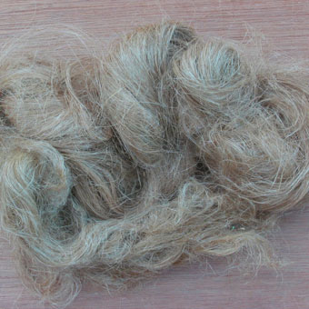
Mitlin’s group decided to see if they could make graphene-like carbons from hemp bast fibers. The fibers come from the inner bark of the plant and often are discarded from Canada’s fast-growing industries that use hemp for clothing, construction materials and other products. …

His team found that if they heated the fibers for 24 hours at a little over 350 degrees Fahrenheit, and then blasted the resulting material with more intense heat, it would exfoliate into carbon nanosheets.

Mitlin’s team built their supercapacitors using the hemp-derived carbons as electrodes and an ionic liquid as the electrolyte. Fully assembled, the devices performed far better than commercial supercapacitors in both energy density and the range of temperatures over which they can work. The hemp-based devices yielded energy densities as high as 12 Watt-hours per kilogram, two to three times higher than commercial counterparts. They also operate over an impressive temperature range, from freezing to more than 200 degrees Fahrenheit.

“We’re past the proof-of-principle stage for the fully functional supercapacitor,” he says. “Now we’re gearing up for small-scale manufacturing.”

I have not been able to confirm the name for Mitlin’s startup but I think it’s called Alta Supercaps (Alta being an abbreviation for Alberta,, amongst other things, and supercaps for supercapacitors) as per the information about a new startup on the Mitlin Group webspace (scroll down to the July 2, 2013 news item) which can still be found on the University of Alberta website (as of Aug. 12, 2014).

For those who would like more technical details, there is this July 2013 article by Mark Crawford for the ASME (American Society of Mechanical Engineers); Note: A link has been removed.

Activated carbons, templated carbons, carbon nanofibers, carbon nanotubes, and graphene have all been intensively studied as materials for supercapacitor electrodes. High manufacturing costs is one issue—another is that the power characteristics of many of these carbons are limited. This is a result of high microporosity, which increases ion transport limitations.

“It is becoming well understood that the key to achieving high power in porous electrodes is to reduce the ion transport limitations” says Mitlin. “Nanomaterials based on graphene and their hybrids have emerged as a new class of promising high-rate electrode candidates—they are, however, too expensive to manufacture compared to activated carbons derived from pyrolysis of agricultural wastes, or from the coking operations.”

Biomass, which mainly contains cellulose and lignin by-products, is widely utilized as a feedstock for producing activated carbons. Mitlin decided to test hemp bast fiber’s unique cellular structure to see if it could produce graphene-like carbon nanosheets.

Hemp fiber waste was pressure-cooked (hydrothermal synthesis) at 180 °C for 24 hours. The resulting carbonized material was treated with potassium hydroxide and then heated to temperatures as high as 800 °C, resulting in the formation of uniquely structured nanosheets. Testing of this material revealed that it discharged 49 kW of power per kg of material—nearly triple what standard commercial electrodes supply, 17 kW/kg.

Mitlin and his team successfully synthesized two-dimensional, yet interconnected, carbon nanosheets with superior electrochemical storage properties comparable to those of state-of-the-art graphene-based electrodes. “We were able to achieve this by employing a biomass precursor with a unique structure—hemp bast fiber,” says Mitlin. “The resultant graphene-like nanosheets possess fundamentally different properties—such as pore size distribution, physical interconnectedness, and electrical conductivity—as compared to conventional biomass-derived activated carbons.”

This image from Wikimedia was used to illustrate the Crawford article,

Hemp bast fiber is a low-cost graphene-like nanomaterial. Image: Wikimedia Commons

Hemp bast fiber is a low-cost graphene-like nanomaterial. Image: Wikimedia Commons

It seems to me that over the last few months there have been more than the usual number of supercapacitor stories, which makes the race to create the one that will break through in the marketplace fascinating to observe.

Two-organ tests (body-on-a-chip) show liver damage possible from nanoparticles

This is the first time I’ve seen testing of two organs for possible adverse effects from nanoparticles. In this case, the researchers were especially interested in the liver. From an Aug. 12, 2014 news item on Azonano,

Nanoparticles in food, sunscreen and other everyday products have many benefits. But Cornell [University] biomedical scientists are finding that at certain doses, the particles might cause human organ damage.

A recently published study in Lab on a Chip by the Royal Society of Chemistry and led by senior research associate Mandy Esch shows that nanoparticles injure liver cells when they are in microfluidic devices designed to mimic organs of the human body. The injury was worse when tested in two-organ systems, as opposed to single organs – potentially raising concerns for humans and animals.

Anne Ju’s Aug. 11, 2014 article for Cornell University’s Chronicle describes the motivation for this work and the research itself in more detail,

“We are looking at the effects of what are considered to be harmless nanoparticles in humans,” Esch said. “These particles are not necessarily lethal, but … are there other consequences? We’re looking at the non-lethal consequences.”

She used 50-nanometer carboxylated polystyrene nanoparticles, found in some animal food sources and considered model inert particles. Shuler’s lab specializes in “body-on-a-chip” microfluidics, which are engineered chips with carved compartments that contain cell cultures to represent the chemistry of individual organs.

In Esch’s experiment, she made a human intestinal compartment, a liver compartment and a compartment to represent surrounding tissues in the body. She then observed the effects of fluorescently labeled nanoparticles as they traveled through the system.

Esch found that both single nanoparticles as well as small clusters crossed the gastrointestinal barrier and reached liver cells, and the liver cells released an enzyme called aspartate transaminase, known to be released during cell death or damage.

It’s unclear exactly what damage is occurring or why, but the results indicate that the nanoparticles must be undergoing changes as they cross the gastrointestinal barrier, and that these alterations may change their toxic potential, Esch said. Long-term consequences for organs in proximity could be a concern, she said.

“The motivation behind this study was twofold: one, to show that multi-organ, in vitro systems give us more information when testing for the interaction of a substance with the human body, and two … to look at nanoparticles because they have a huge potential for medicine, yet adverse effects have not been studied in detail yet,” Esch said.

Mary Macleod’s July 3, 2014 article for Chemistry World features a diagram of the two-organ system and more technical details about the research,

Schematic of the two-organ system [downloaded from http://www.rsc.org/chemistryworld/2014/07/nanoparticle-liver-gastrointestinal-tract-microfluidic-chip]

Schematic of the two-organ system [downloaded from http://www.rsc.org/chemistryworld/2014/07/nanoparticle-liver-gastrointestinal-tract-microfluidic-chip]

HepG2/C3A cells were used to represent the liver, with the intestinal cell co-culture consisting of enterocytes (Caco-2) and mucin-producing (HT29-MTX) cells. Carboxylated polystyrene nanoparticles were fluorescently labelled so their movement between the chambers could be tracked. Levels of aspartate transaminase, a cytosolic enzyme released into the culture medium upon cell death, were measured to give an indication of liver damage.

The study saw that single nanoparticles and smaller nanoparticle aggregates were able to cross the GI barrier and reach the liver cells. The increased zeta potentials of these nanoparticles suggest that crossing the barrier may raise their toxic potential. However, larger nanoparticles, which interact with cell membranes and aggregate into clusters, were stopped much more effectively by the GI tract barrier.

The gastrointestinal tract is an important barrier preventing ingested substances crossing into systemic circulation. Initial results indicate that soluble mediators released upon low-level injury to liver cells may enhance the initial injury by damaging the cells which form the GI tract. These adverse effects were not seen in conventional single-organ tests.

Here’s a link to and a citation for the paper,

Body-on-a-chip simulation with gastrointestinal tract and liver tissues suggests that ingested nanoparticles have the potential to cause liver injury by Mandy B. Esch, Gretchen J. Mahler, Tracy Stokol, and Michael L. Shuler. Lab Chip, 2014,14, 3081-3092 DOI: 10.1039/C4LC00371C First published online 27 Jun 2014

This paper is open access until Aug. 12, 2014.

While this research is deeply concerning, it should be noted the researchers are being very careful in their conclusions as per Ju’s article, “It’s unclear exactly what damage is occurring or why, but the results indicate that the nanoparticles must be undergoing changes as they cross the gastrointestinal barrier, and that these alterations may change their toxic potential … Long-term consequences for organs in proximity could be a concern … .”

Graphene and an artificial retina

A graphene-based artificial retina project has managed to intermingle the European Union’s two major FET (Future and Emerging Technologies) funding projects, 1B Euros each to be disbursed over 10 years, the Graphene Flagship and the Human Brain Project. From an Aug. 7, 2014 Technische Universitaet Muenchen (TUM) news release (also on EurekAlert),

Because of its unusual properties, graphene holds great potential for applications, especially in the field of medical technology. A team of researchers led by Dr. Jose A. Garrido at the Walter Schottky Institut of the TUM is taking advantage of these properties. In collaboration with partners from the Institut de la Vision of the Université Pierre et Marie Curie in Paris and the French company Pixium Vision, the physicists are developing key components of an artificial retina made of graphene.

Retina implants can serve as optical prostheses for blind people whose optical nerves are still intact. The implants convert incident light into electrical impulses that are transmitted to the brain via the optical nerve. There, the information is transformed into images. Although various approaches for implants exist today, the devices are often rejected by the body and the signals transmitted to the brain are generally not optimal.

Already funded by the Human Brain Project as part of the Neurobotics effort, Garrido and his colleagues will now also receive funding from the Graphene Flagship. As of July 2014, the Graphene Flagship has added 86 new partners including TUM according to the news release.

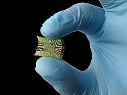
Here’s an image of an ‘invisible’ graphene sensor (a precursor to developing an artificial retina),

Graphene electronics can be prepared on flexible substrates. Only the gold metal leads are visible in the transparent graphene sensor. (Photo: Natalia Hutanu / TUM)

Graphene electronics can be prepared on flexible substrates. Only the gold metal leads are visible in the transparent graphene sensor. (Photo: Natalia Hutanu / TUM)

Artificial retinas were first featured on this blog in an Aug. 18, 2011 posting about video game Deus Ex: Human Revolution which features a human character with artificial sight. The post includes links to a video of a scientist describing an artificial retina trial with 30 people and an Israeli start-up company, ‘Nano Retina’, along with information about ‘Eyeborg’, a Canadian filmmaker who on losing an eye in an accident had a camera implanted in the previously occupied eye socket.

More recently, a Feb. 15, 2013 posting featured news about the US Food and Drug Administration’s decision to allow sale of the first commercial artificial retinas in the US in the context of news about a neuroprosthetic implant in a rat which allowed it to see in the infrared range, normally an impossible feat.

Cyborgs (a presentation) at the American Chemical Society’s 248th meeting

There will be a plethora of chemistry news online over the next few days as the American Society’s (ACS) 248th meeting in San Francisco, CA from Aug. 10 -14, 2014 takes place. Unexpectedly, an Aug. 11, 2014 news item on Azonano highlights a meeting presentation focused on cyborgs,

No longer just fantastical fodder for sci-fi buffs, cyborg technology is bringing us tangible progress toward real-life electronic skin, prosthetics and ultraflexible circuits. Now taking this human-machine concept to an unprecedented level, pioneering scientists are working on the seamless marriage between electronics and brain signaling with the potential to transform our understanding of how the brain works — and how to treat its most devastating diseases.

An Aug. 10, 2014 ACS news release on EurekAlert provides more detail about the presentation (Note: Links have been removed),

“By focusing on the nanoelectronic connections between cells, we can do things no one has done before,” says Charles M. Lieber, Ph.D. “We’re really going into a new size regime for not only the device that records or stimulates cellular activity, but also for the whole circuit. We can make it really look and behave like smart, soft biological material, and integrate it with cells and cellular networks at the whole-tissue level. This could get around a lot of serious health problems in neurodegenerative diseases in the future.”

These disorders, such as Parkinson’s, that involve malfunctioning nerve cells can lead to difficulty with the most mundane and essential movements that most of us take for granted: walking, talking, eating and swallowing.

Scientists are working furiously to get to the bottom of neurological disorders. But they involve the body’s most complex organ — the brain — which is largely inaccessible to detailed, real-time scrutiny. This inability to see what’s happening in the body’s command center hinders the development of effective treatments for diseases that stem from it.

By using nanoelectronics, it could become possible for scientists to peer for the first time inside cells, see what’s going wrong in real time and ideally set them on a functional path again.

For the past several years, Lieber has been working to dramatically shrink cyborg science to a level that’s thousands of times smaller and more flexible than other bioelectronic research efforts. His team has made ultrathin nanowires that can monitor and influence what goes on inside cells. Using these wires, they have built ultraflexible, 3-D mesh scaffolding with hundreds of addressable electronic units, and they have grown living tissue on it. They have also developed the tiniest electronic probe ever that can record even the fastest signaling between cells.

Rapid-fire cell signaling controls all of the body’s movements, including breathing and swallowing, which are affected in some neurodegenerative diseases. And it’s at this level where the promise of Lieber’s most recent work enters the picture.

In one of the lab’s latest directions, Lieber’s team is figuring out how to inject their tiny, ultraflexible electronics into the brain and allow them to become fully integrated with the existing biological web of neurons. They’re currently in the early stages of the project and are working with rat models.

“It’s hard to say where this work will take us,” he says. “But in the end, I believe our unique approach will take us on a path to do something really revolutionary.”

Lieber acknowledges funding from the U.S. Department of Defense, the National Institutes of Health and the U.S. Air Force.

I first covered Lieber’s work in an Aug. 27, 2012 posting  highlighting some good descriptions from Lieber and his colleagues of their work. There’s also this Aug. 26, 2012 article by Peter Reuell in the Harvard Gazette (featuring a very good technical description for someone not terribly familiar with the field but able to grasp some technical information while managing their own [mine] ignorance). The posting and the article provide details about the foundational work for Lieber’s 2014 presentation at the ACS meeting.

Lieber will be speaking next at the IEEE (Institute for Electrical and Electronics Engineers) 14th International Conference on Nanotechnology sometime between August 18 – 21, 2014 in Toronto, Ontario, Canada.

As for some of Lieber’s latest published work, there’s more information in my Feb. 20, 2014 posting which features a link to a citation for the paper (behind a paywall) in question.

Wearable solar panels with perovskite

There was a bit of a flutter online in late July 2014 about solar cell research and perovskite, a material that could replace silicon therefore making solar cells more affordable, which hopefully would lead to greater adoption of the technology. Happily, the publishers of the study seem to have reissued their news release (h/t Aug. 11, 2014 news item on Nanwerk).

From the Wiley online press release Nr. 29/2014,

Textile solar cells are an ideal power source for small electronic devices incorporated into clothing. In the journal Angewandte Chemie, Chinese scientists have now introduced novel solar cells in the form of fibers that can be woven into a textile. The flexible, coaxial cells are based on a perovskite material and carbon nanotubes; they stand out due to their excellent energy conversion efficiency of 3.3 % and their low production cost.

The dilemma for solar cells: they are either inexpensive and inefficient, or they have a reasonable efficiency and are very expensive. One solution may come from solar cells made of perovskite materials, which are less expensive than silicon and do not require any expensive additives. Perovskites are materials with a special crystal structure that is like that of perovskite, a calcium titanate. These structures are often semiconductors and absorb light relatively efficiently. Most importantly, they can move electrons excited by light for long distances within the crystal lattice before they return to their energetic ground state and take up a solid position – a property that is very important in solar cells.

A team led by Hisheng Peng at Fudan University in Shanghai has now developed perovskite solar cells in the form of flexible fibers that can be woven into electronic textiles. Their production process is relatively simple and inexpensive because it uses a solution-based process to build up the layers.

The anode is a fine stainless steel wire coated with a compact n-semiconducting titanium dioxide layer. A layer of porous nanocrystalline titanium dioxide is deposited on top of this. This provides a large surface area for the subsequent deposition of the perovskite material CH3NH3PbI3. This is followed by a layer made of a special organic material. Finally a transparent layer of aligned carbon nanotubes is continuously wound over the whole thing to act as the cathode. The resulting fiber is so fine and flexible that it can be woven into textiles.

The perovskite layer absorbs light, that excites electrons and sets them free, causing a charge separation between the electrons and the formally positively charged “holes” The electrons enter the conducting band of the compact titanium dioxide layer and move to the anode. The “holes” are captured by the organic layer. The large surface area and the high electrical conductivity of the carbon nanotube cathode aid in the rapid conduction of the charges with high photoelectric currents. The fiber solar cell can attain an energy conversion efficiency of 3.3 %, exceeding that of all previous coaxial fiber solar cells made with either dyes or polymers.

Here’s an image used in the press release illustrating the new fiber,

[downloaded from http://www.wiley-vch.de/vch/journals/2002/press/201429press.pdf]

[downloaded from http://www.wiley-vch.de/vch/journals/2002/press/201429press.pdf]

Here’s a link to and a citation for the paper,

Integrating Perovskite Solar Cells into a Flexible Fiber by Longbin Qiu, Jue Deng, Xin Lu, Zhibin Yang, and Prof. Huisheng Peng. Angewandte Chemie International Edition DOI: 10.1002/anie.201404973 Article first published online: 22 JUL 2014

© 2014 WILEY-VCH Verlag GmbH & Co. KGaA, Weinheim

This paper is behind a paywall.

I found a second item about perovskite and solar cells in a May 16, 2014 article by Vicki Marshall for Chemistry World which discussed some research in the UK (Note: Links have been removed),

A lead-free and non-toxic alternative to current perovskite solar-cell technology has been reported by researchers in the UK: tin halide perovskite solar cells. They are also cheaper to manufacture than the silicon solar cells currently dominating the market.

Nakita Noel, part of Henry Snaith’s research team at the University of Oxford, describes how perovskite materials have caused a bit of a whirlwind since they came out in 2009: ‘Everybody that’s working in the solar community is looking to beat silicon.’ Despite the high efficiency of conventional crystalline silicon solar cells (around 20%), high production and installation costs decrease their economic feasibility and widespread use.

The challenge to find a cheaper alternative led to the development of perovskite-based solar cells, as organic–inorganic metal trihalide perovskites have both abundant and cheap starting materials. However, the presence of lead in some semiconductors could create toxicology issues in the future. As Noel puts it ‘every conference you present at somebody is bound to put up their hand and ask “What about the lead – isn’t this toxic?”’

Brian Hardin, co-founder of PLANT PV, US, and an expert in new materials for photovoltaic cells, says the study ‘should be considered a seminal work on alternative perovskites and is extremely valuable to the field as they look to better understand how changes in chemistry affect solar cell performance and stability.’

Here’s a link to and a citation for the UK researchers’ paper,

Lead-free organic–inorganic tin halide perovskites for photovoltaic applications by Nakita K. Noel, Samuel D. Stranks, Antonio Abate, Christian Wehrenfennig, Simone Guarnera, Amir-Abbas Haghighirad, Aditya Sadhana, Giles E. Eperon, Sandeep K. Pathak, Michael B. Johnston, Annamaria Petrozza, Laura M. Herza, and Henry J. Snaith. Energy Environ. Sci., 2014, Advance Article DOI: 10.1039/C4EE01076K First published online 01 May 2014

This article was open access until June 27, 2014 but now it is behind a paywall.

I notice there’s no mention of lead in the materials describing the research paper from the Chinese scientists. Perhaps they were working with lead-free materials.