Monthly Archives: July 2020

Chameleon skin (nanomaterial made of gold nanoparticles) for robots

A June 17, 2020 news item on Nanowerk trumpets research into how robots might be able to sport chameleon-like skin one day,

A new film made of gold nanoparticles changes color in response to any type of movement. Its unprecedented qualities could allow robots to mimic chameleons and octopi — among other futuristic applications.

Unlike other materials that try to emulate nature’s color changers, this one can respond to any type of movement, like bending or twisting. Robots coated in it could enter spaces that might be dangerous or impossible for humans, and offer information just based on the way they look.

For example, a camouflaged robot could enter tough-to-access underwater crevices. If the robot changes color, biologists could learn about the pressures facing animals that live in these environments.

Although some other color-changing materials can also respond to motion, this one can be printed and programmed to display different, complex patterns that are difficult to replicate.

This video from the University of California at Riverside researchers shows the material in action (Note: It gets more interesting after the first 20 secs.),

A June 15, 2020 University of California at Riverside (UCR) news release (also on EurekAlert but published on June 17, 2020) by Jules Bernstein, which originated the news item, delves further,

Nanomaterials are simply materials that have been reduced to an extremely small scale — tens of nanometers in width and length, or, about the size of a virus. When materials like silver or gold become smaller, their colors will change depending on their size, shape, and the direction they face.

“In our case, we reduced gold to nano-sized rods. We knew that if we could make the rods point in a particular direction, we could control their color,” said chemistry professor Yadong Yin. “Facing one way, they might appear red. Move them 45 degrees, and they change to green.”

The problem facing the research team was how to take millions of gold nanorods floating in a liquid solution and get them all to point in the same direction to display a uniform color.

Their solution was to fuse smaller magnetic nanorods onto the larger gold ones. The two different-sized rods were encapsulated in a polymer shield, so that they would remain side by side. That way, the orientation of both rods could be controlled by magnets.

“Just like if you hold a magnet over a pile of needles, they all point in the same direction. That’s how we control the color,” Yin said.

Once the nanorods are dried into a thin film, their orientation is fixed in place and they no longer respond to magnets. “But, if the film is flexible, you can bend and rotate it, and will still see different colors as the orientation changes,” Yin said.

Other materials, like butterfly wings, are shiny and colorful at certain angles, and can also change color when viewed at other angles. However, those materials rely on precisely ordered microstructures, which are difficult and expensive to make for large areas. But this new film can be made to coat the surface of any sized object just as easily as applying spray paint on a house.

Though futuristic robots are an ultimate application of this film, it can be used in many other ways. UC Riverside chemist Zhiwei Li, the first author on this paper, explained that the film can be incorporated into checks or cash as an authentication feature. Under normal lighting, the film is gray, but when you put on sunglasses and look at it through polarized lenses, elaborate patterns can be seen. In addition, the color contrast of the film may change dramatically if you twist the film.

The applications, in fact, are only limited by the imagination. “Artists could use this technology to create fascinating paintings that are wildly different depending on the angle from which they are viewed,” Li said. “It would be wonderful to see how the science in our work could be combined with the beauty of art.”

Here’s a link to and a citation for the paper,

Coupling magnetic and plasmonic anisotropy in hybrid nanorods for mechanochromic responses by Zhiwei Li, Jianbo Jin, Fan Yang, Ningning Song & Yadong Yin. Nature Communications volume 11, Article number: 2883 (2020) DOI: https://doi.org/10.1038/s41467-020-16678-8 Published: 08 June 2020

This paper is open access.

Call for works for a virtual October 2020 ‘Catalyst: A Sci-Art Exhibition’ (Michigan State University)

McKenzie Prillaman both profiles a 2019 sci-art exhibit at Michigan State University (MSU) and publicizes a ‘call for submissions’ for the 2020 edition in a July 10, 2020 posting on artthescience.com (Note: Links have been removed),

The Exploring Life Through SciArt exhibition united the science and art communities of East Lansing, Michigan. Organized by the Michigan State University Science Communication Organization (MSU SciComm) in 2019, this exhibition featured original science-art collaborations created by university science students and local artists.

The artworks, which were exhibited on MSU’s campus from October 2019 to March 2020 then donated to various locations around the city, are now on display in Art the Science’s Polyfield Digital Art Gallery. 

Fine art photographer Jane Kramer helped plant biology student Emily Jennings share her research on chloroplasts, the structures in plants that convert light energy into plant food. Together, they created the work Micrometer by Micrometer, which showcases the size variation in chloroplasts. Jennings’ research examines how chloroplast size impacts the amount of food a plant can produce.

This initiative taught the photographer that participation in sci-art projects didn’t always mean she needed to be behind the camera. “I lent my skills in conceptualizing how to best present Emily’s work in a way that engages and excites the public,” Kramer says. “It was important for me to incorporate her passion for this research into the final piece and bring her message to light.”

Micrometer by Micrometer (2019) by Emily Jennings and Jane Kramer, 6” x 9”, image capture on acrylic

You can see all 18 artist or artist/scientist pairings for a digital exhibit of the work here at the online Polyfield Digital Art Gallery.

The ‘call for submissions’ is an ‘application’ according to the organizers (from the MSU SciComm ‘Catalyst: A Sci-Art Exhibition’ application webpage),

MSU SciComm 2020 Sci-Art Application

This form is meant to provide us with a more detailed picture of your project. Additionally, information provided on this form will help us get you the resources needed for your project’s success. Thank you and happy planning! The theme for this year’s exhibit is Catalyst: A SciArt Exhibit

Due Date: July 31st at 11:59pm

-Noelia Barvo (MSU SciComm SciArt Chair)

Please email questions at msuscicomm.sciart@gmail.com

There are two questions on the form:

Question 1: Please list a short description of yourself including name, background/affiliation, and degree (completed/in progress) as you’d like it to appear online.

Question 2: Are you an MSU student?

YesNo

Should you be concerned that you’re not a student or member of the MSU community, there’s this from Prillaman’s July 10, 2020 posting,

Applications are now open for MSU SciComm’s next exhibition, Catalyst: A Sci-Art Exhibition, scheduled to open virtually in October 2020. The organization hopes to keep growing and work with international artists [emphases mine] to continue making an impact through the beauty of science-art.

Fro the curious, here’s a brief description of the MSU SciComm’s mission on their organization’s homepage (scroll down),

MSU SciComm is a student-run organization that focuses primarily on promoting awareness of science communication across the various disciplines at Michigan State University. We focus on science writing, speaking, art, policy, & outreach.

Our mission is to empower students and young professionals to communicate complex scientific topics in clear and engaging ways. 

Good luck with your submission/application!

Regulating body temperature, graphene-style

I find some illustrations a little difficult to decipher,

Caption: Graphene thermal smart materials. Credit: The University of Manchester

I believe the red in the ‘on/off’ images, signifies heat from the surrounding environment and is not an indicator for body heat and the yellow square in the ‘on’ image indicates the shirt is working and repelling that heat.

Moving on, a June 18, 2020 news item on Nanowerk describes this latest work on a smart textile that can help regulate body temperature when it’s hot,

New research on the two-dimensional (2D) material graphene has allowed researchers to create smart adaptive clothing which can lower the body temperature of the wearer in hot climates.

A team of scientists from The University of Manchester’s National Graphene Institute have created a prototype garment to demonstrate dynamic thermal radiation control within a piece of clothing by utilising the remarkable thermal properties and flexibility of graphene. The development also opens the door to new applications such as, interactive infrared displays and covert infrared communication on textiles.

A June 18, 2020 University of Manchester press release (also on EurekAlert), which originated the news item, provides more detail,

The human body radiates energy in the form of electromagnetic waves in the infrared spectrum (known as blackbody radiation). In a hot climate it is desirable to make use the full extent of the infrared radiation to lower the body temperature that can be achieved by using infrared-transparent textiles. As for the opposite case, infrared-blocking covers are ideal to minimise the energy loss from the body. Emergency blankets are a common example used to deal with treating extreme cases of body temperature fluctuation.

The collaborative team of scientists demonstrated the dynamic transition between two opposite states by electrically tuning the infrared emissivity (the ability to radiate energy) of the graphene layers integrated onto textiles.

One-atom thick graphene was first isolated and explored in 2004 at The University of Manchester. Its potential uses are vast and research has already led to leaps forward in commercial products including; batteries, mobile phones, sporting goods and automotive.

The new research published today in journal Nano Letters, demonstrates that the smart optical textile technology can change its thermal visibility. The technology uses graphene layers to control of thermal radiation from textile surfaces.

Professor Coskun Kocabas, who led the research, said: “Ability to control the thermal radiation is a key necessity for several critical applications such as temperature management of the body in excessive temperature climates. Thermal blankets are a common example used for this purpose. However, maintaining these functionalities as the surroundings heats up or cools down has been an outstanding challenge.”

Prof Kocabas added: “The successful demonstration of the modulation of optical properties on different forms of textile can leverage the ubiquitous use of fibrous architectures and enable new technologies operating in the infrared and other regions of the electromagnetic spectrum for applications including textile displays, communication, adaptive space suits, and fashion”.

This study built on the same group’s previous research using graphene to create thermal camouflage which would fool infrared cameras. The new research can also be integrated into existing mass-manufacture textile materials such as cotton. To demonstrate, the team developed a prototype product within a t-shirt allowing the wearer to project coded messages invisible to the naked eye but readable by infrared cameras.

“We believe that our results are timely showing the possibility of turning the exceptional optical properties of graphene into novel enabling technologies. The demonstrated capabilities cannot be achieved with conventional materials.”

“The next step for this area of research is to address the need for dynamic thermal management of earth-orbiting satellites. Satellites in orbit experience excesses of temperature, when they face the sun, and they freeze in the earth’s shadow. Our technology could enable dynamic thermal management of satellites by controlling the thermal radiation and regulate the satellite temperature on demand.” said Kocabas.

Professor Sir Kostya Novoselov was also involved in the research: “This is a beautiful effect, intrinsically routed in the unique band structure of graphene. It is really exciting to see that such effects give rise to the high-tech applications.” he said.

Advanced materials is one of The University of Manchester’s research beacons – examples of pioneering discoveries, interdisciplinary collaboration and cross-sector partnerships that are tackling some of the biggest questions facing the planet. #ResearchBeacons

Here’s a link to and a citation for the paper,

Graphene-Enabled Adaptive Infrared Textiles by M. Said Ergoktas, Gokhan Bakan, Pietro Steiner, Cian Bartlam, Yury Malevich, Elif Ozden-Yenigun, Guanliang He, Nazmul Karim, Pietro Cataldi, Mark A. Bissett, Ian A. Kinloch, Kostya S. Novoselov, and Coskun Kocabas. Nano Lett. 2020, XXXX, XXX, XXX-XXX DOI: https://doi.org/10.1021/acs.nanolett.0c01694 Publication Date:June 18, 2020 Copyright © 2020 American Chemical Society

This paper is behind a paywall.

A biohybrid artificial synapse that can communicate with living cells

As I noted in my June 16, 2020 posting, we may have more than one kind of artificial brain in our future. This latest work features a biohybrid. From a June 15, 2020 news item on ScienceDaily,

In 2017, Stanford University researchers presented a new device that mimics the brain’s efficient and low-energy neural learning process [see my March 8, 2017 posting for more]. It was an artificial version of a synapse — the gap across which neurotransmitters travel to communicate between neurons — made from organic materials. In 2019, the researchers assembled nine of their artificial synapses together in an array, showing that they could be simultaneously programmed to mimic the parallel operation of the brain [see my Sept. 17, 2019 posting].

Now, in a paper published June 15 [2020] in Nature Materials, they have tested the first biohybrid version of their artificial synapse and demonstrated that it can communicate with living cells. Future technologies stemming from this device could function by responding directly to chemical signals from the brain. The research was conducted in collaboration with researchers at Istituto Italiano di Tecnologia (Italian Institute of Technology — IIT) in Italy and at Eindhoven University of Technology (Netherlands).

“This paper really highlights the unique strength of the materials that we use in being able to interact with living matter,” said Alberto Salleo, professor of materials science and engineering at Stanford and co-senior author of the paper. “The cells are happy sitting on the soft polymer. But the compatibility goes deeper: These materials work with the same molecules neurons use naturally.”

While other brain-integrated devices require an electrical signal to detect and process the brain’s messages, the communications between this device and living cells occur through electrochemistry — as though the material were just another neuron receiving messages from its neighbor.

A June 15, 2020 Stanford University news release (also on EurekAlert) by Taylor Kubota, which originated the news item, delves further into this recent work,

How neurons learn

The biohybrid artificial synapse consists of two soft polymer electrodes, separated by a trench filled with electrolyte solution – which plays the part of the synaptic cleft that separates communicating neurons in the brain. When living cells are placed on top of one electrode, neurotransmitters that those cells release can react with that electrode to produce ions. Those ions travel across the trench to the second electrode and modulate the conductive state of this electrode. Some of that change is preserved, simulating the learning process occurring in nature.

“In a biological synapse, essentially everything is controlled by chemical interactions at the synaptic junction. Whenever the cells communicate with one another, they’re using chemistry,” said Scott Keene, a graduate student at Stanford and co-lead author of the paper. “Being able to interact with the brain’s natural chemistry gives the device added utility.”

This process mimics the same kind of learning seen in biological synapses, which is highly efficient in terms of energy because computing and memory storage happen in one action. In more traditional computer systems, the data is processed first and then later moved to storage.

To test their device, the researchers used rat neuroendocrine cells that release the neurotransmitter dopamine. Before they ran their experiment, they were unsure how the dopamine would interact with their material – but they saw a permanent change in the state of their device upon the first reaction.

“We knew the reaction is irreversible, so it makes sense that it would cause a permanent change in the device’s conductive state,” said Keene. “But, it was hard to know whether we’d achieve the outcome we predicted on paper until we saw it happen in the lab. That was when we realized the potential this has for emulating the long-term learning process of a synapse.”

A first step

This biohybrid design is in such early stages that the main focus of the current research was simply to make it work.

“It’s a demonstration that this communication melding chemistry and electricity is possible,” said Salleo. “You could say it’s a first step toward a brain-machine interface, but it’s a tiny, tiny very first step.”

Now that the researchers have successfully tested their design, they are figuring out the best paths for future research, which could include work on brain-inspired computers, brain-machine interfaces, medical devices or new research tools for neuroscience. Already, they are working on how to make the device function better in more complex biological settings that contain different kinds of cells and neurotransmitters.

Here’s a link to and a citation for the paper,

A biohybrid synapse with neurotransmitter-mediated plasticity by Scott T. Keene, Claudia Lubrano, Setareh Kazemzadeh, Armantas Melianas, Yaakov Tuchman, Giuseppina Polino, Paola Scognamiglio, Lucio Cinà, Alberto Salleo, Yoeri van de Burgt & Francesca Santoro. Nature Materials (2020) DOI: https://doi.org/10.1038/s41563-020-0703-y Published: 15 June 2020

This paper is behind a paywall.

Living with a mind-controlled prosthetic

This could be described as the second half of an October 10, 2014 post (Mind-controlled prostheses ready for real world activities). Five and a half years later, Sweden’s Chalmers University of Technology has announced mind-controlled prosthetics in daily use that feature the sense of touch. From an April 30, 2020 Chalmers University of Technology press release (also on EurekAlert but published April 29, 2020) by Johanna Wilde,

For the first time, people with arm amputations can experience sensations of touch in a mind-controlled arm prosthesis that they use in everyday life. A study in the New England Journal of Medicine reports on three Swedish patients who have lived, for several years, with this new technology – one of the world’s most integrated interfaces between human and machine.

See the film: “The most natural robotic prosthesis in the world” [Should you not have Swedish language skills, you can click on the subtitle option in the video’s settings field]

The advance is unique: the patients have used a mind-controlled prosthesis in their everyday life for up to seven years. For the last few years, they have also lived with a new function – sensations of touch in the prosthetic hand. This is a new concept for artificial limbs, which are called neuromusculoskeletal prostheses – as they are connected to the user’s nerves, muscles, and skeleton.

The research was led by Max Ortiz Catalan, Associate Professor at Chalmers University of Technology, in collaboration with Sahlgrenska University Hospital, University of Gothenburg, and Integrum AB, all in Gothenburg, Sweden. Researchers at Medical University of Vienna in Austria and the Massachusetts Institute of Technology in the USA were also involved.

“Our study shows that a prosthetic hand, attached to the bone and controlled by electrodes implanted in nerves and muscles, can operate much more precisely than conventional prosthetic hands. We further improved the use of the prosthesis by integrating tactile sensory feedback that the patients use to mediate how hard to grab or squeeze an object. Over time, the ability of the patients to discern smaller changes in the intensity of sensations has improved,” says Max Ortiz Catalan.

“The most important contribution of this study was to demonstrate that this new type of prosthesis is a clinically viable replacement for a lost arm. No matter how sophisticated a neural interface becomes, it can only deliver real benefit to patients if the connection between the patient and the prosthesis is safe and reliable in the long term. Our results are the product of many years of work, and now we can finally present the first bionic arm prosthesis that can be reliably controlled using implanted electrodes, while also conveying sensations to the user in everyday life”, continues Max Ortiz Catalan.

Since receiving their prostheses, the patients have used them daily in all their professional and personal activities.

The new concept of a neuromusculoskeletal prosthesis is unique in that it delivers several different features which have not been presented together in any other prosthetic technology in the world:

[1] It has a direct connection to a person’s nerves, muscles, and skeleton.

[2] It is mind-controlled and delivers sensations that are perceived by the user as arising from the missing hand.

[3] It is self-contained; all electronics needed are contained within the prosthesis, so patients do not need to carry additional equipment or batteries.

[4] It is safe and stable in the long term; the technology has been used without interruption by patients during their everyday activities, without supervision from the researchers, and it is not restricted to confined or controlled environments.

The newest part of the technology, the sensation of touch, is possible through stimulation of the nerves that used to be connected to the biological hand before the amputation. Force sensors located in the thumb of the prosthesis measure contact and pressure applied to an object while grasping. This information is transmitted to the patients’ nerves leading to their brains. Patients can thus feel when they are touching an object, its characteristics, and how hard they are pressing it, which is crucial for imitating a biological hand.

“Currently, the sensors are not the obstacle for restoring sensation,” says Max Ortiz Catalan. “The challenge is creating neural interfaces that can seamlessly transmit large amounts of artificially collected information to the nervous system, in a way that the user can experience sensations naturally and effortlessly.”
The implantation of this new technology took place at Sahlgrenska University Hospital, led by Professor Rickard Brånemark and Doctor Paolo Sassu. Over a million people worldwide suffer from limb loss, and the end goal for the research team, in collaboration with Integrum AB, is to develop a widely available product suitable for as many of these people as possible.

“Right now, patients in Sweden are participating in the clinical validation of this new prosthetic technology for arm amputation,” says Max Ortiz Catalan. “We expect this system to become available outside Sweden within a couple of years, and we are also making considerable progress with a similar technology for leg prostheses, which we plan to implant in a first patient later this year.”

More about: How the technology works:

The implant system for the arm prosthesis is called e-OPRA and is based on the OPRA implant system created by Integrum AB. The implant system anchors the prosthesis to the skeleton in the stump of the amputated limb, through a process called osseointegration (osseo = bone). Electrodes are implanted in muscles and nerves inside the amputation stump, and the e-OPRA system sends signals in both directions between the prosthesis and the brain, just like in a biological arm.

The prosthesis is mind-controlled, via the electrical muscle and nerve signals sent through the arm stump and captured by the electrodes. The signals are passed into the implant, which goes through the skin and connects to the prosthesis. The signals are then interpreted by an embedded control system developed by the researchers. The control system is small enough to fit inside the prosthesis and it processes the signals using sophisticated artificial intelligence algorithms, resulting in control signals for the prosthetic hand’s movements.

The touch sensations arise from force sensors in the prosthetic thumb. The signals from the sensors are converted by the control system in the prosthesis into electrical signals which are sent to stimulate a nerve in the arm stump. The nerve leads to the brain, which then perceives the pressure levels against the hand.

The neuromusculoskeletal implant can connect to any commercially available arm prosthesis, allowing them to operate more effectively.

More about: How the artificial sensation is experienced:

People who lose an arm or leg often experience phantom sensations, as if the missing body part remains although not physically present. When the force sensors in the prosthetic thumb react, the patients in the study feel that the sensation comes from their phantom hand. Precisely where on the phantom hand varies between patients, depending on which nerves in the stump receive the signals. The lowest level of pressure can be compared to touching the skin with the tip of a pencil. As the pressure increases, the feeling becomes stronger and increasingly ‘electric’.

I have read elsewhere that one of the most difficult aspects of dealing with a prosthetic is the loss of touch. This has to be exciting news for a lot of people. Here’s a link to and a citation for the paper,

Self-Contained Neuromusculoskeletal Arm Prostheses by Max Ortiz-Catalan, Enzo Mastinu, Paolo Sassu, Oskar Aszmann, and Rickard Brånemark. N Engl J Med 2020; 382:1732-1738 DOI: 10.1056/NEJMoa1917537 Published: April 30, 2020

This paper is behind a paywall.

Nanocellulose films made with liquid-phase fabrication method

I always appreciate a reference to Star Trek and three-dimensional chess was one of my favourite concepts. You’ll find that and more in a May 19, 2020 news item on Nanowerk,

Researchers at The Institute of Scientific and Industrial Research at Osaka University [Japan] introduced a new liquid-phase fabrication method for producing nanocellulose films with multiple axes of alignment. Using 3D-printing methods for increased control, this work may lead to cheaper and more environmentally friendly optical and thermal devices.

Ever since appearing on the original Star Trek TV show in the 1960s, the game of “three-dimensional chess” has been used as a metaphor for sophisticated thinking. Now, researchers at Osaka University can say that they have added their own version, with potential applications in advanced optics and inexpensive smartphone displays.

It’s not exactly three-dimensional chess but this nanocellulose film was produced by 3D printing methods,

Caption: Developed multiaxis nanocellulose-oriented film. Credit: Osaka University

A May 20, 2020 Osaka University press release (also on EurekAlert but dated May 19, 2020), which originated the news item, provides more detail,

Many existing optical devices, including liquid-crystal displays (LCDs) found in older flat-screen televisions, rely on long needle-shaped molecules aligned in the same direction. However, getting fibers to line up in multiple directions on the same device is much more difficult. Having a method that can reliably and cheaply produce optical fibers would accelerate the manufacture of low-cost displays or even “paper electronics”–computers that could be printed from biodegradable materials on demand.

Cellulose, the primary component of cotton and wood, is an abundant renewable resource made of long molecules. Nanocelluloses are nanofibers made of uniaxially aligned cellulose molecular chains that have different optical and heat conduction properties along one direction compared to the another.

In newly published research from the Institute of Scientific and Industrial Research at Osaka University, nanocellulose was harvested from sea pineapples, a kind of sea squirt. They then used liquid-phase 3D-pattering, which combined the wet spinning of nanofibers with the precision of 3D-printing. A custom-made triaxial robot dispensed a nanocellulose aqueous suspension into an acetone coagulation bath.

“We developed this liquid-phase three-dimensional patterning technique to allow for nanocellulose alignment along any preferred axis,” says first author Kojiro Uetani. The direction of the patterns could be programmed so that it formed an alternating checkerboard pattern of vertically- and horizontally-aligned fibers.

To demonstrate the method, a film was sandwiched between two orthogonal polarizing films. Under the proper viewing conditions, a birefringent checkerboard pattern appeared. They also measured the thermal transfer and optical retardation properties.

“Our findings could aid in the development of next-generation optical materials and paper electronics,” says senior author Masaya Nogi. “This could be the start of bottom-up techniques for building sophisticated and energy-efficient optical and thermal materials.”

Here’s a link to and a citation for the paper,

Checkered Films of Multiaxis Oriented Nanocelluloses by Liquid-Phase Three-Dimensional Patterning by Kojiro Uetani, Hirotaka Koga and Masaya Nogi. Nanomaterials 2020, 10(5), 958; DOI: https://doi.org/10.3390/nano10050958 Published: 18 May 2020

This is an open access paper.

Preventing warmed-up vaccines from becoming useless

One of the major problems with vaccines is that they need to be refrigerated. (The Nanopatch, which additionally wouldn’t require needles or syringes, is my favourite proposed solution and it comes from Australia.) This latest research into making vaccines more long-lasting is from the UK and takes a different approach to the problem.

From a June 8, 2020 news item on phys.org,

Vaccines are notoriously difficult to transport to remote or dangerous places, as they spoil when not refrigerated. Formulations are safe between 2°C and 8°C, but at other temperatures the proteins start to unravel, making the vaccines ineffective. As a result, millions of children around the world miss out on life-saving inoculations.

However, scientists have now found a way to prevent warmed-up vaccines from degrading. By encasing protein molecules in a silica shell, the structure remains intact even when heated to 100°C, or stored at room temperature for up to three years.

The technique for tailor-fitting a vaccine with a silica coat—known as ensilication—was developed by a Bath [University] team in collaboration with the University of Newcastle. This pioneering technology was seen to work in the lab two years ago, and now it has demonstrated its effectiveness in the real world too.

Here’s the lead researcher describing her team’s work

Ensilication: success in animal trials from University of Bath on Vimeo.

A June 8, 2020 University of Bath press release (also on EurekAlert) fills in more details about the research,

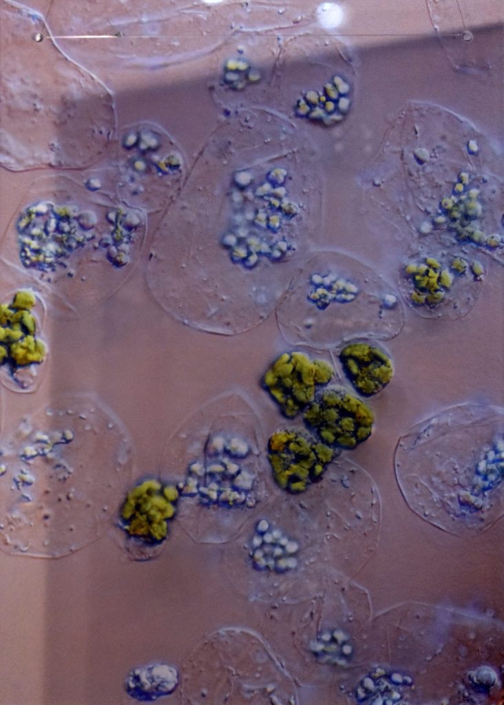
In their latest study, published in the journal Scientific Reports, the researchers sent both ensilicated and regular samples of the tetanus vaccine from Bath to Newcastle by ordinary post (a journey time of over 300 miles, which by post takes a day or two). When doses of the ensilicated vaccine were subsequently injected into mice, an immune response was triggered, showing the vaccine to be active. No immune response was detected in mice injected with unprotected doses of the vaccine, indicating the medicine had been damaged in transit.

Dr Asel Sartbaeva, who led the project from the University of Bath’s Department of Chemistry, said: “This is really exciting data because it shows us that ensilication preserves not just the structure of the vaccine proteins but also the function – the immunogenicity.”

“This project has focused on tetanus, which is part of the DTP (diphtheria, tetanus and pertussis) vaccine given to young children in three doses. Next, we will be working on developing a thermally-stable vaccine for diphtheria, and then pertussis. Eventually we want to create a silica cage for the whole DTP trivalent vaccine, so that every child in the world can be given DTP without having to rely on cold chain distribution.”

Cold chain distribution requires a vaccine to be refrigerated from the moment of manufacturing to the endpoint destination.

Silica is an inorganic, non-toxic material, and Dr Sartbaeva estimates that ensilicated vaccines could be used for humans within five to 15 years. She hopes the technology to silica-wrap proteins will eventually be adopted to store and transport all childhood vaccines, as well as other protein-based products, such as antibodies and enzymes.

“Ultimately, we want to make important medicines stable so they can be more widely available,” she said. “The aim is to eradicate vaccine-preventable diseases in low income countries by using thermally stable vaccines and cutting out dependence on cold chain.”

Currently, up to 50% of vaccine doses are discarded before use due to exposure to suboptimal temperatures. According to the World Health Organisation (WHO), 19.4 million infants did not receive routine life-saving vaccinations in 2018.

Here’s a link to and a citation for the paper,

Ensilicated tetanus antigen retains immunogenicity: in vivo study and time-resolved SAXS characterization by A. Doekhie, R. Dattani, Y-C. Chen, Y. Yang, A. Smith, A. P. Silve, F. Koumanov, S. A. Wells, K. J. Edler, K. J. Marchbank, J. M. H. van den Elsen & A. Sartbaeva. Scientific Reports volume 10, Article number: 9243 (2020) DOI: https://doi.org/10.1038/s41598-020-65876-3 Published 08 June 2020

This paper is open access

Nanopatch update

I tend to lose track as a science gets closer to commercialization since the science news becomes business news and I almost never scan that sector. It’s been about two-and-half years since I featured research that suggested Nanopatch provided more effective polio vaccination than the standard needle and syringe method in a December 20, 2017 post. The latest bits of news have an interesting timeline.

March 2020

Mark Kendal (Wikipedia entry) is the researcher behind the Nanopatch. He’s interviewed in a March 5, 2020 episode (about 20 mins.) in the Pioneers Series (bankrolled by Rolex [yes, the watch company]) on Monocle.com. Coincidentally or not, a new piece of research funded by Vaxxas (the nanopatch company founded by Mark Kendall; on the website you will find a ‘front’ page and a ‘Contact us’ page only) was announced in a March 17, 2020 news item on medical.net,

Vaxxas, a clinical-stage biotechnology company commercializing a novel vaccination platform, today announced the publication in the journal PLoS Medicine of groundbreaking clinical research indicating the broad immunological and commercial potential of Vaxxas’ novel high-density microarray patch (HD-MAP). Using influenza vaccine, the clinical study of Vaxxas’ HD-MAP demonstrated significantly enhanced immune response compared to vaccination by needle/syringe. This is the largest microarray patch clinical vaccine study ever performed.

“With vaccine coated onto Vaxxas HD-MAPs shown to be stable for up to a year at 40°C [emphasis mine], we can offer a truly differentiated platform with a global reach, particularly into low and middle income countries or in emergency use and pandemic situations,” said Angus Forster, Chief Development and Operations Officer of Vaxxas and lead author of the PLoS Medicine publication. “Vaxxas’ HD-MAP is readily fabricated by injection molding to produce a 10 x 10 mm square with more than 3,000 microprojections that are gamma-irradiated before aseptic dry application of vaccine to the HD-MAP’s tips. All elements of device design, as well as coating and QC, have been engineered to enable small, modular, aseptic lines to make millions of vaccine products per week.”

The PLoS publication reported results and analyses from a clinical study involving 210 clinical subjects [emphasis mine]. The clinical study was a two-part, randomized, partially double-blind, placebo-controlled trial conducted at a single Australian clinical site. The clinical study’s primary objective was to measure the safety and tolerability of A/Singapore/GP1908/2015 H1N1 (A/Sing) monovalent vaccine delivered by Vaxxas HD-MAP in comparison to an uncoated Vaxxas HD-MAP and IM [intramuscular] injection of a quadrivalent seasonal influenza vaccine (QIV) delivering approximately the same dose of A/Sing HA protein. Exploratory outcomes were: to evaluate the immune responses to HD-MAP application to the forearm with A/Sing at 4 dose levels in comparison to IM administration of A/Sing at the standard 15 μg HA per dose per strain, and to assess further measures of immune response through additional assays and assessment of the local skin response via punch biopsy of the HD-MAP application sites. Local skin response, serological, mucosal and cellular immune responses were assessed pre- and post-vaccination.

Here’s a link to and a citation for the latest ‘nanopatch’ paper,

Safety, tolerability, and immunogenicity of influenza vaccination with a high-density microarray patch: Results from a randomized, controlled phase I clinical trial by Angus H. Forster, Katey Witham, Alexandra C. I. Depelsenaire, Margaret Veitch, James W. Wells, Adam Wheatley, Melinda Pryor, Jason D. Lickliter, Barbara Francis, Steve Rockman, Jesse Bodle, Peter Treasure, Julian Hickling, Germain J. P. Fernando. DOI: https://doi.org/10.1371/journal.pmed.1003024 PLOS (Public Library of Science) Published: March 17, 2020

This is an open access paper.

May 2020

Two months later, Merck, an American multinational pharmaceutical company, showed some serious interest in the ‘nanopatch’. A May 28, 2020 article by Chris Newmarker for drugdelvierybusiness.com announces the news (Note: Links have been removed),

Merck has exercised its option to use Vaxxas‘ High Density Microarray Patch (HD-MAP) platform as a delivery platform for a vaccine candidate, the companies announced today [Thursday, May 28, 2020].

Also today, Vaxxas announced that German manufacturing equipment maker Harro Höfliger will help Vaxxas develop a high-throughput, aseptic manufacturing line to make vaccine products based on Vaxxas’ HD-MAP technology. Initial efforts will focus on having a pilot line operating in 2021 to support late-stage clinical studies — with a goal of single, aseptic-based lines being able to churn out 5 million vaccine products a week.

“A major challenge in commercializing microarray patches — like Vaxxas’ HD-MAP — for vaccination is the ability to manufacture at industrially-relevant scale, while meeting stringent sterility and quality standards. Our novel device design along with our innovative vaccine coating and quality verification technologies are an excellent fit for integration with Harro Höfliger’s aseptic process automation platforms. Adopting a modular approach, it will be possible to achieve output of tens-of-millions of vaccine-HD-MAP products per week,” Hoey [David L. Hoey, President and CEO of Vaxxas] said.

Vaxxas also claims that the patches can deliver vaccine more efficiently — a positive when people around the world are clamoring for a vaccine against COVID-19. The company points to a recent [March 17, 2020] clinical study in which their micropatch delivering a sixth of an influenza vaccine dose produced an immune response comparable to a full dose by intramuscular injection. A two-thirds dose by HD-MAP generated significantly faster and higher overall antibody responses.

As I noted earlier, this is an interesting timeline.

Final comment

In the end, what all of this means is that there may be more than one way to deal with vaccines and medicines that deteriorate all too quickly unless refrigerated. I wish all of these researchers the best.