Tag Archives: art conservation

Sniffing for art conservation

The American Chemical Society (ACS) has produced a video titled, “How that ‘old book smell’ could save priceless artifacts” according to their Sept. 6, 2016 news release on EurekAlert,

Odor-detecting devices like Breathalyzers have been used for years to determine blood-alcohol levels in drunk drivers. Now, researchers are using a similar method to sniff out the rate of decay in historic art and artifacts. By tracking the chemicals in “old book smell” and similar odors, conservators can react quickly to preserve priceless art and artifacts at the first signs of decay. In this Speaking of Chemistry, Sarah Everts explains how cultural-heritage science uses the chemistry of odors to save books, vintage jewelry and even early Legos. …

Here’s the video,

Heritage Smells, the UK project mentioned in the video, is now completed but it was hosted by the University of Strathclyde and more project information can be found here.

When based on plastic materials, contemporary art can degrade quickly

There’s an intriguing April 1, 2016 article by Josh Fischman for Scientific American about a problem with artworks from the 20th century and later—plastic-based materials (Note: A link has been removed),

Conservators at museums and art galleries have a big worry. They believe there is a good chance the art they showcase now will not be fit to be seen in one hundred years, according to researchers in a project  called Nanorestart. Why? After 1940, artists began using plastic-based material that was a far cry from the oil-based paints used by classical painters. Plastic is also far more fragile, it turns out. Its chemical bonds readily break. And they cannot be restored using techniques historically relied upon by conservators.

So art conservation scientists have turned to nanotechnology for help.

Sadly, there isn’t any detail in Fischman’s article (*ETA June 17, 2016 article [for Fast Company] by Charlie Sorrel, which features some good pictures, a succinct summary of Fischman’s article and a literary reference [Kurt Vonnegut’s Bluebeard]I*) about how nanotechnology is playing or might play a role in this conservation effort. Further investigation into the two projects (NanoRestART and POPART) mentioned by Fischman didn’t provide much more detail about NanoRestART’s science aspect but POPART does provide some details.

NanoRestART

It’s probably too soon (this project isn’t even a year-old) to be getting much in the way of the nanoscience details but NanoRestART has big plans according to its website homepage,

The conservation of this diverse cultural heritage requires advanced solutions at the cutting edge of modern chemistry and material science in an entirely new scientific framework that will be developed within NANORESTART project.

The NANORESTART project will focus on the synthesis of novel poly-functional nanomaterials and on the development of highly innovative restoration techniques to address the conservation of a wide variety of materials mainly used by modern and contemporary artists.

In NANORESTART, enterprises and academic centers of excellence in the field of synthesis and characterization of nano- and advanced materials have joined forces with complementary conservation institutions and freelance restorers. This multidisciplinary approach will cover the development of different materials in response to real conservation needs, the testing of such materials, the assessment of their environmental impact, and their industrial scalability.

NanoRestART’s (NANOmaterials for the REStoration of works of ART) project page spells out their goals in the order in which they are being approached,

The ground-breaking nature of our research can be more easily outlined by focussing on specific issues. The main conservation challenges that will be addressed in the project are:


Conservation challenge 1Cleaning of contemporary painted and plastic surfaces (CC1)

Conservation challenge 2Stabilization of canvases and painted layers in contemporary art (CC2)

Conservation challenge 3Removal of unwanted modern materials (CC3)

Conservation challenge 4Enhanced protection of artworks in museums and outdoors (CC4)

The European Commission provides more information about the project on its CORDIS website’s NanoRestART webpage including the start and end dates for the project and the consortium members,

From 2015-06-01 to 2018-12-01, ongoing project

CHALMERS TEKNISKA HOEGSKOLA AB
Sweden
MIRABILE ANTONIO
France
NATIONALMUSEET
Denmark
CONSIGLIO NAZIONALE DELLE RICERCHE
Italy
UNIVERSITY COLLEGE CORK, NATIONAL UNIVERSITY OF IRELAND, CORK
Ireland
MBN NANOMATERIALIA SPA
Italy
KEMIJSKI INSTITUT
Slovenia
CHEVALIER AURELIA
France
UNIVERSIDADE FEDERAL DO RIO GRANDE DO SUL
Brazil
UNIVERSITA CA’ FOSCARI VENEZIA
Italy
AKZO NOBEL PULP AND PERFORMANCE CHEMICALS AB
Sweden
COMMISSARIAT A L ENERGIE ATOMIQUE ET AUX ENERGIES ALTERNATIVES
France
ARKEMA FRANCE SA
France
UNIVERSIDAD DE SANTIAGO DE COMPOSTELA
Spain
UNIVERSITY COLLEGE LONDON
United Kingdom
ZFB ZENTRUM FUR BUCHERHALTUNG GMBH
Germany
UNIVERSITAT DE BARCELONA
Spain
THE BOARD OF TRUSTEES OF THE TATE GALLERY
United Kingdom
ASSOCIAZIONE ITALIANA PER LA RICERCA INDUSTRIALE – AIRI
Italy
THE ART INSTITUTE OF CHICAGO
United States
MINISTERIO DE EDUCACION, CULTURA Y DEPORTE
Spain
STICHTING HET RIJKSMUSEUM
Netherlands
UNIVERSITEIT VAN AMSTERDAM
Netherlands
UNIVERSIDADE FEDERAL DO RIO DE JANEIRO
Brazil
ACCADEMIA DI BELLE ARTI DI BRERA
Italy

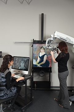
It was a bit surprising to see Brazil and the US as participants but The Art Institute of Chicago has done nanotechnology-enabled conservation in the past as per my March 24, 2014 posting about a Renoir painting. I’m not familiar with the Brazilian organization.

POPART

POPART (Preservation of Plastic Artefacts in museum collections) mentioned by Fischman was a European Commission project which ran from 2008 – 2012. Reports can be found on the CORDIS Popart webpage. The final report has some interesting bits (Note: I have added subheads in the [] square brackets),

To achieve a valid comparison of the various invasive and non-invasive techniques proposed for the identification and characterisation of plastics, a sample collection (SamCo) of plastics artefacts of about 100 standard and reference plastic objects was gathered. SamCo was made up of two kinds of reference materials: standards and objects. Each standard represents the reference material of a ‘pure’ plastic; while each object represents the reference of the same plastic as in the standards, but compounded with pigments, dyestuffs, fillers, anti oxidants, plasticizers etc.  Three partners ICN [Instituut Collectie Nederland], V&A [Victoria and Albert Museum] and Natmus [National Museet] collected different natural and synthetic plastics from the ICN reference collections of plastic objects, from flea markets, antique shops and from private collections and from their own collection to contribute to SamCo, the sample collection for identification by POPART partners. …

As a successive step, the collections of the following museums were surveyed:

-Victoria & Albert Museum (V&A), London, U.K.
-Stedelijk Museum, Amsterdam, The Netherlands
-Musée d’Art Moderne et d’Art Contemporaine (MAMAC) Nice, France
-Musée d’Art moderne, St. Etienne, France
-Musée Galliera, Paris, France

At the V&A approximately 200 objects were surveyed. Good or fair conservation conditions were found for about 85% of the objects, whereas the remaining 15% was in poor or even in unacceptable (3%) conditions. In particular, crazing and delamination of polyurethane faux leather and surface stickiness and darkening of plasticized PVC were observed. The situation at the Stedelijk Museum in Amsterdam was particularly favourable because a previous survey had been done in 1995 so that it was possible to make a comparison with the Popart survey in 2010. A total number of 40 objects, which comprised plastics early dating from the 1930’s until the newer plastics from the 1980’s, were considered and their actual conservation state compared with the 1995 records. Of the objects surveyed in 2010, it can be concluded that 21 remained in the same condition. 13 objects containing PA, PUR, PVC, PP or natural rubber changed due to chemical and physical degradation while works of art containing either PMMA or PS changed due to mechanical damages and incorrect artist’s technique (inappropriate adhesive) into a lesser condition. 6 works of art (containing either PA or PMMA or both) changed into a better condition due to restoration or replacements.  More than 230 objects have been examined in the 3 museums in France. A particular effort was devoted to the identification of the constituting plastics materials. Surveys have been undertaken without any sophisticated equipment, in order to work in museums everyday conditions. Plastics hidden by other materials or by paint layers were not or hardly accessible, it is why the final count of some plastics may be under estimated in the final results. Another outcome is that plastic identification has been made at a general level only, by trying to identify the polymer family each plastic belongs to. Lastly, evidence of chemical degradation processes that do not cause visible or perceptible damage have not been detected and could not be taken in account in the final results.

… The most damaged artefacts resulted constituted by cellulose acetate, cellulose nitrate and PVC.

[Polly (the doll)]

One of the main issues that is of interest for conservators and curators is to assess which kinds of plastics are most vulnerable to deterioration and to what extent they can deteriorate under the environmental conditions normally encountered in museums. Although one might expect that real time deterioration could be ascertained by a careful investigation of museum objects on display or in storage, real objects or artworks may not sampled due to ethical considerations. Therefore, reference objects were prepared by Natmus in the form of a doll (Polly) for simultaneous exposures in different environmental conditions. The doll comprised of 11 different plastics representative of types typically found in modern museum collections. The 16 identical dolls realized were exposed in different places, not only in normal exhibit conditions, but also in some selected extreme conditions to ascertain possible acceleration of the deterioration process. In most cases the environmental parameters were also measured. The dolls were periodically evaluated by visual inspection and in selected cases by instrumental analyses. 

In conclusion the experimental campaign carried out with Polly dolls can be viewed as a pilot study aimed at tackling the practical issues related to the monitoring of real three dimensional plastic artworks and the surrounding environment.

The overall exposure period (one year and half) was sufficient to observe initial changes in the more susceptible polymers, such as polyurethane ethers and esters, and polyamide, with detectable chromatic changes and surface effects. Conversely the other polymers were shown to be stable in the same conditions over this time period.

[Polly as an awareness raising tool]

Last but not least, the educational and communication benefits of an object like Polly facilitated the dissemination of the Popart Project to the public, and increased the awareness of issues associated with plastics in museum collections.

[Cleaning issues]

Mechanical cleaning has long been perceived as the least damaging technique to remove soiling from plastics. The results obtained from POPART suggest that the risks of introducing scratches or residues by mechanical cleaning are measurable. Some plastics were clearly more sensitive to mechanical damage than others. From the model plastics evaluated, HIPS was the most sensitive followed by HDPE, PVC, PMMA and CA. Scratches could not be measured on XPS due to its inhomogeneous surfaces. Plasticised PVC scratched easily, but appeared to repair itself because plasticiser migrated to surfaces and filled scratches.

Photo micrographs revealed that although all 22 cleaning materials evaluated in POPART scratched test plastics, some scratches were sufficiently shallow to be invisible to the naked eye. Duzzit and Scotch Brite sponges as well as all paper based products caused more scratching of surfaces than brushes and cloths. Some cleaning materials, notably Akapad yellow and white sponges, compressed air, latex and synthetic rubber sponges and goat hair brushes left residues on surfaces. These residues were only visible on glass-clear, transparent test plastics such as PMMA. HDPE and HIPS surfaces both had matte and roughened appearances after cleaning with dry-ice. XPS was completely destroyed by the treatment. No visible changes were present on PMMA and PVC.

Of the cleaning methods evaluated, only canned air, natural and synthetic feather duster left surfaces unchanged. Natural and synthetic feather duster, microfiber-, spectacle – and cotton cloths, cotton bud, sable hair brush and leather chamois showed good results when applied to clean model plastics.

Most mechanical cleaning materials induced static electricity after cleaning, causing immediate attraction of dust. It was also noticed that generally when adding an aqueous cleaning agent to a cleaning material, the area scratched was reduced. This implied that cleaning agents also functioned as lubricants. A similar effect was exhibited by white spirit and isopropanol.
Based on cleaning vectors, Judith Hofenk de Graaff detergent, distilled water and Dehypon LS45 were the least damaging cleaning agents for all model plastics evaluated. None of the aqueous cleaning agents caused visible changes when used in combination with the least damaging cleaning materials. Sable hair brush, synthetic feather duster and yellow Akapad sponge were unsuitable for applying aqueous cleaning agents. Polyvinyl acetate sponge swelled in contact with solvents and was only suitable for aqueous cleaning processes.

Based on cleaning vectors, white spirit was the least damaging solvent. Acetone and Surfynol 61 were the most damaging for all model plastics and cannot be recommended for cleaning plastics. Surfynol 61 dissolved polyvinyl acetate sponge and left a milky residue on surfaces, which was particularly apparent on clear PMMA surfaces. Surfynol 61 left residues on surfaces on evaporating and acetone evaporated too rapidly to lubricate cleaning materials thereby increasing scratching of surfaces.

Supercritical carbon dioxide induced discolouration and mechanical damage to the model plastics, particularly to XPS, CA and PMMA and should not be used for conservation cleaning of plastics.

Potential Impact:
Cultural heritage is recognised as an economical factor, the cost of decay of cultural heritage and the risk associated to some material in collection may be high. It is generally estimated that plastics, developed at great numbers since the 20th century’s interbellum, will not survive that long. This means that fewer generations will have access to lasting plastic art for study, contemplation and enjoyment. On the other hand will it normally be easier to reveal a contemporary object’s technological secrets because of better documentation and easier access to artists’ working methods, ideas and intentions. A first more or less world encompassing recognition of the problems involved with museum objects made wholly or in part of plastics was through the conference ‘Saving the twentieth century” held in Ottawa, Canada in 1991. This was followed later by ‘Modern Art, who cares’ in Amsterdam, The Netherlands in 1997, ‘Mortality Immortality? The Legacy of Modern Art’ in Los Angeles, USA in 1998 and, for example much more recent, ‘Plastics –Looking at the future and learning from the Past’ in London, UK in 2007. A growing professional interest in the care of plastics was clearly reflected in the creation of an ICOM-CC working group dedicated to modern materials in 1996, its name change to Modern Materials and Contemporary Art in 2002, and its growing membership from 60 at inception to over 200 at the 16th triennial conference in Lisbon, Portugal in 2011 and tentatively to over 300 as one of the aims put forward in the 2011-2014 programme of that ICOM-CC working group. …

[Intellectual property]

Another element pertaining to conservation of modern art is the copyright of artists that extends at least 50 years beyond their death. Both, damage, value and copyright may influence the way by which damage is measured through scientific analysis, more specifically through the application of invasive or non invasive techniques. Any selection of those will not only have an influence on the extent of observable damage, but also on the detail of information gathered and necessary to explain damage and to suggest conservation measures.

[How much is deteriorating?]

… it is obvious from surveys carried out in several museums in France, the UK and The Netherlands that from 15 to 35 % of what I would then call an average plastic material based collection is in a poor to unacceptable condition. However, some 75 % would require cleaning,

I hope to find out more about how nanotechnology is expected to be implemented in the conservation and preservation of plastic-based art. The NanoRestART project started in June 2015 and hopefully more information will be disseminated in the next year or so.

While it’s not directly related, there was some work with conservation of daguerreotypes (19th century photographic technique) and nanotechnology mentioned in my Nov. 17, 2015 posting which was a followup to my Jan. 10, 2015 posting about the project and the crisis precipitating it.

*ETA June 30, 2016: Here’s clip from a BBC programme, Science in Action broadcast on June 30, 2016 featuring a chat with some of the scientists involved in the NanoRestArt project (Note: This excerpt is from a longer programme and seemingly starts in the middle of a conversation,)

Protecting Disney’s art with an artificial nose

Curators and conservators are acutely aware of how fragile artworks (see my Jan. 10, 2013 posting about a show where curators watched helplessly as daguerreotypes deteriorated) can be so this new technology from Disney is likely to excite a lot of interest. From a March 14, 2016 news item on phys.org,

Original drawings and sketches from Walt Disney Animation Studio’s more than 90-year history—from Steamboat Willie through Frozen—traveled internationally for the first time this summer. This gave conservators the rare opportunity to monitor the artwork with a new state-of-the-art sensor. A team of researchers report today that they developed and used a super-sensitive artificial “nose,” customized specifically to detect pollutants before they could irreversibly damage the artwork.

Here’s a sample of the art work,

Caption: To protect works of art, including this image of Disney's Steamboat Willie, scientists developed an optoelectronic "nose" to sniff out potentially damaging compounds in pollution. Credit: Steamboat Willie, 1928 Animation cel and background © Disney Enterprises, Inc. Courtesy of Walt Disney Animation Research Library

Caption: To protect works of art, including this image of Disney’s Steamboat Willie, scientists developed an optoelectronic “nose” to sniff out potentially damaging compounds in pollution. Credit: Steamboat Willie, 1928 Animation cel and background © Disney Enterprises, Inc. Courtesy of Walt Disney Animation Research Library

A March 14, 2016 American Chemical Society (ACS) news release (also on EurekAlert), provides more detail,

The researchers report on their preservation efforts at the 251st National Meeting & Exposition of the American Chemical Society (ACS). ACS, the world’s largest scientific society, is holding the meeting here through Thursday. It features more than 12,500 presentations on a wide range of science topics.

“Many pollutants that are problematic for human beings are also problematic for works of art,” says Kenneth Suslick, Ph.D. For example, pollutants can spur oxidative damage and acid degradation that, in prints or canvases, lead to color changes or decomposition. “The ability to monitor how much pollution a drawing or painting is exposed to is an important element of art preservation,” he says.

However, works of art are susceptible to damage at far lower pollutant levels than what’s considered acceptable for humans. “The high sensitivity of artists’ materials makes a lot of sense for two reasons,” explains Suslick, who is at the University of Illinois at Urbana-Champaign. “Human beings are capable of healing, which, of course, works of art cannot do. Moreover, human beings have finite lifetimes, whereas ideally works of art should last for future generations.”

To protect valuable works of art from these effects, conservators enclose vulnerable pieces in sealed display cases. But even then, some artists’ materials may “exhale” reactive compounds that accumulate in the cases and damage the art. To counter the accumulation of pollutants, conservators often hide sorbent materials inside display cases that scrub potentially damaging compounds from the enclosed environment. But it is difficult to know precisely when to replace the sorbents.

Suslick, a self-proclaimed “museum hound,” figured he might have an answer. He had already invented an optoelectronic nose — an array of dyes that change color when exposed to various compounds. But it is used largely for biomedical purposes, and it can’t sniff out the low concentrations of pollutants that damage works of art. To redesign the nose with the aim of protecting artwork, he approached scientists at the Getty Conservation Institute (GCI), a private non-profit institution in Los Angeles that works internationally to advance art conservation practice. He proposed that his team devise a sensor several hundred times more sensitive than existing devices used for cultural heritage research. The collaboration took off, and the scientists built a keener nose.

At the time, GCI was involved in a research project with the Walt Disney Animation Research Library to investigate the impact of storage environment on their animation cels, which are transparent sheets that artists drew or painted on before computer animation was developed. Such research ultimately could help extend the life of this important collection. The new sensors would monitor levels of acetic acid and other compounds that emanate from these sheets.

Before the exhibit, “Drawn from Life: The Art of Disney Animation Studios,” hit the road on tour, Suslick recommended placing the sensors in discrete places to monitor the pollution levels both inside and outside of the sealed and framed artworks. If the sensors indicated pollution levels inside the sealed frames were rising, conservators traveling with the Disney exhibit would know to replace the sorbents. An initial analysis of sensor data showed that the sorbents were effective. Suslick says he expects to continue expanding the sensors’ applications in the field of cultural heritage.

Collaborators in the project include Maria LaGasse, a graduate student in Suslick’s lab; Kristen McCormick, art exhibitions and conservation manager at the Walt Disney Animation Research Library; Herant Khanjian, assistant scientist; and Michael Schilling, senior scientist at the Getty Conservation Institute.

I was able to find one museum exhibiting “Drawn from Life: The Art of Disney Animation Studios”; it was the Museum of China which hosted the show from June 30 – August 18, 2015. There are pictures of the exhibit at the Museum of China posted by Leon Ingram here on Behance.

NANOART Research Tool offers affordable paint analysis

There’s some encouraging news for art conservators and authenticators, an affordable nanotech-based kit for greater accuracy analyzing ancient (or old)  paint is one step closer according to a Jan. 11, 2016 notice on CORDIS,

Developed through the EU-funded NANOART project, the new testing kit has already been applied to identify binders such as collagen and ovalbumin in ancient paint, not only in model samples painted in the lab but also in real samples collected from works of art.

‘Once fully completed, our new tool will be made available to conservation scientists from around the world at an affordable cost (an assay can cost around EUR 0.5 per target), which will facilitate greater knowledge about historical works of art and help international museums, restoration art studios and laboratories to plan the best conservation and preventive strategies,’ explains NANOART project coordinator Dr Jesus de la Fuente from the CSIC/University of Zaragoza, Spain.

In addition, the sensitiveness of the project’s new nanotechnology-based methods means that smaller samples are required to be taken from the artwork for analysis. This in itself will help to better preserve our cultural heritage.

In order to characterise ancient paints, experts have often relied on conventional molecular biology methodologies that were developed decades ago. The concept behind the NANOART project was that these techniques could be substituted by more sensitive, inexpensive and faster techniques that take advantage of emerging nanotechnologies.

Furthermore, conventional methods – apart from being expensive – are also only available at a few laboratories, and require specialised personnel and equipment. A key objective of the NANOART project has been to address the cost issue by applying techniques developed for clinical diagnosis. In this way, the project is also highly original as it aims to take latest developments in clinical medicine and apply them to the conservation and preservation of cultural heritage.

‘The innovative nature of the project is also denoted by the fact that there is currently no method or kit available that can be easily used at point-of-care to analyse paints without requiring expensive equipment and extensive training,’ says Ana Claro, research fellow from the INA/University of Zaragoza. ‘With the NANOART kit, the final user will be able to conduct an affordable analysis (in some cases at the cost of only a few euros) by simply following the instructions. Within a four-hour period, the results will be available.’

The potential opportunities opened up by the new analytical nanotechnology are huge. For example, developed in parallel with the NANOART kit, a spin-off company called NanoImmunotech has been launched in order to develop devices to detect bacterial infection in meat using the same technology as used in NANOART.

‘This opens our technology to other applications far from cultural heritage applications,’ says de la Fuente. ‘However, we would like to continue further developing novel uses of NANOART technology for other applications in cultural heritage, and our next step will be to look for funding to develop an even more user friendly device.’

This announcement comes just as the NANOART project is scheduled to be completed (Jan. 31, 2016) according to its webpage on CORDIS.

For those with Spanish language skills, there’s this Jan. 11, 2016 news item on the Catalunya Vanguardista website (I believe the English language version above is a machine translation with this being the original text),

Nanotecnología para analizar pinturas históricas de forma barata y precisa

Empleando nanotecnologías, se ha creado un equipo de diagnóstico clínico destinado a analizar capas de pintura antiguas que podría ahorrar costes a los profesionales de la conservación y permitirles alcanzar mayor precisión.

Cordis / El nuevo equipo de ensayo, desarrollado mediante el proyecto financiado con fondos europeos NANOART, ya se ha empleado en la identificación de aglutinantes como el colágeno y la ovoalbúmina en pinturas históricas. Además, los resultados se han obtenido tanto con muestras pintadas en el laboratorio como con otras extraídas de obras de arte.

«Una vez completemos su desarrollo, nuestra herramienta quedará a disposición de científicos de todo el mundo dedicados a la conservación por un módico precio (cada ensayo costará cerca de medio euro por objetivo). De este modo se obtendrá un conocimiento más profundo sobre las obras de arte históricas y tanto museos como talleres de restauración y laboratorios podrán plantear las estrategias de conservación y prevención idóneas», explicó el coordinador del proyecto, el Dr. Jesús de la Fuente del Instituto de Ciencia de los Materiales —centro mixto dependiente del CSIC y la Universidad de Zaragoza (España)—.Además, la sensibilidad ofrecida por los métodos nanotecnológicos propuestos por el proyecto permite extraer muestras de menor tamaño de las obras de arte, lo cual contribuirá a conservar mejor el patrimonio cultural.Para caracterizar pinturas antiguas, hasta ahora los expertos solían emplear metodologías convencionales de la biología molecular desarrolladas hace decenios. La propuesta del proyecto NANOART pasa por sustituir estas técnicas por otras más sensibles, baratas y rápidas que se valen de las nanotecnologías emergentes.

Es más, los métodos convencionales, además de resultar caros, sólo están a disposición de unos pocos laboratorios que cuentan con equipos y personal especializados. NANOART se propuso sobre todo abaratar los costes mediante el empleo de técnicas de diagnóstico del ámbito clínico. La originalidad de este planteamiento es notoria, pues aprovecha los últimos progresos logrados en medicina clínica para aplicarlos a la conservación y la protección del patrimonio cultural.

«La naturaleza innovadora del proyecto también obedece a la carencia hoy en día de un método o equipo que pueda emplearse con facilidad in situ para analizar pinturas sin necesidad de equipos caros ni formación exhaustiva», afirmó Ana Claro, investigadora del INA de la Universidad de Zaragoza. «Gracias al equipo de NANOART, el usuario final podrá ejecutar ensayos asequibles, en algunos casos por valor de tan sólo unos pocos euros, siguiendo las instrucciones proporcionadas. Los resultados estarán disponibles en cuatro horas».

Las oportunidades que ofrece la nueva nanotecnología analítica son enormes. Por ejemplo, la empresa derivada NanoImmunotech se ha puesto en marcha en paralelo al desarrollo del equipo de NANOART para que cree servicios con los que detectar infecciones bacterianas en la carne mediante los mismos métodos empleados por el proyecto en el ámbito del arte.

«De esta forma se amplían las aplicaciones de la tecnología a otros campos muy alejados del patrimonio cultural», afirmó de la Fuente. «No obstante, seguiremos indagando en nuevos usos de la tecnología de NANOART relacionados con el patrimonio cultural y procederemos ya a buscar fuentes de financiación que nos permitan crear un dispositivo aún más fácil de usar».

I expect the folks at the Canadian Conservation Institute (CCI) and other such insitutions are keeping a close eye on developments of this nature. The institute was mentioned here in the context of a series I wrote on attempts to authenticate a painting, Autumn Harbour, as a Lawren Harris (one of Canada’s Group of Seven painters). My July 14, 2014 post was devoted to a response from Marie-Claude Corbeil to a query about scientific investigation of visual art,

… [the response],

The Canadian Conservation Institute (CCI) has been conducting research into the materials and techniques of Canadian artists (mainly 20th-century artists) since the early 1990s. Databases were created for each artists. At the moment CCI has no such database on Harris.

The CCI is the only institution in Canada carrying out this kind of research. I would add that European conservation institutes or laboratories have a long tradition of conducting this type of research focusing mainly on European art, basically because many were created long before North-American conservation institutes or laboratories were established.

I was quite fascinated by the whole thing and wrote a four-part series about Autumn Harbour, Lawren Harris, and much more, as well as, the July 14, 2014 post, which has links to the Autumn Harbour series along with the response from the CCI and links to articles recommended by Corbeil.

2015 daguerreotype exhibit follows problematic 2005 show

In 2005, curators had a horrifying experience when historical images (daguerreotypes) were deteriorating as the 150-year old images were being displayed in an exhibit titled “Young America.” Some 25 of the photographs were affected, five of them sustaining critical damage. The debacle occasioned a research project involving conservators, physicists, and nanotechnology (see my Jan. 10, 2013 posting for more about the 2005 exhibit and resulting research project).

A new daguerreotype exhibit currently taking place showcases the results of that research according to a Nov. 13, 2015 University of Rochester news release,

In 1839, Louis-Jacques-Mandé Daguerre unveiled one of the world’s first successful photographic mediums: the daguerreotype. The process transformed the human experience by providing a means to capture light and record people, places, and events. The University of Rochester is leading groundbreaking nanotechnology research that explores the extraordinary qualities of this photographic process. A new exhibition in Rush Rhees Library showcases the results of this research, while bridging the gap between the sciences and the humanities. …

… From 2010-2014, a National Science Foundation grant supported nanotechnology research conducted by two University of Rochester scientists—Nicholas Bigelow, Lee A. DuBridge Professor of Physics, and Ralph Wiegandt, visiting research scientist and conservator—who explored how environment impacts the survival of these unique, non-reproducible images. In addition to conservation science and cultural research, Bigelow and Wiegandt are also investigating ways in which the chemical and physical processes used to create daguerreotypes can influence modern nanofabrication and nanotechnology.

“The daguerreotype should be considered one of humankind’s most disruptive technological advances,” Bigelow and Wiegandt said. “Not only was it the first successful imaging medium, it was also the first truly engineered nanotechnology. The daguerreotype was a prescient catalyst to the ensuing cascade of discoveries in physics and chemistry over the latter half of the 19th century and into the 20th.”

Blending the past with the future, the exhibition displays the first known daguerreotype of a Rochester graduating class (1853) alongside a 2015 daguerreotype of current University President Joel Seligman, created by Rochester daguerreotypist Irving Pobboravsky.

Both Bigelow and Wiegandt are mentioned in the 2013 posting describing the research project’s inception.

For anyone who’s in the area of New York state where the University of Rochester is located, the exhibit will run until February 29, 2016 in the Friedlander Lobby of Rush Rhees Library.  Plus, there’s this from the news release,

A special presentation about the scientific advances surrounding the daguerreotype and their relationship to cultural preservation will be led by Bigelow, Wiegandt, and Jim Kuhn, assistant dean for Special Collections and Preservation, on December 14 from 7-9 p.m. in the Hawkins-Carlson Room of Rush Rhees Library. For more information visit: http://www.library.rochester.edu/event/daguerreotype-exhibition or call (585).

There’s no indication that the special presentation will be livestreamed or recorded and made available at a later date.

Nanotechnology and drones for London’s (UK) Old Royal Naval College (ORNC)

It’s an art conservation project where nanotechnology and drones will be employed to help preserve the Old Royal Naval College’s (ORNC) Painted Hall. From an April 12, 2015 news item on Nanotechnology Now,

Plans for a major conservation project to restore the Painted Hall at the Old Royal Naval College (ORNC) in Greenwich, UK, will be announced in the US at an event on 14th May 2015 hosted by the British Consulate General in New York.

The ORNC, Sir Christopher Wren’s twin-domed riverside masterpiece stands on the site of the Greenwich Palace, Henry VIII’s birthplace and favorite royal residence. It is one of the most important ensembles in European baroque architecture.

Following a £2.77 million pledge in November 2014 from the Heritage Lottery Fund, the ORNC are embarking on the second stage of its plans to restore the Painted Hall to its former glory. A further £4 million is required to achieve the full scale of this landmark project.

Cutting-edge technologies are being applied for this conservation project, including drones and nanotechnology-enabled materials.

About the Old Royal Naval College

The Old Royal Naval College (ORNC) in Greenwich was established as the Royal Hospital for Seamen by King William III and Queen Mary II in 1694.

Designed by Sir Christopher Wren, it is one of the most important ensembles in European baroque architecture. From 1705, the Royal Hospital provided modest, wood-lined cabins as accommodation for retired sailors, housing as many as 2,700 residents at its peak in 1814. The last naval pensioners left in 1869, when the site became home to the Royal Naval College, an officers’ training academy, until 1997. When the Navy left, an independent charity was established to conserve the site for present and future generations, and create enjoyment, learning and unique cultural experiences for everyone.

Today this historic landmark is open to the public and is the home of three unique and free to visit attractions; the Painted Hall, the Chapel, and the Discover Greenwich visitor centre.

The Painted Hall is the greatest piece of decorative painting in England and has been described as ‘the Sistine Chapel of the UK’. The walls and ceilings were painted by Sir James Thornhill between 1708 and 1727.

The Chapel of St Peter and St Paul is a neo-classical masterpiece by James ‘Athenian’ Stuart and William Newton. Featuring a Samuel Green organ and an altarpiece painted by Benjamin West, it is one of the finest eighteenth century interiors in existence.

Here’s the Painted Hall and Chapel,


 Gryffindor derivative work: Fpo (talk) - Royal_Naval_College_Greenwich_001.JPG Royal_Naval_College_Greenwich_002.JPG  Creative Commons Attribution-Share Alike 3.0

Gryffindor derivative work: Fpo (talk) – Royal_Naval_College_Greenwich_001.JPG Royal_Naval_College_Greenwich_002.JPG Creative Commons Attribution-Share Alike 3.0

You can find out more about the ORNC here.

Masterpieces seen in a new light

Caption: This image shows: After Raphael 1483 - 1520 probably before 1600 Oil on wood 87 x 61.3 cm Wynn Ellis Bequest, 1876 Credit: © National Gallery, London

Caption: This image shows: After Raphael 1483 – 1520
probably before 1600
Oil on wood
87 x 61.3 cm
Wynn Ellis Bequest, 1876
Credit: © National Gallery, London

An April 13, 2015 Optical Society news release (also on EurekAlert) describes a new technique for ‘seeing’ below the surface of a painting without taking samples,

A painting hanging on the wall in an art gallery tells one story. What lies beneath its surface may tell quite another.

Often in a Rembrandt, a Vermeer, a Leonardo, a Van Eyck, or any other great masterpiece of western art, the layers of paint are covered with varnish, sometimes several coats applied at different times over their history. The varnish was originally applied to protect the paint underneath and make the colors appear more vivid, but over the centuries it can degrade. Conservators carefully clean off the old varnish and replace it with new, but to do this safely it is useful to understand the materials and structure of the painting beneath the surface. Conservation scientists can glean this information by analyzing the hidden layers of paint and varnish.

Now, researchers from Nottingham Trent University’s School of Science and Technology have partnered with the National Gallery in London to develop an instrument capable of non-invasively capturing subsurface details from artwork at a high resolution. Their setup, published in an Optics Express paper, will allow conservators and conservation scientists to more effectively peek beneath the surface of paintings and artifacts to learn not only how the artist built up the original composition, but also what coatings have been applied to it over the years.

Traditionally, analyzing the layers of a painting requires taking a very small physical sample — usually around a quarter of a millimeter across — to view under a microscope. The technique provides a cross-section of the painting’s layers, which can be imaged at high resolution and analyzed to gain detailed information on the chemical composition of the paint, but does involve removing some original paint, even if only a very tiny amount. When studying valuable masterpieces, conservation scientists must therefore sample very selectively from already-damaged areas, often only taking a few minute samples from a large canvas.

More recently, researchers have begun to use non-invasive imaging techniques to study paintings and other historical artifacts. For example, Optical Coherence Tomography (OCT) was originally developed for medical imaging but has also been applied to art conservation. Because it uses a beam of light to scan the intact painting without removing physical samples, OCT allows researchers to analyze the painting more extensively. However, the spatial resolution of commercially-available OCT setups is not high enough to fully map the fine layers of paint and varnish.

The Nottingham Trent University researchers gave OCT an upgrade. “We’re trying to see how far we can go with non-invasive techniques. We wanted to reach the kind of resolution that conventional destructive techniques have reached,” explained Haida Liang, who led the project.

In OCT, a beam of light is split: half is directed towards the sample, and the other half is sent to a reference mirror. The light scatters off both of these surfaces. By measuring the combined signal, which effectively compares the returned light from the sample versus the reference, the apparatus can determine how far into the sample the light penetrated. By repeating this procedure many times across an area, researchers can build up a cross-sectional map of the painting.

Liang and her colleagues used a broadband laser-like light source — a concentrated beam of light containing a wide range of frequencies. The wider frequency range allows for more precise data collection, but such light sources were not commercially available until recently.

Along with a few other modifications, the addition of the broadband light source enabled the apparatus to scan the painting at a higher resolution. When tested on a late 16th-century copy of a Raphael painting, housed at the National Gallery in London, it performed as well as traditional invasive imaging techniques.

“We are able to not only match the resolution but also to see some of the layer structures with better contrast. That’s because OCT is particularly sensitive to changes in refractive index,” said Liang. In some places, the ultra-high resolution OCT setup identified varnish layers that were almost indistinguishable from each other under the microscope.

Eventually, the researchers plan to make their instrument available to other art institutions. It could also be useful for analyzing historical manuscripts, which cannot be physically sampled in the same way that paintings can.

In a parallel paper recently published in Optics Express, the researchers also improved the depth into the painting that their apparatus can scan. The two goals are somewhat at odds: using a longer wavelength light source could enhance the penetration depth, but shorter wavelength light (as used in their current setup) provides the best resolution.

“The next challenge is perhaps to be able to do that in one instrument, as well as to extract chemical information from different layers,” said Liang.

Here are links to and citations for the two recent papers published by Liang and her team,

Ultra-high resolution Fourier domain optical coherence tomography for old master paintings by C. S. Cheung, M. Spring, and H. Liang. Optics Express, Vol. 23, Issue 8, pp. 10145-10157 (2015) http://dx.doi.org/10.1364/OE.23.010145

High resolution Fourier domain optical coherence tomography in the 2 μm wavelength range using a broadband supercontinuum source by C. S. Cheung, J. M. O. Daniel, M. Tokurakawa, W. A. Clarkson, and H. Liang. Optics Express, Vol. 23, Issue 3, pp. 1992-2001 (2015) http://dx.doi.org/10.1364/OE.23.001992

Both papers are open access.

Lawren Harris (Group of Seven), art authentication, and the Canadian Conservation Institute* (addendum to four-part series)

I recently wrote an exhaustive four-part series (links at the end of this posting) featuring Raman spectroscopy testing of an authenticated (Hurdy Gurdy) and a purported (Autumn Harbour) Lawren Harris paintings. During the course of my research, I sent a query to the Canadian Conservation Institute to disprove or confirm my statements regarding Canada and its database of art pigments,

.. According to some informal sources, Canada has a very small (almost nonexistent) data bank of information about pigments used in its important paintings. For example, the federal government’s Canadian Conservation Institute has a very small database of pigments and nothing from Lawren Harris paintings [unconfirmed at time of publication; June 18, 2014 query outstanding] …

Marie-Claude Corbeil, Ph.D. Gestionnaire de la Division de la science de la conservation | Manager of Conservation Science Division, very kindly replied to my query with this on July 10, 2014 (I believe she was on holidays [en vacances] when my query was received in June),

The Canadian Conservation Institute (CCI) has been conducting research into the materials and techniques of Canadian artists (mainly 20th-century artists) since the early 1990s. Databases were created for each artists. At the moment CCI has no such database on Harris.

The CCI is the only institution in Canada carrying out this kind of research. I would add that European conservation institutes or laboratories have a long tradition of conducting this type of research focusing mainly on European art, basically because many were created long before North-American conservation institutes or laboratories were established.

… An important point to make is that scientific investigation is only one part of an authentication study. Authentication should start with stylistic study and research into the provenance of the artwork which are carried out by curators and art historians.

Regarding your question about Raman spectroscopy, I would say that Raman spectroscopy is only one of many techniques that can be used to analyse paint or any other material. At CCI we often use up to six techniques to analyse paint to obtain the full makeup of the sample including pigments, fillers and binding media. I should also add that analysis of material is carried out at CCI to answer questions related to a number of issues, including but not limited to authentication. Analysis is often carried out to understand the degradation of museum objects and works of art, or to provide information required during the course of a conservation treatment.

Thank you for this excellent explanation and for your time.

Art (Lawren Harris and the Group of Seven), science (Raman spectroscopic examinations), and other collisions at the 2014 Canadian Chemistry Conference

Part 1

Part 2

Part 3

Part 4

ETA July 14, 2014 at 1305 PDT: For those who want more information, Ms. Corbeil has provided some articles about the CCI and its Canadian Artists Painting Materials Research Project:

The Canadian Artists’ Painting Materials Project, 1992, J. M. Taylor. (PDF)

CCI 1992 Taylor

Detecting Art Fraud: Sometimes Scientific Examination Can Help, 1993. J. M. Taylor (PDF)

CCI 1993 Taylor
The Canadian Artists Painting Materials Research Project, 1995, Marie-Claude Corbeil (PDF)

CCI 1995 Corbeil

*’Istitute’ changed to ‘Institute’ on Jan. 14, 2016.

Italians and Polish collaborate on nanoscale study of vanishing Da Vinci self-portrait

In addition to a new nondamaging technique to examine paintings (my June 2, 2014 post: Damage-free art authentication and spatially offset Raman spectroscopy [SORS]), there’s a new report in a June 3, 2014 news item on ScienceDaily about a nondamaging technique to examine paper such as the paper on which holds a Da Vinci self-portrait,

One of Leonardo da Vinci’s masterpieces, drawn in red chalk on paper during the early 1500s and widely believed to be a self-portrait, is in extremely poor condition. Centuries of exposure to humid storage conditions or a closed environment has led to widespread and localized yellowing and browning of the paper, which is reducing the contrast between the colors of chalk and paper and substantially diminishing the visibility of the drawing.

A group of researchers from Italy and Poland with expertise in paper degradation mechanisms was tasked with determining whether the degradation process has now slowed with appropriate conservation conditions — or if the aging process is continuing at an unacceptable rate.

Caption: This is Leonardo da Vinci's self-portrait as acquired during diagnostic studies carried out at the Central Institute for the Restoration of Archival and Library Heritage in Rome, Italy. Credit: M. C. Misiti/Central Institute for the Restoration of Archival and Library Heritage, Rome

Caption: This is Leonardo da Vinci’s self-portrait as acquired during diagnostic studies carried out at the Central Institute for the Restoration of Archival and Library Heritage in Rome, Italy.
Credit: M. C. Misiti/Central Institute for the Restoration of Archival and Library Heritage, Rome

The June 3, 2014 American Institute of Physics news release on EurekAlert provides more detail about the work,

… the team developed an approach to nondestructively identify and quantify the concentration of light-absorbing molecules known as chromophores in ancient paper, the culprit behind the “yellowing” of the cellulose within ancient documents and works of art.

“During the centuries, the combined actions of light, heat, moisture, metallic and acidic impurities, and pollutant gases modify the white color of ancient paper’s main component: cellulose,” explained Joanna Łojewska, a professor in the Department of Chemistry at Jagiellonian University in Krakow, Poland. “This phenomenon is known as ‘yellowing,’ which causes severe damage and negatively affects the aesthetic enjoyment of ancient art works on paper.”

Chromophores are the key to understanding the visual degradation process because they are among the chemical products developed by oxidation during aging and are, ultimately, behind the “yellowing” within cellulose. Yellowing occurs when “chromophores within cellulose absorb the violet and blue range of visible light and largely scatter the yellow and red portions — resulting in the characteristic yellow-brown hue,” said Olivia Pulci, a professor in the Physics Department at the University of Rome Tor Vergata.

To determine the degradation rate of Leonardo’s self-portrait, the team created a nondestructive approach that centers on identifying and quantifying the concentration of chromophores within paper. It involves using a reflectance spectroscopy setup to obtain optical reflectance spectra of paper samples in the near-infrared, visible, and near-ultraviolet wavelength ranges.

Once reflectance data is gathered, the optical absorption spectrum of cellulose fibers that form the sheet of paper can be calculated using special spectroscopic data analysis.

Then, computational simulations based on quantum mechanics — in particular, Time-Dependent Density Functional Theory, which plays a key role in studying optical properties in theoretical condensed matter physics — are tapped to calculate the optical absorption spectrum of chromophores in cellulose.

“Using our approach, we were able to evaluate the state of degradation of Leonardo da Vinci’s self-portrait and other paper specimens from ancient books dating from the 15th century,” said Adriano Mosca Conte, a researcher at the University of Rome Tor Vergata. “By comparing the results of ancient papers with those of artificially aged samples, we gained significant insights into the environmental conditions in which Leonardo da Vinci’s self-portrait was stored during its lifetime.”

Their work revealed that the type of chromophores present in Leonardo’s self portrait are “similar to those found in ancient and modern paper samples aged in extremely humid conditions or within a closed environment, which agrees with its documented history,” said Mauro Missori, a researcher at the Institute for Complex Systems, CNR, in Rome, Italy.

One of the most significant implications of their work is that the state of degradation of ancient paper can be measured and quantified by evaluation of the concentrations of chromophores in cellulose fibers. “The periodic repetition of our approach is fundamental to establishing the formation rate of chromophores within the self-portrait. Now our approach can serve as a precious tool to preserve and save not only this invaluable work of art, but others as well,” Conte noted.

Absolutely fascinating stuff to those of use who care about yellowing paper. (Having worked in an archives, I care deeply.) Here’s a link to and a citation for the study,

Visual degradation in Leonardo da Vinci’s iconic self-portrait: A nanoscale study by A. Mosca Conte, O. Pulci, M. C. Misiti, J. Lojewska, L. Teodonio1, C. Violante, and M. Missori. Appl. Phys. Lett. 104, 224101 (2014); http://dx.doi.org/10.1063/1.4879838

This is an open access study.

Richard Van Duyne solves mystery of Renoir’s red with surface-enhanced Raman spectroscopy (SERS) and Canadian scientists uncover forgeries

The only things these two items have in common is that they are concerned with visual art. and with solving mysteries The first item concerns research by Richard Van Duyne into the nature of the red paint used in one of Renoir’s paintings. A February 14, 2014 news item on Azonano describes some of the art conservation work that Van Duyne’s (nanoish) technology has made possible along with details about this most recent work,

Scientists are using powerful analytical and imaging tools to study artworks from all ages, delving deep below the surface to reveal the process and materials used by some of the world’s greatest artists.

Northwestern University chemist Richard P. Van Duyne, in collaboration with conservation scientists at the Art Institute of Chicago, has been using a scientific method he discovered nearly four decades ago to investigate masterpieces by Pierre-Auguste Renoir, Winslow Homer and Mary Cassatt.

Van Duyne recently identified the chemical components of paint, now partially faded, used by Renoir in his oil painting “Madame Léon Clapisson.” Van Duyne discovered the artist used carmine lake, a brilliant but light-sensitive red pigment, on this colorful canvas. The scientific investigation is the cornerstone of a new exhibition at the Art Institute of Chicago.

The Art Institute of Chicago’s exhibition is called, Renoir’s True Colors: Science Solves a Mystery. being held from Feb. 12, 2014 – April 27, 2014. Here is an image of the Renoir painting in question and an image featuring the equipment being used,

Renoir-Madame-Leon-Clapisson.Art Institute of Chicago.

Renoir-Madame-Leon-Clapisson.Art Institute of Chicago.

Renoir and surface-enhanced Raman spectroscopy (SERS). Art Institute of Chicago

Renoir and surface-enhanced Raman spectroscopy (SERS). Art Institute of Chicago

The Feb. 13, 2014 Northwestern University news release (also on EurekAlert) by Megan Fellman, which originated the news item, gives a brief description of Van Duyne’s technique and its impact on conservation at the Art Institute of Chicago (Note: A link has been removed),

To see what the naked eye cannot see, Van Duyne used surface-enhanced Raman spectroscopy (SERS) to uncover details of Renoir’s paint. SERS, discovered by Van Duyne in 1977, is widely recognized as the most sensitive form of spectroscopy capable of identifying molecules.

Van Duyne and his colleagues’ detective work informed the production of a new digital visualization of the painting’s original colors by the Art Institute’s conservation department. The re-colorized reproduction and the original painting (presented in a case that offers 360-degree views) can be viewed side by side at the exhibition “Renoir’s True Colors: Science Solves a Mystery” through April 27 [2014] at the Art Institute.

I first wrote about Van Duyne’s technique in my wiki, The NanoTech Mysteries. From the Scientists get artful page (Note: A footnote was removed),

Richard Van Duyne, then a chemist at Northwestern University, developed the technique in 1977. Van Duyne’s technology, based on Raman spectroscopy which has been around since the 1920s, is called surface-enhanced Raman spectroscopy’ or SERS “[and] uses laser light and nanoparticles of precious metals to interact with molecules to show the chemical make-up of a particular dye.”

This next item is about forgery detection. A March 5, 2014 news release on EurekAlert describes the latest developments,

Gallery owners, private collectors, conservators, museums and art dealers face many problems in protecting and evaluating their collections such as determining origin, authenticity and discovery of forgery, as well as conservation issues. Today these problems are more accurately addressed through the application of modern, non-destructive, “hi-tech” techniques.

Dmitry Gavrilov, a PhD student in the Department of Physics at the University of Windsor (Windsor, Canada), along with Dr. Roman Gr. Maev, the Department of Physics Professor at the University of Windsor (Windsor, Canada) and Professor Dr. Darryl Almond of the University of Bath (Bath, UK) have been busy applying modern techniques to this age-old field. Infrared imaging, thermography, spectroscopy, UV fluorescence analysis, and acoustic microscopy are among the innovative approaches they are using to conduct pre-restoration analysis of works of art. Some fascinating results from their applications are published today in the Canadian Journal of Physics.

Since the early 1900s, using infrared imaging in various wave bands, scientists have been able to see what parts of artworks have been retouched or altered and sometimes even reveal the artist’s original sketches beneath layers of the paint. Thermography is a relatively new approach in art analysis that allows for deep subsurface investigation to find defects and past reparations. To a conservator these new methods are key in saving priceless works from further damage.

Gavrilov explains, “We applied new approaches in processing thermographic data, materials spectra data, and also the technique referred to as craquelure pattern analysis. The latter is based on advanced morphological processing of images of surface cracks. These cracks, caused by a number of factors such as structure of canvas, paints and binders used, can uncover important clues on the origins of a painting.”

“Air-coupled acoustic imaging and acoustic microscopy are other innovative approaches which have been developed and introduced into art analysis by our team under supervision of Dr. Roman Gr. Maev. The technique has proven to be extremely sensitive to small layer detachments and allows for the detection of early stages of degradation. It is based on the same principles as medical and industrial ultrasound, namely, the sending a sound wave to the sample and receiving it back. ”

Spectroscopy is a technique that has been useful in the fight against art fraud. It can determine chemical composition of pigments and binders, which is essential information in the hands of an art specialist in revealing fakes. As described in the paper, “…according to the FBI, the value of art fraud, forgery and theft is up to $6 billion per year, which makes it the third most lucrative crime in the world after drug trafficking and the illegal weapons trade.”

One might wonder how these modern applications can be safe for delicate works of art when even flash photography is banned in art galleries. The authors discuss this and other safety concerns, describing both historic and modern-day implications of flash bulbs and exhibit illumination and scientific methods. As the paper concludes, the authors suggest that we can expect that the number of “hi-tech” techniques will only increase. In the future, art experts will likely have a variety of tools to help them solve many of the mysteries hiding beneath the layers.

Here’s a link to and a citation for the paper,

A review of imaging methods in analysis of works of art: Thermographic imaging method in art analysis by D. Gavrilov, R.Gr. Maev, and D.P. Almond. Canadian Journal of Physics, 10.1139/cjp-2013-0128

This paper is open access.