DNA, the stuff of life, may very well also pack quite the jolt for engineers trying to advance the development of tiny, low-cost electronic devices.
Much like flipping your light switch at home — only on a scale 1,000 times smaller than a human hair — an ASU [Arizona State University]-led team has now developed the first controllable DNA switch to regulate the flow of electricity within a single, atomic-sized molecule. The new study, led by ASU Biodesign Institute researcher Nongjian Tao, was published in the advanced online journal Nature Communications.
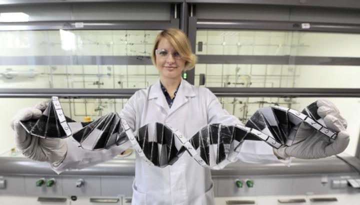
DNA, the stuff of life, may very well also pack quite the jolt for engineers trying to advance the development of tiny, low-cost electronic devices. Courtesy: ASU
“It has been established that charge transport is possible in DNA, but for a useful device, one wants to be able to turn the charge transport on and off. We achieved this goal by chemically modifying DNA,” said Tao, who directs the Biodesign Center for Bioelectronics and Biosensors and is a professor in the Fulton Schools of Engineering. “Not only that, but we can also adapt the modified DNA as a probe to measure reactions at the single-molecule level. This provides a unique way for studying important reactions implicated in disease, or photosynthesis reactions for novel renewable energy applications.”
Engineers often think of electricity like water, and the research team’s new DNA switch acts to control the flow of electrons on and off, just like water coming out of a faucet.
Previously, Tao’s research group had made several discoveries to understand and manipulate DNA to more finely tune the flow of electricity through it. They found they could make DNA behave in different ways — and could cajole electrons to flow like waves according to quantum mechanics, or “hop” like rabbits in the way electricity in a copper wire works —creating an exciting new avenue for DNA-based, nano-electronic applications.
Tao assembled a multidisciplinary team for the project, including ASU postdoctoral student Limin Xiang and Li Yueqi performing bench experiments, Julio Palma working on the theoretical framework, with further help and oversight from collaborators Vladimiro Mujica (ASU) and Mark Ratner (Northwestern University).
To accomplish their engineering feat, Tao’s group, modified just one of DNA’s iconic double helix chemical letters, abbreviated as A, C, T or G, with another chemical group, called anthraquinone (Aq). Anthraquinone is a three-ringed carbon structure that can be inserted in between DNA base pairs but contains what chemists call a redox group (short for reduction, or gaining electrons or oxidation, losing electrons).
These chemical groups are also the foundation for how our bodies’ convert chemical energy through switches that send all of the electrical pulses in our brains, our hearts and communicate signals within every cell that may be implicated in the most prevalent diseases.
The modified Aq-DNA helix could now help it perform the switch, slipping comfortably in between the rungs that make up the ladder of the DNA helix, and bestowing it with a new found ability to reversibly gain or lose electrons.
Through their studies, when they sandwiched the DNA between a pair of electrodes, they careful [sic] controlled their electrical field and measured the ability of the modified DNA to conduct electricity. This was performed using a staple of nano-electronics, a scanning tunneling microscope, which acts like the tip of an electrode to complete a connection, being repeatedly pulled in and out of contact with the DNA molecules in the solution like a finger touching a water droplet.
“We found the electron transport mechanism in the present anthraquinone-DNA system favors electron “hopping” via anthraquinone and stacked DNA bases,” said Tao. In addition, they found they could reversibly control the conductance states to make the DNA switch on (high-conductance) or switch-off (low conductance). When anthraquinone has gained the most electrons (its most-reduced state), it is far more conductive, and the team finely mapped out a 3-D picture to account for how anthraquinone controlled the electrical state of the DNA.
For their next project, they hope to extend their studies to get one step closer toward making DNA nano-devices a reality.
“We are particularly excited that the engineered DNA provides a nice tool to examine redox reaction kinetics, and thermodynamics the single molecule level,” said Tao.
Gate-controlled conductance switching in DNA by Limin Xiang, Julio L. Palma, Yueqi Li, Vladimiro Mujica, Mark A. Ratner, & Nongjian Tao. Nature Communications 8, Article number: 14471 (2017) doi:10.1038/ncomms14471 Published online: 20 February 2017
Caption: A polymer negative of a sequence of the genetic code, chemically active and able to bind complementary nucleobases, has been created by researchers from the Institute of Physical Chemistry of the Polish Academy of Sciences in Warsaw. Credit: IPC PAS, Grzegorz Krzyzewski
Those are very large hands! In any event, I think they left out the word ‘model’ when describing what the researcher is holding.
A Jan. 19, 2017 news item on phys.org announces the research from the Institute of Physical Chemistry of the Polish Academy of Sciences (IPC PAS),
In a carefully designed polymer, researchers at the Polish Academy of Sciences have imprinted a sequence of a single strand of DNA. The resulting negative remained chemically active and was capable of binding the appropriate nucleobases of a genetic code. The polymer matrix—the first of its type—thus functioned exactly like a sequence of real DNA.
A Jan. 18, 2017 IPC PAS press release, which originated the news item, provides more detail about the breakthrough and explains how it could lead to synthetic genetics,
Imprinting of chemical molecules in a polymer, or molecular imprinting, is a well-known method that has been under development for many years. However, no-one has ever before used it to construct a polymer chain complementing a sequence of a single strand of DNA. This feat has just been accomplished by researchers from the Institute of Physical Chemistry of the Polish Academy of Sciences (IPC PAS) in Warsaw in collaboration with the University of North Texas (UNT) in Denton, USA, and the University of Milan in Italy. In an appropriately selected polymer, they reproduced a genetically important DNA sequence, constructed of six nucleobases.
Typically, molecular imprinting is accomplished in several steps. The molecules intended for imprinting are first placed to a solution of monomers (i.e. the basic “building blocks” from which the future polymer is to be formed). The monomers are selected so as to automatically arrange themselves around the molecules being imprinted. Next, the resulting complex is electrochemically polymerized and then the imprinted molecules are extracted from the fixed structure. This process results in a polymer structure with molecular cavities matching the original molecules with their size and shape, and even their local chemical properties.
“Using molecular imprinting, we can produce, e.g. recognition films for chemical sensors, capturing molecules of only a specific chemical compound from the surroundings – since only these molecules fit into the existing molecular cavities. However, there’s no rose without a thorn. Molecular imprinting is perfect for smaller chemical molecules, but the larger the molecule, the more difficult it is to imprint it accurately into the polymer,” explains Prof. Wlodzimierz Kutner (IPC PAS).
Molecules of deoxyribonucleic acid, or DNA, are really large: their lengths are of the order of centimetres. These molecules generally consist of of two long strands, paired up with each other. A single strand is made up of nucleotides with multiple repetitions, each of which contains one of the nucleobases: adenine (A), guanine (G), cytosine (C), or thymine (T). The bases on both strands are not arranged freely: adenine on one strand always corresponds to thymine on the other, and guanine to cytosine. So, when we have one thread, we can always recreate its complement, which is the second strand.
The complementarity of nucleobases in DNA strands is very important for cells. Not only does it increase the permanence of the record of the genetic code (damage in one strand can be repaired based on the construction of the other), but it also makes it possible to transfer it from DNA to RNA in the process known as transcription. Transcription is the first step in the synthesis of proteins.
“Our idea was to try to imprint in the polymer a sequence of a single-stranded DNA. At the same time, we wanted to reproduce not only the shape of the strand, but also the sequential order of the constituent nucleobases,” says Dr. Agnieszka Pietrzyk-Le (IPC PAS).
In the study, financed on the Polish side by grants from the Foundation for Polish Science and the National Centre for Science, researchers from the IPC PAS used sequences of the genetic code known as TATAAA. This sequence plays an important biological role: it participates in deciding on the activation of the gene behind it. TATAAA is found in most eukaryotic cells (those containing a nucleus); in humans it is present in about every fourth gene.
A key step of the research was to design synthetic monomers undergoing electrochemical polymerization. These had to be capable of accurately surrounding the imprinted molecule in such a way that each of the adenines and thymines on the DNA strand were accompanied by their complementary bases. The mechanical requirements were also important, because after polymerization the matrix had to be stable. Suitable monomers were synthesized by the group of Prof. Francis D’Souza (UNT).
“When all the reagents and apparatus have been prepared, the imprinting itself of the TATAAA oligonucleotide is not especially complicated. The most important processes take place automatically in solutions in no more than a few dozen minutes. Finally, on the electrode used for electropolymerization, we obtain a layer of conductive polymer with molecular cavities where the nucleobases are arranged in the TTTATA sequence, that is, complementary to the extracted original”, describes doctoral student Katarzyna Bartold (IPC PAS).
Do polymer matrices prepared in this manner really reconstruct the original sequence of the DNA chain? To answer this question, at the IPC PAS careful measurements were carried out on the properties of the new polymers and a series of experiments was performed that confirmed the interaction of the polymers with various nucleobases in solutions. The results leave no doubt: the polymer DNA negative really is chemically active and selectively binds the TATAAA oligonucleotide, correctly reproducing the sequence of nucleobases.
The possibility of the relatively simple and low-cost production of stable polymer equivalents of DNA sequences is an important step in the development of synthetic genetics, especially in terms of its widespread applications in biotechnology and molecular medicine. If an improvement in the method developed at the IPC PAS is accomplished in the future, it will be possible to reproduce longer sequences of the genetic code in polymer matrices. This opens up inspiring perspectives associated not only with learning about the details of the process of transcription in cells or the construction of chemosensors for applications in nanotechnologies operating on chains of DNA, but also with the permanent archiving and replicating of the genetic code of different organisms.
In the quest for smaller and smaller, DNA (deoxyribonucleic acid) is being exploited as never before. From a Nov. 9, 2016 news item on phys.org,
Tinier than the AIDS virus—that is currently the circumference of the smallest transistors. The industry has shrunk the central elements of their computer chips to fourteen nanometers in the last sixty years. Conventional methods, however, are hitting physical boundaries. Researchers around the world are looking for alternatives. One method could be the self-organization of complex components from molecules and atoms. Scientists at the Helmholtz-Zentrum Dresden-Rossendorf (HZDR) and Paderborn University have now made an important advance: the physicists conducted a current through gold-plated nanowires, which independently assembled themselves from single DNA strands. …
At first glance, it resembles wormy lines in front of a black background. But what the electron microscope shows up close is that the nanometer-sized structures connect two electrical contacts. Dr. Artur Erbe from the Institute of Ion Beam Physics and Materials Research is pleased about what he sees. “Our measurements have shown that an electrical current is conducted through these tiny wires.” This is not necessarily self-evident, the physicist stresses. We are, after all, dealing with components made of modified DNA. In order to produce the nanowires, the researchers combined a long single strand of genetic material with shorter DNA segments through the base pairs to form a stable double strand. Using this method, the structures independently take on the desired form.
“With the help of this approach, which resembles the Japanese paper folding technique origami and is therefore referred to as DNA-origami, we can create tiny patterns,” explains the HZDR researcher. “Extremely small circuits made of molecules and atoms are also conceivable here.” This strategy, which scientists call the “bottom-up” method, aims to turn conventional production of electronic components on its head. “The industry has thus far been using what is known as the ‘top-down’ method. Large portions are cut away from the base material until the desired structure is achieved. Soon this will no longer be possible due to continual miniaturization.” The new approach is instead oriented on nature: molecules that develop complex structures through self-assembling processes.
Golden Bridges Between Electrodes
The elements that thereby develop would be substantially smaller than today’s tiniest computer chip components. Smaller circuits could theoretically be produced with less effort. There is, however, a problem: “Genetic matter doesn’t conduct a current particularly well,” points out Erbe. He and his colleagues have therefore placed gold-plated nanoparticles on the DNA wires using chemical bonds. Using a “top-down” method – electron beam lithography — they subsequently make contact with the individual wires electronically. “This connection between the substantially larger electrodes and the individual DNA structures have come up against technical difficulties until now. By combining the two methods, we can resolve this issue. We could thus very precisely determine the charge transport through individual wires for the first time,” adds Erbe.
As the tests of the Dresden researchers have shown, a current is actually conducted through the gold-plated wires — it is, however, dependent on the ambient temperature. “The charge transport is simultaneously reduced as the temperature decreases,” describes Erbe. “At normal room temperature, the wires function well, even if the electrons must partially jump from one gold particle to the next because they haven’t completely melded together. The distance, however, is so small that it currently doesn’t even show up using the most advanced microscopes.” In order to improve the conduction, Artur Erbe’s team aims to incorporate conductive polymers between the gold particles. The physicist believes the metallization process could also still be improved.
He is, however, generally pleased with the results: “We could demonstrate that the gold-plated DNA wires conduct energy. We are actually still in the basic research phase, which is why we are using gold rather than a more cost-efficient metal. We have, nevertheless, made an important stride, which could make electronic devices based on DNA possible in the future.”
This work on a new technique for producing nanofibers comes from Harvard University’s School of Engineering and Applied Sciences and the Wyss Institute for Biologically Inspired Engineering (also at Harvard University). From an Oct. 10, 2016 news item on phys.org,
Fibrous materials—known for their toughness, durability and pliability—are used in everything from bulletproof vests to tires, filtration systems and cellular scaffolds for tissue engineering and regenerative medicine.
The properties of these materials are such that the smaller the fibers are, the stronger and tougher they become. But making certain fibers very small has been an engineering challenge.
Now, researchers from the Harvard John A. Paulson School of Engineering and Applied Sciences (SEAS) and the Wyss Institute for Biologically Inspired Engineering at Harvard have developed a new method to make and collect nanofibers and control their size and morphology. This could lead to stronger, more durable bulletproof vests and armor and more robust cellular scaffolding for tissue repair.
Nanofibers are smaller than one micrometer in diameter. Most nanofiber production platforms rely on dissolving polymers in a solution, which then evaporates as the fiber forms.
Rotary Jet-Spinning (RJS), the technique developed by Kit Parker’s Disease Biophysics Group, works likes a cotton candy machine. Parker is Tarr Family Professor of Bioengineering and Applied Physics at SEAS and a Core Member of the Wyss Institute. A liquid polymer solution is loaded into a reservoir and pushed out through a tiny opening by centrifugal force as the device spins. As the solution leaves the reservoir, the solvent evaporates and the polymers solidify and elongate into small, thin fibers.
“This advance is important because it allows us to manufacture ballistic protection that is much lighter, more flexible and more functional than what is available today,” said Parker, who in addition to his Harvard role is a lieutenant colonel in the United States Army Reserve and was motivated by his own combat experiences in Afghanistan. “Not only could it save lives but for the warfighter, it also could help reduce the repetitive injury motions that soldiers, sailors, marines and airmen have suffered over the last 15 years of the war on terror.”
“Rotary Jet-Spinning is great for most polymer fibers you want to make,” said Grant Gonzalez, a graduate student at SEAS and first author of the paper. “However, some fibers require a solvent that doesn’t evaporate easily. Para-aramid, the polymer used in Kevlar® for example, is dissolved in sulfuric acid, which doesn’t evaporate off. The solution just splashes against the walls of the device without forming fibers.”
Other methods, such as electrospinning, which uses an electric field to pull the polymer into a thin fiber, also have poor results with Kevlar and other polymers such as alginate used for tissue scaffolding and DNA.
The Harvard team overcame these challenges by developing a wet-spinning platform, which uses the same principles as the RJS system but relies on precipitation rather than evaporation to separate the solvent from the polymer.
In this system, called immersion Rotary Jet-Spinning (iRJS), when the polymer solution shoots out of the reservoir, it first passes through an area of open air, where the polymers elongate and the chains align. Then the solution hits a liquid bath that removes the solvent and precipitates the polymers to form solid fibers. Since the bath is also spinning — like water in a salad spinner — the nanofibers follow the stream of the vortex and wrap around a rotating collector at the base of the device.
Using this system, the team produced Nylon, DNA, alginate and ballistic resistant para-aramid nanofibers. The team could tune the fiber’s diameter by changing the solution concentration, the rotational speed and the distance the polymer traveled from the reservoir to the bath.
“By being able to modulate fiber strength, we can create a cellular scaffold that can mimic skeleton muscle and native tissues,” said Gonzalez. “This platform could enable us to create a wound dressing out of alginate material or seed and mature cells on scaffolding for tissue engineering.”
Because the fibers were collected by a spinning vortex, the system also produced well-aligned sheets of nanofibers, which is important for scaffolding and ballistic resistant materials.
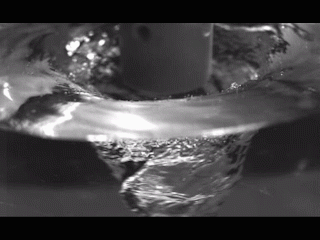
This is the ‘candy floss’ technique at work,
Rotary Jet-Spinning (RJS) works likes a cotton candy machine. A liquid polymer solution is loaded into a reservoir and pushed out through a tiny opening by centrifugal force as the device spins. As the solution leaves the reservoir, the solvent evaporates and the polymers solidify and elongate into small, thin fibers. Courtesy: Harvard University
Basically, scientists at Duke University (US) have created an analog computer at the nanoscale, which can perform basic arithmetic. From an Aug. 23, 2016 news item on ScienceDaily,
Often described as the blueprint of life, DNA contains the instructions for making every living thing from a human to a house fly.
But in recent decades, some researchers have been putting the letters of the genetic code to a different use: making tiny nanoscale computers.
In a new study, a Duke University team led by professor John Reif created strands of synthetic DNA that, when mixed together in a test tube in the right concentrations, form an analog circuit that can add, subtract and multiply as they form and break bonds.
Rather than voltage, DNA circuits use the concentrations of specific DNA strands as signals.
Other teams have designed DNA-based circuits that can solve problems ranging from calculating square roots to playing tic-tac-toe. But most DNA circuits are digital, where information is encoded as a sequence of zeroes and ones.
Instead, the new Duke device performs calculations in an analog fashion by measuring the varying concentrations of specific DNA molecules directly, without requiring special circuitry to convert them to zeroes and ones first.
…
Unlike the silicon-based circuits used in most modern day electronics, commercial applications of DNA circuits are still a long way off, Reif said.
For one, the test tube calculations are slow. It can take hours to get an answer.
“We can do some limited computing, but we can’t even begin to think of competing with modern-day PCs or other conventional computing devices,” Reif said.
But DNA circuits can be far tinier than those made of silicon. And unlike electronic circuits, DNA circuits work in wet environments, which might make them useful for computing inside the bloodstream or the soupy, cramped quarters of the cell.
The technology takes advantage of DNA’s natural ability to zip and unzip to perform computations. Just like Velcro and magnets have complementary hooks or poles, the nucleotide bases of DNA pair up and bind in a predictable way.
The researchers first create short pieces of synthetic DNA, some single-stranded and some double-stranded with single-stranded ends, and mix them in a test tube.
When a single strand encounters a perfect match at the end of one of the partially double-stranded ones, it latches on and binds, displacing the previously bound strand and causing it to detach, like someone cutting in on a dancing couple.
The newly released strand can in turn pair up with other complementary DNA molecules downstream in the circuit, creating a domino effect.
The researchers solve math problems by measuring the concentrations of specific outgoing strands as the reaction reaches equilibrium.
To see how their circuit would perform over time as the reactions proceeded, Reif and Duke graduate student Tianqi Song used computer software to simulate the reactions over a range of input concentrations. They have also been testing the circuit experimentally in the lab.
Besides addition, subtraction and multiplication, the researchers are also designing more sophisticated analog DNA circuits that can do a wider range of calculations, such as logarithms and exponentials.
Conventional computers went digital decades ago. But for DNA computing, the analog approach has its advantages, the researchers say. For one, analog DNA circuits require fewer strands of DNA than digital ones, Song said.
Analog circuits are also better suited for sensing signals that don’t lend themselves to simple on-off, all-or-none values, such as vital signs and other physiological measurements involved in diagnosing and treating disease.
The hope is that, in the distant future, such devices could be programmed to sense whether particular blood chemicals lie inside or outside the range of values considered normal, and release a specific DNA or RNA — DNA’s chemical cousin — that has a drug-like effect.
Reif’s lab is also beginning to work on DNA-based devices that could detect molecular signatures of particular types of cancer cells, and release substances that spur the immune system to fight back.
“Even very simple DNA computing could still have huge impacts in medicine or science,” Reif said.
Here’s a link to and a citation for the paper,
Analog Computation by DNA Strand Displacement Circuits by Tianqi Song, Sudhanshu Garg, Reem Mokhtar, Hieu Bui, and John Reif. ACS Synth. Biol., 2016, 5 (8), pp 898–912 DOI: 10.1021/acssynbio.6b00144 Publication Date (Web): July 01, 2016
An Aug. 15, 2016 news item on ScienceDaily announces research into graphene nanoribbons and their DNA (deoxyribonucleic acid)-like properties,
Graphene nanoribbons (GNRs) bend and twist easily in solution, making them adaptable for biological uses like DNA analysis, drug delivery and biomimetic applications, according to scientists at Rice University.
Knowing the details of how GNRs behave in a solution will help make them suitable for wide use in biomimetics, according to Rice physicist Ching-Hwa Kiang, whose lab employed its unique capabilities to probe nanoscale materials like cells and proteins in wet environments. Biomimetic materials are those that imitate the forms and properties of natural materials.
Graphene nanoribbons can be thousands of times longer than they are wide. They can be produced in bulk by chemically “unzipping” carbon nanotubes, a process invented by Rice chemist and co-author James Tour and his lab.
Their size means they can operate on the scale of biological components like proteins and DNA, Kiang said. “We study the mechanical properties of all different kinds of materials, from proteins to cells, but a little different from the way other people do,” she said. “We like to see how materials behave in solution, because that’s where biological things are.” Kiang is a pioneer in developing methods to probe the energy states of proteins as they fold and unfold.
She said Tour suggested her lab have a look at the mechanical properties of GNRs. “It’s a little extra work to study these things in solution rather than dry, but that’s our specialty,” she said.
Nanoribbons are known for adding strength but not weight to solid-state composites, like bicycle frames and tennis rackets, and forming an electrically active matrix. A recent Rice project infused them into an efficient de-icer coating for aircraft.
But in a squishier environment, their ability to conform to surfaces, carry current and strengthen composites could also be valuable.
“It turns out that graphene behaves reasonably well, somewhat similar to other biological materials. But the interesting part is that it behaves differently in a solution than it does in air,” she said. The researchers found that like DNA and proteins, nanoribbons in solution naturally form folds and loops, but can also form helicoids, wrinkles and spirals.
Kiang, Wijeratne [Sithara Wijeratne, Rice graduate now a postdoctoral researcher at Harvard University] and Jingqiang Li, a co-author and student in the Kiang lab, used atomic force microscopy to test their properties. Atomic force microscopy can not only gather high-resolution images but also take sensitive force measurements of nanomaterials by pulling on them. The researchers probed GNRs and their precursors, graphene oxide nanoribbons.
The researchers discovered that all nanoribbons become rigid under stress, but their rigidity increases as oxide molecules are removed to turn graphene oxide nanoribbons into GNRs. They suggested this ability to tune their rigidity should help with the design and fabrication of GNR-biomimetic interfaces.
“Graphene and graphene oxide materials can be functionalized (or modified) to integrate with various biological systems, such as DNA, protein and even cells,” Kiang said. “These have been realized in biological devices, biomolecule detection and molecular medicine. The sensitivity of graphene bio-devices can be improved by using narrow graphene materials like nanoribbons.”
Wijeratne noted graphene nanoribbons are already being tested for use in DNA sequencing, in which strands of DNA are pulled through a nanopore in an electrified material. The base components of DNA affect the electric field, which can be read to identify the bases.
The researchers saw nanoribbons’ biocompatibility as potentially useful for sensors that could travel through the body and report on what they find, not unlike the Tour lab’s nanoreporters that retrieve information from oil wells.
Further studies will focus on the effect of the nanoribbons’ width, which range from 10 to 100 nanometers, on their properties.
Here’s a link to and a citation for the paper,
Detecting the Biopolymer Behavior of Graphene Nanoribbons in Aqueous Solution by Sithara S. Wijeratne, Evgeni S. Penev, Wei Lu, Jingqiang Li, Amanda L. Duque, Boris I. Yakobson, James M. Tour, & Ching-Hwa Kiang. Scientific Reports 6, Article number: 31174 (2016) doi:10.1038/srep31174 Published online: 09 August 2016
In the last few years, there’s been a veritable plethora of movies (and television shows in Canada and the US) that are about science and technology or have a significant component or investigate the social impact. The trend does not seem to be slowing.
This first of two parts features the film, *Hidden* Figures, and a play being turned into a film, Photograph 51. The second part features the evolving Theranos story and plans to turn it into a film, The Man Who Knew Infinity, a film about an Indian mathematician, the science of the recent all woman Ghostbusters, and an ezine devoted to science films.
For the following movie tidbits, I have David Bruggeman to thank.
Hidden Figures
From David’s June 21, 2016 post on his Pasco Phronesis blog (Note: A link has been removed),
Hidden Figures is a fictionalized treatment of the book of the same name written by Margot Lee Shetterly (and underwritten by the Sloan Foundation). Neither the book nor the film are released yet. The book is scheduled for a September release, and the film currently has a January release date in the U.S.
Both the film and the book focus on the story of African American women who worked as computers for the government at the Langley National Aeronautic Laboratory in Hampton, Virginia. The women served as human computers, making the calculations NASA needed during the Space Race. While the book features four women, the film is focused on three: Katherine Johnson (recipient of the Presidential Medal of Freedom), Dorothy Vaughan, and Mary Jackson. They are played by, respectively, Taraji P. Henson, Octavia Spencer, and Janelle Monae. Other actors in the film include Kevin Costner, Kirsten Dunst, Aldis Hodge, and Jim Parsons. The film is directed by Theodore Melfi, and the script is by Allison Schroeder.
*ETA Oct. 6, 2016: The book ‘Hidden Figures’ is nonfiction while the movie is a fictionalized adaptation based on a true story.*
According to imdb.com, the movie’s release date is Dec. 25, 2016 (this could change again).
The history for ‘human computers’ stretches back to the 17th century, at least. From the Human Computer entry in Wikipedia (Note: Links have been removed),
The term “computer”, in use from the early 17th century (the first known written reference dates from 1613),[1] meant “one who computes”: a person performing mathematical calculations, before electronic computers became commercially available. “The human computer is supposed to be following fixed rules; he has no authority to deviate from them in any detail.” (Turing, 1950) Teams of people were frequently used to undertake long and often tedious calculations; the work was divided so that this could be done in parallel.
Prior to NASA, a team of women in the 19th century in the US were known as Harvard Computers (from the Wikipedia entry; Note: Links have been removed),
Edward Charles Pickering (director of the Harvard Observatory from 1877 to 1919) decided to hire women as skilled workers to process astronomical data. Among these women were Williamina Fleming, Annie Jump Cannon, Henrietta Swan Leavitt and Antonia Maury. This staff came to be known as “Pickering’s Harem” or, more respectfully, as the Harvard Computers.[1] This was an example of what has been identified as the “harem effect” in the history and sociology of science.
It seems that several factors contributed to Pickering’s decision to hire women instead of men. Among them was the fact that men were paid much more than women, so he could employ more staff with the same budget. This was relevant in a time when the amount of astronomical data was surpassing the capacity of the Observatories to process it.[2]
The first woman hired was Williamina Fleming, who was working as a maid for Pickering. It seems that Pickering was increasingly frustrated with his male assistants and declared that even his maid could do a better job. Apparently he was not mistaken, as Fleming undertook her assigned chores efficiently. When the Harvard Observatory received in 1886 a generous donation from the widow of Henry Draper, Pickering decided to hire more female staff and put Fleming in charge of them.[3]
While it’s not thrilling to find out that Pickering was content to exploit the women he was hiring, he deserves kudos for recognizing that women could do excellent work and acting on that recognition. When you consider the times, Pickering’s was an extraordinary act.
Getting back to Hidden Figures, an Aug.15, 2016 posting by Kathleen for Lainey Gossip celebrates the then newly released trailer for the movie,
If you’ve been watching the Olympics [Rio 2016], you know how much the past 10 days have been an epic display of #BlackGirlMagic. Fittingly, the trailer for Hidden Figures was released last night during Sunday’s Olympic coverage. It’s the story of three brilliant African American women, played by Taraji P Henson, Octavia Spencer and Janelle Monae, who made history by serving as the brains behind the NASA launch of astronaut John Glenn into orbit in 1962.
Three black women helped launch a dude into space in the 60s. AT NASA. Think about how America treated black women in the 60s. As Katherine Johnson, played by Taraji P Henson, jokes in the trailer, they were still sitting at the back of the bus. In 1962 Malcolm X said, “The most disrespected person in America is the Black woman, the most unprotected person in America is the Black woman. The most neglected person in America is the Black woman.” These women had to face that truth every day and they still rose to greatness. I’m obsessed with this story.
Overall, the trailer is good. I like the pace and the performances look strong. …
…
I’m most excited for Hidden Figures (as Lainey pointed out, this title is THE WORST) because black girls are being celebrated for their brains on screen. That is rare. When the trailer aired, my brother Sam texted me, “WHOA, a smart black girl movie!”
*ETA Sept. 5, 2016: Aran Shetterly contacted me to say this:
What you may not know is that the term “Hidden Figures” is a specific reference to flight science. It tested a pilot’s ability to pick out a simple figure from a set of more complex, difficult to see images. http://www.militaryaptitudetests.com/afoqt/
Thank you Mr. Shetterly!
Photograph 51 (the Rosalind Franklin story)
Also in David’s June 21, 2016 post is a mention of Photograph 51, a play and soon-to-be film about Rosalind Franklin, the discovery of the double helix, and a science controversy. I first wrote about Photograph 51 in a Jan. 16, 2012 posting (scroll down about 50% of the way) regarding an international script writing competition being held in Dublin, Ireland. At the time, I noted that Anna Ziegler’s play, Photograph 51 had won a previous competition cycle of the screenwriting competition. I wrote again about the play in a Sept. 2, 2015 posting about its London production (Sept. 5 – Nov. 21, 2015) featuring actress Nicole Kidman.
The versions of the Franklin story with which I’m familiar paint her as the wronged party, ignored and unacknowledged by the scientists (Francis, Crick, James Watson, and Maurice Wilkins) who got all the glory and the Nobel Prize. Stephen Curry in a Sept. 16, 2015 posting on the Guardian science blogs suggests the story may not be quite as simple as that (Note: A link has been removed),
Ziegler [Anna Ziegler, playwright] is up front in admitting that she has rearranged facts to suit the drama. This creates some oddities of chronology and motive for those familiar with the history. I know of no suggestion of romantic interest in Franklin from Wilkins, or of a separation of Crick from his wife in the aftermath of his triumph with Watson in solving the DNA structure. There is no mention in the play of the fact that Franklin published her work (and the famous photograph 51) in the journal Nature alongside Watson and Crick’s paper and one by Wilkins. Nor does the audience hear of the international recognition that Franklin enjoyed in her own right between 1953 and her untimely death in 1958, not just for her involvement in DNA, but also for her work on the structure of coal and of viruses.
Published long after her death, The Double Helix is widely thought to treat Franklin unfairly. In the minds of many she remains the wronged woman whose pioneering results were taken by others to solve DNA and win the Nobel prize. But the real story – many elements of which come across strongly in the play – is more complex*.
…
Franklin is a gifted experimentalist. Her key contributions to the discovery were in improving methods for taking X-ray pictures of and discovering the distinct A and B conformations of DNA. But it becomes clear that her methodical, meticulous approach to data analysis – much to Wilkins’ impotent frustration – eventually allows the Kings ‘team’ to be overtaken by the bolder, intuitive stratagem of Watson and Crick.
…
Curry’s piece is a good read and provides insight into the ways temperament affects how science is practiced.
Interestingly, there was a 1987 dramatization of the ‘double helix or life story’ (from the Life Story entry on Wikipedia; Note: Links have been removed),
The film tells the story of the rivalries of the two teams of scientists attempting to discover the structure of DNA. Francis Crick and James D. Watson at Cambridge University and Maurice Wilkins and Rosalind Franklin at King’s College London.
The film manages to convey the loneliness and competitiveness of scientific research but also educates the viewer as to how the structure of DNA was discovered. In particular, it explores the tension between the patient, dedicated laboratory work of Franklin and the sometimes uninformed intuitive leaps of Watson and Crick, all played against a background of institutional turf wars, personality conflicts and sexism. In the film Watson jokes, plugging the path of intuition: “Blessed are they who believed before there was any evidence.” The film also shows why Watson and Crick made their discovery, overtaking their competitors in part by reasoning from genetic function to predict chemical structure, thus helping to establish the then still-nascent field of molecular biology.
You can find out more about the stars, crew, and cast here on imdb.com
In addition to Life Story, the dramatization is also sometimes titled as ‘The Race for the Double Helix’ or the ‘Double Helix’.
Getting back to Photograph 51 (the film), Michael Grandage who directed the stage play will also direct the film. Grandage just made his debut as a film director with ‘Genius’ starring Colin Firth and Jude Law. According to this June 23, 2016 review by Sarah on Laineygossip.com, he stumbled a bit by casting British and Australian actors as Americans,
The first hurdle to clear with Genius, the feature film debut of English theater director Michael Grandage, is that everyone is played by Brits and Aussies, and by “everyone” I mean some of the most towering figures of American literature. You cast the best actor for the role and a good actor can convince you they’re anyone, so it shouldn’t really matter, but there is something profoundly odd about watching a parade of Lit 101 All Stars appear on screen and struggle with American accents. …
That kind of casting should not be a problem with Photograph 51 where the action takes place with British personalities.
McMaster University (Ontario, Canada) researchers have developed a technique for using DNA (deoxyribonucleic acid) as a sensor according to a July 7, 2016 news item on ScienceDaily,
Researchers at McMaster University have established a way to harness DNA as the engine of a microscopic “machine” they can turn on to detect trace amounts of substances that range from viruses and bacteria to cocaine and metals.
“It’s a completely new platform that can be adapted to many kinds of uses,” says John Brennan, director of McMaster’s Biointerfaces Insitute and co-author of a paper in the journal Nature Communications that describes the technology. “These DNA nano-architectures are adaptable, so that any target should be detectable.”
DNA is best known as a genetic material, but is also a very programmable molecule that lends itself to engineering for synthetic applications.
The new method shapes separately programmed pieces of DNA material into pairs of interlocking circles.
The first remains inactive until it is released by the second, like a bicycle wheel in a lock. When the second circle, acting as the lock, is exposed to even a trace of the target substance, it opens, freeing the first circle of DNA, which replicates quickly and creates a signal, such as a colour change.
“The key is that it’s selectively triggered by whatever we want to detect,” says Brennan, who holds the Canada Research Chair in Bioanalytical Chemistry and Biointerfaces. “We have essentially taken a piece of DNA and forced it to do something it was never designed to do. We can design the lock to be specific to a certain key. All the parts are made of DNA, and ultimately that key is defined by how we build it.”
The idea for the “DNA nanomachine” comes from nature itself, explains co-author Yingfu Li, who holds the Canada Research Chair in Nucleic Acids Research.
“Biology uses all kinds of nanoscale molecular machines to achieve important functions in cells,” Li says. “For the first time, we have designed a DNA-based nano-machine that is capable of achieving ultra-sensitive detection of a bacterial pathogen.”
The DNA-based nanomachine is being further developed into a user-friendly detection kit that will enable rapid testing of a variety of substances, and could move to clinical testing within a year.
A close cousin to DNA (deoxyribonucleic acid), RNA (ribonucleic acid) is a communicator according to a July 4, 2016 news item on ScienceDaily describing how a team at the Massachusetts Institute of Technology (MIT) managed to image RNA more precisely,
Cells contain thousands of messenger RNA molecules, which carry copies of DNA’s genetic instructions to the rest of the cell. MIT engineers have now developed a way to visualize these molecules in higher resolution than previously possible in intact tissues, allowing researchers to precisely map the location of RNA throughout cells.
Key to the new technique is expanding the tissue before imaging it. By making the sample physically larger, it can be imaged with very high resolution using ordinary microscopes commonly found in research labs.
“Now we can image RNA with great spatial precision, thanks to the expansion process, and we also can do it more easily in large intact tissues,” says Ed Boyden, an associate professor of biological engineering and brain and cognitive sciences at MIT, a member of MIT’s Media Lab and McGovern Institute for Brain Research, and the senior author of a paper describing the technique in the July 4, 2016 issue of Nature Methods.
A July 4, 2016 MIT news release (also on EurekAlert), which originated the news item, explains why scientists want a better look at RNA and how the MIT team accomplished the task,
Studying the distribution of RNA inside cells could help scientists learn more about how cells control their gene expression and could also allow them to investigate diseases thought to be caused by failure of RNA to move to the correct location.
Boyden and colleagues first described the underlying technique, known as expansion microscopy (ExM), last year, when they used it to image proteins inside large samples of brain tissue. In a paper appearing in Nature Biotechnology on July 4, the MIT team has now presented a new version of the technology that employs off-the-shelf chemicals, making it easier for researchers to use.
MIT graduate students Fei Chen and Asmamaw Wassie are the lead authors of the Nature Methods paper, and Chen and graduate student Paul Tillberg are the lead authors of the Nature Biotechnology paper.
A simpler process
The original expansion microscopy technique is based on embedding tissue samples in a polymer that swells when water is added. This tissue enlargement allows researchers to obtain images with a resolution of around 70 nanometers, which was previously possible only with very specialized and expensive microscopes. However, that method posed some challenges because it requires generating a complicated chemical tag consisting of an antibody that targets a specific protein, linked to both a fluorescent dye and a chemical anchor that attaches the whole complex to a highly absorbent polymer known as polyacrylate. Once the targets are labeled, the researchers break down the proteins that hold the tissue sample together, allowing it to expand uniformly as the polyacrylate gel swells.
In their new studies, to eliminate the need for custom-designed labels, the researchers used a different molecule to anchor the targets to the gel before digestion. This molecule, which the researchers dubbed AcX, is commercially available and therefore makes the process much simpler.
AcX can be modified to anchor either proteins or RNA to the gel. In the Nature Biotechnology study, the researchers used it to anchor proteins, and they also showed that the technique works on tissue that has been previously labeled with either fluorescent antibodies or proteins such as green fluorescent protein (GFP).
“This lets you use completely off-the-shelf parts, which means that it can integrate very easily into existing workflows,” Tillberg says. “We think that it’s going to lower the barrier significantly for people to use the technique compared to the original ExM.”
Using this approach, it takes about an hour to scan a piece of tissue 500 by 500 by 200 microns, using a light sheet fluorescence microscope. The researchers showed that this technique works for many types of tissues, including brain, pancreas, lung, and spleen.
Imaging RNA
In the Nature Methods paper, the researchers used the same kind of anchoring molecule but modified it to target RNA instead. All of the RNAs in the sample are anchored to the gel, so they stay in their original locations throughout the digestion and expansion process.
After the tissue is expanded, the researchers label specific RNA molecules using a process known as fluorescence in situ hybridization (FISH), which was originally developed in the early 1980s and is widely used. This allows researchers to visualize the location of specific RNA molecules at high resolution, in three dimensions, in large tissue samples.
This enhanced spatial precision could allow scientists to explore many questions about how RNA contributes to cellular function. For example, a longstanding question in neuroscience is how neurons rapidly change the strength of their connections to store new memories or skills. One hypothesis is that RNA molecules encoding proteins necessary for plasticity are stored in cell compartments close to the synapses, poised to be translated into proteins when needed.
With the new system, it should be possible to determine exactly which RNA molecules are located near the synapses, waiting to be translated.
“People have found hundreds of these locally translated RNAs, but it’s hard to know where exactly they are and what they’re doing,” Chen says. “This technique would be useful to study that.”
Boyden’s lab is also interested in using this technology to trace the connections between neurons and to classify different subtypes of neurons based on which genes they are expressing.
There’s a brief (30 secs.), silent video illustrating the work (something about a ‘Brainbow Hippocampus’) made available by MIT,
Here’s a link to and a citation for the paper,
Nanoscale imaging of RNA with expansion microscopy by Fei Chen, Asmamaw T Wassie, Allison J Cote, Anubhav Sinha, Shahar Alon, Shoh Asano, Evan R Daugharthy, Jae-Byum Chang, Adam Marblestone, George M Church, Arjun Raj, & Edward S Boyden. Nature Methods (2016) doi:10.1038/nmeth.3899 Published online 04 July 2016
After publishing a June 15, 2016 post about taking DNA (deoxyribonucleic acid) beyond genetics, it seemed like a good to publish a companion piece featuring a more technical explanation of at least one way DNA might provide the base for living computers and robots. From a June 13, 2016 BrookHaven National Laboratory news release (also on EurekAlert),
A cube, an octahedron, a prism–these are among the polyhedral structures, or frames, made of DNA that scientists at the U.S. Department of Energy’s (DOE) Brookhaven National Laboratory have designed to connect nanoparticles into a variety of precisely structured three-dimensional (3D) lattices. The scientists also developed a method to integrate nanoparticles and DNA frames into interconnecting modules, expanding the diversity of possible structures.
These achievements, described in papers published in Nature Materials and Nature Chemistry, could enable the rational design of nanomaterials with enhanced or combined optical, electric, and magnetic properties to achieve desired functions.
“We are aiming to create self-assembled nanostructures from blueprints,” said physicist Oleg Gang, who led this research at the Center for Functional Nanomaterials (CFN), a DOE Office of Science User Facility at Brookhaven. “The structure of our nanoparticle assemblies is mostly controlled by the shape and binding properties of precisely designed DNA frames, not by the nanoparticles themselves. By enabling us to engineer different lattices and architectures without having to manipulate the particles, our method opens up great opportunities for designing nanomaterials with properties that can be enhanced by precisely organizing functional components. For example, we could create targeted light-absorbing materials that harness solar energy, or magnetic materials that increase information-storage capacity.”
The news release goes on to describe the frames,
Gang’s team has previously exploited DNA’s complementary base pairing–the highly specific binding of bases known by the letters A, T, G, and C that make up the rungs of the DNA double-helix “ladder”–to bring particles together in a precise way. Particles coated with single strands of DNA link to particles coated with complementary strands (A binds with T and G binds with C) while repelling particles coated with non-complementary strands.
They have also designed 3D DNA frames whose corners have single-stranded DNA tethers to which nanoparticles coated with complementary strands can bind. When the scientists mix these nanoparticles and frames, the components self-assemble into lattices that are mainly defined by the shape of the designed frame. The Nature Materials paper describes the most recent structures achieved using this strategy.
“In our approach, we use DNA frames to promote the directional interactions between nanoparticles such that the particles connect into specific configurations that achieve the desired 3D arrays,” said Ye Tian, lead author on the Nature Materials paper and a member of Gang’s research team. “The geometry of each particle-linking frame is directly related to the lattice type, though the exact nature of this relationship is still being explored.”
So far, the team has designed five polyhedral frame shapes–a cube, an octahedron, an elongated square bipyramid, a prism, and a triangular bypyramid–but a variety of other shapes could be created.
“The idea is to construct different 3D structures (buildings) from the same nanoparticle (brick),” said Gang. “Usually, the particles need to be modified to produce the desired structures. Our approach significantly reduces the structure’s dependence on the nature of the particle, which can be gold, silver, iron, or any other inorganic material.”
Nanoparticles (yellow balls) capped with short single-stranded DNA (blue squiggly lines) are mixed with polyhedral DNA frames (from top to bottom): cube, octahedron, elongated square bipyramid, prism, and triangular bipyramid. The frames’ vertices are encoded with complementary DNA strands for nanoparticle binding. When the corresponding frames and particles mix, they form a framework. Courtesy of Brookhaven National Laboratory
There’s also a discussion about how DNA origami was used to design the frames,
To design the frames, the team used DNA origami, a self-assembly technique in which short synthetic strands of DNA (staple strands) are mixed with a longer single strand of biologically derived DNA (scaffold strand). When the scientists heat and cool this mixture, the staple strands selectively bind with or “staple” the scaffold strand, causing the scaffold strand to repeatedly fold over onto itself. Computer software helps them determine the specific sequences for folding the DNA into desired shapes.
The folding of the single-stranded DNA scaffold introduces anchoring points that contain free “sticky” ends–unpaired strings of DNA bases–where nanoparticles coated with complementary single-strand tethers can attach. These sticky ends can be positioned anywhere on the DNA frame, but Gang’s team chose the corners so that multiple frames could be connected.
For each frame shape, the number of DNA strands linking a frame corner to an individual nanoparticle is equivalent to the number of edges converging at that corner. The cube and prism frames have three strands at each corner, for example. By making these corner tethers with varying numbers of bases, the scientists can tune the flexibility and length of the particle-frame linkages.
The interparticle distances are determined by the lengths of the frame edges, which are tens of nanometers in the frames designed to date, but the scientists say it should be possible to tailor the frames to achieve any desired dimensions.
The scientists verified the frame structures and nanoparticle arrangements through cryo-electron microscopy (a type of microscopy conducted at very low temperatures) at the CFN and Brookhaven’s Biology Department, and x-ray scattering at the National Synchrotron Light Source II (NSLS-II), a DOE Office of Science User Facility at Brookhaven.
The team started with a relatively simple form (from the news release),
In the Nature Chemistry paper, Gang’s team described how they used a similar DNA-based approach to create programmable two-dimensional (2D), square-like DNA frames around single nanoparticles.
DNA strands inside the frames provide coupling to complementary DNA on the nanoparticles, essentially holding the particle inside the frame. Each exterior side of the frame can be individually encoded with different DNA sequences. These outer DNA strands guide frame-frame recognition and connection.
Gang likens these DNA-framed nanoparticle modules to Legos whose interactions are programmed: “Each module can hold a different kind of nanoparticle and interlock to other modules in different but specific ways, fully determined by the complementary pairing of the DNA bases on the sides of the frame.”
In other words, the frames not only determine if the nanoparticles will connect but also how they will connect. Programming the frame sides with specific DNA sequences means only frames with complementary sequences can link up.
Mixing different types of modules together can yield a variety of structures, similar to the constructs that can be generated from Lego pieces. By creating a library of the modules, the scientists hope to be able to assemble structures on demand.
Finally, the discussion turns to the assembly of multifuctional nanomaterials (from the news release),
The selectivity of the connections enables different types and sizes of nanoparticles to be combined into single structures.
The geometry of the connections, or how the particles are oriented in space, is very important to designing structures with desired functions. For example, optically active nanoparticles can be arranged in a particular geometry to rotate, filter, absorb, and emit light–capabilities that are relevant for energy-harvesting applications, such as display screens and solar panels.
By using different modules from the “library,” Gang’s team demonstrated the self-assembly of one-dimensional linear arrays, “zigzag” chains, square-shaped and cross-shaped clusters, and 2D square lattices. The scientists even generated a simplistic nanoscale model of Leonardo da Vinci’s Vitruvian Man.
“We wanted to demonstrate that complex nanoparticle architectures can be self-assembled using our approach,” said Gang.
Again, the scientists used sophisticated imaging techniques–electron and atomic force microscopy at the CFN and x-ray scattering at NSLS-II–to verify that their structures were consistent with the prescribed designs and to study the assembly process in detail.
“Although many additional studies are required, our results show that we are making advances toward our goal of creating designed matter via self-assembly, including periodic particle arrays and complex nanoarchitectures with freeform shapes,” said Gang. “Our approach is exciting because it is a new platform for nanoscale manufacturing, one that can lead to a variety of rationally designed functional materials.”
Here’s an image illustrating among other things da Vinci’s Vitruvian Man,
A schematic diagram (left) showing how a nanoparticle (yellow ball) is incorporated within a square-like DNA frame. The DNA strands inside the frame (blue squiggly lines) are complementary to the DNA strands on the nanoparticle; the colored strands on the outer edges of the frame have different DNA sequences that determine how the DNA-framed nanoparticle modules can connect. The architecture shown (middle) is a simplistic nanoscale representation of Leonardo da Vinci’s Vitruvian Man, assembled from several module types. The scientists used atomic force microscopy to generate the high-magnification image of this assembly (right). Courtesy Brookhaven National Laboratory
I enjoy the overviews provided by various writers and thinkers in the field but it’s details such as these that are often most compelling to me.