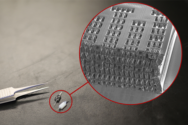
You can mend a broken heart this valentine’s day now that researchers invented a new hydrogel that can be used to heal damaged heart tissue and improve cancer treatments.
University of Waterloo chemical engineering researcher Dr. Elisabeth Prince teamed up with researchers from the University of Toronto and Duke University to design the synthetic material made using cellulose nanocrystals [CNC], which are derived from wood pulp.
The material is engineered to replicate the fibrous nanostructures and properties of human tissues, thereby recreating its unique biomechanical properties.
“Cancer is a diverse disease and two patients with the same type of cancer will often respond to the same treatment in very different ways,” Prince said. “Tumour organoids are essentially a miniaturized version of an individual patient’s tumour that can be used for drug testing, which could allow researchers to develop personalized therapies for a specific patient.”
As director of the Prince Polymer Materials Lab, Prince designs synthetic biomimetic hydrogels for biomedical applications. The hydrogels have a nanofibrous architecture with large pores for nutrient and waste transport, which affect mechanical properties and cell interaction.
Prince, a professor in Waterloo’s Department of Chemical Engineering, utilized these human-tissue mimetic hydrogels to promote the growth of small-scale tumour replicas derived from donated tumour tissue.
She aims to test the effectiveness of cancer treatments on the mini-tumour organoids before administering the treatment to patients, potentially allowing for personalized cancer therapies. This research was conducted alongside Professor David Cescon at the Princess Margaret Cancer Center.
Prince’s research group at Waterloo is developing similar biomimetic hydrogels to be injectable for drug delivery and regenerative medical applications as Waterloo researchers continue to lead health innovation in Canada.
Her research aims to use injected filamentous hydrogel material to regrow heart tissue damaged after a heart attack. She used nanofibers as a scaffolding for the regrowth and healing of damaged heart tissue.
“We are building on the work that I started during my PhD to design human-tissue mimetic hydrogels that can be injected into the human body to deliver therapeutics and repair the damage caused to the heart when a patient suffers a heart attack,” Prince said.
Prince’s research is unique as most gels currently used in tissue engineering or 3D cell culture don’t possess this nanofibrous architecture. Prince’s group uses nanoparticles and polymers as building blocks for materials and develops chemistry for nanostructures that accurately mimic human tissues.
The next step in Prince’s research is to use conductive nanoparticles to make electrically conductive nanofibrous gels that can be used to heal heart and skeletal muscle tissue.
That is a pretty stunning image and this March 15, 2022 news item on phys.org provides an explanation of what you see (Note: A link has been removed),
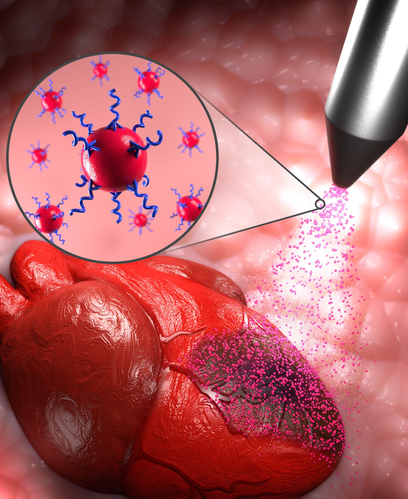
Could a spritz of super-tiny particles of gold and peptides on a damaged heart potentially provide minimally invasive, on-the-spot repair?
Cutting-edge research led by University of Ottawa Faculty of Medicine Associate Professors Dr. Emilio Alarcon and Dr. Erik Suuronen suggests a spray-on technology using customized nanoparticles of one of the world’s most precious metals offers tremendous therapeutic potential and could eventually help save many lives. Cardiovascular diseases are the leading cause of death globally, claiming roughly 18 million lives each year.
In a paper recently published online in ACS Nano, a peer-reviewed journal that highlighted the new research on its supplementary cover, Dr. Alarcon and his team of fellow investigators suggest that this approach might one day be used in conjunction with coronary artery bypass surgeries. That’s the most common type of heart surgery.
The therapy tested by the researchers – which was sprayed on the hearts of lab mice – used very low concentrations of peptide-modified particles of gold created in the laboratory. From the nozzle of a miniaturized spraying apparatus, the material can be evenly painted on the surface of a heart within a few seconds.
Gold nanoparticles have been shown to have some unusual properties and are highly chemically reactive. For years, researchers have been employing gold nanoparticles – so tiny they are undetectable by the human eye – in such a wide range of technologies that it’s become an area of intense research interest.
In this case, the custom-made nanogold modified with peptides—a short chain of amino acids —was sprayed on the hearts of lab mice. The research found that the spray-on therapy not only resulted in an increase in cardiac function and heart electrical conductivity but that there was no off-target organ infiltration by the tiny gold particles.
“That’s the beauty of this approach. You spray, then you wait a couple of weeks, and the animals are doing just fine compared to the controls,” says Dr. Alarcon, who is part of the Faculty of Medicine’s Department of Biochemistry, Microbiology and Immunology and also Director of the Bio-nanomaterials Chemistry and Engineering Laboratory at the University of the Ottawa Heart Institute.
Dr. Alarcon says that not only does the data suggest that the therapeutic action of the spray-on nanotherapeutic is highly effective, but its application is far simpler than other regenerative approaches for treating an infarcted heart.
At first, the observed improvement of cardiac function and electrical signal propagation in the hearts of tested mice was hard for the team to believe. But repeated experiments delivered the same positive results, according to Dr. Alarcon, who is part of the Faculty of Medicine’s Department of Biochemistry, Microbiology and Immunology and Director of the Bio-nanomaterials Chemistry and Engineering Laboratory at the University of Ottawa Heart Institute.
To validate the exciting findings in mice, the team is now seeking to adapt this technology to minimally invasive procedures that will expedite testing in large animal models, such as rabbits and pigs.
Dr. Alarcon praised the research culture at uOttawa and the Heart Institute, saying that the freedom to explore is paramount. “When you have an environment where you are allowed to make mistakes and criticize, that really drives discoveries,” he says.
The team involved in the paper includes researchers from uOttawa and the University of Talca in Chile. Part of the work was funded by the Canadian government’s New Frontiers in Research Fund, which was launched in 2018 and supports transformative high risk/high reward research led by Canadian researchers working with local and international partners.
If successful the hope is that ‘human-on-a-chip’ will replace most, if not all, animal testing. This July 3, 2019 Hesperos news release (also on EurekAlert) suggests scientists are making serious gains in the drive to replace animal testing (Note: For anyone having difficulty with the terms, pharmacokinetics and pharmacodynamics, there are definitions towards the end of this posting, which may prove helpful),
Hesperos Inc., pioneers* of the “human-on-a-chip” in vitro system has announced the use of its innovative multi-organ model to successfully measure the concentration and metabolism of two known cardiotoxic small molecules over time, to accurately describe the drug behavior and toxic effects in vivo. The findings further support the potential of body-on-a-chip systems to transform the drug discovery process.
In a study published in Nature Scientific Reports, in collaboration with AstraZeneca, Hesperos described how they used a pumpless heart model and a heart:liver system to evaluate the temporal pharmacokinetic/pharmacodynamic (PKPD) relationship for terfenadine, an antihistamine that was banned due to toxic cardiac effects, as well as determine its mechanism of toxicity.
The study found there was a time-dependent, drug-induced response in the heart model. Further experiments were conducted, adding a metabolically competent liver module to the Hesperos Human-on-a-Chip® system to observe what happened when terfenadine was converted to fexofenadine. By doing so, the researchers were able to determine the driver of the pharmacodynamic (PD) effect and develop a mathematical model to predict the effect of terfenadine in preclinical species. This is the first time an in vitro human-on-a-chip system has been shown to predict in vivo outcomes, which could be used to predict clinical trial outcomes in the future.
“The ability to examine PKPD relationships in vitro would enable us to understand compound behavior prior to in vivo testing, offering significant cost and time savings,” said Dr. Shuler, President and CEO, Hesperos, Inc and Professor Emeritus, Cornell University. “We are excited about the potential of this technology to help us ensure that potential new drug candidates have a higher probability of success during the clinical trial process.”
Understanding the inter-relationship between pharmacokinetics (PK), the drug’s time course for absorption, distribution, metabolism and excretion, and PD, the biological effect of a drug, is crucial in drug discovery and development. Scientists have learned that the maximum drug effect is not always driven by the peak drug concentration. In some cases, time is a critical factor influencing drug effect, but often this concentration-effect-time relationship only comes to light during the advanced stages of the preclinical program. In addition, often the data cannot be reliably extrapolated to humans.
“It is costly and time consuming to discover that potential drug candidates may have poor therapeutic qualities preventing their onward progression,” said James Hickman, Chief Scientist at Hesperos and Professor at the University of Central Florida. “Being able to define this during early drug discovery will be a valuable contribution to the optimization of potential new drug candidates.”
As demonstrated with the terfenadine experiment, the PKPD modelling approach was critical for understanding both the flux of compound between compartments as well as the resulting PD response in the context of dynamic exposure profiles of both parent and metabolite, as indicated by Dr. Shuler.
In order to test the viability of their system in a real-world drug discovery setting, the Hesperos team collaborated with scientists at AstraZeneca, to test one of their failed small molecules, known to have a CV [cardiovscular?] risk.
One of the main measurements used to assess the electrical properties of the heart is the QT interval, which approximates the time taken from when the cardiac ventricles start to contract to when they finish relaxing. Prolongation of the QT interval on the electrocardiogram can lead to a fatal arrhythmia known as Torsade de Pointes. Consequently, it is a mandatory requirement prior to first-in-human administration of potential new drug candidates that their ability to inhibit the hERG channel (a biomarker for QT prolongation) is investigated.
In the case of the AstraZeneca molecule, the molecule was assessed for hERG inhibition early on, and it was concluded to have a low potential to cause in vivo QT prolongation up to 100 μM. In later pre-clinical testing, the QT interval increased by 22% at a concentration of just 3 μM. Subsequent investigations found that a major metabolite was responsible. Hesperos was able to detect a clear PD effect at concentrations above 3 μM and worked to determine the mechanism of toxicity of the molecule.
The ability of these systems to assess cardiac function non-invasively in the presence of both parent molecule and metabolite over time, using multiplexed and repeat drug dosing regimes, provides an opportunity to run long-term studies for chronic administration of drugs to study their potential toxic effects.
Hesperos, Inc. is the first company spun out from the Tissue Chip Program at NCATS (National Center for Advancing Translational Sciences), which was established in 2011 to address the long timelines, steep costs and high failure rates associated with the drug development process. Hesperos currently is funded through NCATS’ Small Business Innovation Research program to undertake these studies and make tissue chips technology available as a service based company.
“The application of tissue chip technology in drug testing can lead to advances in predicting the potential effects of candidate medicines in people,” said Danilo Tagle, Ph.D., associate director for special initiatives at NCATS.
###
About Hesperos Hesperos, Inc. is a leader in efforts to characterize an individual’s biology with human-on-a-chip microfluidic systems. Founders Michael L. Shuler and James J. Hickman have been at the forefront of every major scientific discovery in this realm, from individual organ-on-a-chip constructs to fully functional, interconnected multi-organ systems. With a mission to revolutionize toxicology testing as well as efficacy evaluation for drug discovery, the company has created pumpless platforms with serum-free cellular mediums that allow multi-organ system communication and integrated computational PKPD modeling of live physiological responses utilizing functional readouts from neurons, cardiac, muscle, barrier tissues and neuromuscular junctions as well as responses from liver, pancreas and barrier tissues. Created from human stem cells, the fully human systems are the first in vitro solutions that accurately utilize in vitro systems to predict in vivo functions without the use of animal models, as featured in Science. More information is available at http://www. hesperosinc.com
Years ago I went to a congress focused on alternatives to animal testing (August 22, 2014 posting) and saw a video of heart cells in a petri dish (in vitro) beating in a heartlike rhythm. It was something like this,
ipscira Published on Oct 17, 2010 https://www.youtube.com/watch?v=BqzW9Jq-OVA
I found it amazing as did the scientist who drew my attention to it. After, it’s just a collection of heart cells. How do they start beating and keep time with each other?
Getting back to the latest research, here’s a link and a citation for the paper,
On the potential of in vitro organ-chip models to define temporal pharmacokinetic-pharmacodynamic relationships by Christopher W. McAleer, Amy Pointon, Christopher J. Long, Rocky L. Brighton, Benjamin D. Wilkin, L. Richard Bridges, Narasimham Narasimhan Sriram, Kristin Fabre, Robin McDougall, Victorine P. Muse, Jerome T. Mettetal, Abhishek Srivastava, Dominic Williams, Mark T. Schnepper, Jeff L. Roles, Michael L. Shuler, James J. Hickman & Lorna Ewart. Scientific Reports volume 9, Article number: 9619 (2019) DOI: https://doi.org/10.1038/s41598-019-45656-4 Published: 03 July 2019
Integrative pharmacology is a discipline that builds an understanding of the inter-relationship between pharmacokinetics (PK), the drug’s time course for absorption, distribution, metabolism and excretion and pharmacodynamics (PD), the biological effect of a drug. In drug discovery, this multi-variate approach guides medicinal chemists to modify structural properties of a drug molecule to improve its chance of becoming a medicine in a process known as “lead optimization”.
…
*More than one person and more than one company and more than one country claims pioneer status where ‘human-on-a-chip’ is concerned.
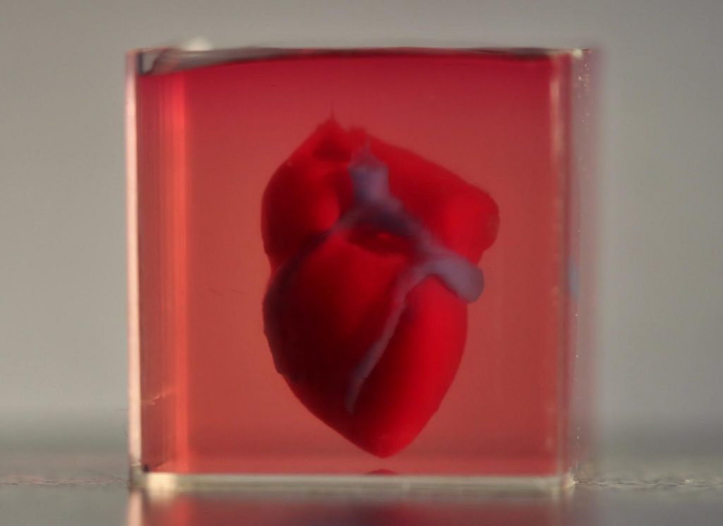
In a major medical breakthrough, Tel Aviv University researchers have “printed” the world’s first 3D vascularised engineered heart using a patient’s own cells and biological materials. Their findings were published on April 15 [2019] in a study in Advanced Science.
Until now, scientists in regenerative medicine — a field positioned at the crossroads of biology and technology — have been successful in printing only simple tissues without blood vessels.
“This is the first time anyone anywhere has successfully engineered and printed an entire heart replete with cells, blood vessels, ventricles and chambers,” says Prof. Tal Dvir of TAU’s School of Molecular Cell Biology and Biotechnology, Department of Materials Science and Engineering, Center for Nanoscience and Nanotechnology and Sagol Center for Regenerative Biotechnology, who led the research for the study.
Heart disease is the leading cause of death among both men and women in the United States. Heart transplantation is currently the only treatment available to patients with end-stage heart failure. Given the dire shortage of heart donors, the need to develop new approaches to regenerate the diseased heart is urgent.
“This heart is made from human cells and patient-specific biological materials. In our process these materials serve as the bioinks, substances made of sugars and proteins that can be used for 3D printing of complex tissue models,” Prof. Dvir says. “People have managed to 3D-print the structure of a heart in the past, but not with cells or with blood vessels. Our results demonstrate the potential of our approach for engineering personalized tissue and organ replacement in the future.
Research for the study was conducted jointly by Prof. Dvir, Dr. Assaf Shapira of TAU’s Faculty of Life Sciences and Nadav Moor, a doctoral student in Prof. Dvir’s lab.
“At this stage, our 3D heart is small, the size of a rabbit’s heart, [emphasis mine] ” explains Prof. Dvir. “But larger human hearts require the same technology.”
For the research, a biopsy of fatty tissue was taken from patients. The cellular and a-cellular materials of the tissue were then separated. While the cells were reprogrammed to become pluripotent stem cells, the extracellular matrix (ECM), a three-dimensional network of extracellular macromolecules such as collagen and glycoproteins, were processed into a personalized hydrogel that served as the printing “ink.”
After being mixed with the hydrogel, the cells were efficiently differentiated to cardiac or endothelial cells to create patient-specific, immune-compatible cardiac patches with blood vessels and, subsequently, an entire heart.
According to Prof. Dvir, the use of “native” patient-specific materials is crucial to successfully engineering tissues and organs.
“The biocompatibility of engineered materials is crucial to eliminating the risk of implant rejection, which jeopardizes the success of such treatments,” Prof. Dvir says. “Ideally, the biomaterial should possess the same biochemical, mechanical and topographical properties of the patient’s own tissues. Here, we can report a simple approach to 3D-printed thick, vascularized and perfusable cardiac tissues that completely match the immunological, cellular, biochemical and anatomical properties of the patient.”
The researchers are now planning on culturing the printed hearts in the lab and “teaching them to behave” like hearts, Prof. Dvir says. They then plan to transplant the 3D-printed heart in animal models.
“We need to develop the printed heart further,” he concludes. “The cells need to form a pumping ability; they can currently contract, but we need them to work together. Our hope is that we will succeed and prove our method’s efficacy and usefulness.
“Maybe, in ten years, there will be organ printers in the finest hospitals around the world, and these procedures will be conducted routinely.”
Growing the heart to human size and getting the cells to work together so the heart will pump makes it seem like the 10 years Dvir imagines as the future date when there will be organ printers in hospitals routinely printing up hearts seems a bit optimistic. Regardless, I hope he’s right. Bravo to these Israeli researchers!
I wish the folks at the University of British Columbia (UBC) would include more technical/scientific information in their news releases about research. For those who do like a little more technical information, I included the paper’s abstract at the end of this post.
Researchers at UBC have created the first-ever nanocomposite biomaterial heart-valve developed to reduce or eliminate complications related to heart transplants.
By using a newly developed technique, the researchers were able to build a more durable valve that enables the heart to adapt faster and more seamlessly.
Assistant Professor Hadi Mohammadi runs the Heart Valve Performance Laboratory (HVPL) through UBC Okanagan’s School of Engineering. Lead author on the study, he says the newly developed valve is an example of a transcatheter heart valve, a promising new branch of cardiology. These valves are unique because they can be inserted into a patient through small incisions rather than opening a patient’s chest–a procedure that is generally safer and much less invasive.
“Existing transcatheter heart valves are made of animal tissues, most often the pericardium membrane from a cow’s heart, and have had only moderate success to date,” explains Mohammadi. “The problem is that they face significant implantation risks and can lead to coronary obstruction and acute kidney injury.”
The new valve solves that problem by using naturally derived nanocomposites–a material assembled with a variety of very small components–including gels, vinyl and cellulose. The combination of their new material with the non-invasive nature of transcatheter heart valves makes this new design very promising for use with high-risk patients, according to Mohammadi.
“Not only is the material important but the design and construction of our valve means that it lowers stress on the valve by as much as 40 per cent compared to valves currently available,” says Dylan Goode, a graduate researcher at the HVPL. “It is uniquely manufactured in one continuous form, so it gains strength and flexibility to withstand the circulatory complications that can arise following transplantation.”
Working with researchers from Kelowna General Hospital and Western University, the valve will now undergo vigorous testing to perfect its material composition and design. The testing will include human heart simulators and large animal in-vivo studies. If successful, the valve will then proceed to clinical patient testing.
“This has the potential to become the new standard in heart valve replacement and to provide a safer, longer-term solution for many patients.”
The new design was highlighted in a paper published this month in the Journal of Engineering in Medicine with financial support from the Natural Sciences and Engineering Research Council of Canada [NSERC] .
Here’s a link to and a citation for the paper,
Proposed percutaneous aortic valve prosthesis made of cryogel by Hadi Mohammadi, Dylan Goode, Guy Fradet, Kibret Mequanint. Proceedings of the Institution of Mechanical Engineers, Part H: Journal of Engineering in Medicine, 2019; 095441191983730 DOI: 10.1177/0954411919837302 First Published March 20, 2019
This paper is behind a paywall.
As promised, here’s the abstract,
Transcatheter heart valves are promising for high-risk patients. Generally, their leaflets are made of pericardium stented in a Nitinol basket. Despite their relative success, they are associated with significant complications such as valve migration, implantation risks, stroke, coronary obstruction, myocardial infraction, acute kidney injury (which all are due to the release of detached solid calcific pieces in to the blood stream) and expected issues existing with tissue valves such as leaflet calcification. This study is an attempt to fabricate the first ever polymeric percutaneous valves made of cryogel following the geometry and mechanical properties of porcine aortic valve to address some of the above-mentioned shortcomings. A novel, one-piece, tricuspid percutaneous valve, consisting of leaflets made entirely from the hydrogel, polyvinyl alcohol cryogel reinforced by bacterial cellulose natural nanocomposite, attached to a Nitinol basket was developed and demonstrated. Following the natural geometry of the valve, a novel approach was applied based on the revolution about an axis of a hyperboloid shape. The geometry was modified based on avoiding sharp warpage of leaflets and removal of the central opening orifice area of the valve when valve is fully closed using the finite element analysis. The modified geometry was replaced by a cloud of (control) points and was essentially converted to Bezier surfaces for further adjustment. A cavity mold was then designed and fabricated to form the valve. The fabricated valve was sewn into the Nitinol basket which is covered by Dacron cloth. The models presented in this study merit further development and revisions for both aortic and mitral positions.
So, this new valve partially consists of bacterial cellulose and the design is based on porcine (pig) valves. Cellulose is the most abundant organic material on earth and if it forms part of the nanocomposite, I’d expect to see the word ‘nanocellulose’ mentioned somewhere. What puzzles me is the ‘bacterial cellulose’, a term that is unfamiliar to me. Anyone who cares to clarify the matter for me, please feel free to leave a comment.
Regarding the pig valve, I understand that heart patients who require valves have a choice of a pig valve or a mechanical valve. Apparently, people with porcine valves don’t need to take drugs to counteract rejection amongst other advantages but the valves do have a shorter life span (10 to 15 years) in addition to the other shortcomings mentioned in the abstract.
Assuming I properly understand the abstract, this ‘nanocomposite’ valve could combine the advantages of the mechanical and porcine valves while offering more durability than either one.
Again, should anyone care to increase my understanding of the valves and the advantages of this new one, please do leave a comment.
Also known as human-on-a-chip, the 10-organ body-on-a-chip was being discussed at the 9th World Congress on Alternatives to Animal Testing in the Life Sciences in 2014 in Prague, Czech Republic (see this July 1, 2015 posting for more). At the time, scientists were predicting success at achieving their goal of 10 organs on-a-chip in 2017 (the best at the time was four organs). Only a few months past that deadline, scientists from the Massachusetts Institute of Technology (MIT) seem to have announced a ’10 organ chip’ in a March 14, 2018 news item on ScienceDaily,
MIT engineers have developed new technology that could be used to evaluate new drugs and detect possible side effects before the drugs are tested in humans. Using a microfluidic platform that connects engineered tissues from up to 10 organs, the researchers can accurately replicate human organ interactions for weeks at a time, allowing them to measure the effects of drugs on different parts of the body.
Such a system could reveal, for example, whether a drug that is intended to treat one organ will have adverse effects on another.
“Some of these effects are really hard to predict from animal models because the situations that lead to them are idiosyncratic,” says Linda Griffith, the School of Engineering Professor of Teaching Innovation, a professor of biological engineering and mechanical engineering, and one of the senior authors of the study. “With our chip, you can distribute a drug and then look for the effects on other tissues, and measure the exposure and how it is metabolized.”
These chips could also be used to evaluate antibody drugs and other immunotherapies, which are difficult to test thoroughly in animals because they are designed to interact with the human immune system.
David Trumper, an MIT professor of mechanical engineering, and Murat Cirit, a research scientist in the Department of Biological Engineering, are also senior authors of the paper, which appears in the journal Scientific Reports. The paper’s lead authors are former MIT postdocs Collin Edington and Wen Li Kelly Chen.
Modeling organs
When developing a new drug, researchers identify drug targets based on what they know about the biology of the disease, and then create compounds that affect those targets. Preclinical testing in animals can offer information about a drug’s safety and effectiveness before human testing begins, but those tests may not reveal potential side effects, Griffith says. Furthermore, drugs that work in animals often fail in human trials.
“Animals do not represent people in all the facets that you need to develop drugs and understand disease,” Griffith says. “That is becoming more and more apparent as we look across all kinds of drugs.”
Complications can also arise due to variability among individual patients, including their genetic background, environmental influences, lifestyles, and other drugs they may be taking. “A lot of the time you don’t see problems with a drug, particularly something that might be widely prescribed, until it goes on the market,” Griffith says.
As part of a project spearheaded by the Defense Advanced Research Projects Agency (DARPA), Griffith and her colleagues decided to pursue a technology that they call a “physiome on a chip,” which they believe could offer a way to model potential drug effects more accurately and rapidly. To achieve this, the researchers needed new equipment — a platform that would allow tissues to grow and interact with each other — as well as engineered tissue that would accurately mimic the functions of human organs.
Before this project was launched, no one had succeeded in connecting more than a few different tissue types on a platform. Furthermore, most researchers working on this kind of chip were working with closed microfluidic systems, which allow fluid to flow in and out but do not offer an easy way to manipulate what is happening inside the chip. These systems also require external pumps.
The MIT team decided to create an open system, which essentially removes the lid and makes it easier to manipulate the system and remove samples for analysis. Their system, adapted from technology they previously developed and commercialized through U.K.-based CN BioInnovations, also incorporates several on-board pumps that can control the flow of liquid between the “organs,” replicating the circulation of blood, immune cells, and proteins through the human body. The pumps also allow larger engineered tissues, for example tumors within an organ, to be evaluated.
Complex interactions
The researchers created several versions of their chip, linking up to 10 organ types: liver, lung, gut, endometrium, brain, heart, pancreas, kidney, skin, and skeletal muscle. Each “organ” consists of clusters of 1 million to 2 million cells. These tissues don’t replicate the entire organ, but they do perform many of its important functions. Significantly, most of the tissues come directly from patient samples rather than from cell lines that have been developed for lab use. These so-called “primary cells” are more difficult to work with but offer a more representative model of organ function, Griffith says.
Using this system, the researchers showed that they could deliver a drug to the gastrointestinal tissue, mimicking oral ingestion of a drug, and then observe as the drug was transported to other tissues and metabolized. They could measure where the drugs went, the effects of the drugs on different tissues, and how the drugs were broken down. In a related publication, the researchers modeled how drugs can cause unexpected stress on the liver by making the gastrointestinal tract “leaky,” allowing bacteria to enter the bloodstream and produce inflammation in the liver.
Kevin Healy, a professor of bioengineering and materials science and engineering at the University of California at Berkeley, says that this kind of system holds great potential for accurate prediction of complex adverse drug reactions.
“While microphysiological systems (MPS) featuring single organs can be of great use for both pharmaceutical testing and basic organ-level studies, the huge potential of MPS technology is revealed by connecting multiple organ chips in an integrated system for in vitro pharmacology. This study beautifully illustrates that multi-MPS “physiome-on-a-chip” approaches, which combine the genetic background of human cells with physiologically relevant tissue-to-media volumes, allow accurate prediction of drug pharmacokinetics and drug absorption, distribution, metabolism, and excretion,” says Healy, who was not involved in the research.
Griffith believes that the most immediate applications for this technology involve modeling two to four organs. Her lab is now developing a model system for Parkinson’s disease that includes brain, liver, and gastrointestinal tissue, which she plans to use to investigate the hypothesis that bacteria found in the gut can influence the development of Parkinson’s disease.
Other applications include modeling tumors that metastasize to other parts of the body, she says.
“An advantage of our platform is that we can scale it up or down and accommodate a lot of different configurations,” Griffith says. “I think the field is going to go through a transition where we start to get more information out of a three-organ or four-organ system, and it will start to become cost-competitive because the information you’re getting is so much more valuable.”
The research was funded by the U.S. Army Research Office and DARPA.
Caption: MIT engineers have developed new technology that could be used to evaluate new drugs and detect possible side effects before the drugs are tested in humans. Using a microfluidic platform that connects engineered tissues from up to 10 organs, the researchers can accurately replicate human organ interactions for weeks at a time, allowing them to measure the effects of drugs on different parts of the body. Credit: Felice Frankel
Here’s a link to and a citation for the paper,
Interconnected Microphysiological Systems for Quantitative Biology and Pharmacology Studies by Collin D. Edington, Wen Li Kelly Chen, Emily Geishecker, Timothy Kassis, Luis R. Soenksen, Brij M. Bhushan, Duncan Freake, Jared Kirschner, Christian Maass, Nikolaos Tsamandouras, Jorge Valdez, Christi D. Cook, Tom Parent, Stephen Snyder, Jiajie Yu, Emily Suter, Michael Shockley, Jason Velazquez, Jeremy J. Velazquez, Linda Stockdale, Julia P. Papps, Iris Lee, Nicholas Vann, Mario Gamboa, Matthew E. LaBarge, Zhe Zhong, Xin Wang, Laurie A. Boyer, Douglas A. Lauffenburger, Rebecca L. Carrier, Catherine Communal, Steven R. Tannenbaum, Cynthia L. Stokes, David J. Hughes, Gaurav Rohatgi, David L. Trumper, Murat Cirit, Linda G. Griffith. Scientific Reports, 2018; 8 (1) DOI: 10.1038/s41598-018-22749-0 Published online:
This paper which describes testing for four-, seven-, and ten-organs-on-a-chip, is open access. From the paper’s Discussion,
In summary, we have demonstrated a generalizable approach to linking MPSs [microphysiological systems] within a fluidic platform to create a physiome-on-a-chip approach capable of generating complex molecular distribution profiles for advanced drug discovery applications. This adaptable, reusable system has unique and complementary advantages to existing microfluidic and PDMS-based approaches, especially for applications involving high logD substances (drugs and hormones), those requiring precise and flexible control over inter-MPS flow partitioning and drug distribution, and those requiring long-term (weeks) culture with reliable fluidic and sampling operation. We anticipate this platform can be applied to a wide range of problems in disease modeling and pre-clinical drug development, especially for tractable lower-order (2–4) interactions.
I have two items about cardiac research in Ontario. Not strictly speaking about nanotechnology, the two items do touch on topics covered here before, 3D organs and stem cells.
Matters of the heart can be complicated, but York University scientists have found a way to create 3D heart tissue that beats in synchronized harmony, like a heart in love, that will lead to better understanding of cardiac health, and improved treatments.
York U chemistry Professor Muhammad Yousaf and his team of grad students have devised a way to stick three different types of cardiac cells together, like Velcro, to make heart tissue that beats as one.
Until now, most 2D and 3D in vitro tissue did not beat in harmony and required scaffolding for the cells to hold onto and grow, causing limitations. In this research, Yousaf and his team made a scaffold free beating tissue out of three cell types found in the heart – contractile cardiac muscle cells, connective tissue cells and vascular cells.
The researchers believe this is the first 3D in vitro cardiac tissue with three cell types that can beat together as one entity rather than at different intervals.
“This breakthrough will allow better and earlier drug testing, and potentially eliminate harmful or toxic medications sooner,” said Yousaf of York U’s Faculty of Science.
In addition, the substance used to stick cells together (ViaGlue), will provide researchers with tools to create and test 3D in vitro cardiac tissue in their own labs to study heart disease and issues with transplantation. Cardiovascular associated diseases are the leading cause of death globally and are responsible for 40 per cent of deaths in North America.
“Making in vitro 3D cardiac tissue has long presented a challenge to scientists because of the high density of cells and muscularity of the heart,” said Dmitry Rogozhnikov, a chemistry PhD student at York. “For 2D or 3D cardiac tissue to be functional it needs the same high cellular density and the cells must be in contact to facilitate synchronized beating.”
Although the 3D cardiac tissue was created at a millimeter scale, larger versions could be made, said Yousaf, who has created a start-up company OrganoLinX to commercialize the ViaGlue reagent and to provide custom 3D tissues on demand.
Ontario Institute for Regenerative Medicine and its heart stem cell research
Steven Erwood has written about how Toronto has become a centre for certain kinds of cardiac research by focusing on specific researchers in a Feb. 13, 2017 posting on the Ontario Institute for Regenerative Medicine’s expression blog (Note: Links have been removed),
You may have heard that Paris is the city of love, but you might not know that Toronto specializes in matters of the heart, particularly broken hearts.
Dr. Ren Ke Li, an investigator with the Ontario Institute for Regenerative Medicine, established his lab at the Toronto General Hospital Research Institute in 1993 hoping to find a way to replace the muscle cells, or cardiomyocytes, that are lost after a heart attack. Specifically, Li hoped to transplant a collection of cells, called stem cells, into a heart damaged by a heart attack. Stem cells have the power to differentiate into virtually any cell type, so if Li could coax them to become cardiomyocytes, they could theoretically reverse the damage caused by the heart attack.
Over the years, Li’s experiments using stem cells to regenerate and repair damaged heart tissue, which progressed all the way through to human clinical trials, pushed Li to rethink his approach to heart repair. Most of the transplanted cells failed to engraft to the host tissue and many of those that did successfully integrate into the patient’s heart remained non-contractile, sitting still beside the rest of the beating heart muscle. Despite this, the treatments were still proving beneficial — albeit less beneficial than Li had hoped. These cells weren’t replacing the lost cardiomyocytes, but they were still helping the patient recover. Li was then just beginning to reveal something that is now well described: transplanting exogenous stem cells (originating outside the patient) onto damaged tissue stimulated the endogenous stem cells to repair that damage. These transplanted stem cells were changing the behaviour of the patient’s own stem cells, enhancing their response to injury.
Li calls this process “rejuvenation” — arguing that the reason older populations can’t recover from cardiac injury is because they have fewer stem cells, and those stem cells have lost their ability to repair and regenerate damaged tissue over time. Li argues that the positive effects he was seeing in his experiments and clinical trials was a restoration or reversal of age-related deterioration in repair capability — a rejuvenation of the aged heart.
Li, alongside fellow OIRM [Ontario Institute for Regenerative Medicine] researcher and cardiac surgeon at Toronto General Hospital, Dr. Richard Weisel, dedicated a large part of their research effort to understanding this process. Weisel explains, “We put young cells into old animals, and we can get them to respond to a heart attack like a young person — which is remarkable!”
A team of researchers led by the duo published an article in Basic Research in Cardiology last month describing a new method to rejuvenate the aged heart, and characterizing this rejuvenation at the molecular and cellular level.
…
Successfully advancing this research to the clinic is where Weisel thinks Toronto provides a unique advantage. “We have the ability to do the clinical trials — the same people who are working on these projects [in the lab], can also take them into the clinic, and a lot of other places in the world [the clinicians and the researchers] are separate. We’ve been doing that for all the areas of stem cell research.” This unique set of circumstances, Weisel argues, more readily allows for a successful transition from research to clinical practice.
But an integrated research and clinical environment isn’t all the city has to offer to those looking to make substantial progress in stem cell therapies. Dr. Michael Laflamme, OIRM researcher and a leading authority on stem cell therapies for cardiac repair, called his decision to relocate to Toronto from the University of Washington in Seattle “a no-brainer”.
Laflamme focuses on improving the existing approaches to exogenous stem cell transplantation in cardiac repair and believes that solving the problems Li faced in his early experiments is just a matter of finding the right cell type. Laflamme, in an ongoing preclinical trial funded by OIRM, is differentiating stem cells in a bioreactor into ventricular cardiomyocytes, the specific type of cell lost after a heart attack, and delivering those cells directly to the scar tissue in hopes of turning it back into muscle. Laflamme is optimistic these ventricular cardiomyocytes might be just the cell type he’s looking for. Using these cells in animal models, although in a mixture of other cardiac cell types, Laflamme explains, “We’ve shown that those cells will stably engraft and they actually become electrically integrated with the rest of the tissue — they will [beat] in synchrony with the rest of the heart.”
Laflamme states that “Toronto is the place where we can get this stuff done better and we can get it done faster,” citing the existing Toronto-based expertise in both the differentiation of stem cells and the biotechnological means to scale these processes as being unparalleled elsewhere in the world.
It’s not only academic researchers and clinicians that recognize Toronto’s potential to advance regenerative medicine and stem cell therapy. Pharmaceutical giant Bayer, partnered with San Francisco-based venture capital firm Versant Ventures, announced last December a USD 225 million investment in a stem cell biotechnology company called BlueRock Therapeutics — the second largest investment of it’s kind in the history of the biotechnology industry. …
There’s substantially to more Erwood’s piece in the original posting.
One final thought, I wonder if there is a possibility that York University’s ViaGlue might be useful in the work talking place at Ontario Institute for Regenerative Medicine. I realize the two institutions are in the same city but do the researchers even know about each other’s work?
Usually called ‘human-on-a-chip’, a team at the University of Toronto have developed a two-organ ‘person on a chip’ according to a March 7, 2016 news item on phys.org (Note: Links have been removed),
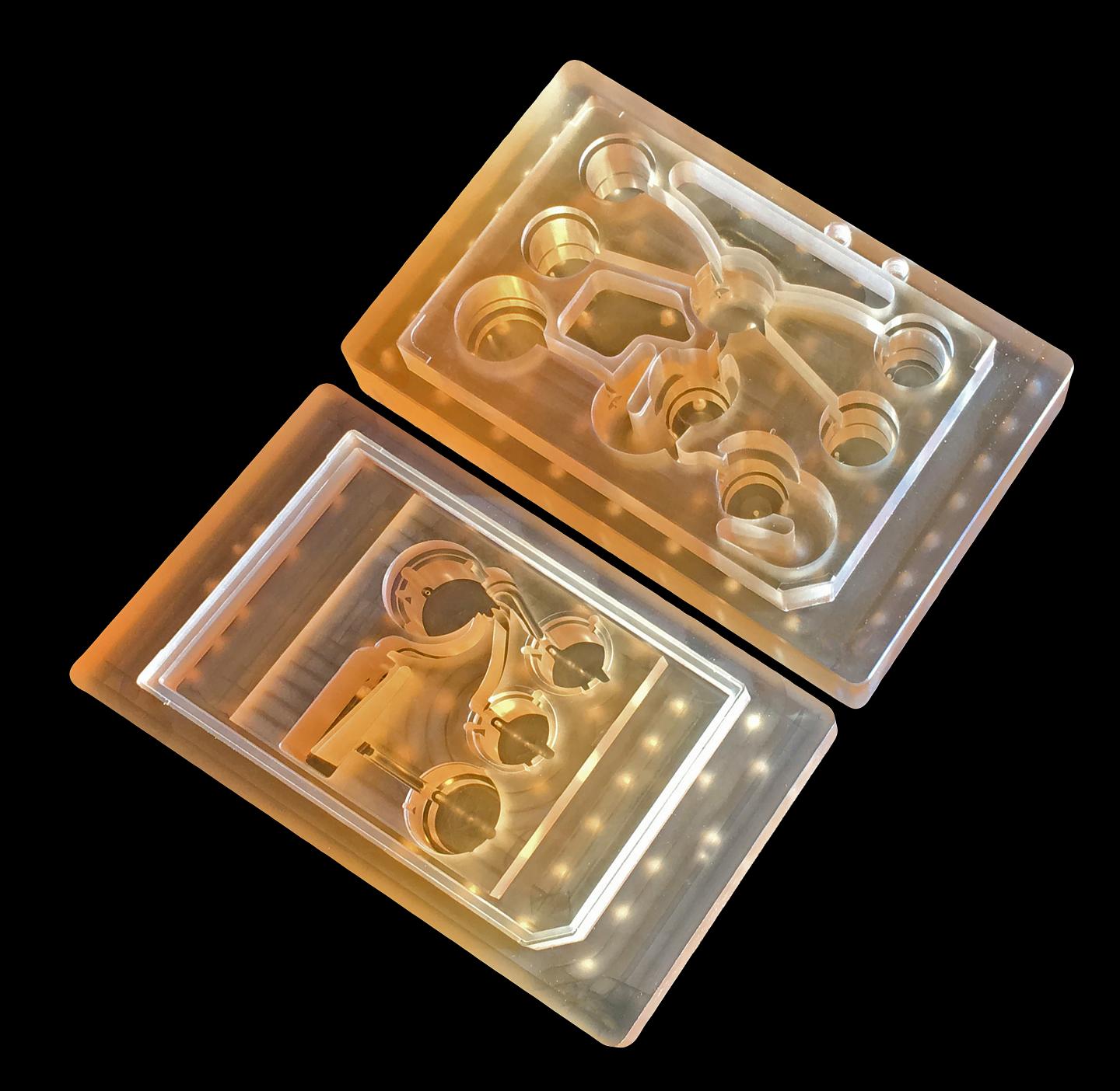
Researchers at U of T [University of Toronto] Engineering have developed a new way of growing realistic human tissues outside the body. Their “person-on-a-chip” technology, called AngioChip, is a powerful platform for discovering and testing new drugs, and could eventually be used to repair or replace damaged organs.
Professor Milica Radisic (IBBME, ChemE), graduate student Boyang Zhang and the rest of the team are among those research groups around the world racing to find ways to grow human tissues in the lab, under conditions that mimic a real person’s body. They have developed unique methods for manufacturing small, intricate scaffolds for individual cells to grow on. These artificial environments produce cells and tissues that resemble the real thing more closely than those grown lying flat in a petri dish.
The team’s recent creations have included BiowireTM—an innovative method of growing heart cells around a silk suture—as well as a scaffold for heart cells that snaps together like sheets of Velcro. But AngioChip takes tissue engineering to a whole new level. “It’s a fully three-dimensional structure complete with internal blood vessels,” says Radisic. “It behaves just like vasculature, and around it there is a lattice for other cells to attach and grow.” …
Zhang built the scaffold out of POMaC, a polymer that is both biodegradable and biocompatible. The scaffold is built out of a series of thin layers, stamped with a pattern of channels that are each about 50 to 100 micrometres wide. The layers, which resemble the computer microchips, are then stacked into a 3D structure of synthetic blood vessels. As each layer is added, UV light is used to cross-link the polymer and bond it to the layer below.
When the structure is finished, it is bathed in a liquid containing living cells. The cells quickly attach to the inside and outside of the channels and begin growing just as they would in the human body.
“Previously, people could only do this using devices that squish the cells between sheets of silicone and glass,” says Radisic. “You needed several pumps and vacuum lines to run just one chip. Our system runs in a normal cell culture dish, and there are no pumps; we use pressure heads to perfuse media through the vasculature. The wells are open, so you can easily access the tissue.”
Using the platform, the team has built model versions of both heart and liver tissues that function like the real thing. “Our liver actually produced urea and metabolized drugs,” says Radisic. They can connect the blood vessels of the two artificial organs, thereby modelling not just the organs themselves, but the interactions between them. They’ve even injected white blood cells into the vessels and watched as they squeezed through gaps in the vessel wall to reach the tissue on the other side, just as they do in the human body.
The news release also mentions potential markets and the work that needs to be accomplished before AngioChip is available for purchase,
AngioChip has great potential in the field of pharmaceutical testing. Current drug-testing methods, such as animal testing and controlled clinical trials, are costly and fraught with ethical concerns. Testing on lab-grown human tissues would provide a realistic model at a fraction of the cost, but this area of research is still in its infancy. “In the last few years, it has become possible to order cultures of human cells for testing, but they’re grown on a plate, a two-dimensional environment,” says Radisic. “They don’t capture all the functional hallmarks of a real heart muscle, for example.”
A more realistic platform like AngioChip could enable drug companies to detect dangerous side effects and interactions between organ compartments long before their products reach the market, saving countless lives. It could also be used to understand and validate the effectiveness of current drugs and even to screen libraries of chemical compounds to discover new drugs. Through TARA Biosystems Inc., a spin-off company co-founded by Radisic, the team is already working on commercializing the technology.
In future, Radisic envisions her lab-grown tissues being implanted into the body to repair organs damaged by disease. Because the cells used to seed the platform can come from anyone, the new tissues could be genetically identical to the intended host, reducing the risk of organ rejection. Even in its current form, the team has shown that the AngioChip can be implanted into a living animal, its artificial blood vessels connected to a real circulatory system. The polymer scaffolding itself simply biodegrades after several months.
The team still has much work to do. Each AngioChip is currently made by hand; if the platform is to be used industrially, the team will need to develop high-throughput manufacturing methods to create many copies at once. Still, the potential is obvious. “It really is multifunctional, and solves many problems in the tissue engineering space,” says Radisic. “It’s truly next-generation.”
Here’s a link to and a citation for the paper,
Biodegradable scaffold with built-in vasculature for organ-on-a-chip engineering and direct surgical anastomosis by Boyang Zhang, Miles Montgomery, M. Dean Chamberlain, Shinichiro Ogawa, Anastasia Korolj, Aric Pahnke, Laura A. Wells, Stéphane Massé, Jihye Kim, Lewis Reis, Abdul Momen, Sara S. Nunes, Aaron R. Wheeler, Kumaraswamy Nanthakumar, Gordon Keller, Michael V. Sefton, & Milica Radisic. Nature Materials (2016) doi:10.1038/nmat4570 Published online 07 March 2016
This paper is behind a paywall.
The researchers have made two images illustrating their work available. There’s this still image,
These tiny polymer scaffolds contain channels that are about 100 micrometres wide, about the same diameter as a human hair. When seeded with cells, the channels act as artificial blood vessels. By mimicking tissues in the human heart and other organs, these scaffolds provide a new way to test drugs for potentially dangerous side effects. (Image: Tyler Irving/Boyang Zhang/Kevin Soobrian)
Perhaps more intriguing is this one,
When seeded with heart cells, the flexible polymer scaffold contracts with a regular rhythm, just like real heart tissue. (Image: Boyang Zhang)
I have mentioned ‘human-on-a-chip’ projects many times here and as the news release writer notes, there is an international race. My July 1, 2015 posting (cross-posted from the June 30, 2015 posting [Testing times: the future of animal alternatives] on the International Innovation blog [a CORDIS-listed project dissemination partner for FP7 and H2020 projects]) notes a couple of those projects,
Organ-on-a-chip projects use stem cells to create human tissues that replicate the functions of human organs. Discussions about human-on-a-chip activities – a phrase used to describe 10 interlinked organ chips – were a highlight of the 9th World Congress on Alternatives to Animal Testing held in Prague, Czech Republic, last year. One project highlighted at the event was a joint US National Institutes of Health (NIH), US Food and Drug Administration (FDA) and US Defense Advanced Research Projects Agency (DARPA) project led by Dan Tagle that claimed it would develop functioning human-on-a-chip by 2017. However, he and his team were surprisingly close-mouthed and provided few details making it difficult to assess how close they are to achieving their goal.
By contrast, Uwe Marx – Leader of the ‘Multi-Organ-Chip’ programme in the Institute of Biotechnology at the Technical University of Berlin and Scientific Founder of TissUse, a human-on-a-chip start-up company – claims to have sold two-organ chips. He also claims to have successfully developed a four-organ chip and that he is on his way to building a human-on-a-chip. Though these chips remain to be seen, if they are, they will integrate microfluidics, cultured cells and materials patterned at the nanoscale to mimic various organs, and will allow chemical testing in an environment that somewhat mirrors a human.
As for where the University of Toronto efforts fit into the race, I don’t know for sure. It’s the first time I’ve come across a reference to liver tissue producing urea but I believe there’s at least one other team in China which has achieved a three-dimensional, more lifelike aspect for liver tissue in my Jan. 29, 2016 posting ‘Constructing a liver’.
University of Washington researchers have shown that a favorable electrical property is present in a type of protein found in organs that repeatedly stretch and retract, such as the lungs, heart and arteries. These findings are the first that clearly track this phenomenon, called ferroelectricity, occurring at the molecular level in biological tissues.
The news release gives a brief description of ferroelectricity and describes the research team’s latest work with biological tissues,
Ferroelectricity is a response to an electric field in which a molecule switches from having a positive to a negative charge. This switching process in synthetic materials serves as a way to power computer memory chips, display screens and sensors. This property only recently has been discovered in animal tissues and researchers think it may help build and support healthy connective tissues in mammals.
A research team led by Li first discovered ferroelectric properties in biological tissues in 2012, then in 2013 found that glucose can suppress this property in the body’s connective tissues, wherever the protein elastin is present. But while ferroelectricity is a proven entity in synthetic materials and has long been thought to be important in biological functions, its actual existence in biology hasn’t been firmly established.
This study proves that ferroelectric switching happens in the biological protein elastin. When the researchers looked at the base structures within the protein, they saw similar behavior to the unit cells of solid-state materials, where ferroelectricity is well understood.
“When we looked at the smallest structural unit of the biological tissue and how it was organized into a larger protein fiber, we then were able to see similarities to the classic ferroelectric model found in solids,” Li said.
The researchers wanted to establish a more concrete, precise way of verifying ferroelectricity in biological tissues. They used small samples of elastin taken from a pig’s aorta and poled the tissues using an electric field at high temperatures. They then measured the current with the poling field removed and found that the current switched direction when the poling electric field was switched, a sign of ferroelectricity.
They did the same thing at room temperature using a laser as the heat source, and the current also switched directions.
Then, the researchers tested for this behavior on the smallest-possible unit of elastin, called tropoelastin, and again observed the phenomenon. They concluded that this switching property is “intrinsic” to the molecular make-up of elastin.
The next step is to understand the biological and physiological significance of this property, Li said. One hypothesis is that if ferroelectricity helps elastin stay flexible and functional in the body, a lack of it could directly affect the hardening of arteries.
“We may be able to use this as a very sensitive technique to detect the initiation of the hardening process at a very early stage when no other imaging technique will be able to see it,” Li said.
The team also is looking at whether this property plays a role in normal biological functions, perhaps in regulating the growth of tissue.
Co-authors are Pradeep Sharma at the University of Houston, Yanhang Zhang at Boston University, and collaborators at Nanjing University and the Chinese Academy of Sciences.
Here’s a link to and a citation for the research paper,
Ferroelectric switching of elastin by Yuanming Liu, Hong-Ling Cai, Matthew Zelisko, Yunjie Wang, Jinglan Sun, Fei Yan, Feiyue Ma, Peiqi Wang, Qian Nataly Chen, Hairong Zheng, Xiangjian Meng, Pradeep Sharma, Yanhang Zhang, and Jiangyu Li. Proceedings of the National Academy of Sciences (PNAS) doi: 10.1073/pnas.1402909111
This paper is behind a paywall.
I think this is a new practice. There is a paragraph on the significance of this work (follow the link to the paper),
Ferroelectricity has long been speculated to have important biological functions, although its very existence in biology has never been firmly established. Here, we present, to our knowledge, the first macroscopic observation of ferroelectric switching in a biological system, and we elucidate the origin and mechanism underpinning ferroelectric switching of elastin. It is discovered that the polarization in elastin is intrinsic at the monomer level, analogous to the unit cell level polarization in classical perovskite ferroelectrics. Our findings settle a long-standing question on ferroelectric switching in biology and establish ferroelectricity as an important biophysical property of proteins. We believe this is a critical first step toward resolving its physiological significance and pathological implications.
The stem cell scientists at the National University of Ireland (NUI) and Trinity College Dublin’s CRANN (Centre for Research on Adaptive Nanostructures and Nanodevices) aren’t making hearts out of carbon nanotubes but they are using the particles to stimulate stem cells into becoming heart-like.The Sept. 19, 2012 news item on Nanowerk provides context for this work,
Stem cell scientists have capitalised on the electrical properties of a widely used nanomaterial to develop cells which may allow the regeneration of cardiac cells. The breakthrough has been led by a team of scientists at the Regenerative Medicine Institute (REMEDI) at the National University of Ireland Galway in conjunction with Trinity College Dublin.
Heart disease is the leading cause of death in Ireland. Once damaged by heart attack, cardiac muscle has very little capacity for self-repair and at present there are no clinical treatments available to repair damaged cardiac muscle tissue.
Over the last 10 years, there has been tremendous interest in developing a cell-based therapy to address this problem. Since the use of a patient’s own heart cells is not a viable clinical option, many researchers are working to try to find an alternative source of cells that could be used for cardiac tissue repair.
The NUI Sept. 19, 2012 news release, which originated the news item, describes how carbon nanotubes have properties similar to certain heart cells and how the researchers decided to exploit that similarity,
The researchers recognised that carbon nanotubes, a widely used nanoparticle, is reactive to electrical stimulation. They then used these nanomaterials to create cells with the characteristics of cardiac progenitors, a special type of cell found in the heart, from adult stem cells.
“The electrical properties of the nanomaterial triggered a response in the mesenchymal (adult) stem cells, which we sourced from human bone marrow. In effect, they became electrified, which made them morph into more cardiac-like cells”, explains Valerie Barron of REMEDI at National University of Ireland Galway. “This is a totally new approach and provides a ready-source of tailored cells, which have the potential to be used as a new clinical therapy. Excitingly, this symbiotic strategy lays the foundation stone for other electroactive tissue repair applications, and can be readily exploited for other clinically challenging areas such as in the brain and the spinal cord.”
The team’s collaborator at CRANN, Professor Werner Blau made a comment I found a bit odd (from the NUI news release),
“It is great to see two decades of our pioneering nanocarbon research here at TCD come to fruition in a way that addresses a major global health problem. Hopefully many people around the world will ultimately benefit from it. Some of our carbon nanotube research has been patented by TCD and is being licensed to international companies in material science, electronics and health care,” said Professor Blau.
I’m not a big fan of the current patenting regimes which seem to have been turned into innovation-killing machines. As for patenting medicines and medical devices, I recall that Frederick Banting and Charles Best who discovered insulin refused to patent the discovery as they believed it would constrain access.
I appreciate that businesses need to make money and scientists need money to do their work and so on but this blind rush to patent discoveries seems a little misguided to me and it might be a good time to consider new business and economic models.