This is another one of my roundups. The focus is mostly mathematics.
Art Rapture 2017 September 22 – 23, 2017
Mark Ollinger claims to be presenting math-influenced street art at Vancouver’s 2017 Art Rapture in a Sept. 12, 2017 article by Joanna Riquett for the Daily Hive,
…
Tell us about your style, how would you describe it?
My work is very much based in mathematics, I come up with what I would call formulas and punch information into these formulas and an image will end up coming out. If I was to label my work as a genre, I would say I’m trying to blend optical art and graffiti.
…
What is your process for creating artwork?
I have like a 50+ step process to coming up with and producing a piece, haha. There are a lot of different things involved.

…
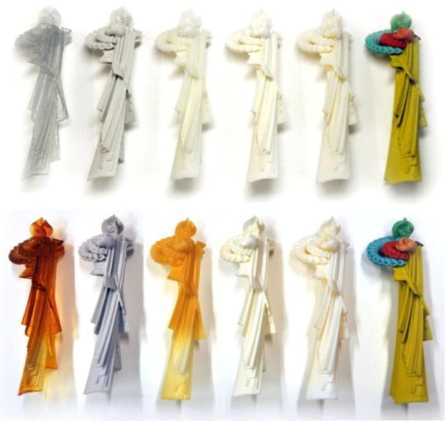
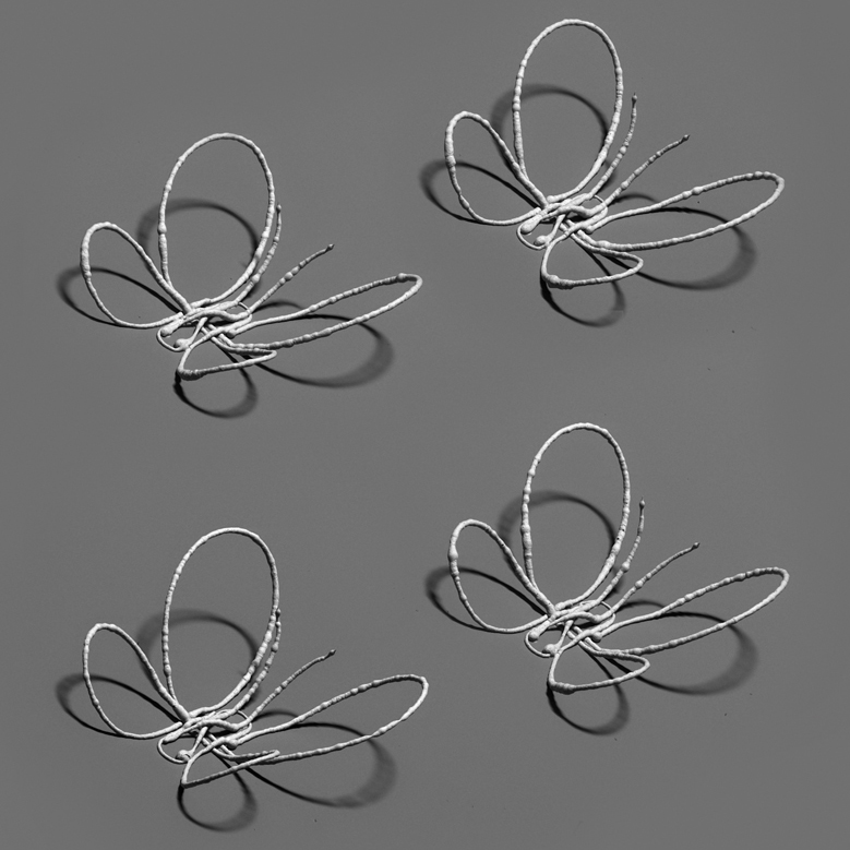
I wonder if he’s using 3D printers for the pieces at 2017 Art Rapture; it certainly fits with his comment about formulas (maybe he meant algorithms?) and that piece looks like it might be a polymer, a dominant material in 3D printing (biological inks are also used in 3D printing for medical research).
Ollinger’s artist description on the Kimoto Gallery website has this about the former Calgarian now living in Vancouver,
Mark Ollinger’s work begins with how we communicate. From the macroscopic study of language to the microscopic analysis of their constituent alphabets, Mark explores the conditions of language that underly our inner thoughts and outer expressions. He channels the meaning, sound, and physical appearance of words into sculptural elements whose geometric intricacies reflect the complexity of human linguistic [sic]. From here it becomes a cerebral exercise, a visual representation of an acoustic ephemerality. Drawing on the critical analysis of universal laws and fundamental frameworks, Mark seeks to visualize the phenomenology of the three dimensional world through the usage of physical and mathematical formulae.
This description has a lot of ideas but none are particularly well developed. It’s hard to tell if this is someone who’s struggling to communicate a complex set of well formed ideas or if the ideas are still being formed.
Ollinger’s biography on the Hetringer Kiss Gallery website (they represent him n Calgary) is a bit more informative about his past accomplishments,
Mark was born in 1988 and grew up in Calgary, Alberta, Canada. After graduating high school Mark began freelancing in graphic design and illustration and working at a silkscreen print shop for a year. Mark then started his own project, Duality Clothing, a clothing line that became his full-time endeavor. During this time Mark was painting daily and six years after owning and operating Duality Clothing Mark decided to pursue his passion for creating paintings and sculptural works. In 2015 he embarked on an ongoing body of work of unsanctioned public sculptural installations in Canada and abroad including Melbourne, Los Angeles, Toronto and Montreal. Self-taught, with no formal training Mark now works full time as an artist. He has gallery representation in Vancouver and Calgary and has been based in Vancouver, British Columbia since 2009.
Taming Infinities (or On Coin Tosses, Atoms and Forest Fires); a math lecture and event in Toronto, Ontario
The Fields Medal in Mathematics is a very big deal and the 2017 Fields Medal Symposium is being held in Toronto from Oct. 16 – 19, 2017,
Description
The 2017 Fields Medal Symposium will be centered on the work of Martin Hairer (Fields Medal 2014), and its current and potential impact.
The Scientific Program is intended for a wide audience, including graduate students, mathematicians in other research areas, and scientists who use mathematics in an important way.
Schedule: 9:30 am – 4:30 pm daily – see detailed schedule below
Location: The Fields Institute (222 College St., Toronto, ON)
The Symposium comprises four separate events:
- Student Workshop (October 15): aims to provide students with a good general exposure to probability a better understanding of what they will encounter during the Scientific Program, including regularity structures and their applications.
- Scientific Program (October 16-19): see description above.
- Public Opening (October 16): features a public lecture by Martin Hairer entitled Taming Infinities. This public lecture will be held at the MaRS Discovery District.
- Student Night (October 17): offers the opportunity for undergraduate and high school students to attend an evening talk by Martin Hairer entitled On Coin Tosses, Atoms and Forest Fires [perhaps the title has been changed?] — followed by networking (with plenty of time for questions/answers) and pizza.
Events will be broadcast live online whenever possible.
Registration Instruction:
All events are free to attend, but registration is required.
See the sidebar to only register for the Scientific Program and/or Student Workshop and below to register for the other events.
Register HERE for the Public Opening
Register HERE for the Student Night
I have more information about the public lecture by Martin Hairer inf the following file,
2017 Public Opening (PDF)
For those who don’t care to download the file,
Taming Infinities
Some physical and mathematical theories have the unfortunate feature that if one takes them at face value, many quantities of interest appear to be infinite! What’s worse, this doesn’t just happen for some exotic theories, but in the standard theories describing some of the most fundamental aspects of nature.
MaRS Auditorium
101 College Street
Toronto, ON
FREE EVENT • REGISTRATION REQUIRED
https://2017fmspublicopening.eventbrite.ca
Join us for the Public Opening of the 2017 Fields Medal Symposium, featuring a general audience presentation by Professor Martin Hairer (Fields Medal 2014).
Welcome and introductions by distinguished guests from academia and government:
• Cheryl Regehr, Vice-President and Provost, University of Toronto
• The Honourable Reza Moridi, Minister of Research, Innovation and Science, Ontario
• Sylvia Serfaty, Professor, Courant Institute of Mathematical Sciences, NYU
Fields Medals are awarded every four years by the International Mathematical Union to the most distinguished mathematicians age 40 or under. In the absence of a Nobel Prize in mathematics, the Fields Medal is regarded as the highest professional honor a mathematician can attain. Each year, the Fields Institute holds the Fields Medal Symposium to showcase and celebrate the work of a Fields Medallist. The Symposium
brings together brilliant researchers to support and further the research areas of Fields Medallists. It also raises public awareness of mathematics, the Fields Medal, and inspires new and upcoming researchers.
This event is a unique opportunity for the general public and academics to engage with this prominent scholar, his ideas, and cutting-edge research in mathematics.
h/t the Sept. 14, 2017 announcement from the ArtSci Salon.
Math is for solving puzzles
Jennifer Ruef, assistant professor of education studies at the University of Oregon, promises to make you a math whiz in four easy steps in her Sept. 11, 2017 essay for The Conversation (Note: Links have been removed),
…
What do you think of when you think about mathematics? Perhaps you think about x’s and y’s, intractable fractions, or nonsensical word problems. The cartoonist Gary Larson once depicted hell’s library as containing only giant tomes of word problems. You know, “If a train leaves New York…”
I was trained as a mathematician, and I will let you in on a trade secret: That is not what mathematics is, nor where it lives. It’s true that learning mathematics often involves solving problems, but it should focus on the joy of solving puzzles, rather than memorizing rules.
…
For many reasons, not the least of which is that [George] Pólya died in 1985, you will meet him as I did – through his wildly successful “How to Solve It.” Penned in 1945, this book went on to sell over one million copies and was translated into 17 languages.
As a mathematician, Pólya worked on a wide range of problems, including the study of heuristics, or how to solve problems. When you read “How to Solve It,” it feels like you’re taking a guided tour of Pólya’s mind. This is because his writing is metacognitive – he writes about how he thinks about thinking. And metacognition is often the heart of problem solving.
Pólya’s problem solving plan breaks down to four simple steps:
- Make sure you understand the problem.
- Make a plan to solve the problem.
- Carry out the plan.
- Check your work to test your answer.
There it is. Problem solving in the palm of your hand – math reduced to four steps.
Here’s a classic problem from research on mathematics education done by Jean Lave. A man, let’s call him John, is making ¾ of a recipe that calls for 2/3 cup of cottage cheese. What do you think John did? What would you do?
If you’re like me, you might immediately dive into calculations, perhaps struggling with what the fractions mean, working to remember the rules for arithmetic. That’s what John seemed to do, at first. But then he had a Eureka! moment.
John measured 2/3 cup of cottage cheese, then dumped it onto a cutting board. He patted the cheese into a circle and drew lines into it, one vertical, one horizontal, dividing the cheese patty into quarters. He then carefully pushed one quarter of the cottage cheese back into its container. Voilá! Three-quarters of 2/3 cup of cottage cheese remained.
John is a mathematician and problem solver. First, he understood the problem: He needed ¾ of what the recipe called for, which was 2/3 cup. Then, he made a plan, most likely visualizing in his head how he would measure and divide the cottage cheese. Finally, he carried out the plan.
Did he check his answer? That remains unclear, but we can check the validity of his work for him. Did he indeed end up with ¾ of 2/3 cup of cottage cheese? Yes, because the full amount was reduced by one-quarter, leaving three-quarters.
…
Ruef goes on to the discuss another solution, using pictures, for the same problem before she summarizes this delightful essay. (h/t Sept. 12, 2017 news item on phys.org)
Science Literacy Week in Canada (Sept. 18 – 24, 2017)
Thanks to Ingenium (formerly the Canada Science and Technology Museum Corporation) and its What’s up @ the Museums Sept. 2013 newsletter (received via email), I found out about Canada’s Science Literacy Week being held from Sept. 18-24, 2017. You can check here for online events and for events in your area.
Getting back to Ingenium, they are holding a Science Literacy Week programme at the Canada Aviation and Space Museum in Ottawa,
Demonstrations and activities:
Tours
Explore the Museum’s world-renowned collection in one of three ways:
Cockpit Interpretation Centre
Want the experience of flight but prefer to keep your feet on the ground? Here is your chance to sit at the controls of an aircraft without taking off! Friendly, knowledgeable Museum staff will assist you in boarding one of two cockpits:
- the Tutor (CT-114)
- the MIG-21
Cessna 150 Cockpit
Sit at the controls of a Cessna 150. Practise takeoffs and landings, and get a feel for what it’s like to fly this popular civilian airplane.
Flight Simulators
The Museum offers two flight simulators, open to the public at an additional cost. Climb aboard a replica of an F-16 cockpit and test your flight skills! Or, try the Redbird full-motion platform simulator, used to train pilots around the world.
Ejection Seat Demonstration
Monday to Saturday at 11:30 a.m. and Sundays at 4 p.m.
Learn how an ejection seat can save a pilot’s life.
Breathing on Mars

Saturdays and Sundays at 1 p.m.
Could humans breathe on Mars? Learn what the atmosphere on Mars is made of.
Shaping the Future in Space

Saturdays at 4 p.m. and Sundays at 11:30 a.m.
Discover new experiments that could improve human living conditions in Space.
Final comments
First, it’s interesting to note how many women are involved in the Fields Medal Symposium as speakers and that the essay on mathematics is by a woman. Second, while the programme at the Canada Aviation and Space Museum is being listed on their Science Literacy Week page, these demonstrations, etc. do not seem to have been developed specially for Science Literacy Week. In any event, there you have it!