Tag Archives: nanoparticles

Faster diagnostics with nanoparticles and magnetic phenomenon discovered 170 years ago

A Jan. 19, 2017 news item on ScienceDaily announces some new research from the University of Central Florida (UCF),

A UCF researcher has combined cutting-edge nanoscience with a magnetic phenomenon discovered more than 170 years ago to create a method for speedy medical tests.

The discovery, if commercialized, could lead to faster test results for HIV, Lyme disease, syphilis, rotavirus and other infectious conditions.

“I see no reason why a variation of this technique couldn’t be in every hospital throughout the world,” said Shawn Putnam, an assistant professor in the University of Central Florida’s College of Engineering & Computer Science.

A Jan. 19, 2017 UCF news release by Mark Schlueb, which originated the news item,  provides more technical detail,

At the core of the research recently published in the academic journal Small are nanoparticles – tiny particles that are one-billionth of a meter. Putnam’s team coated nanoparticles with the antibody to BSA, or bovine serum albumin, which is commonly used as the basis of a variety of diagnostic tests.

By mixing the nanoparticles in a test solution – such as one used for a blood test – the BSA proteins preferentially bind with the antibodies that coat the nanoparticles, like a lock and key.

That reaction was already well known. But Putnam’s team came up with a novel way of measuring the quantity of proteins present. He used nanoparticles with an iron core and applied a magnetic field to the solution, causing the particles to align in a particular formation. As proteins bind to the antibody-coated particles, the rotation of the particles becomes sluggish, which is easy to detect with laser optics.

The interaction of a magnetic field and light is known as Faraday rotation, a principle discovered by scientist Michael Faraday in 1845. Putnam adapted it for biological use.

“It’s an old theory, but no one has actually applied this aspect of it,” he said.

Other antigens and their unique antibodies could be substituted for the BSA protein used in the research, allowing medical tests for a wide array of infectious diseases.

The proof of concept shows the method could be used to produce biochemical immunology test results in as little as 15 minutes, compared to several hours for ELISA, or enzyme-linked immunosorbent assay, which is currently a standard approach for biomolecule detection.

Here’s a link to and a citation for the paper,

High-Throughput, Protein-Targeted Biomolecular Detection Using Frequency-Domain Faraday Rotation Spectroscopy by Richard J. Murdock, Shawn A. Putnam, Soumen Das, Ankur Gupta, Elyse D. Z. Chase, and Sudipta Seal. Small DOI: 10.1002/smll.201602862 Version of Record online: 16 JAN 2017

© 2017 WILEY-VCH Verlag GmbH & Co. KGaA, Weinheim

This paper is behind a paywall.

Nanoparticles can activate viruses lying dormant in lung cells

The nanoparticles in question are from combustion engines, which means that we are exposed to them. One other note, the testing has not been done on humans but rather on cells. From a Jan. 16, 2017 news item on ScienceDaily,

Nanoparticles from combustion engines can activate viruses that are dormant in in lung tissue cells. This is the result of a study by researchers of Helmholtz Zentrum München, a partner in the German Center for Lung Research (DZL), which has now been published in the journal Particle and Fibre Toxicology.

To evade the immune system, some viruses hide in cells of their host and persist there. In medical terminology, this state is referred to as a latent infection. If the immune system becomes weakened or if certain conditions change, the viruses become active again, begin to proliferate and destroy the host cell. A team of scientists led by Dr. Tobias Stöger of the Institute of Lung Biology and Prof. Dr. Heiko Adler, deputy head of the research unit Lung Repair and Regeneration at Helmholtz Zentrum München, now report that nanoparticles can also trigger this process.

A Jan. 16, 2017 Helmholtz Zentrum München press release (also on EurekAlert), which originated the news item, provides more detail,

“From previous model studies we already knew that the inhalation of nanoparticles has an inflammatory effect and alters the immune system,” said study leader Stöger. Together with his colleagues Heiko Adler and Prof. Dr. Philippe Schmitt-Kopplin, he showed that “an exposure to nanoparticles can reactivate latent herpes viruses in the lung.”

Specifically, the scientists tested the influence of nanoparticles typically generated by fossil fuel combustion in an experimental model for a particular herpes virus infection. They detected a significant increase in viral proteins, which are only produced with active virus proliferation. “Metabolic and gene expression analyses also revealed patterns resembling acute infection,” said Philippe Schmitt-Kopplin, head of the research unit Analytical BioGeoChemistry (BGC). Moreover, further experiments with human cells demonstrated that Epstein-Barr viruses are also ‘awakened’ when they come into contact with the nanoparticles.

Potential approach for chronic lung diseases

In further studies, the research team would like to test whether the results can also be transferred to humans. “Many people carry herpes viruses, and patients with idiopathic pulmonary fibrosis are particularly affected,” said Heiko Adler. “If the results are confirmed in humans, it would be important to investigate the molecular process of the reactivation of latent herpes viruses induced by particle inhalation. Then we could try to influence this pathway therapeutically.”

Special cell culture models shall therefore elucidate the exact mechanism of virus reactivation by nanoparticles. “In addition,” Stöger said, ”in long-term studies we would like to investigate to what extent  repeated nanoparticle exposure with corresponding virus reactivation leads to chronic inflammatory and remodeling processes in the lung.”

Further Information

Background:
In 2015 another group at the Helmholtz Zentrum München demonstrated how the Epstein-Barr virus  hides in human cells. In March 2016 researchers also showed that microRNAs silence immune alarm signals of cells infected with the Epstein-Barr virus.

Original Publication:
Sattler, C. et al. (2016): Nanoparticle exposure reactivates latent herpesvirus and restores a signature of acute infection. Particle and Fibre Toxicology, DOI 10.1186/s12989-016-0181-1

Here’s a link to and a citation for the paper on investigating latent herpes virus,

Nanoparticle exposure reactivates latent herpesvirus and restores a signature of acute infection by Christine Sattler, Franco Moritz, Shanze Chen, Beatrix Steer, David Kutschke, Martin Irmler, Johannes Beckers, Oliver Eickelberg, Philippe Schmitt-Kopplin, Heiko Adler. Particle and Fibre Toxicology201714:2 DOI: 10.1186/s12989-016-0181-1 Published: 10 January 2017

©  The Author(s). 2017

This paper is open access and, so too, is the 2016 paper.

Going underground to observe atoms in a bid for better batteries

A Jan. 16, 2017 news item on ScienceDaily describes what lengths researchers at Stanford University (US) will go to in pursuit of their goals,

In a lab 18 feet below the Engineering Quad of Stanford University, researchers in the Dionne lab camped out with one of the most advanced microscopes in the world to capture an unimaginably small reaction.

The lab members conducted arduous experiments — sometimes requiring a continuous 30 hours of work — to capture real-time, dynamic visualizations of atoms that could someday help our phone batteries last longer and our electric vehicles go farther on a single charge.

Toiling underground in the tunneled labs, they recorded atoms moving in and out of nanoparticles less than 100 nanometers in size, with a resolution approaching 1 nanometer.

A Jan. 16, 2017 Stanford University news release (also on EurekAlert) by Taylor Kubota, which originated the news item, provides more detail,

“The ability to directly visualize reactions in real time with such high resolution will allow us to explore many unanswered questions in the chemical and physical sciences,” said Jen Dionne, associate professor of materials science and engineering at Stanford and senior author of the paper detailing this work, published Jan. 16 [2017] in Nature Communications. “While the experiments are not easy, they would not be possible without the remarkable advances in electron microscopy from the past decade.”

Their experiments focused on hydrogen moving into palladium, a class of reactions known as an intercalation-driven phase transition. This reaction is physically analogous to how ions flow through a battery or fuel cell during charging and discharging. Observing this process in real time provides insight into why nanoparticles make better electrodes than bulk materials and fits into Dionne’s larger interest in energy storage devices that can charge faster, hold more energy and stave off permanent failure.

Technical complexity and ghosts

For these experiments, the Dionne lab created palladium nanocubes, a form of nanoparticle, that ranged in size from about 15 to 80 nanometers, and then placed them in a hydrogen gas environment within an electron microscope. The researchers knew that hydrogen would change both the dimensions of the lattice and the electronic properties of the nanoparticle. They thought that, with the appropriate microscope lens and aperture configuration, techniques called scanning transmission electron microscopy and electron energy loss spectroscopy might show hydrogen uptake in real time.

After months of trial and error, the results were extremely detailed, real-time videos of the changes in the particle as hydrogen was introduced. The entire process was so complicated and novel that the first time it worked, the lab didn’t even have the video software running, leading them to capture their first movie success on a smartphone.

Following these videos, they examined the nanocubes during intermediate stages of hydrogenation using a second technique in the microscope, called dark-field imaging, which relies on scattered electrons. In order to pause the hydrogenation process, the researchers plunged the nanocubes into an ice bath of liquid nitrogen mid-reaction, dropping their temperature to 100 degrees Kelvin (-280 F). These dark-field images served as a way to check that the application of the electron beam hadn’t influenced the previous observations and allowed the researchers to see detailed structural changes during the reaction.

“With the average experiment spanning about 24 hours at this low temperature, we faced many instrument problems and called Ai Leen Koh [co-author and research scientist at Stanford’s Nano Shared Facilities] at the weirdest hours of the night,” recalled Fariah Hayee, co-lead author of the study and graduate student in the Dionne lab. “We even encountered a ‘ghost-of-the-joystick problem,’ where the joystick seemed to move the sample uncontrollably for some time.”

While most electron microscopes operate with the specimen held in a vacuum, the microscope used for this research has the advanced ability to allow the researchers to introduce liquids or gases to their specimen.

“We benefit tremendously from having access to one of the best microscope facilities in the world,” said Tarun Narayan, co-lead author of this study and recent doctoral graduate from the Dionne lab. “Without these specific tools, we wouldn’t be able to introduce hydrogen gas or cool down our samples enough to see these processes take place.”

Pushing out imperfections

Aside from being a widely applicable proof of concept for this suite of visualization techniques, watching the atoms move provides greater validation for the high hopes many scientists have for nanoparticle energy storage technologies.

The researchers saw the atoms move in through the corners of the nanocube and observed the formation of various imperfections within the particle as hydrogen moved within it. This sounds like an argument against the promise of nanoparticles but that’s because it’s not the whole story.

“The nanoparticle has the ability to self-heal,” said Dionne. “When you first introduce hydrogen, the particle deforms and loses its perfect crystallinity. But once the particle has absorbed as much hydrogen as it can, it transforms itself back to a perfect crystal again.”

The researchers describe this as imperfections being “pushed out” of the nanoparticle. This ability of the nanocube to self-heal makes it more durable, a key property needed for energy storage materials that can sustain many charge and discharge cycles.

Looking toward the future

As the efficiency of renewable energy generation increases, the need for higher quality energy storage is more pressing than ever. It’s likely that the future of storage will rely on new chemistries and the findings of this research, including the microscopy techniques the researchers refined along the way, will apply to nearly any solution in those categories.

For its part, the Dionne lab has many directions it can go from here. The team could look at a variety of material compositions, or compare how the sizes and shapes of nanoparticles affect the way they work, and, soon, take advantage of new upgrades to their microscope to study light-driven reactions. At present, Hayee has moved on to experimenting with nanorods, which have more surface area for the ions to move through, promising potentially even faster kinetics.

Here’s a link to and a citation for the paper,

Direct visualization of hydrogen absorption dynamics in individual palladium nanoparticles by Tarun C. Narayan, Fariah Hayee, Andrea Baldi, Ai Leen Koh, Robert Sinclair, & Jennifer A. Dionne. Nature Communications 8, Article number: 14020 (2017) doi:10.1038/ncomms14020 Published online: 16 January 2017

This paper is open access.

Investigating nanoparticles and their environmental impact for industry?

It seems the Center for the Environmental Implications of Nanotechnology (CEINT) at Duke University (North Carolina, US) is making an adjustment to its focus and opening the door to industry, as well as, government research. It has for some years (my first post about the CEINT at Duke University is an Aug. 15, 2011 post about its mesocosms) been focused on examining the impact of nanoparticles (also called nanomaterials) on plant life and aquatic systems. This Jan. 9, 2017 US National Science Foundation (NSF) news release (h/t Jan. 9, 2017 Nanotechnology Now news item) provides a general description of the work,

We can’t see them, but nanomaterials, both natural and manmade, are literally everywhere, from our personal care products to our building materials–we’re even eating and drinking them.

At the NSF-funded Center for Environmental Implications of Nanotechnology (CEINT), headquartered at Duke University, scientists and engineers are researching how some of these nanoscale materials affect living things. One of CEINT’s main goals is to develop tools that can help assess possible risks to human health and the environment. A key aspect of this research happens in mesocosms, which are outdoor experiments that simulate the natural environment – in this case, wetlands. These simulated wetlands in Duke Forest serve as a testbed for exploring how nanomaterials move through an ecosystem and impact living things.

CEINT is a collaborative effort bringing together researchers from Duke, Carnegie Mellon University, Howard University, Virginia Tech, University of Kentucky, Stanford University, and Baylor University. CEINT academic collaborations include on-going activities coordinated with faculty at Clemson, North Carolina State and North Carolina Central universities, with researchers at the National Institute of Standards and Technology and the Environmental Protection Agency labs, and with key international partners.

The research in this episode was supported by NSF award #1266252, Center for the Environmental Implications of NanoTechnology.

The mention of industry is in this video by O’Brien and Kellan, which describes CEINT’s latest work ,

Somewhat similar in approach although without a direction reference to industry, Canada’s Experimental Lakes Area (ELA) is being used as a test site for silver nanoparticles. Here’s more from the Distilling Science at the Experimental Lakes Area: Nanosilver project page,

Water researchers are interested in nanotechnology, and one of its most commonplace applications: nanosilver. Today these tiny particles with anti-microbial properties are being used in a wide range of consumer products. The problem with nanoparticles is that we don’t fully understand what happens when they are released into the environment.

The research at the IISD-ELA [International Institute for Sustainable Development Experimental Lakes Area] will look at the impacts of nanosilver on ecosystems. What happens when it gets into the food chain? And how does it affect plants and animals?

Here’s a video describing the Nanosilver project at the ELA,

You may have noticed a certain tone to the video and it is due to some political shenanigans, which are described in this Aug. 8, 2016 article by Bartley Kives for the Canadian Broadcasting Corporation’s (CBC) online news.

Nanoparticle ‘caterpillars’ and immune system ‘crows’

This University of Colorado work fits in nicely with other efforts to ensure that nanoparticle medical delivery systems get to their destinations. From a Dec. 19, 2016 news item on phys.org,

In the lab, doctors can attach chemotherapy to nanoparticles that target tumors, and can use nanoparticles to enhance imaging with MRI, PET and CT scans. Unfortunately, nanoparticles look a lot like pathogens – introducing nanoparticles to the human body can lead to immune system activation in which, at best, nanoparticles are cleared before accomplishing their purpose, and at worst, the onset of dangerous allergic reaction. A University of Colorado Cancer Center paper published today [Dec. 19, 2016] in the journal Nature Nanotechnology details how the immune system recognizes nanoparticles, potentially paving the way to counteract or avoid this detection.

Specifically, the study worked with dextran-coated iron oxide nanoparticles, a promising and versatile class of particles used as drug-delivery vehicles and MRI contrast enhancers in many studies. As their name implies, the particles are tiny flecks of iron oxide encrusted with sugar chains.

“We used several sophisticated microscopy approaches to understand that the particles basically look like caterpillars,” says Dmitri Simberg, PhD, investigator at the CU Cancer Center and assistant professor in the Skaggs School of Pharmacy and Pharmaceutical Sciences, the paper’s senior author.

The comparison is striking: the iron oxide particle is the caterpillar’s body, which is surrounded by fine hairs of dextran.

Caption: University of Colorado Cancer Study shows how nanoparticles activate the complement system, potentially paving the way for expanded use of these technologies.
Credit: University of Colorado Cancer Center

A Dec. 19, 2016 University of Colorado news release on EurekAlert, which originated the news item, describes the work in more detail,

If Simberg’s dextran-coated iron oxide nanoparticles are caterpillars, then the immune system is a fat crow that would eat them – that is, if it can find them. In fact, the immune system has evolved for exactly this purpose – to find and “eat” foreign particles – and rather than one homogenous entity is actually composed of a handful of interrelated systems, each specialized to counteract a specific form of invading particle.

Simberg’s previous work shows that it is the immune subcomponent called the complement system that most challenges nanoparticles. Basically, the complement system is a group of just over 30 proteins that circulate through the blood and attach to invading particles and pathogens. In humans, complement system activation requires that three proteins come together on a particle -C3b, Bb and properdin – which form a stable complex called C3-convertase.

“The whole complement system activation starts with the assembly of C3-convertase,” Simberg says. “In this paper, we ask the question of how the complement proteins actually recognize the nanoparticle surface. How is this whole reaction triggered?”

First, it was clear that the dextran coating that was supposed to protect the nanoparticles from human complement attack was not doing its job. Simberg and colleagues could see complement proteins literally invade the barrier of dextran hairs.

“Electron microscopy images show protein getting inside the particle to touch the iron oxide core,” Simberg says.

In fact, as long as the nanoparticle coating allowed the nanoparticle to absorb proteins from blood, the C3 convertase was assembled and activated on these proteins. The composition of the coating was irrelevant – if any blood protein was able to bind to nanoparticles, it always led to complement activation. Moreover, Simberg and colleagues also showed that complement system activation is a dynamic and ongoing process – blood proteins and C3 convertase constantly dissociate from nanoparticles, and new proteins and C3 convertases bind to the particles, continuing the cascade of immune system activation. The group also demonstrated that this dynamic assembly of complement proteins occurs not only in the test tubes but also in living organisms as particles circulate in blood.

Simberg suggests that the work points to challenges and three possible strategies to avoid complement system activation by nanoparticles: “First, we could try to change the nanoparticle coating so that it can’t absorb proteins, which is a difficult task; second, we could better understand the composition of proteins absorbed from blood on the particle surface that allow it to bind complement proteins; and third, there are natural inhibitors of complement activation – for example blood Factor H – but in the context of nanoparticles, it’s not strong enough to stop complement activation. Perhaps we could get nanoparticles to attract more Factor H to decrease this activation.”

At one point, the concept of nanomedicine seemed as if it would be simple – engineers and chemists would make a nanoparticle with affinity for tumor tissue and then attach a drug molecule to it. Or they would inject nanoparticles into patients that would improve the resolution of diagnostic imaging. When the realities associated with the use of nanoparticles in the landscape of the human immune system proved more challenging, many researchers realized the need to step back from possible clinical use to better understand the mechanisms that challenge nanoparticle use.

“This basic groundwork is absolutely necessary,” says Seyed Moein Moghimi, PhD, nanotechnologist at Durham University, UK, and the coauthor of the Simberg paper. “It’s essential that we learn to control the process of immune recognition so that we can bridge between the promise that nanoparticles demonstrate in the lab and their use with real patients in the real world.”

Here’s a link to and a citation for the paper,

Complement proteins bind to nanoparticle protein corona and undergo dynamic exchange in vivo by Fangfang Chen, Guankui Wang, James I. Griffin, Barbara Brenneman, Nirmal K. Banda, V. Michael Holers, Donald S. Backos, LinPing Wu, Seyed Moein Moghimi, & Dmitri Simberg. Nature Nanotechnology  (2016) doi:10.1038/nnano.2016.269 19 December 2016

This paper is behind a paywall.

I have a few previous postings about nanoparticles as drug delivery systems which have yet to fulfill their promise. There’s the April 27, 2016 posting (How many nanoparticle-based drugs does it take to kill a cancer tumour? More than 1%) and the Sept. 9, 2016 posting (Discovering how the liver prevents nanoparticles from reaching cancer cells).

The character of water: both types

This is to use an old term, ‘mindblowing’. Apparently, there are two types of the liquid we call water according to a Nov. 10, 2016 news item on phys.org,

There are two types of liquid water, according to research carried out by an international scientific collaboration. This new peculiarity adds to the growing list of strange phenomena in what we imagine is a simple substance. The discovery could have implications for making and using nanoparticles as well as in understanding how proteins fold into their working shape in the body or misfold to cause diseases such as Alzheimer’s or CJD [Creutzfeldt-Jakob Disease].

A Nov. 10, 2016 Inderscience Publishers news release, which originated the news item, expands on the theme,

Writing in the International Journal of Nanotechnology, Oxford University’s Laura Maestro and her colleagues in Italy, Mexico, Spain and the USA, explain how the physical and chemical properties of water have been studied for more than a century and revealed some odd behavior not seen in other substances. For instance, when water freezes it expands. By contrast, almost every other known substance contracts when it is cooled. Water also exists as solid, liquid and gas within a very small temperature range (100 degrees Celsius) whereas the melting and boiling points of most other compounds span a much greater range.

Many of water’s bizarre properties are due to the molecule’s ability to form short-lived connections with each other known as hydrogen bonds. There is a residual positive charge on the hydrogen atoms in the V-shaped water molecule either or both of which can form such bonds with the negative electrons on the oxygen atom at the point of the V. This makes fleeting networks in water possible that are frozen in place when the liquid solidifies. They bonds are so short-lived that they do not endow the liquid with any structure or memory, of course.

The team has looked closely at several physical properties of water like its dielectric constant (how well an electric field can permeate a substance) or the proton-spin lattice relaxation (the process by which the magnetic moments of the hydrogen atoms in water can lose energy having been excited to a higher level). They have found that these phenomena seem to flip between two particular characters at around 50 degrees Celsius, give or take 10 degrees, i.e. from 40 to 60 degrees Celsius. The effect is that thermal expansion, speed of sound and other phenomena switch between two different states at this crossover temperature.

These two states could have important implications for studying and using nanoparticles where the character of water at the molecule level becomes important for the thermal and optical properties of such particles. Gold and silver nanoparticles are used in nanomedicine for diagnostics and as antibacterial agents, for instance. Moreover, the preliminary findings suggest that the structure of liquid water can strongly influence the stability of proteins and how they are denatured at the crossover temperature, which may well have implications for understanding protein processing in the food industry but also in understanding how disease arises when proteins misfold.

Here’s a link to and a citation for the paper,

On the existence of two states in liquid water: impact on biological and nanoscopic systems
by L.M. Maestro, M.I. Marqués, E. Camarillo, D. Jaque, J. García Solé, J.A. Gonzalo, F. Jaque, Juan C. Del Valle, F. Mallamace, H.E. Stanley.
International Journal of Nanotechnology (IJNT), Vol. 13, No. 8/9, 2016 DOI: 10.1504/IJNT.2016.079670

This paper is behind a paywall.

Mimicking rain and sun to test plastic for nanoparticle release

One of Canada’s nanotechnology experts once informed a House of Commons Committee on Health that nanoparticles encased in plastic (he was talking about cell phones) weren’t likely to harm you except in two circumstances (when workers were using them in the manufacturing process and when the product was being disposed of). Apparently, under some circumstances, that isn’t true any more. From a Sept. 30, 2016 news item on Nanowerk,

If the 1967 film “The Graduate” were remade today, Mr. McGuire’s famous advice to young Benjamin Braddock would probably be updated to “Plastics … with nanoparticles.” These days, the mechanical, electrical and durability properties of polymers—the class of materials that includes plastics—are often enhanced by adding miniature particles (smaller than 100 nanometers or billionths of a meter) made of elements such as silicon or silver. But could those nanoparticles be released into the environment after the polymers are exposed to years of sun and water—and if so, what might be the health and ecological consequences?

A Sept. 30, 2016 US National Institute of Standards and Technology (NIST) news release, which originated the news item, describes how the research was conducted and its results (Note: Links have been removed),

In a recently published paper (link is external), researchers from the National Institute of Standards and Technology (NIST) describe how they subjected a commercial nanoparticle-infused coating to NIST-developed methods for accelerating the effects of weathering from ultraviolet (UV) radiation and simulated washings of rainwater. Their results indicate that humidity and exposure time are contributing factors for nanoparticle release, findings that may be useful in designing future studies to determine potential impacts.

In their recent experiment, the researchers exposed multiple samples of a commercially available polyurethane coating containing silicon dioxide nanoparticles to intense UV radiation for 100 days inside the NIST SPHERE (Simulated Photodegradation via High-Energy Radiant Exposure), a hollow, 2-meter (7-foot) diameter black aluminum chamber lined with highly UV reflective material that bears a casual resemblance to the Death Star in the film “Star Wars.” For this study, one day in the SPHERE was equivalent to 10 to 15 days outdoors. All samples were weathered at a constant temperature of 50 degrees Celsius (122 degrees Fahrenheit) with one group done in extremely dry conditions (approximately 0 percent humidity) and the other in humid conditions (75 percent humidity).

To determine if any nanoparticles were released from the polymer coating during UV exposure, the researchers used a technique they created and dubbed “NIST simulated rain.” Filtered water was converted into tiny droplets, sprayed under pressure onto the individual samples, and then the runoff—with any loose nanoparticles—was collected in a bottle. This procedure was conducted at the beginning of the UV exposure, at every two weeks during the weathering run and at the end. All of the runoff fluids were then analyzed by NIST chemists for the presence of silicon and in what amounts. Additionally, the weathered coatings were examined with atomic force microscopy (AFM) and scanning electron microscopy (SEM) to reveal surface changes resulting from UV exposure.

Both sets of coating samples—those weathered in very low humidity and the others in very humid conditions—degraded but released only small amounts of nanoparticles. The researchers found that more silicon was recovered from the samples weathered in humid conditions and that nanoparticle release increased as the UV exposure time increased. Microscopic examination showed that deformations in the coating surface became more numerous with longer exposure time, and that nanoparticles left behind after the coating degraded often bound together in clusters.

“These data, and the data from future experiments of this type, are valuable for developing computer models to predict the long-term release of nanoparticles from commercial coatings used outdoors, and in turn, help manufacturers, regulatory officials and others assess any health and environmental impacts from them,” said NIST research chemist Deborah Jacobs, lead author on the study published in the Journal of Coatings Technology and Research (link is external).

Here’s a link to and a citation for the paper,

Surface degradation and nanoparticle release of a commercial nanosilica/polyurethane coating under UV exposure by Deborah S. Jacobs, Sin-Ru Huang, Yu-Lun Cheng, Savelas A. Rabb, Justin M. Gorham, Peter J. Krommenhoek, Lee L. Yu, Tinh Nguyen, Lipiin Sung. J Coat Technol Res (2016) 13: 735. doi:10.1007/s11998-016-9796-2 First published online 13 July 2016

This paper is behind a paywall.

For anyone interested in the details about the House of Commons nano story I told at the start of this post, here’s the June 23, 2010 posting where I summarized the hearing on nanotechnology. If you scroll down about 50% of the way, you’ll find Dr. Nils Petersen’s (then director of Canada’s National Institute of Nanotechnology) comments about nanoparticles being encased. The topic had been nanosunscreens and he was describing the conditions under which he believed nanoparticles could be dangerous.

Tattoo therapy for chronic disease?

It’s good to wake up to something truly new. In this case, it’s using tattoos and nanoparticles for medical applications. From a Sept. 22, 2016 news item on ScienceDaily,

A temporary tattoo to help control a chronic disease might someday be possible, according to scientists at Baylor College of Medicine [Texas, US] who tested antioxidant nanoparticles created at Rice University [Texas, US].

A Sept. 22, 2016 Rice University news release, which originated the news item, provides more information and some good explanations of the terms used (Note: Links have been removed),

A proof-of-principle study led by Baylor scientist Christine Beeton published today by Nature’s online, open-access journal Scientific Reports shows that nanoparticles modified with polyethylene glycol are conveniently choosy as they are taken up by cells in the immune system.

That could be a plus for patients with autoimmune diseases like multiple sclerosis, one focus of study at the Beeton lab. “Placed just under the skin, the carbon-based particles form a dark spot that fades over about one week as they are slowly released into the circulation,” Beeton said.

T and B lymphocyte cells and macrophages are key components of the immune system. However, in many autoimmune diseases such as multiple sclerosis, T cells are the key players. One suspected cause is that T cells lose their ability to distinguish between invaders and healthy tissue and attack both.

In tests at Baylor, nanoparticles were internalized by T cells, which inhibited their function, but ignored by macrophages. “The ability to selectively inhibit one type of cell over others in the same environment may help doctors gain more control over autoimmune diseases,” Beeton said.

“The majority of current treatments are general, broad-spectrum immunosuppressants,” said Redwan Huq, lead author of the study and a graduate student in the Beeton lab. “They’re going to affect all of these cells, but patients are exposed to side effects (ranging) from infections to increased chances of developing cancer. So we get excited when we see something new that could potentially enable selectivity.” Since the macrophages and other splenic immune cells are unaffected, most of a patient’s existing immune system remains intact, he said.

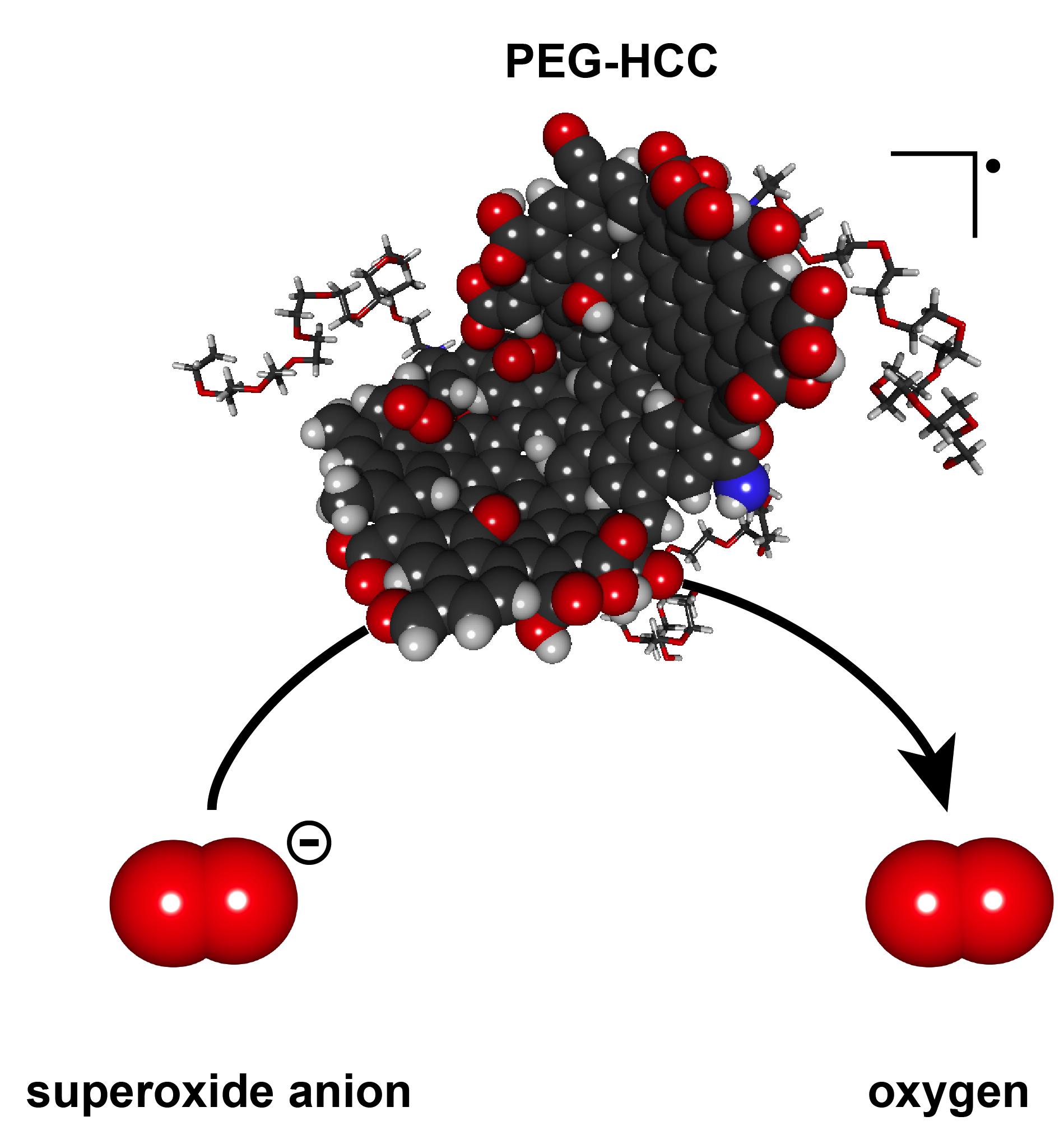
The soluble nanoparticles synthesized by the Rice lab of chemist James Tour have shown no signs of acute toxicity in prior rodent studies, Huq said. They combine polyethylene glycol with hydrophilic carbon clusters, hence their name, PEG-HCCs. The carbon clusters are 35 nanometers long, 3 nanometers wide and an atom thick, and bulk up to about 100 nanometers in globular form with the addition of PEG. They have proven to be efficient scavengers of reactive oxygen species called superoxide molecules, which are expressed by cells the immune system uses to kill invading microorganisms.

T cells use superoxide in a signaling step to become activated. PEG-HCCs remove this superoxide from the T cells, preventing their activation without killing the cells.

Beeton became aware of PEG-HCCs during a presentation by former Baylor graduate student Taeko Inoue, a co-author of the new study. “As she talked, I was thinking, ‘That has to work in models of multiple sclerosis,’” Beeton said. “I didn’t have a good scientific rationale, but I asked for a small sample of PEG-HCCs to see if they affected immune cells.

“We found they affected the T lymphocytes and not the other splenic immune cells, like the macrophages. It was completely unexpected,” she said.

The Baylor lab’s tests on animal models showed that small amounts of PEG-HCCs injected under the skin are slowly taken up by T lymphocytes, where they collect and inhibit the cell’s function. They also found the nanoparticles did not remain in T cells and dispersed within days after uptake by the cells.

“That’s an issue because you want a drug that’s in the system long enough to be effective, but not so long that, if you have a problem, you can’t remove it,” Beeton said. “PEG-HCCs can be administered for slow release and don’t stay in the system for long. This gives us much better control over the circulating half-life.”

“The more we study the abilities of these nanoparticles, the more surprised we are at how useful they could be for medical applications,” Tour said. The Rice lab has published papers with collaborators at Baylor and elsewhere on using functionalized nanoparticles to deliver cancer drugs to tumors and to quench the overproduction of superoxides after traumatic brain injuries.

Beeton suggested delivering carbon nanoparticles just under the skin rather than into the bloodstream would keep them in the system longer, making them more available for uptake by T cells. And the one drawback – a temporary but visible spot on the skin that looks like a tattoo – could actually be a perk to some.

“We saw it made a black mark when we injected it, and at first we thought that’s going to be a real problem if we ever take it into the clinic,” Beeton said. “But we can work around that. We can inject into an area that’s hidden, or use micropattern needles and shape it.

“I can see doing this for a child who wants a tattoo and could never get her parents to go along,” she said. “This will be a good way to convince them.”

The research was supported by Baylor College of Medicine, the National Multiple Sclerosis Society, National Institutes of Health, the Dan L. Duncan Cancer Center, John S. Dunn Gulf Coast Consortium for Chemical Genomics and the U.S. Army-funded Traumatic Brain Injury Consortium.

That’s an interesting list of funders at the end of the news release.

Here’s a link to and a citation for the paper,

Preferential uptake of antioxidant carbon nanoparticles by T lymphocytes for immunomodulation by Redwan Huq, Errol L. G. Samuel, William K. A. Sikkema, Lizanne G. Nilewski, Thomas Lee, Mark R. Tanner, Fatima S. Khan, Paul C. Porter, Rajeev B. Tajhya, Rutvik S. Patel, Taeko Inoue, Robia G. Pautler, David B. Corry, James M. Tour, & Christine Beeton. Scientific Reports 6, Article number: 33808 (2016) doi:10.1038/srep33808 Published online: 22 September 2016

This paper is open access.

Here’s an image provided by the researchers,

Polyethylene glycol-hydrophilic carbon clusters developed at Rice University were shown to be selectively taken up by T cells, which inhibits their function, in tests at Baylor College of Medicine. The researchers said the nanoparticles could lead to new strategies for controlling autoimmune diseases like multiple sclerosis. (Credit: Errol Samuel/Rice University) - See more at: http://news.rice.edu/2016/09/22/tattoo-therapy-could-ease-chronic-disease/#sthash.sIfs3b0S.dpuf

Polyethylene glycol-hydrophilic carbon clusters developed at Rice University were shown to be selectively taken up by T cells, which inhibits their function, in tests at Baylor College of Medicine. The researchers said the nanoparticles could lead to new strategies for controlling autoimmune diseases like multiple sclerosis. (Credit: Errol Samuel/Rice University)

Counteracting chemotherapy resistance with nanoparticles that mimic salmonella

Given the reputation that salmonella (for those who don’t know, it’s a toxin you don’t want to find in your food) has, a nanoparticle which mimics its effects has a certain cachet. An Aug. 22, 2016 news item on Nanowerk,

Researchers at the University of Massachusetts Medical School have designed a nanoparticle that mimics the bacterium Salmonella and may help to counteract a major mechanism of chemotherapy resistance.

Working with mouse models of colon and breast cancer, Beth McCormick, Ph.D., and her colleagues demonstrated that when combined with chemotherapy, the nanoparticle reduced tumor growth substantially more than chemotherapy alone.

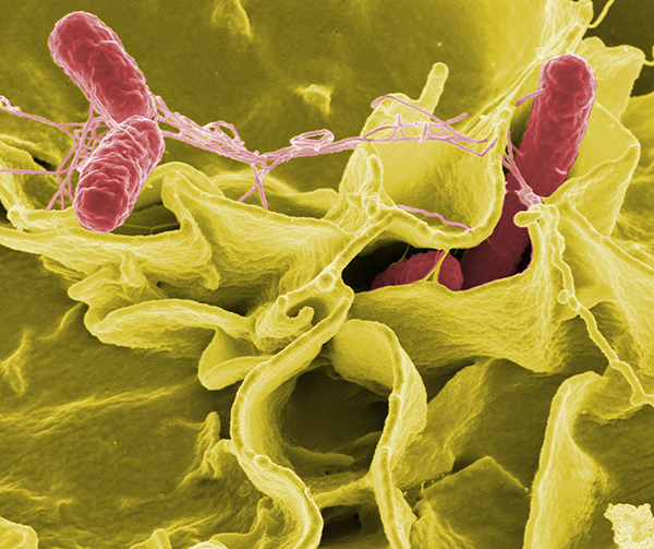
Credit: Rocky Mountain Laboratories,NIAID,NIHColor-enhanced scanning electron micrograph showing Salmonella typhimurium (red) invading cultured human cells.

Credit: Rocky Mountain Laboratories,NIAID,NIHColor-enhanced scanning electron micrograph showing Salmonella typhimurium (red) invading cultured human cells.

An Aug. 22, 2016 US National Institute of Cancer news release, which originated the news item, explains the research in more detail,

A membrane protein called P-glycoprotein (P-gp) acts like a garbage chute that pumps waste, foreign particles, and toxins out of cells. P-gp is a member of a large family of transporters, called ATP-binding cassette (ABC) transporters, that are active in normal cells but also have roles in cancer and other diseases. For instance, cancer cells can co-opt P-gp to rid themselves of chemotherapeutic agents, severely limiting the efficacy of these drugs.

In previous work, Dr. McCormick and her colleagues serendipitously discovered that Salmonella enterica, a bacterium that causes food poisoning, decreases the amount of P-gp on the surface of intestinal cells. Because Salmonella has the capacity to grow selectively in cancer cells, the researchers wondered whether there was a way to use the bacterium to counteract chemotherapy resistance caused by P-gp.

“While trying to understand how Salmonella invades the human host, we made this other observation that may be relevant to cancer therapeutics and multidrug resistance,” explained Dr. McCormick.

Salmonella and Cancer Cells

To determine the specific bacterial component responsible for reducing P-gp levels, the researchers engineered multiple Salmonella mutant strains and tested their effect on P-gp levels in colon cells. They found that a Salmonella strain lacking the bacterial protein SipA was unable to reduce P-gp levels in the colon of mice or in a human colon cancer cell line. Salmonella secretes SipA, along with other proteins, to help the bacterium invade human cells.

The researchers then showed that treatment with SipA protein alone decreased P-gp levels in cell lines of human colon cancer, breast cancer, bladder cancer, and lymphoma.

Because P-gp can pump drugs out of cells, the researchers next sought to determine whether SipA treatment would prevent cancer cells from expelling chemotherapy drugs.

When they treated human colon cancer cells with the chemotherapy agents doxorubicin or vinblastine, with or without SipA, they found that the addition of SipA increased drug retention inside the cells. SipA also increased the cancer cells’ sensitivity to both drugs, suggesting that it could possibly be used to enhance chemotherapy.

“Through millions of years of co-evolution, Salmonella has figured out a way to remove this transporter from the surface of intestinal cells to facilitate host infection,” said Dr. McCormick. “We capitalized on the organism’s ability to perform that function.”

A Nanoparticle Mimic

It would not be feasible to infect people with the bacterium, and SipA on its own will likely deteriorate quickly in the bloodstream, coauthor Gang Han, Ph.D., of the University of Massachusetts Medical School, explained in a press release. The researchers therefore fused SipA to gold nanoparticles, generating what they refer to as a nanoparticle mimic of Salmonella. They designed the nanoparticle to enhance the stability of SipA, while retaining its ability to interact with other proteins.

In an effort to target tumors without harming healthy tissues, the researchers used a nanoparticle of specific size that should only be able to access the tumor tissue due to its “leaky” architecture. “Because of this property, we are hoping to be able to avoid negative effects to healthy tissues,” said Dr. McCormick. Another benefit of this technology is that the nanoparticle can be modified to enhance tumor targeting and minimize the potential for side effects, she added.

The researchers showed that this nanoparticle was 100 times more effective than SipA protein alone at reducing P-gp levels in a human colon cancer cell line. The enhanced function of the nanoparticle is likely due to stabilization of SipA, explained the researchers.

The team then tested the nanoparticle in a mouse model of colon cancer, because this cancer type is known to express high levels of P-gp. When they treated tumor-bearing mice with the nanoparticle plus doxorubicin, P-gp levels dropped and the tumors grew substantially less than in mice treated with the nanoparticle or doxorubicin alone. The researchers observed similar results in a mouse model of human breast cancer.

There are concerns about the potential effect of nanoparticles on normal tissues. “P-gp has evolved as a defense mechanism” to rid healthy cells of toxic molecules, said Suresh Ambudkar, Ph.D., deputy chief of the Laboratory of Cell Biology in NCI’s Center for Cancer Research. It plays an important role in protecting cells of the blood-brain barrier, liver, testes, and kidney. “So when you try to interfere with that, you may create problems,” he said.

The researchers, however, found no evidence of nanoparticle accumulation in the brain, heart, kidney, or lungs of mice, nor did it appear to cause toxicity. They did observe that the nanoparticles accumulated in the liver and spleen, though this was expected because these organs filter the blood, said Dr. McCormick.

Moving Forward

The research team is moving forward with preclinical studies of the SipA nanoparticle to test its safety and toxicity, and to establish appropriate dosage levels.

However, Dr. Ambudkar noted, “the development of drug resistance in cancer cells is a multifactorial process. In addition to the ABC transporters, other phenomena are involved, such as drug metabolism.” And because there is a large family of ABC transporters, one transporter can compensate if another is blocked, he explained.

For the last 25 years, clinical trials with drugs that inhibit P-gp have failed to overcome chemotherapy resistance, Dr. Ambudkar said. Tackling the issue of multidrug resistance in cancer, he continued, “is not something that can be solved easily.”

Dr. McCormick and her team are also pursuing research to better characterize and understand the biology of SipA. “We are not naïve about the complexity of the problem,” she said. “However, if we know more about the biology, we believe we can ultimately make a better drug.”

Here’s a link to and a citation for the paper,

A Salmonella nanoparticle mimic overcomes multidrug resistance in tumours by Regino Mercado-Lubo, Yuanwei Zhang, Liang Zhao, Kyle Rossi, Xiang Wu, Yekui Zou, Antonio Castillo, Jack Leonard, Rita Bortell, Dale L. Greiner, Leonard D. Shultz, Gang Han, & Beth A. McCormick. Nature Communications 7, Article number: 12225  doi:10.1038/ncomms12225 Published 25 July 2016

This paper is open access.

Nanoparticles could make blood clot faster

It was the 252nd meeting for the American Chemical Society from Aug. 21 – 25, 2016 and that meant a flurry of news about the latest research. From an Aug. 23, 2016 news item on Nanowerk,

Whether severe trauma occurs on the battlefield or the highway, saving lives often comes down to stopping the bleeding as quickly as possible. Many methods for controlling external bleeding exist, but at this point, only surgery can halt blood loss inside the body from injury to internal organs. Now, researchers have developed nanoparticles that congregate wherever injury occurs in the body to help it form blood clots, and they’ve validated these particles in test tubes and in vivo [animal testing].

The researchers will present their work today [Aug. 22, 2016] at the 252nd National Meeting & Exposition of the American Chemical Society (ACS). ACS, the world’s largest scientific society, is holding the meeting here through Thursday. It features more than 9,000 presentations on a wide range of science topics.

An Aug. 22, 2016 American Chemical Society (ACS) news release (also on EurekAlert), which originated the news item, provided more detail,

“When you have uncontrolled internal bleeding, that’s when these particles could really make a difference,” says Erin B. Lavik, Sc.D. “Compared to injuries that aren’t treated with the nanoparticles, we can cut bleeding time in half and reduce total blood loss.”

Trauma remains a top killer of children and younger adults, and doctors have few options for treating internal bleeding. To address this great need, Lavik’s team developed a nanoparticle that acts as a bridge, binding to activated platelets and helping them join together to form clots. To do this, the nanoparticle is decorated with a molecule that sticks to a glycoprotein found only on the activated platelets.

Initial studies suggested that the nanoparticles, delivered intravenously, helped keep rodents from bleeding out due to brain and spinal injury, Lavik says. But, she acknowledges, there was still one key question: “If you are a rodent, we can save your life, but will it be safe for humans?”

As a step toward assessing whether their approach would be safe in humans, they tested the immune response toward the particles in pig’s blood. If a treatment triggers an immune response, it would indicate that the body is mounting a defense against the nanoparticle and that side effects are likely. The team added their nanoparticles to pig’s blood and watched for an uptick in complement, a key indicator of immune activation. The particles triggered complement in this experiment, so the researchers set out to engineer around the problem.

“We made a battery of particles with different charges and tested to see which ones didn’t have this immune-response effect,” Lavik explains. “The best ones had a neutral charge.” But neutral nanoparticles had their own problems. Without repulsive charge-charge interactions, the nanoparticles have a propensity to aggregate even before being injected. To fix this issue, the researchers tweaked their nanoparticle storage solution, adding a slippery polymer to keep the nanoparticles from sticking to each other.

Lavik also developed nanoparticles that are stable at higher temperatures, up to 50 degrees Celsius (122 degrees Fahrenheit). This would allow the particles to be stored in a hot ambulance or on a sweltering battlefield.

In future studies, the researchers will test whether the new particles activate complement in human blood. Lavik also plans to identify additional critical safety studies they can perform to move the research forward. For example, the team needs to be sure that the nanoparticles do not cause non-specific clotting, which could lead to a stroke. Lavik is hopeful though that they could develop a useful clinical product in the next five to 10 years.

It’s not unusual for scientists to give an estimate of 5 – 10 years before their science reaches the market.  Another popular range is 3 – 5 years.