Tag Archives: nanoparticles

Discovering how the liver prevents nanoparticles from reaching cancer cells

There’s a lot of excitement about nanoparticles as enabling a precise drug delivery system but to date results have been disappointing as a team of researchers at the University of Toronto (Canada) noted recently (see my April 27, 2016 posting). According to those researchers, one of the main problems with the proposed nanoparticle drug delivery system is that we don’t understand how the body delivers materials to cells and disappointingly few nanoparticles (less than 1%) make their way to tumours. That situation may be changing.

An Aug. 19, 2016 news item on Nanowerk announces the latest research from the University of Toronto,

The emerging field of nanomedicine holds great promise in the battle against cancer. Particles the size of protein molecules can be customized to carry tumour-targeting drugs and destroy cancer cells without harming healthy tissue.

But here’s the problem: when nanoparticles are administered into the body, more than 99 per cent of them become trapped in non-targeted organs, such as the liver and spleen. These nanoparticles are not delivered to the site of action to carry out their intended function.

To solve this problem, researchers at the University of Toronto and the University Health Network have figured out how the liver and spleen trap intact nanoparticles as they move through the organ. “If you want to unlock the promise of nanoparticles, you have to understand and solve the problem of the liver,” says Dr. Ian McGilvray, a transplant surgeon at the Toronto General Hospital and scientist at the Toronto General Research Institute (TGRI).

An Aug. 15, 2016 University of Toronto news release by Luke Ng, which originated the news item, expands on the theme,

In a recent paper in the journal Nature Materials, the researchers say that as nanoparticles move through the liver sinusoid, the flow rate slows down 1,000 times, which increases the interaction of the nanoparticles all of types of liver cells. This was a surprising finding because the current thought is that Kupffer cells, responsible for toxin breakdown in the liver, are the ones that gobbles [sic] up the particles.  This study found that liver B-cells and liver sinusoidal endothelial cells are also involved and that the cell phenotype also matters.

“We know that the liver is the principle organ controlling what gets absorbed by our bodies and what gets filtered out—it governs our everyday biological functions,” says Dr. Kim Tsoi (… [and] research partner Sonya MacParland), a U of T orthopaedic surgery resident, and a first author of the paper, who completed her PhD in biomedical engineering with Warren Chan (IBBME). “But nanoparticle drug delivery is a newer approach and we haven’t had a clear picture of how they interact with the liver—until now.”

Tsoi and MacParland first examined both the speed and location of their engineered nanoparticles as they moved through the liver.

“This gives us a target to focus on,” says MacParland, an immunology post-doctoral fellow at U of T and TGRI. “Knowing the specific cells to modify will allow us to eventually deliver more of the nanoparticles to their intended target, attacking only the pathogens or tumours, while bypassing healthy cells.”

“Many prior studies that have tried to reduce nanomaterial clearance in the liver have focused on the particle design itself,” says Chan. “But our work now gives greater insight into the biological mechanisms underpinning our experimental observations — now we hope to use our fundamental findings to help design nanoparticles that work with the body, rather than against it.”

Here’s a link to and a citation for the paper,

Mechanism of hard-nanomaterial clearance by the liver by Kim M. Tsoi, Sonya A. MacParland, Xue-Zhong Ma, Vinzent N. Spetzler, Juan Echeverri, Ben Ouyang, Saleh M. Fadel, Edward A. Sykes, Nicolas Goldaracena, Johann M. Kaths, John B. Conneely, Benjamin A. Alman, Markus Selzner, Mario A. Ostrowski, Oyedele A. Adeyi, Anton Zilman, Ian D. McGilvray, & Warren C. W. Chan. Nature Materials (2016) doi:10.1038/nmat4718 Published online 15 August 2016

This paper is behind a paywall.
Open access to nanoparticles and nanocomposites

One of the major issues for developing nanotechnology-enabled products is access to nanoparticles and nanocomposites. For example, I’ve had a number of requests from entrepreneurs for suggestions as to how to access cellulose nanocrystals (CNC) so they can develop a product idea. (It’s been a few years since the last request and I hope that means it’s easier to get access to CNC.)

Regardless, access remains a problem and the European Union has devised a solution which allows open access to nanoparticles and nanocomposites through project Co-Pilot. The announcement was made in a May 10, 2016 news item on Nanowerk (Note: A link has been removed),

“What opportunities does the nanotechnology provide in general, provide nanoparticles for my products and processes?” So far, this question cannot be answered easily. Preparation and modification of nanoparticles and the further processing require special technical infrastructure and complex knowledge. For small and medium businesses the construction of this infrastructure “just on luck” is often not worth it. Even large companies shy away from the risks. As a result many good ideas just stay in the drawer.

A simple and open access to high-class infrastructure for the reliable production of small batches of functionalized nanoparticles and nanocomposites for testing could ease the way towards new nano-based products for chemical and pharmaceutical companies. The European Union has allocated funds for the construction of a number of pilot lines and open-access infrastructure within the framework of the EU project CoPilot.

A May 9, 2016 Fraunhofer-Institut für Silicatforschung press release, which originated the news item, offers greater description,

A simple and open access to high-class infrastructure for the reliable production of small batches of functionalized nanoparticles and nanocomposites for testing could ease the way towards new nano-based products for chemical and pharmaceutical companies. The European Union has allocated funds for the construction of a number of pilot lines and open-access infrastructure within the framework of the EU project CoPilot. A consortium of 13 partners from research and industry, including nanotechnology specialist TNO from the Netherlands and the Fraunhofer Institute for Silicate Research ISC from Wuerzburg, Germany as well as seven nanomaterial manufacturers, is currently setting up the pilot line in Wuerzburg. First, they establish the particle production, modification and compounding on pilot scale based on four different model systems. The approach enables maximum variability and flexibility for the pilot production of various particle systems and composites. Two further open access lines will be established at TNO in Eindhoven and at the Sueddeutsche Kunststoffzentrum SKZ in Selb.

The “nanoparticle kitchen”

Essential elements of the pilot line in Wuerzburg are the particle synthesis in batches up to 100 liters, modification and separation methods such as semi-continuous operating centrifuge and in-line analysis and techniques for the uniform and agglomeration free incorporation of nanoparticles into composites. Dr. Karl Mandel, head of Particle Technology of Fraunhofer ISC, compares the pilot line with a high-tech kitchen: “We provide the top-notch equipment and the star chefs to synthesize a nano menu à la carte as well as nanoparticles according to individual requests. Thus, companies can test their own receipts – or our existing receipts – before they practice their own cooking or set up their nano kitchen.”

In the future, the EU project offers companies a contact point if they want to try their nano idea and require enough material for sampling and estimation of future production costs. This can, on the one hand, minimize the development risk, on the other hand, it maximizes the flexibility and production safety. To give lots of companies the opportunity to influence direction and structure/formation/setup of the nanoparticle kitchen, the project partners will offer open meetings on a regular basis.

I gather Co-Pilot has been offering workshops. The next is in July 2016 according to the press release,

The next workshop in this context takes place at Fraunhofer ISC in Wuerzburg, 7h July 2016. The partners present the pilot line and the first results of the four model systems – double layered hydroxide nanoparticle polymer composites for flame inhibiting fillers, titanium dioxide nanoparticles for high refractive index composites, magnetic particles for innovative catalysts and hollow silica composites for anti-glare coatings. Interested companies can find more information about the upcoming workshop on the website of the project www.h2020copilot.eu and on the website of Fraunhofer ISC www.isc.fraunhofer.de that hosts the event.

I tracked down a tiny bit more information about the July 2016 workshop in a May 2, 2016 Co-Pilot press release,

On July 7 2016, the CoPilot project partners give an insight view of the many new functionalization and applications of tailored nanoparticles in the workshop “The Nanoparticle Kitchen – particles und functions à la carte”, taking place in Wuerzburg, Germany. Join the Fraunhofer ISC’s lab tour of the “Nanoparticle Kitchen”, listen to the presentations of research institutes and industry and discuss your ideas with experts. Nanoparticles offer many options for today’s and tomorrow’s products.

More about program and registration soon on this [CoPilot] website!

I wonder if they’re considering this open access to nanoparticles and nanocomposites approach elsewhere?

Mass production of nanoparticles?

With all the years of nanotechnology and nanomaterials research it seems strange that mass production of nanoparticles is still very much in the early stages as a Feb. 24, 2016 news item on phys.org points out,

Nanoparticles – tiny particles 100,000 times smaller than the width of a strand of hair – can be found in everything from drug delivery formulations to pollution controls on cars to HD TV sets. With special properties derived from their tiny size and subsequently increased surface area, they’re critical to industry and scientific research.

They’re also expensive and tricky to make.

Now, researchers at USC [University of Southern California] have created a new way to manufacture nanoparticles that will transform the process from a painstaking, batch-by-batch drudgery into a large-scale, automated assembly line.

A Feb. 24, 2016 USC news release (also on EurekAlert) by Robert Perkins, which originated the news item, offers additional insight,

Consider, for example, gold nanoparticles. They have been shown to easily penetrate cell membranes without causing any damage — an unusual feat given that most penetrations of cell membranes by foreign objects can damage or kill the cell. Their ability to slip through the cell’s membrane makes gold nanoparticles ideal delivery devices for medications to healthy cells or fatal doses of radiation to cancer cells.

However, a single milligram of gold nanoparticles currently costs about $80 (depending on the size of the nanoparticles). That places the price of gold nanoparticles at $80,000 per gram while a gram of pure, raw gold goes for about $50.

“It’s not the gold that’s making it expensive,” Malmstadt [Noah Malmstadt of the USC Viterbi School of Engineering] said. “We can make them, but it’s not like we can cheaply make a 50-gallon drum full of them.”

A fluid situation

At this time, the process of manufacturing a nanoparticle typically involves a technician in a chemistry lab mixing up a batch of chemicals by hand in traditional lab flasks and beakers.

The new technique used by Brutchey [Richard Brutchey of the USC Dornsife College of Letters, Arts and Sciences] and Malmstadt instead relies on microfluidics — technology that manipulates tiny droplets of fluid in narrow channels.

“In order to go large scale, we have to go small,” Brutchey said.

Really small.

The team 3-D printed tubes about 250 micrometers in diameter, which they believe to be the smallest, fully enclosed 3-D printed tubes anywhere. For reference, your average-sized speck of dust is 50 micrometers wide.

They they built a parallel network of four of these tubes, side-by-side, and ran a combination of two nonmixing fluids (like oil and water) through them. As the two fluids fought to get out through the openings, they squeezed off tiny droplets. Each of these droplets acted as a micro-scale chemical reactor in which materials were mixed and nanoparticles were generated. Each microfluidic tube can create millions of identical droplets that perform the same reaction.

This sort of system has been envisioned in the past, but it hasn’t been able to be scaled up because the parallel structure meant that if one tube got jammed, it would cause a ripple effect of changing pressures along its neighbors, knocking out the entire system. Think of it like losing a single Christmas light in one of the old-style strands — lose one and you lose them all.

Brutchey and Malmstadt bypassed this problem by altering the geometry of the tubes themselves, shaping the junction between the tubes such that the particles come out a uniform size and the system is immune to pressure changes.

Here’s a link to and a citation for the paper,

Flow invariant droplet formation for stable parallel microreactors by Carson T. Riche, Emily J. Roberts, Malancha Gupta, Richard L. Brutchey & Noah Malmstadt. Nature Communications 7, Article number: 10780 doi:10.1038/ncomms10780 Published 23 February 2016

This is an open access paper.

Combining gold and palladium for catalytic and plasmonic octopods

Hopefully I did not the change meaning when I made the title for this piece more succinct. In any event, this research comes from the always prolific Rice University in Texas, US (from a Nov. 30, 2015 news item on Nanotechnology Now),

Catalysts are substances that speed up chemical reactions and are essential to many industries, including petroleum, food processing and pharmaceuticals. Common catalysts include palladium and platinum, both found in cars’ catalytic converters. Plasmons are waves of electrons that oscillate in particles, usually metallic, when excited by light. Plasmonic metals like gold and silver can be used as sensors in biological applications and for chemical detection, among others.

Plasmonic materials are not the best catalysts, and catalysts are typically very poor for plasmonics. But combining them in the right way shows promise for industrial and scientific applications, said Emilie Ringe, a Rice assistant professor of materials science and nanoengineering and of chemistry who led the study that appears in Scientific Reports.

“Plasmonic particles are magnets for light,” said Ringe, who worked on the project with colleagues in the U.S., the United Kingdom and Germany. “They couple with light and create big electric fields that can drive chemical processes. By combining these electric fields with a catalytic surface, we could further push chemical reactions. That’s why we’re studying how palladium and gold can be incorporated together.”

The researchers created eight-armed specks of gold and coated them with a gold-palladium alloy. The octopods proved to be efficient catalysts and sensors.

A Nov. 30, 2015 Rice University news release (also on EurekAlert), which originated the news item, expands on the theme,

“If you simply mix gold and palladium, you may end up with a bad plasmonic material and a pretty bad catalyst, because palladium does not attract light like gold does,” Ringe said. “But our particles have gold cores with palladium at the tips, so they retain their plasmonic properties and the surfaces are catalytic.”

Just as important, Ringe said, the team established characterization techniques that will allow scientists to tune application-specific alloys that report on their catalytic activity in real time.

The researchers analyzed octopods with a variety of instruments, including Rice’s new Titan Themis microscope, one of the most powerful electron microscopes in the nation. “We confirmed that even though we put palladium on a particle, it’s still capable of doing everything that a similar gold shape would do. That’s really a big deal,” she said.

“If you shine a light on these nanoparticles, it creates strong electric fields. Those fields enhance the catalysis, but they also report on the catalysis and the molecules present at the surface of the particles,” Ringe said.

The researchers used electron energy loss spectroscopy, cathodoluminescence and energy dispersive X-ray spectroscopy to make 3-D maps of the electric fields produced by exciting the plasmons. They found that strong fields were produced at the palladium-rich tips, where plasmons were the least likely to be excited.

Ringe expects further research will produce multifunctional nanoparticles in a variety of shapes that can be greatly refined for applications. Her own Rice lab is working on a metal catalyst to turn inert petroleum derivatives into backbone molecules for novel drugs.

Here’s a link to and a citation for the paper,

Resonances of nanoparticles with poor plasmonic metal tips by Emilie Ringe, Christopher J. DeSantis, Sean M. Collins, Martial Duchamp, Rafal E. Dunin-Borkowski, Sara E. Skrabalak, & Paul A. Midgley.  Scientific Reports 5, Article number: 17431 (2015)  doi:10.1038/srep17431 Published online: 30 November 2015

This is an open access paper,

At the root of nanotechnology: advances in dentistry

I couldn’t resist the dental wordplay in my headline. Strictly speaking this posting features a research paper that is looking into dentistry’s nanotechnology-enabled future. From an Oct. 19, 2015 news item on phys.org,

Have a cavity? Ask your dentist about filling it with a mixture of nanoparticles including silica and zirconia. These white fillings (known as nano-composite resins) resemble teeth better than their metal alternatives and are less likely to come loose or fracture teeth. This is just the beginning argue Brazilian scientists in a review of “nanodentistry,” published October 19 [2015] in Trends in Biotechnology. Next-generation dental materials incorporating nanotechnology aim to help teeth self-heal, rebuild enamel, and protect against bacterial infections.

An Oct. 19, 2015 Cell Press news release on EurekAlert, which originated the news item, expands on the theme,

“Nanotechnology can be faced sometimes as a paradigm that promised a lot and delivered very little,” says senior author Nelson Durán of the Universidade Estadual de Campinas. “The evolution of dental materials though nanotechnology is real and remarkable, reflecting on a billionaire market. In this way, dentistry was in fact one of the most benefited areas from the development of nanotechnology.”

Since the introduction of nano-composite resins a decade ago, engineers have been exploring how else nanotechnology can safely be used in the dentist’s office. Products could include antimicrobial adhesives made up of carbon nanotubes–creating a kind of wearable toothpaste–or quantum dots combined with cancer-specific antibodies that can be applied inside the mouth, emitting light if they detect any troublesome cells.

“The remineralization of enamel and dentin with the use of nanoparticles (incorporated in different vehicles), a key issue for improving the quality and longevity of resin restorations, is being currently investigated,” says co-author Amauri Jardim de Paula of Universidade Federal do Ceará. “A future perspective is that nanoparticles incorporated in dental materials will prevent and/or control oral diseases through their long-term release and action.”

Although nanodental technologies have evolved quickly, safety and cost will be barriers to getting them on the market. Some nanomaterials might be toxic to healthy cells, so any new nanomaterials to be used for dentistry would need formal pre-clinical and clinical trials before they can receive approval. Patients will also need to be told that a treatment will use materials in the nanometer size range and should be aware of any possible side effects. This new technology could also be expensive, and insurance companies may not want to foot the bill if treatments could be considered cosmetic; composite resins, for example, are still an out-of-pocket cost.

The review authors believe these hurdles can be overcome, however, and that new nanodental products should be available within a few years.

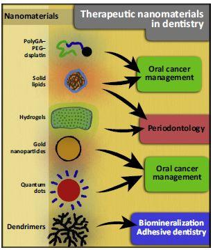
The research has been illustrated,

Caption: This schematic represents the current use and perspectives on the use of nanomaterials on therapeutic dentistry. Credit: Padovania et al./Trends in Biotechnology 2015

Caption: This schematic represents the current use and perspectives on the use of nanomaterials on therapeutic dentistry.
Credit: Padovania et al./Trends in Biotechnology 2015

Here’s a link to and a citation for the paper,

Advances in Dental Materials through Nanotechnology: Facts, Perspectives and Toxicological Aspects by Gislaine C. Padovani, Victor P. Feitosa, Salvatore Sauro, Franklin R. Tay, Gabriela Durán, Amauri J. Paula, & Nelson Durán. Trends in Biotechnology – Cell DOI: http://dx.doi.org/10.1016/j.tibtech.2015.09.005 Publication stage: In Press Corrected Proof Published online Oct. 19, 2015

The paper appears to be open access.

Nanotechnology risk perceptions in 2015 from Australia

I haven’t stumbled across a study on the perceptions of risk and nanotechnology in quite a while.  Before commenting on this latest research from the University of Sydney, here’s a link to and a citation for this new Australian study, which is an open access paper,

Perceptions of risk from nanotechnologies and trust in stakeholders: a cross sectional study of public, academic, government and business attitudes by Adam Capon, James Gillespie, Margaret Rolfe, and Wayne Smith. BMC Public Health 2015, 15:424 Published April 26, 2015  DOI: 10.1186/s12889-015-1795-1

According to the authors, this is the first study that surveyed the general public, academics, government officials, and business people with an eye to distinguishing any differences that might exist in their attitudes,

Our study proposes to extend and develop the knowledge base regarding perceptions of risk from nanotechnology and trust by stakeholders. To do this we use a standardised questionnaire across all the stakeholders surveyed. Secondly we examine stakeholder groups beyond highly published scientists and people attending nano conferences/working in nano laboratories that had previously been surveyed to include academic, government and business stakeholders. These three groups were chosen not just for their expertise, but because they represent the interplay of stakeholders most likely to shape policy in this field. Thirdly we seek and report on views of general risk perception (to health) and for specific products (food, cosmetics and sunscreens, medicines, pesticides, tennis racquets and computers) which broadly represent Australian regulatory arms [22]. Finally we explore several trust actors (health department, scientists, journalists and politicians), all of who have the ability to shape policy.

Our study aims to test six hypotheses. First, very little targeted research has been undertaken on differing stakeholder views of risks from nanotechnology. To explore this we hypothesise that public perceptions of risks from nanotechnology will be greater than those held by ‘experts’. Second, existing studies suggest that food and health applications of nanotechnology are likely to arouse more controversy [23]. We will test the hypothesis that the public, academics, government and business respondents will all perceive a higher level of risk in nanotechnologies that penetrate or have close and prolonged contact with the body. Three, there is inconsistent evidence that increased familiarity with nanotechnology is associated with differing perceptions of nanotechnologies [24]. Our third hypothesis proposes that public self-reported familiarity with nanotechnology will be associated with a reduction in risk perception. This relationship will be found with each of the nano products in the study. Four, the public holds less trust in the government agencies with responsibility for regulating nanotechnology than that expressed by people working in nanotechnology based industries/researching nanotechnology [23]. Our fourth hypothesis tests the evidence for this proposition. We hypothesise that the trust the public vests in scientists, the health department, journalists and politicians will be less than those held by business, academic, and government respondents who have an interest in nanotechnology.

The last two hypotheses expand on hypothesis four, examining the trust of the public in greater detail. Studies have shown that the Australian public are more likely to trust scientists and scientific institutions, followed by government agencies with industry and mass media holding the least amount of trust [25],[26]. In our fifth hypothesis we test the proposition that the public will have greatest trust in scientists, followed by the health department with trust in journalists and politicians below these two. Finally, public trust in business leaders [27], science and consumer protection agencies [28] and government agencies [29] have all been associated with decreased nano risk perception. Examining other stakeholders, the greater trust that people working in nanotechnology based industries or researching nanotechnology had with scientists and government agencies, the less they perceived risk from nanotechnology [23],[30]. Our sixth hypothesis is that significant negative associations exist between the trust the public vest in scientists, health department, journalists and politicians and perceived risk of nanotechnology, both when this risk is considered to health and across all risk applications. Understanding this relationship between trust and risk perception is an important avenue for risk communication and education.

As interesting as I find methodology I’m going to skip most of it and focus on the sample size and demographics,

The surveys consisted of 1355 public, 301 academic, 19 government and 21 business responses. Gender representation of the weighted public survey population was comparable to the June 2012 Australian population estimates of approximately 50% male and female. Gender representationa for academic and business responses was more likely to be male (≈70%) while the gender of government respondents was almost evenly balanced.

Three hundred and ninety eight public respondents (30%) were categorised as having no familiarity with nanotechnology, while 528 (39%) were categorised as having some familiarity and 422 (31%) as having moderate familiarity with nanotechnology.

Amongst the academic responses, the best represented area of research (38%) was in the field of nanomaterials. Nanocharacterisation, nanofabrication, nanobiotechnology/nanomedicine, nanoscale theory/computation, nanophotonics, and nanoelectronics/nanomagnetics represented between 15% to 4% per discipline in descending order. The least represented discipline was translational nanoresearch (2%), of which half were involved in nanotoxicology and the other either in ethical or social research on risk/public attitudes/public impact or did not provide a sub specialisation. Of the business responses the greatest percentage of business involvement was in nanomaterial manufacture, importation or research (33% – 23%). Importation of products containing nanomaterials, waste collection/processing and legal issues had little representation. The highest representation of government respondents was health and safety (37%) followed by communication/social impact (26%), business development (16%) and environment (11%).

The analysis of the results is well worth reading,

The Australian public perceives greater risks from manufactured nanomaterials and shows less trust in scientists and the health department to provide protection from possible health effects than academic, business and government stakeholders in the nanotechnology sector. Food applications and cosmetics/sunscreens loom high on the list of public concerns, although medicines and pesticides are also causes of public concern. Policy makers should be aware of these risk and trust disparities and address public sentiment by treating nanotechnology applications in the higher risk areas with greater caution. Risk communication is best placed in the hands of trusted scientists.

I am a little surprised that no mention was made of the nanosunscreen situation of 2012 where a research study found that 13% (originally reported as 17%) of Australians surveyed said they didn’t use any sunscreens due to fear of nanoparticles. I have the story in my Feb. 9, 2012 posting. Be sure to read through to the end as there were a couple of updates.

Microbubbles reform into nanoparticles after bursting

It seems researchers at the Toronto-based (Canada), Princess Margaret Cancer Centre, have developed a new theranostic tool made of microbubbles used for imaging that are then burst into nanoparticles delivering therapeutics. From a March 30, 2015 news item on phys.org,

Biomedical researchers led by Dr. Gang Zheng at Princess Margaret Cancer Centre have successfully converted microbubble technology already used in diagnostic imaging into nanoparticles that stay trapped in tumours to potentially deliver targeted, therapeutic payloads.

The discovery, published online today [March 30, 2015] in Nature Nanotechnology, details how Dr. Zheng and his research team created a new type of microbubble using a compound called porphyrin – a naturally occurring pigment in nature that harvests light.

A March 30, 2015 University Health Network news release on EurekAlert, which originated the news item, describes the laboratory research on mice,

In the lab in pre-clinical experiments, the team used low-frequency ultrasound to burst the porphyrin containing bubbles and observed that they fragmented into nanoparticles. Most importantly, the nanoparticles stayed within the tumour and could be tracked using imaging.

“Our work provides the first evidence that the microbubble reforms into nanoparticles after bursting and that it also retains its intrinsic imaging properties. We have identified a new mechanism for the delivery of nanoparticles to tumours, potentially overcoming one of the biggest translational challenges of cancer nanotechnology. In addition, we have demonstrated that imaging can be used to validate and track the delivery mechanism,” says Dr. Zheng, Senior Scientist at the Princess Margaret and also Professor of Medical Biophysics at the University of Toronto.

Conventional microbubbles, on the other hand, lose all intrinsic imaging and therapeutic properties once they burst, he says, in a blink-of-an-eye process that takes only a minute or so after bubbles are infused into the bloodstream.

“So for clinicians, harnessing microbubble to nanoparticle conversion may be a powerful new tool that enhances drug delivery to tumours, prolongs tumour visualization and enables them to treat cancerous tumours with greater precision.”

For the past decade, Dr. Zheng’s research focus has been on finding novel ways to use heat, light and sound to advance multi-modality imaging and create unique, organic nanoparticle delivery platforms capable of transporting cancer therapeutics directly to tumours.

Interesting development, although I suspect there are many challenges yet to be met such as ensuring the microbubbles consistently arrive at their intended destination in sufficient mass to be effective both for imaging purposes and, later, as nanoparticles for drug delivery purposes.

Here’s a link to and citation for the paper,

In situ conversion of porphyrin microbubbles to nanoparticles for multimodality imaging by Elizabeth Huynh, Ben Y. C. Leung, Brandon L. Helfield, Mojdeh Shakiba, Julie-Anne Gandier, Cheng S. Jin, Emma R. Master, Brian C. Wilson, David E. Goertz, & Gang Zheng. Nature Nanotechnology (2015) doi:10.1038/nnano.2015.25 Published online 30 March 2015

This paper is behind a paywall but a free preview is available via ReadCube Access.

This is one of those times where I’m including the funding agencies and the ‘About’ portions of the news release,

The research published today was funded by the Canadian Institutes of Health Research (CIHR) Frederick Banting and Charles Best Canada Graduate Scholarship, the Emerging Team Grant on Regenerative Medicine and Nanomedicine co-funded by the CIHR and the Canadian Space Agency, the Natural Sciences and Engineering Research Council of Canada, the Ontario Institute for Cancer Research, the International Collaborative R&D Project of the Ministry of Knowledge Economy, South Korea, the Joey and Toby Tanenbaum/Brazilian Ball Chair in Prostate Cancer Research, the Canada Foundation for Innovation and The Princess Margaret Cancer Foundation.

About Princess Margaret Cancer Centre, University Health Network

The Princess Margaret Cancer Centre has achieved an international reputation as a global leader in the fight against cancer and delivering personalized cancer medicine. The Princess Margaret, one of the top five international cancer research centres, is a member of the University Health Network, which also includes Toronto General Hospital, Toronto Western Hospital and Toronto Rehabilitation Institute. All are research hospitals affiliated with the University of Toronto. For more information, go to http://www.theprincessmargaret.ca or http://www.uhn.ca .

I was not expecting to see South Korea or Brazil mentioned in the funding. Generally, when multiple countries are funding research, their own research institutions are also involved. As for the Princess Margaret Cancer Centre being one of the top five such centres internationally, I wonder how these rankings are determined.

Nanoparticles in 3D courtesy of x-rays

A Feb. 4, 2015 Deutsches Elektronen-Synchrotron (DESY) press release (also on EurekAlert) announces a 3D first,

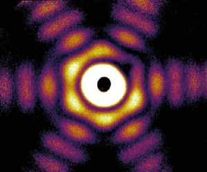
For the first time, a German-American research team has determined the three-dimensional shape of free-flying silver nanoparticles, using DESY’s X-ray laser FLASH. The tiny particles, hundreds of times smaller than the width of a human hair, were found to exhibit an unexpected variety of shapes, as the physicists from the Technical University (TU) Berlin, the University of Rostock, the SLAC National Accelerator Laboratory in the United States and from DESY report in the scientific journal Nature Communications. Besides this surprise, the results open up new scientific routes, such as direct observation of rapid changes in nanoparticles.

The press release goes on to describe the work in more detail,

“The functionality of nanoparticles is linked to their geometric form, which is often very difficult to determine experimentally,” explains Dr. Ingo Barke from the University of Rostock. “This is particularly challenging when they are present as free particles, that is, in the absence of contact with a surface or a liquid.”

The nanoparticle shape can be revealed from the characteristic way how it scatters X-ray light. Therefore, X-ray sources like DESY’s FLASH enable a sort of super microscope into the nano-world. So far, the spatial structure of nanoparticles has been reconstructed from multiple two-dimensional images, which were taken from different angles. This procedure is uncritical for particles on solid substrates, as the images can be taken from many different angles to uniquely reconstruct their three-dimensional shape.

“Bringing nanoparticles into contact with a surface or a liquid can significantly alter the particles, such that you can no longer see their actual form,” says Dr. Daniela Rupp from the TU Berlin. A free particle, however, can only be measured one time in flight before it either escapes or is destroyed by the intense X-ray light. Therefore, the scientists looked for a way to record the entire structural information of a nanoparticle with a single X-ray laser pulse.

To achieve this goal, the scientists led by Prof. Thomas Möller from the TU Berlin and Prof. Karl-Heinz Meiwes-Broer and Prof. Thomas Fennel from the University of Rostock employed a trick. Instead of taking usual small-angle scattering images, the physicists recorded the scattered X-rays in a wide angular range. “This approach virtually captures the structure from many different angles simultaneously from a single laser shot,” explains Fennel.

The researchers tested this method on free silver nanoparticles with diameters of 50 to 250 nanometres (0.00005 to 0.00025 millimetres). The experiment did not only verify the feasibility of the tricky method, but also uncovered the surprising result that large nanoparticles exhibit a much greater variety of shapes than expected.

The shape of free nanoparticles is a result of different physical principles, particularly the particles’ effort to minimize their energy. Consequently, large particles composed of thousands or millions of atoms often yield predictable shapes, because the atoms can only be arranged in a particular way to obtain an energetically favourable state.

In their experiment, however, the researchers observed numerous highly symmetrical three-dimensional shapes, including several types known as Platonic and Archimedean bodies. Examples include the truncated octahedron (a body consisting of eight regular hexagons and six squares) and the icosahedron (a body made up of twenty equilateral triangles). The latter is actually only favourable for extremely small particles consisting of few atoms, and its occurrence with free particles of this size was previously unknown. “The results show that metallic nanoparticles retain a type of memory of their structure, from the early stages of growth to a yet unexplored size range,” emphasizes Barke.

Due to the large variety of shapes, it was especially important to use a fast computational method so that the researchers were capable of mapping the shape of each individual particle. The scientists used a two-step process: the rough shape was determined first and then refined using more complex simulations on a super computer. This approach turned out to be so efficient that it could not only determine various shapes reliably, but could also differentiate between varying orientations of the same shape.

This new method for determining the three-dimensional shape and orientation of nanoparticles with a single X-ray laser shot opens up a wide spectrum of new research directions. In future projects, particles could be directly “filmed” in three dimensions during growth or during phase changes. “The ability to directly film the reaction of a nanoparticle to an intense flash of X-ray light has been a dream for many physicists – this dream could now come true, even in 3D!,” emphasises Rupp.

The researchers have provided an image showing their work,

Caption: This is a wide-angle X-ray diffraction image of a truncated twinned tetrahedra nanoparticle. Credit: Hannes Hartmann/University of Rostock

Caption: This is a wide-angle X-ray diffraction image of a truncated twinned tetrahedra nanoparticle.
Credit: Hannes Hartmann/University of Rostock

Here’s a link to and a citation for the paper,

The 3D-architecture of individual free ​silver nanoparticles captured by X-ray scattering by Ingo Barke, Hannes Hartmann, Daniela Rupp, Leonie Flückiger, Mario Sauppe, Marcus Adolph, Sebastian Schorb, Christoph Bostedt, Rolf Treusch, Christian Peltz, Stephan Bartling, Thomas Fennel, Karl-Heinz Meiwes-Broer, & Thomas Möller. Nature Communications 6, Article number: 6187 doi:10.1038/ncomms7187 Published 04 February 2015

This article is open access.

Of airborne nanomaterials, bacterial microbiomes, viral microbiomes, and paper sensors

There’s a Jan. 14, 2015 news item on Nanowerk from the Virginia Polytechnic Institute (Virginia Tech) which is largely a personal profile featuring some basic information (useful for those new to the topic) about airborne nanoparticles (Note: A link has been removed),

The Harvard educated undergraduate [Linsey Marr,  professor of civil and environmental engineering, Virginia Tech] who obtained her Ph.D. from University of California at Berkeley and trained as a postdoctoral researcher with a Nobel laureate of chemistry at MIT is now among a handful of researchers in the world who are addressing concerns about engineered nanomaterials in the atmosphere.

Marr is part of the National Science Foundation’s Center for the Environmental Implications of Nanotechnology and her research group has characterized airborne nanoparticles at every point of their life cycle. This cycle includes production at a commercial manufacturing facility, use by consumers in the home, and disposal via incineration.

A Jan. 14, 2015 Virginia Tech news release, which originated the news item, quotes Marr on the current thinking about airborne nanoparticles,

“Results have shown that engineered nanomaterials released into the air are often aggregated with other particulate matter, such as combustion soot or ingredients in consumer spray products, and that the size of such aggregates may range from smaller than 10 nanometers to larger than 10 microns,” Marr revealed. She was referring to studies completed by research group members Marina Quadros Vance of Florianopolis, Brazil, a research scientist with the Virginia Tech Institute of Critical Technology and Applied Science, and Eric Vejerano, of Ligao, Philippines, a post-doctoral associate in civil and environmental engineering.

Size matters if these aggregates are inhaled.

Another concern is the reaction of a nanomaterial such as a fullerene with ozone at environmentally relevant concentration levels. Marr’s graduate student, Andrea Tiwari, of Mankato, Minnesota, said the resulting changes in fullerene could lead to enhanced toxicity.

The story then segues into airborne pathogens and viruses eventually honing in on virus microbiomes and bacterial microbiomes (from the news release),

Marr is a former Ironman triathlete who obviously has strong interests in what she is breathing into her own body. So it would be natural for her to expand her study of engineered nanoparticles traveling in the atmosphere to focus on airborne pathogens.

She did so by starting to consider the influenza virus as an airborne pollutant. She applied the same concepts and tools used for studying environmental contaminants and ambient aerosols to the examination of the virus.

She looked at viruses as “essentially self-assembled nanoparticles that are capable of self-replication.”

Her research team became the first to measure influenza virus concentrations in ambient air in a children’s day care center and on airplanes. When they conducted their studies, the Virginia Tech researchers collected samples from a waiting room of a health care center, two toddlers’ rooms and one babies’ area of a childcare center, as well as three cross-country flights between Roanoke, Virginia., and San Francisco. They collected 16 samples between Dec. 10, 2009 and Apr. 22, 2010.

“Half of the samples were confirmed to contain aerosolized influenza A viruses,” Marr said. The childcare samples were the most infected at 75 percent. Next, airplane samples reached 67 percent contamination, and health center numbers came in at 33 percent.

This study serves as a foundation for new work started about a year ago in her lab.

Marr collaborated with Aaron J. Prussin II, of Blacksburg, Virginia, and they successfully secured for him a postdoctoral fellowship from the Alfred P. Sloan Foundation to characterize the bacterial and viral microbiome — the ecological community of microorganisms — of the air in a daycare center.

They are now attempting to determine seasonal changes of both the viral microbiome and the bacterial microbiome in a daycare setting, and examine how changes in the microbiome are related to naturally occurring changes in the indoor environment.

“Little is known about the viral component of the microbiome and it is important because viruses are approximately 10 times more abundant than bacteria, and they help shape the bacterial community. Research suggests that viruses do have both beneficial and harmful interactions with bacteria,” Prussin said.

With Prussin and Marr working together they hope to verify their hypothesis that daycare centers harbor unique, dynamic microbiomes with plentiful bacteria and viruses. They are also looking at what seasonal changes might bring to a daycare setting.

They pointed to the effect of seasonal changes because in previous work, Marr, her former graduate student Wan Yang, of Shantou, China, and Elankumaran Subbiah, a virologist in the biomedical sciences and pathobiology department of the Virginia-Maryland College of Veterinary Medicine, measured the influenza A virus survival rate at various levels of humidity.

Their 2012 study presented for the first time the relationship between the influenza A virus viability in human mucus and humidity over a large range of relative humidities, from 17 percent to 100 percent. They found the viability of the virus was highest when the relative humidity was either close to 100 percent or below 50 percent. The results in human mucus may help explain influenza’s seasonality in different regions.

According to the news release Marr and her colleagues have developed a fast and cheap technology for detection of airborne pathogens (Note: A link has been removed),

With the urgent need to understand the dynamics of airborne pathogens, especially as one considers the threats of bioterrorism, pandemic influenza, and other emerging infectious diseases, Marr said “a breakthrough technology is required to enable rapid, low-cost detection of pathogens in air.”

Along with Subbiah and Peter Vikesland,  professor of civil and environmental engineering, they want to develop readily deployable, inexpensive, paper-based sensors for airborne pathogen detection.

In 2013 they received funding of almost $250,000 from Virginia Tech’s Institute for Critical Technology and Applied Science, a supporter of the clustering of research groups, to support their idea of creating paper-based sensors based on their various successes to date.

Marr explained the sensors “would use a sandwich approach. The bottom layer is paper containing specialized DNA that will immobilize the virus. The middle layer is the virus, which sticks to the specialized DNA on the bottom layer. The top layer is additional specialized DNA that sticks to the virus. This DNA is attached to gold nanoparticles that are easily detectable using a technique known as Raman microscopy.”

They key to their approach is that it combines high-tech with low-tech in the hopes of keeping the assay costs low. Their sampling method will use a bicycle pump, and low cost paper substrates. They hope that they will be able to incorporate smart-phone based signal transduction for the detection. Using this approach, they believe “even remote corners of the world” would be able to use the technique.

Vikesland previously received funding from the Gates Foundation to detect the polio virus via paper-based diagnostics. Polio is still found in countries on the continents of Asia and Africa.

I have previously mentioned Linsey Marr in an Oct. 18, 2013 post about the revival of the Nanotechnology Consumer Products Inventory (originally developed by the Project for Emerging Nanotechnologies) by academics at Virginia Tech and first mentioned CEINT in an Aug. 15, 2011 post about a special project featuring a mesocosm at Duke University (North Carolina).

Two books: Science Ruining Everything and Nanoparticles before nanotechnology

With 20 days left of its Kickstarter campaign, the SCIENCE: Ruining Everything Since 1543 (an SMBC [Saturday Morning Breakfast Cereal] Collection) has amassed $184, 562 for a project with a goal of $20,000.  Some of the more expensive incentives have been snapped but there are still lots of choices. From the campaign page,

SMBC (short for “Saturday Morning Breakfast Cereal”) is a daily-updated comic strip about all sorts of topics. Its author, Zach Weinersmith, is a giant dork who also has many other geeky projects such as producing SMBC Theater, writing for Snowflakes, his science blog the Weinerworks, and his science-themed podcast The Weekly Weinersmith (which he co-hosts with his wife, the parasitologist Kelly Weinersmith).

So it will come as no huge surprise that this, the third SMBC printed collection, is a compendium of his finest science-related strips.

Phil Plait in a Jan. 23, 2013 article for Slate.com describes the project and his involvement,

Today, Zach [Weiner] announced a new and exciting project: He’s collecting his science comics into a single compendium which he’s calling “Science: Ruining Everything Since 1543”. And it’s not just old comics; he’s also written some new ones for the book. He’s creating this book as a Kickstarter project. Give him money, and when it’s done he’ll send you the book.

And there’s more, too: He asked some well-known scientists on the web to send him a personal story about science in their lives, regaling how it’s affected them, and Zach will draw them up as a comic to put in the book. And guess who he asked? Well, the only one he told me about is me, but he assures me there are others. So I sent him an All-True Tale of Bad Astronomy Past, and he created a multi-panel comic about it.

Here’s a representative image of the artwork,

Science: Ruining Everything Since 1543. It's true. Image credit: Zach Weiner [downloaded from http://www.slate.com/blogs/bad_astronomy/2013/01/23/saturday_morning_breakfast_cereal_new_science_book_of_web_comics.html]

Science: Ruining Everything Since 1543. It’s true.
Image credit: Zach Weiner [downloaded from http://www.slate.com/blogs/bad_astronomy/2013/01/23/saturday_morning_breakfast_cereal_new_science_book_of_web_comics.html]

If you go to Phil Plait’s article , you can will find some panels from the collection or you can visit the Science: Ruining Everything campaign page to view a video presentation and learn more about the various incentives.

Meanwhile, the folks at the Nanowiki website have published their third book, Nanoparticles Before Nanotechnology. From the Nanowiki introduction page,

This year we chose the subject of Nanoparticles Before Nanotechnology simply because we were suspicious that nanoparticles are too often, in scientific and non-scientific circles, perceived as invented, while we think they should be understood as discovered.

Nanomaterials are claimed to be so new that people become scared. However, before nanotechnology, one can find nanoparticles in the works of both nature and Man, although they passed unnoticed by us until recently. The origin of nanoparticles in nature is basically i) biogenic, ii) geogenic (and also at the bio-inorganic interface), or iii) cosmogenic, while the nanoparticles produced by men come unintentionally from origins as burning wood and oil or unnoticed in crafted stuff such as cosmetics and colored glass and ceramics.

Additionally, to bring nano closer to society, following the same demystifying aims, we present a set of experiments, Hands On, in which some basics and useful nanoscale phenomena can be easily observed, such as preparing photonic crystals that look like opals, disinfecting water, or harvesting energy.

You can find many options (paper and various e-pub choices)  for getting the book if you follow the link provided previously or you can access the low res web version here as I did. Gorgeous images festoon the book and I wish they’d listed the sources or credits for them. They are truly stunning, even in low res. As for the text, I think they’ve provided a thoughtful compilation of information and organized it very well (I scanned the book quickly). I particularly appreciate having links so I can easily check out the sources for myself and I found a few things I didn’t know about, always a thrill.

In common with most emerging technology topics, knowledge about nanoparticles and other aspects of nanotechnology can change quickly as new data extends or contradicts what was previously believed. Within that context, this is one of the best resources I’ve seen and I’m thankful these folks took the time to pull this book together.