Monthly Archives: April 2017

Canadian Science Policy Centre hosts panel discussion on April 18, 2017 about the April 22, 2017 US March for Science

Coming soon (April 22, 2017) to a city near you is a US ‘March for Science’. The big one will be held in Washington, DC but some 400 satellite marches are planned in cities across the US and around the world.

The Canadian Science Policy Centre has organized two panel discussions (one in Toronto and one in Ottawa) as a prelude to those cities’ marches,

A ‘March for Science’ is set to take place in over 400 locations around the world, including in Ottawa and Toronto, on April 22nd [2017]. The Canadian Science Policy Centre (CSPC) invites you to attend public panels discussing the implications of the march.

To RSVP for the Ottawa event [4:30 pm – 6 pm EDT], please click here

To RSVP for the Toronto event [4:30 – 6:30 pm EDT] please click here

The Ottawa panel features:

Paul Dufour

Paul Dufour is a Fellow and Adjunct Professor at the Institute for Science, Society and Policy in the University of Ottawa and science policy Principal with PaulicyWorks in Gatineau, Québec. He is on the Board of Directors of the graduate student led Science Policy Exchange based in Montréal, and is member of the Investment Committee for Grand Challenges Canada. Paul Dufour has been senior advisor in science policy with several Canadian agencies and organizations over the course of the past 30 years. Among these: Senior Program Specialist with the International Development Research Centre, and interim Executive Director at the former Office of the National Science Advisor to the Canadian Government advising on international S&T matters and broad questions of R&D policy directions for the country. Mr. Dufour lectures regularly on science policy, has authored numerous articles on international S&T relations, and Canadian innovation policy. He is series co-editor of the Cartermill Guides to World Science and is the author of the Canada chapter for the UNESCO 2015 Science Report released in November 2015.

Dr. Kristin Baetz

Dr. Kristin Baetz is a Canada Research Chair in Chemical and Functional Genomics, Director of the Ottawa Institute of Systems Biology at uOttawa, President of the Canadian Society for Molecular Biosciences.

Katie Gibbs

Katie Gibbs is a scientist, community organizer and advocate for science and evidence-based policies. While completing her PhD at the University of Ottawa researching threats to endangered species, she was the lead organizer of the ‘Death of Evidence’ rally which was one of the largest science rallies in Canadian history. Katie is a co-founder and Executive Director of Evidence for Democracy, a national, non-partisan, not-for- profit organization that promotes science integrity and the transparent use of evidence in government decision-making. She has a diverse background organizing and managing various causes and campaigns including playing an integral role in Elizabeth May’s winning election campaign in 2011. Katie is frequently asked to comment on science policy issues and has been quoted and published in numerous media outlets, including the CBC, The Hill Times, the Globe and Mail and the National Post.

Professor Kathryn O’Hara

Professor Kathryn O’Hara has been a faculty member in the School of Journalism and Communication at Carleton University since 2001. She is the first person to hold the School’s CTV Chair in Science Broadcast Journalism, the first such chair of its kind in anglophone Canada. A long-standing broadcast journalist, Professor O’Hara is the former consumer columnist with CBC’s Midday , a former co- anchor of CBC’s Newsday in Ottawa, and the former host of Later the Same Day , CBC Radio Toronto’s “drive-home” program. Her work has also appeared on CBC’s Quirks and Quarks and Ideas programs. Three years before coming to Carleton University, Professor O’Hara was an independent health and science producer for outlets such as RTE and CBC. She serves on the Science and Technology Advisory Boards for Environment Canada and Health Canada and chairs the EC panel on Environment and Health. She is an Associate Professor with the Carleton School of Journalism and Communication.

The Toronto panel is organized a little differently:

Canadian Science Policy Centre in collaboration with Ryerson University’s Faculty of Science presents a panel discussion on the ‘March for Science’. Join us for coffee/tea and light refreshment at 4:00pm followed by the panel discussion at 4:30pm.

Light reception sponsored by Ryerson University’s Faculty of Science

Dr. Imogen Coe

Dr. Imogen R. Coe is currently the Dean of the Faculty of Science at Ryerson University. Imogen possesses a doctorate (Ph.D.) and masters degree in Biology from the University of Victoria, B.C. and a bachelor’s degree from Exeter University in the U.K.  She is an affiliate scientist with Li Ka Shing Knowledge Institute, Keenan Research Centre at St. Michael’s Hospital which is where her research program is located.  She is an accomplished cell biologist and is internationally known for her work on membrane transport proteins (transporters) that are the route of entry into cells for a large class of anti-cancer, anti-viral and anti-parasite drugs.  She has served on NSERC, CIHR and NCIC scientific review panels and continues to supervise research projects of undergraduates, graduate students, postdoctoral fellows and research associates in her group. More about her research can be found  at her research website.

Mehrdad Hariri

Mehrdad Hariri is the founder and CEO of Canadian Science Policy Centre. The Centre is becoming the HUB for science technology and innovation policy in the country. He established the first national annual Canadian Science Policy Conference (CSPC), a forum dedicated to the Canadian Science Technology and Innovation (STI) Policy issues. The Conference engages stakeholders from the science and innovation field, academia and government in discussions of policy issues at the intersection of science and society. Now in its 9th year, CSPC has become the most comprehensive national forum on science and innovation policy issues.

Dr. Jim Woodgett

In his dual roles as Investigator and Director of Research of the Lunenfeld-Tanenbaum Research Institute, Dr. Jim Woodgett applies his visionary approach to research into the manipulation of cell processes to treat certain cancers, diabetes and neurodegenerative conditions, and to ensuring that discoveries made by the world-renowned Institute are applied to patient care. Dr. Woodgett is interested in the causes and treatment of breast cancer, colorectal cancer, diabetes, Alzheimer Disease and bipolar disorder. What links this apparently broad range of diseases is their common basis in disruption of the lines of communication within the cells, or the signalling pathways. By studying the ways in which components of these pathways are mutated and transformed by disease, Dr. Woodgett can identify new and more effective therapeutic targets. Study of the WNT pathway, which contains a number of genes which account for about 90% of human colon cancer, is a particular area of interest. Recent advancements made by Dr. Woodgett’s team in adult stem cell division pave the way for scientists to harvest large quantities of these specialized cells which hold great promise for the treatment and cure of life- threatening illnesses.

Margrit Eichler

Margrit Eichler is Professor emerita of Sociology and Equity Studies at OISE/UT. Her over 200 publications deal, among other topics, with feminist methodology, gender issues, public health, environmental issues, and paid and unpaid work. She is a fellow Fellow of the Royal Society of Canada and the European Academy of Sciences. Since her retirement, she has been active in various citizens’ organizations, including as Secretary of Science for Peace and as President of the advocacy group Our Right to Know.

Ivan Semeniuk [science writer for Globe & Mail newspaper]

Dan Weaver

Dan Weaver is a Ph.D. candidate at the U of T Dept. of Physics. His research involves collecting and analyzing atmospheric measurements taken at the Polar Environment Atmospheric Research Laboratory (PEARL) on Ellesmere Island, Nunavut. He is also involved in the validation of satellites such as Canada’s Atmospheric Chemistry Experiment.In 2012, Dan was at PEARL for fieldwork when the federal government cut science funding that supported PEARL and other research programs across the country. He started a campaign called Save PEARL to advocate for continued funding for climate and Arctic atmospheric research. Dan joined Evidence for Democracy to advocate for science and evidence-based decision-making in 2013 and is a member of its Board of Directors. Dan is also a member of the Toronto March for Science organizing committee.

Toronto tickets are going faster than Ottawa tickets.

I’m feeling just a bit indignant; there are not just two Canadian satellite marches as you might expect given how this notice is written up. There are 18! Eight provinces are represented with marches in Calgary (Alberta), Montréal (Québec), Prince George (British Columbia), Vancouver (British Columbia), Edmonton (Alberta), Winnipeg (Manitoba), Halifax (Nova Scotia), London (Ontario), Windsor (Ontario),  Hamilton (Ontario), Ottawa (Ontario), Toronto (Ontario), Victoria (British Columbia), Lethbridge (Alberta), St. John’s (Newfoundland and Labrador), Kitchener-Waterloo (Ontario), Sudbury (Ontario), and Saskatoon (Saskatchewan). Honestly, these folks in Ontario seem to have gotten quite insular. In any event, you can figure out how to join in by clicking here.

For those who might appreciate some cogent insight into the current science situation in the US (and an antidote to what I suspect will be a great deal of self-congratulation on these April 18, 2017 CSPC panels), there’s an April 14, 2017 article by Jason Lloyd for Slate.com (Note: Links have been removed),

The most prominent response to the situation will come April 22 [2017], as science advocates—including members of major organizations like the Union of Concerned Scientists, the American Geophysical Union, and the American Association for the Advancement of Science—“walk out of the lab and into the streets” for the first-ever March for Science. Modeled in part on January’s record-breaking Women’s March, organizers have planned a march in Washington and satellite marches in more than 400 cities across six continents. The March for Science is intended to be the largest assemblage of science advocates in history.

Too bad it will likely undermine their cause.

The goals of organizers and participants are varied and worthy, but its critics—most prominently the president himself—will smear the march as simply anti-Trump or anti-Republican partisanship. Whether that’s true is beside the point, and scientists who are keen to participate ought to do so without worrying that they’re sullying their objectivity. The many communities distressed by the actions of this administration should of course exercise their right to protest, and the March for Science may inspire deeper social and political engagement.

But participants must understand that the social and political context in which this march takes place means that it cannot produce the outcomes intended by its organizers. The officially nonpartisan march embodies in miniature the larger challenges that confront the scientific enterprise in its relationship with a society that’s undergoing profound and often distressing changes.

Let’s start by looking at what the largest representative of the scientific community, the American Association for the Advancement of Science, intends by endorsing the march. According to the AAAS’s statement of support, the march will help:

…  protect the rights of scientists to pursue and communicate their inquiries unimpeded, expand the placement of scientists throughout the government, build public policies upon scientific evidence, and support broad educational efforts to expand public understanding of the scientific process.

In other words, scientists want support for instructing—not involving—the public in the scientific process, a greater influence on policymaking, and no political accountability. That’s a pretty audacious power play, and it’s easy to see how critics might cast the march’s intent as a privileged group seeking to protect and enhance its privileges. The thing is, they wouldn’t be entirely wrong.

As science policy journalist Colin Macilwain points out in Nature, scientists and other members of the technocratic class have generally enjoyed stable, middle-class employment and society’s respect and admiration for most of the past 70 years. They have benefited from scientific and technological progress while mostly remaining insulated from the collateral damage wrought by creative destruction. Federal funding has remained generous under progressive and conservative governments and through economic booms and busts. Scientists possess a variety of relatively comfortable perches from which they can express their ideas and shape public policy.

But there are a lot of people to whom the past seven decades have not been nearly so kind. They’ve struggled to find and keep well-paying jobs in a world in which technological advancement has decoupled economic growth from employment opportunities. They’ve lost a sense of having their voices heard in policymaking, as governance and regulation becomes increasingly complex. To see a select group of people and institutions profit from this complexity has, understandably, bred resentment throughout post-industrial countries.

So what should scientists do to safeguard and support their community instead? A good first step would be to acknowledge the scope and depth of the problem. The biggest issue confronting science is not a malicious and incompetent executive, or a research enterprise that might receive less generous funding than it’s enjoyed in the past. The critical challenge—and one that will still be relevant long after Donald Trump has gone back to making poor real estate decisions—is figuring out how scientists can build an enduring relationship with all segments of the American public, so that discounting, defunding, or vilifying scientists’ important work is politically intolerable.

This does not excuse whatever appalling policies Trump will no doubt seek to implement, against which scientists should speak out forcefully in the language of public values like free speech. They did this successfully against requests for the names of Department of Energy employees who attended U.N. climate talks and the clampdown on federal agencies’ external communications. But over the longer term, scientists need to improve their connection to the public and articulate their importance to society in a way that resonates with all Americans.

Academia can also challenge the insularity of scientific practice (and not just in the sciences). Instead of an overriding focus on publishing and grants, renewed attention to teaching could train more students in academic rigor and critical appraisal of, among other things, the false claims of a populist demagogue. With research universities scattered throughout the country, academics should be incentivized to improve ties with people who might otherwise consider scientists to be condescending eggheads who only give them bad news about the climate or the economy. University medical centers and military bases provide great models for these types of strong local relationships.

Finally, scientists and technologists must also attend to the social implications of their research. This includes anticipating and mitigating the socioeconomic effects of their innovations (here’s looking at you, Silicon Valley) by allocating resources to address problems they may exacerbate, such as inequality and job loss. The high-level discussion around CRISPR, the revolutionary gene-editing technology, is a good example of both the opportunity for and difficulty of responsible innovation. This process might be made more effective by bringing the public into scientific practice and policymaking using the tools of citizen science and deliberative democracy, rather than simply telling people what scientists are doing or explaining what policymakers have already decided.

If you have the time, please read Lloyd’s piece in its entirety. The piece has certainly generated a fair number of comments (121 when I last looked).

I have run a couple of posts which feature some well-meaning advice for our southern neighbours from Canadians along with my suggestion that they might not be as helpful as we hope.

Jan. 27, 2017 posting (scroll down past the internship announcement, about 15% of the way down)

Feb. 13, 2017 posting

Curcumin gel for burns and scalds

The curcumin debate continues (see my  Jan. 26, 2017 posting titled: Curcumin: a scientific literature review concludes health benefits may be overstated for more about that). In the meantime, scientists at the University of California at Los Angeles’ (UCLA) David Geffen School of Medicine found that curcumin gel might be effective as a treatment for burns. From a March 14, 2017 Pensoft Publishers news release on EurekAlert (Note: Links have been removed),

What is the effect of Topical Curcumin Gel for treating burns and scalds? In a recent research paper, published in the open access journal BioDiscovery, Dr. Madalene Heng, Clinical Professor of Dermatology at the David Geffen School of Medicine, stresses that use of topical curcumin gel for treating skin problems, like burns and scalds, is very different, and appears to work more effectively, when compared to taking curcumin tablets by mouth for other conditions.

“Curcumin gel appears to work much better when used on the skin because the gel preparation allows curcumin to penetrate the skin, inhibit phosphorylase kinase and reduce inflammation,” explains Dr Heng.

In this report, use of curcumin after burns and scalds were found to reduce the severity of the injury, lessen pain and inflammation, and improve healing with less than expected scarring, or even no scarring, of the affected skin. Dr. Heng reports her experience using curcumin gel on such injuries using three examples of patients treated after burns and scalds, and provides a detailed explanation why topical curcumin may work on such injuries.

Curcumin is an ingredient found in the common spice turmeric. Turmeric has been used as a spice for centuries in many Eastern countries and gives well known dishes, such as curry, their typical yellow-gold color. The spice has also been used for cosmetic and medical purposes for just as long in these countries.

In recent years, the medicinal value of curcumin has been the subject of intense scientific studies, with publication numbering in the thousands, looking into the possible beneficial effects of this natural product on many kinds of affliction in humans.

This study published reports that topical curcumin gel applied soon after mild to moderate burns and scalds appears to be remarkably effective in relieving symptoms and improved healing of the affected skin.

“When taken by mouth, curcumin is very poorly absorbed into the body, and may not work as well,” notes Dr. Heng. “Nonetheless, our tests have shown that when the substance is used in a topical gel, the effect is notable.”

The author of the study believes that the effectiveness of curcumin gel on the skin – or topical curcumin – is related to its potent anti-inflammatory activity. Based on studies that she has done both in the laboratory and in patients over 25 years, the key to curcumin’s effectiveness on burns and scalds is that it is a natural inhibitor of an enzyme called phosphorylase kinase.

This enzyme in humans has many important functions, including its involvement in wound healing. Wound healing is the vital process that enables healing of tissues after injury. The process goes through a sequence of acute and chronic inflammatory events, during which there is redness, swelling, pain and then healing, often with scarring in the case of burns and scalds of the skin. The sequence is started by the release of phosphorylase kinase about 5 mins after injury, which activates over 200 genes that are involved in wound healing.

Dr. Heng uses curcumin gel for burns, scalds and other skin conditions as complementary treatment, in addition to standard treatment usually recommended for such conditions.

Caption: These are results from 5 days upon application of curcumin gel to burns, and results after 6 weeks. Credit: Dr. Madalene Heng

Here’s a link to and a citation for the paper,

Phosphorylase Kinase Inhibition Therapy in Burns and Scalds by Madalene Heng. BioDiscovery 20: e11207 (24 Feb 2017) https://doi.org/10.3897/biodiscovery.20.e1120

This paper is in an open access journal.

Bio-based standup pouches (food packaging) made from cellulose

CAPTION: VTT has developed lightweight 100% bio-based stand-up pouches with high technical performance. (Photo by VTT)

A March 14, 2017 news item on ScienceDaily describes a new nanocellulose-based product developed by the Technical Research Centre of Finland (VTT),

VTT Technical Research Centre of Finland Ltd has developed lightweight 100% bio-based stand-up pouches with high technical performance. High performance in both oxygen, grease and mineral oil barrier properties has been reached by using different biobased coatings on paper substrate. The pouches exploit VTT’s patent pending high consistency enzymatic fibrillation of cellulose (HefCel) technology.

A March 14, 2017 VTT press release (also on EurekAlert), which originated the news item, describes why the researchers want to change how food is packaged,

“One-third of food produced for human consumption is lost or wasted globally. Packaging with efficient barrier properties is a crucial factor in the reduction of the food loss. Our solution offers an environmentally friendly option for the global packaging industry”, says Senior Scientist Jari Vartiainen of VTT.

VTT’s HefCel technology provides a low-cost method for the production of nanocellulose resulting in a tenfold increase in the solids content of nanocellulose. Nanocellulose has been shown to be potentially very useful for a number of future technical applications. The densely packed structure of nanocellulose films and coatings enable their outstanding oxygen, grease and mineral oil barrier properties.

HefCel technology exploits industrial enzymes and simple mixing technology as tools to fibrillate cellulose into nanoscale fibrils without the need for high energy consuming process steps. The resulting nanocellulose is in the consistency of 15-25% when traditional nanocellulose production methods result in 1-3% consistency.

The stand-up pouch is the fastest growing type of packaging, growing at a rate of 6.5% per year from 2015-2020. Fossil-based plastic films still dominate the packaging market. However, the development of environmentally friendly new materials is of growing importance. Nanocellulose has been shown to be potentially very useful for a number of future technical applications.

VTT has solid expertise in various bio-based raw materials and their application technologies for producing bio-based coatings, films and even multilayered structures both at lab-scale and pilot-scale. A versatile set of piloting facilities are available from raw material sourcing through processing to application testing and demonstration.

I’m glad to hear they’re finding uses for nanocellulose and I keep wondering when Canadian scientists who at one point were leaders in developing crystal nanocellulose (CNC or sometimes known as nanocrystalline cellulose [NCC]) will be making announcements about potential products.

A new class of artificial retina

If I read the news release rightly (keep scrolling), this particular artificial retina does not require a device outside the body (e.g. specially developed eyeglasses) to capture an image to be transmitted to the implant. This new artificial retina captures the image directly.

The announcement of a new artificial retina is made in a March 13, 2017 news item on Nanowerk (Note: A link has been removed),

A team of engineers at the University of California San Diego and La Jolla-based startup Nanovision Biosciences Inc. have developed the nanotechnology and wireless electronics for a new type of retinal prosthesis that brings research a step closer to restoring the ability of neurons in the retina to respond to light. The researchers demonstrated this response to light in a rat retina interfacing with a prototype of the device in vitro.

They detail their work in a recent issue of the Journal of Neural Engineering (“Towards high-resolution retinal prostheses with direct optical addressing and inductive telemetry”). The technology could help tens of millions of people worldwide suffering from neurodegenerative diseases that affect eyesight, including macular degeneration, retinitis pigmentosa and loss of vision due to diabetes

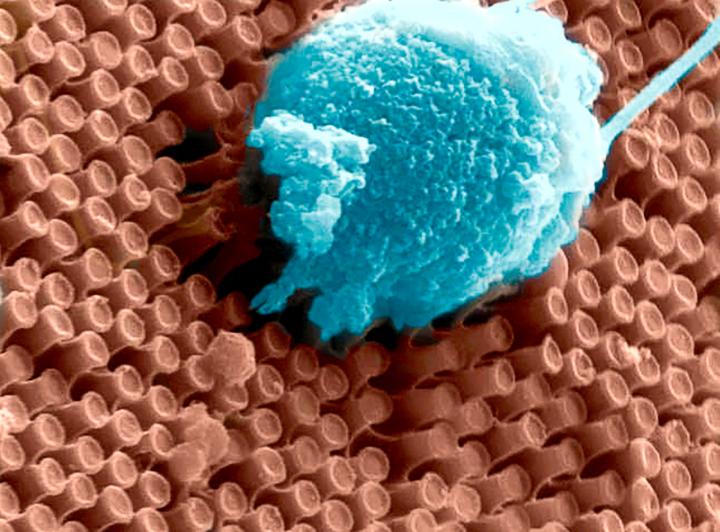
Caption: These are primary cortical neurons cultured on the surface of an array of optoelectronic nanowires. Here a neuron is pulling the nanowires, indicating the the cell is doing well on this material. Credit: UC San Diego

A March 13, 2017 University of California at San Diego (UCSD) news release (also on EurekAlert) by Ioana Patringenaru, which originated the news item, details the new approach,

Despite tremendous advances in the development of retinal prostheses over the past two decades, the performance of devices currently on the market to help the blind regain functional vision is still severely limited–well under the acuity threshold of 20/200 that defines legal blindness.

“We want to create a new class of devices with drastically improved capabilities to help people with impaired vision,” said Gabriel A. Silva, one of the senior authors of the work and professor in bioengineering and ophthalmology at UC San Diego. Silva also is one of the original founders of Nanovision.

The new prosthesis relies on two groundbreaking technologies. One consists of arrays of silicon nanowires that simultaneously sense light and electrically stimulate the retina accordingly. The nanowires give the prosthesis higher resolution than anything achieved by other devices–closer to the dense spacing of photoreceptors in the human retina. The other breakthrough is a wireless device that can transmit power and data to the nanowires over the same wireless link at record speed and energy efficiency.

One of the main differences between the researchers’ prototype and existing retinal prostheses is that the new system does not require a vision sensor outside of the eye [emphasis mine] to capture a visual scene and then transform it into alternating signals to sequentially stimulate retinal neurons. Instead, the silicon nanowires mimic the retina’s light-sensing cones and rods to directly stimulate retinal cells. Nanowires are bundled into a grid of electrodes, directly activated by light and powered by a single wireless electrical signal. This direct and local translation of incident light into electrical stimulation makes for a much simpler–and scalable–architecture for the prosthesis.

The power provided to the nanowires from the single wireless electrical signal gives the light-activated electrodes their high sensitivity while also controlling the timing of stimulation.

“To restore functional vision, it is critical that the neural interface matches the resolution and sensitivity of the human retina,” said Gert Cauwenberghs, a professor of bioengineering at the Jacobs School of Engineering at UC San Diego and the paper’s senior author.

Wireless telemetry system

Power is delivered wirelessly, from outside the body to the implant, through an inductive powering telemetry system developed by a team led by Cauwenberghs.

The device is highly energy efficient because it minimizes energy losses in wireless power and data transmission and in the stimulation process, recycling electrostatic energy circulating within the inductive resonant tank, and between capacitance on the electrodes and the resonant tank. Up to 90 percent of the energy transmitted is actually delivered and used for stimulation, which means less RF wireless power emitting radiation in the transmission, and less heating of the surrounding tissue from dissipated power.

The telemetry system is capable of transmitting both power and data over a single pair of inductive coils, one emitting from outside the body, and another on the receiving side in the eye. The link can send and receive one bit of data for every two cycles of the 13.56 megahertz RF signal; other two-coil systems need at least 5 cycles for every bit transmitted.

Proof-of-concept test

For proof-of-concept, the researchers inserted the wirelessly powered nanowire array beneath a transgenic rat retina with rhodopsin P23H knock-in retinal degeneration. The degenerated retina interfaced in vitro with a microelectrode array for recording extracellular neural action potentials (electrical “spikes” from neural activity).

The horizontal and bipolar neurons fired action potentials preferentially when the prosthesis was exposed to a combination of light and electrical potential–and were silent when either light or electrical bias was absent, confirming the light-activated and voltage-controlled responsivity of the nanowire array.

The wireless nanowire array device is the result of a collaboration between a multidisciplinary team led by Cauwenberghs, Silva and William R. Freeman, director of the Jacobs Retina Center at UC San Diego, UC San Diego electrical engineering professor Yu-Hwa Lo and Nanovision Biosciences.

A path to clinical translation

Freeman, Silva and Scott Thorogood, have co-founded La Jolla-based Nanovision Biosciences, a partner in this study, to further develop and translate the technology into clinical use, with the goal of restoring functional vision in patients with severe retinal degeneration. Animal tests with the device are in progress, with clinical trials following.

“We have made rapid progress with the development of the world’s first nanoengineered retinal prosthesis as a result of the unique partnership we have developed with the team at UC San Diego,” said Thorogood, who is the CEO of Nanovision Biosciences.

Here’s a link to and a citation for the paper,

Towards high-resolution retinal prostheses with direct optical addressing and inductive telemetry by Sohmyung Ha, Massoud L Khraiche, Abraham Akinin, Yi Jing, Samir Damle, Yanjin Kuang, Sue Bauchner, Yu-Hwa Lo, William R Freeman, Gabriel A Silva.Journal of Neural Engineering, Volume 13, Number 5 DOI: https://doi.org/10.1088/1741-2560/13/5/056008

Published 16 August 2016 • © 2016 IOP Publishing Ltd

I’m not sure why they waited so long to make the announcement but, in any event, this paper is behind a paywall.

A new platform for culturing stem cells: a Multiplexed Artificial Cellular Microenvironment array

Japanese scientists have developed a more precise method for culturing stem cells according to a March 14, 2017 news item on Nanowerk,

A team of researchers in Japan has developed a new platform for culturing human pluripotent stem cells that provides far more control of culture conditions than previous tools by using micro and nanotechnologies.

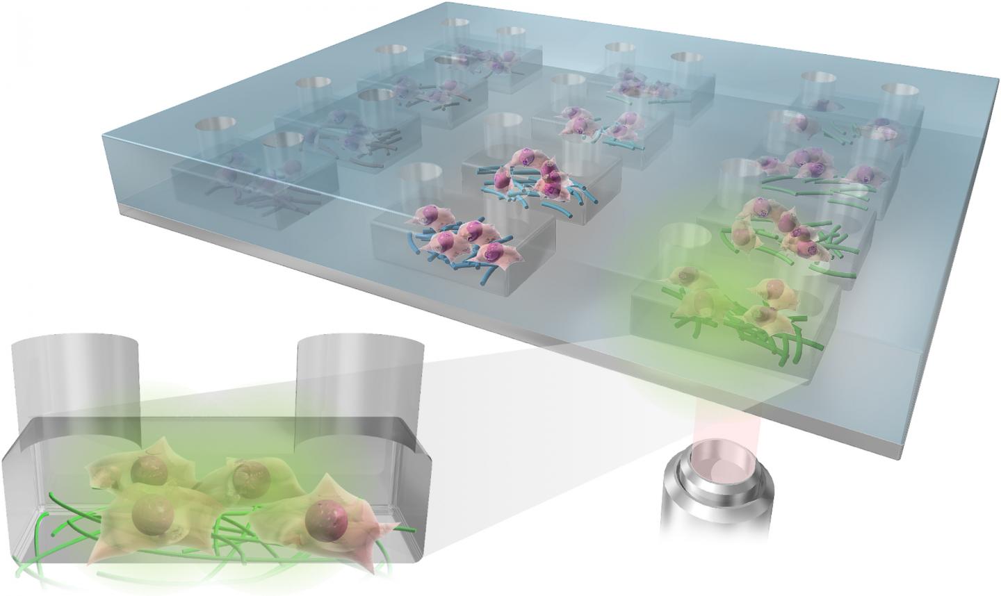
The Multiplexed Artificial Cellular Microenvironment (MACME) array places nanofibres, mimicking cellular matrices, into fluid-filled micro-chambers of precise sizes, which mimic extracellular environments.

Caption: The Multiplexed Artificial Cellular Microenvironment (MACME) array, consisted with a microfluidic structure and nanofibre array for mimicking cellular microenvironments. Credit: Kyoto University iCeMS

A March 17, 2017 Kyoto University press release (also on EurekAlert), which originated the news item, explains the research in more detail,

Human pluripotent stems cells (hPSCs) hold great promise for tissue engineering, regenerative medicine and cell-based therapies because they can become any type of cell. The environment surrounding the cells plays a major role in determining what tissues they become, if they replicate into more cells, or die. However, understanding these interactions has been difficult because researchers have lacked tools that work on the appropriate scale.

Often, stem cells are cultured in a cell culture medium in small petri dishes. While factors such as medium pH levels and nutrients can be controlled, the artificial set up is on the macroscopic scale and does not allow for precise control of the physical environment surrounding the cells.

The MACME array miniaturizes this set up, culturing stem cells in rows of micro-chambers of cell culture medium. It also takes it a step further by placing nanofibers in these chambers to mimic the structures found around cells.

Led by Ken-ichiro Kamei of Kyoto University’s Institute for Integrated Cell-Material Sciences (iCeMS), the team tested a variety of nanofiber materials and densities, micro-chamber heights and initial stem cell densities to determine the best combination that encourages human pluripotent stem cells to replicate.

They stained the cells with several fluorescent markers and used a microscope to see if the cells died, replicated or differentiated into tissues.

Their analysis revealed that gelatin nanofibers and medium-sized chambers that create medium seed cell density provided the best environment for the stem cells to continue to multiply. The quantity and density of neighboring cells strongly influences cell survival.

The array is an “optimal and powerful approach for understanding how environmental cues regulate cellular functions,” the researchers conclude in a recently published paper in the journal Small.

This array appears to be the first time multiple kinds of extracellular environments can be mounted onto a single device, making it much easier to compare how different environments influence cells.

The MACME array could substantially reduce experiment costs compared to conventional tools, in part because it is low volume and requires less cell culture medium. The array does not require any special equipment and is compatible with both commonly used laboratory pipettes and automated pipette systems for performing high-throughput screening.

Here’s a link to and a citation for the paper,

Microfluidic-Nanofiber Hybrid Array for Screening of Cellular Microenvironments by Ken-ichiro Kamei, Yasumasa Mashimo, Momoko Yoshioka, Yumie Tokunaga, Christopher Fockenberg, Shiho Terada, Yoshie Koyama, Minako Nakajima, Teiko Shibata-Seki, Li Liu, Toshihiro Akaike, Eiry Kobatake, Siew-Eng How, Motonari Uesugi, and Yong Chen. Small DOI: 10.1002/smll.201603104 Version of Record online: 8 MAR 2017

© 2017 WILEY-VCH Verlag GmbH & Co. KGaA, Weinheim

This paper is behind a paywall.

Regrowing bone

The ability to grow bone or bone-like material could change life substantially for people with certain kinds of injuries. Scientists at Northwestern University and the University of Chicago have been able to regrow bone in a skull (according to a March 8, 2017 Northwestern University news release (also on EurekAlert),

A team of researchers repaired a hole in a mouse’s skull by regrowing “quality bone,” a breakthrough that could drastically improve the care of people who suffer severe trauma to the skull or face.

The work by a joint team of Northwestern Engineering and University of Chicago researchers was a resounding success, showing that a potent combination of technologies was able to regenerate the skull bone with supporting blood vessels in just the discrete area needed without developing scar tissue — and more rapidly than with previous methods.

“The results are very exciting,” said Guillermo Ameer, professor of biomedical engineering at Northwestern’s McCormick School of Engineering, and professor of surgery at Feinberg School of Medicine.

Supported by the China Scholarship Council, National Institute of Dental and Craniofacial Research, Chicago Community Trust, and National Center for Advancing Translational Sciences, the research was published last week in the journal PLOS One. Russell Reid, associate professor of surgery at the University of Chicago Medical Center, is the article’s corresponding author. Reid, his long-time collaborator Dr. Tong-Chuan He, and colleagues in Hyde Park brought the surgical and biological knowledge and skills. Zari P. Dumanian, affiliated with the medical center’s surgery department, was the paper’s first author.

“This project was a true collaborative team effort in which our Regenerative Engineering Laboratory provided the biomaterials expertise,” Ameer said.

Injuries or defects in the skull or facial bones are very challenging to treat, often requiring the surgeon to graft bone from the patient’s pelvis, ribs, or elsewhere, a painful procedure in itself. Difficulties increase if the injury area is large or if the graft needs to be contoured to the angle of the jaw or the cranial curve.

But if all goes well with this new approach, it may make painful bone grafting obsolete.

In the experiment, the researchers harvested skull cells from the mouse and engineered them to produce a potent protein to promote bone growth. They then used Ameer’s hydrogel, which acted like a temporary scaffolding, to deliver and contain these cells to the affected area. It was the combination of all three technologies that proved so successful, Ameer said.

Using calvaria or skull cells from the subject meant the body didn’t reject those cells.

The protein, BMP9, has been shown to promote bone cell growth more rapidly than other types of BMPs. Importantly, BMP9 also appeared to improve the creation of blood vessels in the area. Being able to safely deliver skull cells that are capable of rapidly regrowing bone in the affected site, in vivo as opposed to using them to grow bone in the laboratory, which would take a very long time, promises a therapy that might be more “surgeon friendly, if you will, and not too complicated to scale up for the patients,” Ameer said.

The scaffolding developed in Ameer’s laboratory, which is a material based on citric acid and called PPCN-g, is a liquid that when warmed to body temperature becomes a gel-like elastic material. “When applied, the liquid, which contains cells capable of producing bone, will conform to the shape of the bone defect to make a perfect fit,” Ameer said. “It then stays in place as a gel, localizing the cells to the site for the duration of the repair.” As the bone regrows, the PPCN-g is reabsorbed by the body.

“What we found is that these cells make natural-looking bone in the presence of the PPCN-g,” Ameer said. “The new bone is very similar to normal bone in that location.”

In fact, the three-part method was successful on a number of fronts: The regenerated bone was better quality, the bone growth was contained to the area defined by the scaffolding, the area healed much more quickly, and the new and old bone were continuous with no scar tissue.

The potential, if the procedure can be adapted to treat people that suffered trauma from car accidents or aggressive cancers that have affected the skull or face, would be huge, and give surgeons a much-sought-after option.

“The reconstruction procedure is a lot easier when you can harvest a few cells, make them produce the BMP9 protein, mix them in the PPCN-g solution, and apply it to the bone defect site to jump-start the new bone growth process where you want it.” Ameer said.

Ameer cautioned that the technology is years away to being used in humans, but added, “We did show proof of concept that we can heal large defects in the skull that would normally not heal on their own using a protein, cells and a new material that come together in a completely new way. Our team is very excited about these findings and the future of reconstructive surgery.”

Here’s a link and a citation for the paper,

Repair of critical sized cranial defects with BMP9-transduced calvarial cells delivered in a thermoresponsive scaffold by Zari P. Dumanian, Viktor Tollemar, Jixing Ye, Minpeng Lu, Yunxiao Zhu, Junyi Liao, Guillermo A. Ameer, Tong-Chuan He, Russell R. Reid. PLOS http://dx.doi.org/10.1371/journal.pone.0172327 Published: March 1, 2017

This is an open access paper.

Formation of a time (temporal) crystal

It’s a crystal arranged in time according to a March 8, 2017 University of Texas at Austin news release (also on EurekAlert), Note: Links have been removed,

Salt, snowflakes and diamonds are all crystals, meaning their atoms are arranged in 3-D patterns that repeat. Today scientists are reporting in the journal Nature on the creation of a phase of matter, dubbed a time crystal, in which atoms move in a pattern that repeats in time rather than in space.

The atoms in a time crystal never settle down into what’s known as thermal equilibrium, a state in which they all have the same amount of heat. It’s one of the first examples of a broad new class of matter, called nonequilibrium phases, that have been predicted but until now have remained out of reach. Like explorers stepping onto an uncharted continent, physicists are eager to explore this exotic new realm.

“This opens the door to a whole new world of nonequilibrium phases,” says Andrew Potter, an assistant professor of physics at The University of Texas at Austin. “We’ve taken these theoretical ideas that we’ve been poking around for the last couple of years and actually built it in the laboratory. Hopefully, this is just the first example of these, with many more to come.”

Some of these nonequilibrium phases of matter may prove useful for storing or transferring information in quantum computers.

Potter is part of the team led by researchers at the University of Maryland who successfully created the first time crystal from ions, or electrically charged atoms, of the element ytterbium. By applying just the right electrical field, the researchers levitated 10 of these ions above a surface like a magician’s assistant. Next, they whacked the atoms with a laser pulse, causing them to flip head over heels. Then they hit them again and again in a regular rhythm. That set up a pattern of flips that repeated in time.

Crucially, Potter noted, the pattern of atom flips repeated only half as fast as the laser pulses. This would be like pounding on a bunch of piano keys twice a second and notes coming out only once a second. This weird quantum behavior was a signature that he and his colleagues predicted, and helped confirm that the result was indeed a time crystal.

The team also consists of researchers at the National Institute of Standards and Technology, the University of California, Berkeley and Harvard University, in addition to the University of Maryland and UT Austin.

Frank Wilczek, a Nobel Prize-winning physicist at the Massachusetts Institute of Technology, was teaching a class about crystals in 2012 when he wondered whether a phase of matter could be created such that its atoms move in a pattern that repeats in time, rather than just in space.

Potter and his colleague Norman Yao at UC Berkeley created a recipe for building such a time crystal and developed ways to confirm that, once you had built such a crystal, it was in fact the real deal. That theoretical work was announced publically last August and then published in January in the journal Physical Review Letters.

A team led by Chris Monroe of the University of Maryland in College Park built a time crystal, and Potter and Yao helped confirm that it indeed had the properties they predicted. The team announced that breakthrough—constructing a working time crystal—last September and is publishing the full, peer-reviewed description today in Nature.

A team led by Mikhail Lukin at Harvard University created a second time crystal a month after the first team, in that case, from a diamond.

Here’s a link to and a citation for the paper,

Observation of a discrete time crystal by J. Zhang, P. W. Hess, A. Kyprianidis, P. Becker, A. Lee, J. Smith, G. Pagano, I.-D. Potirniche, A. C. Potter, A. Vishwanath, N. Y. Yao, & C. Monroe. Nature 543, 217–220 (09 March 2017) doi:10.1038/nature21413 Published online 08 March 2017

This paper is behind a paywall.

Nanomedicine and an enhanced uptake of nanoparticles

It’s nice to know that a step forward has been taken with regard to improving uptake in  nanoparticle-based drug delivery (see my April 27, 2016 posting titled: How many nanoparticle-based drugs does it take to kill a cancer tumour? More than 1% for insight into the difficulties of f nanoparticle-based drug delivery systems).

Here’s the latest move forward in a March 8, 2017 news item on Nanowerk (Note: A link has been removed),

Nanotechnology has become a growing part of medical research in recent years, with scientists feverishly working to see if tiny particles could revolutionize the world of drug delivery.

But many questions remain about how to effectively transport those particles and associated drugs to cells.
In an article published today in Scientific Reports (“Enhanced cellular uptake of size-separated lipophilic silicon nanoparticles”), FSU Associate Professor of Biological Science Steven Lenhert takes a step forward in the understanding of nanoparticles and how they can best be used to deliver drugs.

After conducting a series of experiments, Lenhert and his colleagues found that it may be possible to boost the efficacy of medicine entering target cells via a nanoparticle.

A March 8, 2017 Florida State University news release by Kathleen Haughney, which originated the news item, provides more detail about the research (an international collaboration involving the University of Toronto [Canada] and the Karlsruhe Institute of Technology [Germany]),

“We can enhance how cells take them up and make more drugs more potent,” Lenhert said.

Initially, Lenhert and his colleagues from the University of Toronto and the Karlsruhe Institute of Technology  wanted to see what happened when they encapsulated silicon nanoparticles in liposomes — or small spherical sacs of molecules — and delivered them to HeLa cells, a standard cancer cell model.

The initial goal was to test the toxicity of silicon-based nanoparticles and get a better understanding of its biological activity.

Silicon is a non-toxic substance and has well-known optical properties that allow their nanostructures to appear fluorescent under an infrared camera, where tissue would be nearly transparent. Scientists believe it has enormous potential as a delivery agent for drugs as well as in medical imaging.

But there are still questions about how silicon behaves at such a small size.

“Nanoparticles change properties as they get smaller, so scientists want to understand the biological activity,” Lenhert said. “For example, how does shape and size affect toxicity?”

Scientists found that 10 out of 18 types of the particles, ranging from 1.5 nanometers to 6 nanometers, were significantly more toxic than crude mixtures of the material.

At first, scientists believed this could be a setback, but they then discovered the reason for the toxicity levels. The more toxic fragments also had enhanced cellular uptake.

That information is more valuable long term, Lenhert said, because it means they could potentially alter nanoparticles to enhance the potency of a given therapeutic.

The work also paves the way for researchers to screen libraries of nanoparticles to see how cells react.

“This is an essential step toward the discovery of novel nanotechnology based therapeutics,” Lenhert said. “There’s big potential here for new therapeutics, but we need to be able to test everything first.”

Here’s a link to and a citation for the paper,

Enhanced cellular uptake of size-separated lipophilic silicon nanoparticles by Aubrey E. Kusi-Appiah, Melanie L. Mastronardi, Chenxi Qian, Kenneth K. Chen, Lida Ghazanfari, Plengchart Prommapan, Christian Kübel, Geoffrey A. Ozin, & Steven Lenhert. Scientific Reports 7, Article number: 43731 (2017) doi:10.1038/srep43731 Published online: 08 March 2017

This paper is open access.

Multicolor, electrochromic glass

Electrochromic (changes color to block light and heat) glass could prove to be a significant market by 2020 according to a March 8, 2017 news item on phys.org,

Rice University’s latest nanophotonics research could expand the color palette for companies in the fast-growing market for glass windows that change color at the flick of an electric switch.

In a new paper in the American Chemical Society journal ACS Nano, researchers from the laboratory of Rice plasmonics pioneer Naomi Halas report using a readily available, inexpensive hydrocarbon molecule called perylene to create glass that can turn two different colors at low voltages.

“When we put charges on the molecules or remove charges from them, they go from clear to a vivid color,” said Halas, director of the Laboratory for Nanophotonics (LANP), lead scientist on the new study and the director of Rice’s Smalley-Curl Institute. “We sandwiched these molecules between glass, and we’re able to make something that looks like a window, but the window changes to different types of color depending on how we apply a very low voltage.”

Adam Lauchner, an applied physics graduate student at Rice and co-lead author of the study, said LANP’s color-changing glass has polarity-dependent colors, which means that a positive voltage produces one color and a negative voltage produces a different color.

“That’s pretty novel,” Lauchner said. “Most color-changing glass has just one color, and the multicolor varieties we’re aware of require significant voltage.”

Glass that changes color with an applied voltage is known as “electrochromic,” and there’s a growing demand for the light- and heat-blocking properties of such glass. The projected annual market for electrochromic glass in 2020 has been estimated at more $2.5 billion.

A March 8, 2017 Rice University news release (also on EurekAlert), which originated the news item, provides more detail about the research,

Lauchner said the glass project took almost two years to complete, and he credited co-lead author Grant Stec, a Rice undergraduate researcher, with designing the perylene-containing nonwater-based conductive gel that’s sandwiched between glass layers.

“Perylene is part of a family of molecules known as polycyclic aromatic hydrocarbons,” Stec said. “They’re a fairly common byproduct of the petrochemical industry, and for the most part they are low-value byproducts, which means they’re inexpensive.”

Grant Stec and Adam Lauchner

Grant Stec and Adam Lauchner of Rice University’s Laboratory for Nanophotonics have used an inexpensive hydrocarbon molecule called perylene to create a low-voltage, multicolor, electrochromic glass. (Photo by Jeff Fitlow/Rice University)

There are dozens of polycyclic aromatic hydrocarbons (PAHs), but each contains rings of carbon atoms that are decorated with hydrogen atoms. In many PAHs, carbon rings have six sides, just like the rings in graphene, the much-celebrated subject of the 2010 Nobel Prize in physics.

“This is a really cool application of what started as fundamental science in plasmonics,” Lauchner said.

A plasmon is [a] wave of energy, a rhythmic sloshing in the sea of electrons that constantly flow across the surface of conductive nanoparticles. Depending upon the frequency of a plasmon’s sloshing, it can interact with and harvest the energy from passing light. In dozens of studies over the past two decades, Halas, Rice physicist Peter Nordlander and colleagues have explored both the basic physics of plasmons and potential applications as diverse as cancer treatment, solar-energy collection, electronic displays and optical computing.

The quintessential plasmonic nanoparticle is metallic, often made of gold or silver, and precisely shaped. For example, gold nanoshells, which Halas invented at Rice in the 1990s, consist of a nonconducting core that’s covered by a thin shell of gold.

Grant Stec, Naomi Halas and Adam Lauchner

Student researchers Grant Stec (left) and Adam Lauchner (right) with Rice plasmonics pioneer Naomi Halas, director of Rice University’s Laboratory for Nanophotonics. (Photo by Jeff Fitlow/Rice University)

“Our group studies many kinds of metallic nanoparticles, but graphene is also conductive, and we’ve explored its plasmonic properties for several years,” Halas said.

She noted that large sheets of atomically thin graphene have been found to support plasmons, but they emit infrared light that’s invisible to the human eye.

“Studies have shown that if you make graphene smaller and smaller, as you go down to nanoribbons, nanodots and these little things called nanoislands, you can actually get graphene’s plasmon closer and closer to the edge of the visible regime,” Lauchner said.

In 2013, then-Rice physicist Alejandro Manjavacas, a postdoctoral researcher in Nordlander’s lab, showed that the smallest versions of graphene — PAHs with just a few carbon rings — should produce visible plasmons. Moreover, Manjavacas calculated the exact colors that would be emitted by different types of PAHs.

“One of the most interesting things was that unlike plasmons in metals, the plasmons in these PAH molecules were very sensitive to charge, which suggested that a very small electrical charge would produce dramatic colors,” Halas said.

Electrochromic glass that glass that turns from clear to black

Rice University researchers demonstrated a new type of glass that turns from clear to black when a low voltage is applied. The glass uses a combination of molecules that block almost all visible light when they each gain a single electron. (Photo by Jeff Fitlow/Rice University)

Lauchner said the project really took off after Stec joined the research team in 2015 and created a perylene formulation that could be sandwiched between sheets of conductive glass.

In their experiments, the researchers found that applying just 4 volts was enough to turn the clear window greenish-yellow and applying negative 3.5 volts turned it blue. It took several minutes for the windows to fully change color, but Halas said the transition time could easily be improved with additional engineering.

Stec said the team’s other window, which turns from clear to black, was produced later in the project.

“Dr. Halas learned that one of the major hurdles in the electrochromic device industry was making a window that could be clear in one state and completely black in another,” Stec said. “We set out to do that and found a combination of PAHs that captured no visible light at zero volts and almost all visible light at low voltage.”

Here’s a link to and a citation for the paper,

Multicolor Electrochromic Devices Based on Molecular Plasmonics by Grant J. Stec, Adam Lauchner, Yao Cui, Peter Nordlander, and Naomi J. Halas. ACS Nano, Article ASAP DOI: 10.1021/acsnano.7b00364 Publication Date (Web): February 22, 2017

Copyright © 2017 American Chemical Society

This paper is behind a paywall.

Quantum device provides capabilities of Dr. Who’s sonic screwdriver and Star Trek’s tricorder

I think these Australian scientists are bigger fans of Dr. Who than Star Trek if I read this March 8, 2017 news item on Nanowerk rightly (Note: A link has been removed),

Physicists have designed a handheld device inspired by the sonic screwdriver in Doctor Who and the tricorder in Star Trek that will use the power of MRI and mass spectrometry to perform a chemical analysis of objects (Nano Letters, “Nanomechanical Sensing Using Spins in Diamond”).

The sonic screwdriver is a tool used in Doctor Who to scan and identify matter, among other functions, while the multi-purpose tricorder in Star Trek can provide a detailed analysis of living things.

This video confirms the scientists’ Dr. Who fanhood,

A March 8, 2017 Australian National University (ANU) news release, which originated the news item, provides more technical detail about the research,

Lead researcher Dr Marcus Doherty from ANU said the team had proven the concept of a diamond-based quantum device to perform similar functions to these science fiction tools and would now develop a prototype.

“Laboratories and hospitals will have the power to do full chemical analyses to solve complex problems with our device that they can afford and move around easily,” said Dr Doherty from the ANU Research School of Physics and Engineering (RSPE).

“This device is going to enable many people to use powerful instruments like molecular MRI machines and mass spectrometers much more readily.”

Dr Doherty said medical researchers could use the device to weigh and identify complex molecules such as proteins, which drive diseases, such as cancer, and cures for those diseases.

“Every great advance for microscopy has driven scientific revolution,” he said.

“Our invention will help to solve many complex problems in a wide range of areas, including medical, environmental and biosecurity research.”

Molecular MRI is a form of the common medical imaging technology that is capable of identifying the chemical composition of individual molecules, while mass spectrometers measure the masses within a sample.

Co-researcher Michael Barson said the device would use tiny defects in a diamond to measure the mass and chemical composition of molecules with advanced quantum techniques borrowed from atomic clocks and gravitational wave detectors.

“For the mass spectrometry, when a molecule attaches to the diamond device, its mass changes, which changes the frequency, and we measure the change in frequency using the defects in the diamond,” said Mr Barson, a PhD student from RSPE.

“For the MRI, we are looking at how the magnetic fields in the molecule will influence the defects as well.”

Here’s a link to and a citation for the paper,

Nanomechanical Sensing Using Spins in Diamond by Michael S. J. Barson, Phani Peddibhota, Preeti Ovartchaiyapong, Kumaravelu Ganesan, Richard L. Taylor, Matthew Gebert, Zoe Mielens, Berndt Koslowski, David A. Simpson, Liam P. McGuinness, Jeffrey McCallum, Steven Prawer, Shinobu Onoda, Takeshi Ohshima, Ania C. Bleszynski Jayich, Fedor Jelezko, Neil B. Manson, and Marcus W. Doherty. Nano Lett., 2017, 17 (3), pp 1496–1503 DOI: 10.1021/acs.nanolett.6b04544 Publication Date (Web): February 1, 2017

Copyright © 2017 American Chemical Society

This paper is behind a paywall.