Tag Archives: microfluidics

Science inspired by superheroes, Ant-Man and the Wasp

It’s interesting to see scientists take science fiction and use it as inspiration; something which I think happens more often than we know. After all, when someone asks where you got an idea, it can be difficult to track down the thought process that started it all.

Scientists at Virginia Tech (Virginia Polytechnic Institute and State University) are looking for a new source of inspiration after offering a close examination of how insect-size superheroes, Ant-Man and the Wasp might breathe. From a December 11, 2018 news item on phys.org (Note: A link has been removed),

Max Mikel-Stites and Anne Staples were searching for a sequel.

This summer, Staples, an associate professor in the Department of Biomedical Engineering and Mechanics in the College of Engineering, and graduate student Mikel-Stites published a paper in the inaugural issue of the Journal of Superhero Science titled, “Ant-Man and the Wasp: Microscale Respiration and Microfluidic Technology.”

Now, they needed a new hero.

The two were working with a team of graduate students, brainstorming who could be the superhero subject for their next scientific inquiry. Superman? Batgirl? Aquaman?

Mikel-Stites lobbied for an investigation of Dazzler’s sonoluminescent powers. Staples was curious how Mera, The Princes sof Atlantis, used her hydrokinetic powers.

It turns out, comic books are a great inspiration for scientific discovery.

This month, Mikel-Stites is presenting the findings of their paper at the American Physical Society’s Division of Fluid Dynamics meeting.

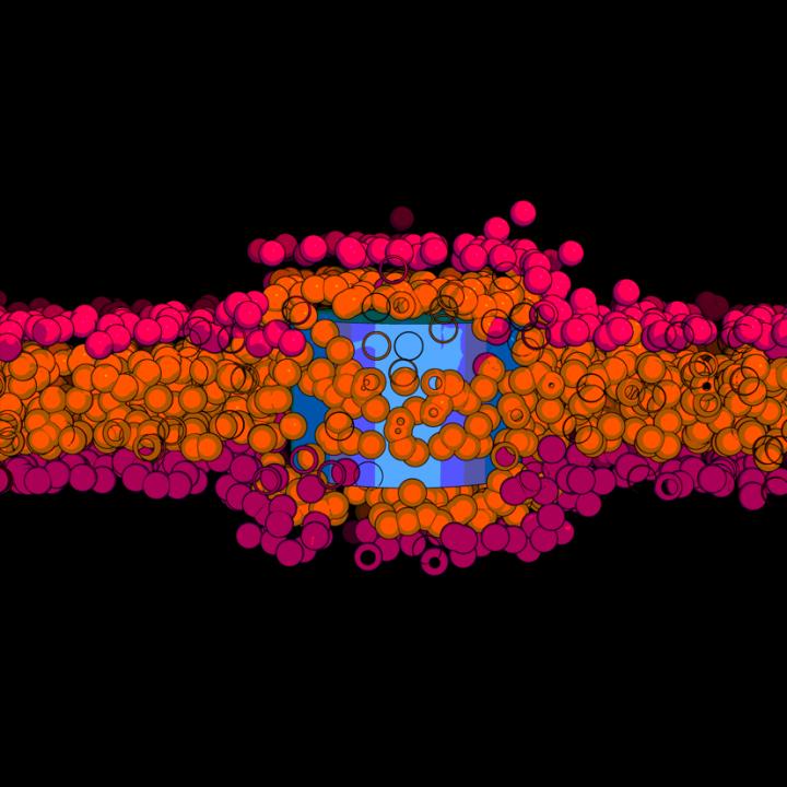
The wonder team’s paper looked at how Ant-Man and the Wasp breathe when they shrink down to insect-size and Staples’ lab studied how fluids flow in nature. Insects naturally move fluids and gases efficiently at tiny scales. If engineers can learn how insects breathe, they can use the knowledge to invent new microfluidic technologies.

A November 2018 Virginia Tech news release (also on EurekAlert but published on December 11, 2018) by Nancy Dudek describes the ‘Ant-Man and Wasp respiratory project’ before revealing the inspiration for the team’s new project,

“Before the 2018 ‘Ant-Man and the Wasp’ movie, my lab was already wondering about insect-scale respiration,” said Staples. “I wanted to get people to appreciate different breathing mechanisms.”

For most of Mikel-Stites’ life, he had been nit-picking at the “science” in science-fiction movies.

“I couldn’t watch ‘Armageddon’ once they got up to space station Mir and there was artificial gravity. Things like that have always bothered me. But for ‘Ant-Man and the Wasp’ it was worse,” he said.

Staples and Mikel-Stites decided to join forces to research Ant-Man’s microscale respiration.

Mikel-Stites was stung by what he dubbed “the altitude problem or death-zone dilemma.” For Ant-Man and the Wasp to shrink down to insect size and still breathe, they would have to overcome an atmospheric density similar to the top of Mt. Everest. Their tiny bodies would also require higher metabolisms. For their survival, the Marvel comic universe had to give Ant-Man and the Wasp superhero technologies.

“I thought it would be fun to find a solution for how this small-scale respiration would work,”said Mikel-Stites.”I started digging through Ant-Man’s history. I looped through scenes in the 2015 movie where we could address the physics. Then I did the same thing with trailers from the 2018 movie. I used that to make a list of problems and a list of solutions.”

Ant-Man and the Wasp solve the altitude problem with their superhero suits. In their publication, Mikel-Stites and Staples write that the masks in Ant-Man and the Wasp’s suits contain “a combination of an air pump, a compressor, and a molecular filter including Pym particle technology,” that allows them to breathe while they are insect-sized.

“This publication showed how different physics phenomena can dominate at different size scales, how well-suited organisms are for their particular size, and what happens when you start altering that,” said Mikel-Stites. “It also shows that Hollywood doesn’t always get it right when it comes to science!”

Their manuscript was accepted by the Journal of Superhero Science before the release of the sequel, “Ant-Man and the Wasp.” Mikel-Stites was concerned the blockbuster might include new technologies or change Ant-Man’s canon. If the Marvel comic universe changed between the 2015 ‘Ant-Man’ movie and the sequel, their hypotheses would be debunked and they would be forced to retract their paper.

“I went to the 2018 movie before the manuscript came out in preprint so that if the movie contradicted us we could catch it. But the 2018 movie actually supported everything we had said, which was really nice,” said Mikel-Stites. Most moviegoers simply watched the special effects and left the theater entertained. But Mikel-Stitesleft the movie with confirmation of the paper’s hypotheses.

The Staples lab members are not the only ones interested in tiny technologies. From lab-on-a-chip microfluidic devices to nanoparticles that deliver drugs directly to cells, consumers will ultimately benefit from this small scientific field that delivers big results.

“In both the movies and science, shrinking is a common theme and has been for the last 50-60 years. This idea is something that we all like to think about. Given enough time, we can reach the point where science can take it from the realms of magic into something that we actually have an explanation for,” Mikel-Stites said.

In fact, the Staples lab group has already done just that.

While Mikel-Stites is presenting his superhero science at the APS meeting, his colleague Krishnashis Chatterjee, who recently completed his Ph.D. in engineering mechanics will be presenting his research on fabricating and testing four different insect-inspired micro-fluidic devices.

From fiction to function, the Staples lab likes to have fun along the way.

“I think that it is really important to connect with people and be engaged in science with topics they already know about. With this superhero science paper I wanted to support this mission,” Staples said.

And who did the lab mates choose for their next superhero science subject? The Princess of Atlantis, Mera. They hope they can publish another superhero science paper that really makes waves.

Here’s a link to and a citation for the paper,

Ant-Man and The Wasp: Microscale Respiration and Microfluidic Technology by Anne Staples and Maxwell Mikel-Stites. Superhero Science and Technology (SST) Vol 1 No 1 (2018): https://doi.org/10.24413/sst.2018.1.2474 July 2018 ISSN 2588-7637

This paper is open access.

And, just because the idea of a superhero science journal tickles my fancy, here’s a little more from the journal’s About webpage,

Serial title
Superhero Science and Technolog

Focus and Scope
Superhero Science and Technology (SST) is multi-disciplinary journal that considers new research in the fields of science, technology, engineering and ethics motivated and presented using the superhero genre.

The superhero genre has become one of the most popular in modern cinema. Since the 2000 film X-Men, numerous superhero-themed films based on characters from Marvel Comics and DC Comics have been released. Films such as The Avengers, Iron Man 3, Avengers: Age of Ultron and Captain America: Civil War have all earned in excess of $1 billion dollars at the box office, thus demonstrating their relevance in modern society and popular culture.

Of particular interest for Superhero Science and Technology are articles that motivate new research by using the platform of superheroes, supervillains, their superpowers, superhero/supervillain exploits in Hollywood blockbuster films or superhero/supervillain adventures from comic books. Articles should be written in a manner so that they are accessible to both the academic community and the interested non-scientist i.e. general public, given the popularity of the superhero genre.

Dissemination of content using this approach provides a potential for the researcher to communicate their work to a larger audience, thus increasing their visibility and outreach within and outside of the academic domain.

The scope of the journal includes but is not limited to:
Genetic editing approaches;
Innovations in the field of robotics;
New and advanced materials;
Additive Manufacturing i.e. 3D printing, for both bio and non-bio applications;
Advancements in bio-chemical processing;
Biomimicry technologies;
Space physics, astrophysical and cosmological research;
Developments in propulsion systems;
Responsible innovation;
Ethical issues pertaining to technologies and their use for human enhancement or augmentation.

Open Access Policy
SST is licensed under a Creative Commons Attribution 4.0 International (CC BY 4.0) licence. You are free to use the work, but you have to attribute (refer to) the work in the manner specified by the author or licensor (but not in any way that suggests that they endorse you or your use of the work). The easiest way to refer to an article is to use the HOWTO CITE tool that you’ll find alongside each article in the right sidebar.

I also looked up the editorial team, from the journal’s Editorial Team webpage,

Editor-in-Chief
Dr. Barry W. Fitzgerald, TU Delft, the Netherlands
Editorial Board
Prof. Wim Briels, University of Twente, the Netherlands
Dr. Ian Clancy, University of Limerick, Ireland
Dr. Neil Clancy, University College London, UK
Dr. Tom Hunt, University of Kent, UK
Ass. Prof. Johan Padding, TU Delft, the Netherlands
Ass. Prof. Aimee van Wynsberghe, TU Delft, the Netherlands
Prof. Ilja Voets, TU Eindhoven, the Netherlands


For anyone unfamiliar with the abbreviation, TU stands for University of Technology or Technische Universiteit in Dutch.

Membrane stretching as a new transport mechanism for nanomaterials

This work comes from Catalonia, Spain by way of a collaboration between Chinese, German, and, of course, Spanish scientists. From a December 12, 2018 Universitat Rovira i Virgili press release (also on EurekAlert),

Increasing awareness of bioeffects and toxicity of nanomaterials interacting with cells puts in focus the mechanisms by which nanomaterials can cross lipid membranes. Apart from well-discussed energy-dependent endocytosis for large objects and passive diffusion through membranes by solute molecules, there can exist other transport mechanisms based on physical principles. Based on this hypothesis, the team of theoretical physics at Universitat Rovira i Virgili in Tarragona, led by Dr. Vladimir Baulin, designed a research project to investigate the interaction between nanotube and lipid membranes. In computer simulations, the researchers studied what they call a “model bilayer”, composed only by one type of lipids. Based on their calculations, the team of Dr. Baulin observed that ultra -short nanotube (10nm length) can insert perpendicularly to the lipid bilayer core.

They observed that these nanotubes stay trapped in the cell membrane, as commonly accepted by the scientific community. But a surprise appears when they stretched their model cell membrane, then inserted nanotubes which were trapped in the bilayer, suddenly started to escape from the bilayer on both sides. This means that it is possible to control the transport of nanomaterial across a cell membrane by tuning the membrane tension.

This is where Dr. Baulin contacted Dr. Jean-Baptiste Fleury at the Saarland University (Germany) to confirm this mechanism and to study experimentally this tension-mediated transport phenomena. Dr. Fleury and his team, designed a microfluidic experiment with a well-controlled phospholipid bilayer, an experimental model for cell membranes and added ultra-small carbon nanotubes (10nm in length) in solution. The nanotubes had an adsorbed lipid monolayer that guarantees their stable dispersion and prevent their clustering. Using a combination of optical fluorescent microscopy and electrophysiological measurements, the team of Dr. Fleury could follow individual nanotube crossing a bilayer and unravel their pathway on a molecular level. And as predicted by the simulations, they observed that nanotubes inserted into the bilayer by dissolving their lipid coating into the artificial membrane. When a tension of 4mN/m was applied to the bilayer, nanotubes spontaneously escaped the bilayer just in few milliseconds, while at lower tensions nanotubes remain trapped inside the membrane.

This discovery of translocation of tiny nanotubes through barriers protecting cells, i.e. lipid bilayer, may raise concerns about safety of nanomaterials for public health and suggest new mechanical mechanisms to control the drug delivery.

Caption: Nanotubes trapped inside the membrane. Credit: © URV

Here’s a link to and a citation for the paper,

Tension-Induced Translocation of an Ultrashort Carbon Nanotube through a Phospholipid Bilayer by Yachong Guo, Marco Werner, Ralf Seemann, Vladimir A. Baulin, and Jean-Baptiste Fleury. ACS Nano, Article ASAP DOI: 10.1021/acsnano.8b04657 Publication Date (Web): November 19, 2018

Copyright © 2018 American Chemical Society

This paper is behind a paywall.

A bioengineered robot hand with its own nervous system: machine/flesh and a job opening

A November 14, 2017 news item on phys.org announces a grant for a research project which will see engineered robot hands combined with regenerative medicine to imbue neuroprosthetic hands with the sense of touch,

The sense of touch is often taken for granted. For someone without a limb or hand, losing that sense of touch can be devastating. While highly sophisticated prostheses with complex moving fingers and joints are available to mimic almost every hand motion, they remain frustratingly difficult and unnatural for the user. This is largely because they lack the tactile experience that guides every movement. This void in sensation results in limited use or abandonment of these very expensive artificial devices. So why not make a prosthesis that can actually “feel” its environment?

That is exactly what an interdisciplinary team of scientists from Florida Atlantic University and the University of Utah School of Medicine aims to do. They are developing a first-of-its-kind bioengineered robotic hand that will grow and adapt to its environment. This “living” robot will have its own peripheral nervous system directly linking robotic sensors and actuators. FAU’s College of Engineering and Computer Science is leading the multidisciplinary team that has received a four-year, $1.3 million grant from the National Institute of Biomedical Imaging and Bioengineering of the [US] National Institutes of Health for a project titled “Virtual Neuroprosthesis: Restoring Autonomy to People Suffering from Neurotrauma.”

A November14, 2017 Florida Atlantic University (FAU) news release by Gisele Galoustian, which originated the news item, goes into more detail,

With expertise in robotics, bioengineering, behavioral science, nerve regeneration, electrophysiology, microfluidic devices, and orthopedic surgery, the research team is creating a living pathway from the robot’s touch sensation to the user’s brain to help amputees control the robotic hand. A neuroprosthesis platform will enable them to explore how neurons and behavior can work together to regenerate the sensation of touch in an artificial limb.

At the core of this project is a cutting-edge robotic hand and arm developed in the BioRobotics Laboratory in FAU’s College of Engineering and Computer Science. Just like human fingertips, the robotic hand is equipped with numerous sensory receptors that respond to changes in the environment. Controlled by a human, it can sense pressure changes, interpret the information it is receiving and interact with various objects. It adjusts its grip based on an object’s weight or fragility. But the real challenge is figuring out how to send that information back to the brain using living residual neural pathways to replace those that have been damaged or destroyed by trauma.

“When the peripheral nerve is cut or damaged, it uses the rich electrical activity that tactile receptors create to restore itself. We want to examine how the fingertip sensors can help damaged or severed nerves regenerate,” said Erik Engeberg, Ph.D., principal investigator, an associate professor in FAU’s Department of Ocean and Mechanical Engineering, and director of FAU’s BioRobotics Laboratory. “To accomplish this, we are going to directly connect these living nerves in vitro and then electrically stimulate them on a daily basis with sensors from the robotic hand to see how the nerves grow and regenerate while the hand is operated by limb-absent people.”

For the study, the neurons will not be kept in conventional petri dishes. Instead, they will be placed in  biocompatible microfluidic chambers that provide a nurturing environment mimicking the basic function of living cells. Sarah E. Du, Ph.D., co-principal investigator, an assistant professor in FAU’s Department of Ocean and Mechanical Engineering, and an expert in the emerging field of microfluidics, has developed these tiny customized artificial chambers with embedded micro-electrodes. The research team will be able to stimulate the neurons with electrical impulses from the robot’s hand to help regrowth after injury. They will morphologically and electrically measure in real-time how much neural tissue has been restored.

Jianning Wei, Ph.D., co-principal investigator, an associate professor of biomedical science in FAU’s Charles E. Schmidt College of Medicine, and an expert in neural damage and regeneration, will prepare the neurons in vitro, observe them grow and see how they fare and regenerate in the aftermath of injury. This “virtual” method will give the research team multiple opportunities to test and retest the nerves without any harm to subjects.

Using an electroencephalogram (EEG) to detect electrical activity in the brain, Emmanuelle Tognoli, Ph.D., co-principal investigator, associate research professor in FAU’s Center for Complex Systems and Brain Sciences in the Charles E. Schmidt College of Science, and an expert in electrophysiology and neural, behavioral, and cognitive sciences, will examine how the tactile information from the robotic sensors is passed onto the brain to distinguish scenarios with successful or unsuccessful functional restoration of the sense of touch. Her objective: to understand how behavior helps nerve regeneration and how this nerve regeneration helps the behavior.

Once the nerve impulses from the robot’s tactile sensors have gone through the microfluidic chamber, they are sent back to the human user manipulating the robotic hand. This is done with a special device that converts the signals coming from the microfluidic chambers into a controllable pressure at a cuff placed on the remaining portion of the amputated person’s arm. Users will know if they are squeezing the object too hard or if they are losing their grip.

Engeberg also is working with Douglas T. Hutchinson, M.D., co-principal investigator and a professor in the Department of Orthopedics at the University of Utah School of Medicine, who specializes in hand and orthopedic surgery. They are developing a set of tasks and behavioral neural indicators of performance that will ultimately reveal how to promote a healthy sensation of touch in amputees and limb-absent people using robotic devices. The research team also is seeking a post-doctoral researcher with multi-disciplinary experience to work on this breakthrough project.

Here’s more about the job opportunity from the FAU BioRobotics Laboratory job posting, (I checked on January 30, 2018 and it seems applications are still being accepted.)

Post-doctoral Opportunity

Dated Posted: Oct. 13, 2017

The BioRobotics Lab at Florida Atlantic University (FAU) invites applications for a NIH NIBIB-funded Postdoctoral position to develop a Virtual Neuroprosthesis aimed at providing a sense of touch in amputees and limb-absent people.

Candidates should have a Ph.D. in one of the following degrees: mechanical engineering, electrical engineering, biomedical engineering, bioengineering or related, with interest and/or experience in transdisciplinary work at the intersection of robotic hands, biology, and biomedical systems. Prior experience in the neural field will be considered an advantage, though not a necessity. Underrepresented minorities and women are warmly encouraged to apply.

The postdoctoral researcher will be co-advised across the department of Mechanical Engineering and the Center for Complex Systems & Brain Sciences through an interdisciplinary team whose expertise spans Robotics, Microfluidics, Behavioral and Clinical Neuroscience and Orthopedic Surgery.

The position will be for one year with a possibility of extension based on performance. Salary will be commensurate with experience and qualifications. Review of applications will begin immediately and continue until the position is filled.

The application should include:

  1. a cover letter with research interests and experiences,
  2. a CV, and
  3. names and contact information for three professional references.

Qualified candidates can contact Erik Engeberg, Ph.D., Associate Professor, in the FAU Department of Ocean and Mechanical Engineering at eengeberg@fau.edu. Please reference AcademicKeys.com in your cover letter when applying for or inquiring about this job announcement.

You can find the apply button on this page. Good luck!

Liquid biopsy chip that uses carbon nanotubes in place of microfluidics

They’re calling this a breakthrough technology in a Dec. 15, 2016 news item on ScienceDaily,

A chip developed by mechanical engineers at Worcester Polytechnic Institute (WPI) [UK] can trap and identify metastatic cancer cells in a small amount of blood drawn from a cancer patient. The breakthrough technology uses a simple mechanical method that has been shown to be more effective in trapping cancer cells than the microfluidic approach employed in many existing devices.

The WPI device uses antibodies attached to an array of carbon nanotubes at the bottom of a tiny well. Cancer cells settle to the bottom of the well, where they selectively bind to the antibodies based on their surface markers (unlike other devices, the chip can also trap tiny structures called exosomes produced by cancers cells). This “liquid biopsy,” described in a recent issue of the journal Nanotechnology, could become the basis of a simple lab test that could quickly detect early signs of metastasis and help physicians select treatments targeted at the specific cancer cells identified.

A Dec. 15, 2016 WPI press release (also on EurekAlert), which originated the news item, explains the breakthrough in more detail (Note: Links have been removed),

Metastasis is the process by which a cancer can spread from one organ to other parts of the body, typically by entering the bloodstream. Different types of tumors show a preference for specific organs and tissues; circulating breast cancer cells, for example, are likely to take root in bones, lungs, and the brain. The prognosis for metastatic cancer (also called stage IV cancer) is generally poor, so a technique that could detect these circulating tumor cells before they have a chance to form new colonies of tumors at distant sites could greatly increase a patient’s survival odds.

“The focus on capturing circulating tumor cells is quite new,” said Balaji Panchapakesan, associate professor of mechanical engineering at WPI and director of the Small Systems Laboratory. “It is a very difficult challenge, not unlike looking for a needle in a haystack. There are billions of red blood cells, tens of thousands of white blood cells, and, perhaps, only a small number of tumor cells floating among them. We’ve shown how those cells can be captured with high precision.”

The device developed by Panchapakesan’s team includes an array of tiny elements, each about a tenth of an inch (3 millimeters) across. Each element has a well, at the bottom of which are antibodies attached to carbon nanotubes. Each well holds a specific antibody that will bind selectively to one type of cancer cell type, based on genetic markers on its surface. By seeding elements with an assortment of antibodies, the device could be set up to capture several different cancer cells types using a single blood sample. In the lab, the researchers were able to fill a total of 170 wells using just under 0.3 fluid ounces (0.85 milliliter) of blood. Even with that small sample, they captured between one and a thousand cells per device, with a capture efficiency of between 62 and 100 percent.

In a paper published in the journal Nanotechnology [“Static micro-array isolation, dynamic time series classification, capture and enumeration of spiked breast cancer cells in blood: the nanotube–CTC chip”], Panchapakesan’s team, which includes postdoctoral researcher Farhad Khosravi, the paper’s lead author, and researchers at the University of Louisville and Thomas Jefferson University, describe a study in which antibodies specific for two markers of metastatic breast cancer, EpCam and Her2, were attached to the carbon nanotubes in the chip. When a blood sample that had been “spiked” with cells expressing those markers was placed on the chip, the device was shown to reliably capture only the marked cells.

In addition to capturing tumor cells, Panchapakesan says the chip will also latch on to tiny structures called exosomes, which are produced by cancers [sic] cells and carry the same markers. “These highly elusive 3-nanometer structures are too small to be captured with other types of liquid biopsy devices, such as microfluidics, due to shear forces that can potentially destroy them,” he noted. “Our chip is currently the only device that can potentially capture circulating tumor cells and exosomes directly on the chip, which should increase its ability to detect metastasis. This can be important because emerging evidence suggests that tiny proteins excreted with exosomes can drive reactions that may become major barriers to effective cancer drug delivery and treatment.”

Panchapakesan said the chip developed by his team has additional advantages over other liquid biopsy devices, most of which use microfluidics to capture cancer cells. In addition to being able to capture circulating tumor cells far more efficiently than microfluidic chips (in which cells must latch onto anchored antibodies as they pass by in a stream of moving liquid), the WPI device is also highly effective in separating cancer cells from the other cells and material in the blood through differential settling.

While the initial tests with the chip have focused on breast cancer, Panchapakesan says the device could be set up to detect a wide range of tumor types, and plans are already in the works for development of an advanced device as well as testing for other cancer types, including lung and pancreas cancer. He says he envisions a day when a device like his could be employed not only for regular follow ups for patients who have had cancer, but in routine cancer screening.

“Imagine going to the doctor for your yearly physical,” he said. “You have blood drawn and that one blood sample can be tested for a comprehensive array of cancer cell markers. Cancers would be caught at their earliest stage and other stages of development, and doctors would have the necessary protein or genetic information from these captured cells to customize your treatment based on the specific markers for your cancer. This would really be a way to put your health in your own hands.”

“White blood cells, in particular, are a problem, because they are quite numerous in blood and they can be mistaken for cancer cells,” he said. “Our device uses what is called a passive leukocyte depletion strategy. Because of density differences, the cancer cells tend to settle to the bottom of the wells (and this only happens in a narrow window), where they encounter the antibodies. The remainder of the blood contents stays at the top of the wells and can simply be washed away.”

Here’s a link to and a citation for the paper,

Static micro-array isolation, dynamic time series classification, capture and enumeration of spiked breast cancer cells in blood: the nanotube–CTC chip by Farhad Khosravi, Patrick J Trainor, Christopher Lambert, Goetz Kloecker, Eric Wickstrom, Shesh N Rai, and Balaji Panchapakesan. Nanotechnology, Volume 27, Number 44 DOI http://dx.doi.org/10.1088/0957-4484/27/44/44LT03 Published 29 September 2016

© 2016 IOP Publishing Ltd

This paper is open access.

Sutures that can gather data wirelessly

Are sutures which gather data hackable? It’s a little early to start thinking about that issue as this seems to be brand new research. A July 18, 2016 news item on ScienceDaily tells more,

For the first time, researchers led by Tufts University engineers have integrated nano-scale sensors, electronics and microfluidics into threads — ranging from simple cotton to sophisticated synthetics — that can be sutured through multiple layers of tissue to gather diagnostic data wirelessly in real time, according to a paper published online July 18 [2016] in Microsystems & Nanoengineering. The research suggests that the thread-based diagnostic platform could be an effective substrate for a new generation of implantable diagnostic devices and smart wearable systems.

A July 18, 2016 Tufts University news release (also on EurekAlert), which originated the news item, provides more detail,

The researchers used a variety of conductive threads that were dipped in physical and chemical sensing compounds and connected to wireless electronic circuitry to create a flexible platform that they sutured into tissue in rats as well as in vitro. The threads collected data on tissue health (e.g. pressure, stress, strain and temperature), pH and glucose levels that can be used to determine such things as how a wound is healing, whether infection is emerging, or whether the body’s chemistry is out of balance. The results were transmitted wirelessly to a cell phone and computer.

The three-dimensional platform is able to conform to complex structures such as organs, wounds or orthopedic implants.

While more study is needed in a number of areas, including investigation of long-term biocompatibility, researchers said initial results raise the possibility of optimizing patient-specific treatments.

“The ability to suture a thread-based diagnostic device intimately in a tissue or organ environment in three dimensions adds a unique feature that is not available with other flexible diagnostic platforms,” said Sameer Sonkusale, Ph.D., corresponding author on the paper and director of the interdisciplinary Nano Lab in the Department of Electrical and Computer Engineering at Tufts School of Engineering. “We think thread-based devices could potentially be used as smart sutures for surgical implants, smart bandages to monitor wound healing, or integrated with textile or fabric as personalized health monitors and point-of-care diagnostics.”

Until now, the structure of substrates for implantable devices has essentially been two-dimensional, limiting their usefulness to flat tissue such as skin, according to the paper. Additionally, the materials in those substrates are expensive and require specialized processing.

Here’s a link to and a citation for the paper,

A toolkit of thread-based microfluidics, sensors, and electronics for 3D tissue embedding for medical diagnostics by Pooria Mostafalu, Mohsen Akbari, Kyle A. Alberti, Qiaobing Xu, Ali Khademhosseini, & Sameer R. Sonkusale. Microsystems & Nanoengineering 2, Article number: 16039 (2016) doi:10.1038/micronano.2016.39 Published online 18 July 2016

This paper is open access.

Combat cells (Robot Wars for cells) and a plea from Concordia University

Students at Concordia University (located in Montréal, Québec, Canada) are requesting help (financial or laboratory supplies) for their submission to  the 2016 iGEM (International Genetically Engineered Machine) competition.

Here’s a little about their entry (from a June 16, 2016 request received via email),

For this year’s project, we plan to design a biological system that mimics the concept of the popular TV series Robot Wars. We will be engineering cellular species to wear nanoparticles as battle shields and then use microfluidics to guide them through an obstacle course leading to a battledome, where both cells will engage into a duel. Essentially, we want to test the interactions between nanoparticles and cell membranes, as well as their protective abilities against varying environmental conditions and other equipped cells. The method in which we will adapt Robot Wars for synthetic biology is by creating a web series that will visualize the cell battle and communicate the research behind it. This web series will serve as an entertaining  medium to educate and inspire the audience to develop an interest in science. We are incorporating the emerging fields of  synthetic biology, nanotechnology and microfluidics to make this process possible.  Furthermore, this study will contribute to the advancement of nanotechnology, an interdisciplinary field aiming to make applicable improvements in other fields such as medicine, optics and cosmetics.

Here’s a little more about iGEM (from the organization’s homepage),

The iGEM Foundation is dedicated to education and competition, advancement of synthetic biology, and the development of open community and collaboration.

The main program at the iGEM Foundation is the International Genetically Engineered Machine (iGEM) Competition. The iGEM Competition is the premiere student competition in Synthetic Biology. Since 2004, participants of the competition have experienced education, teamwork, sharing, and more in a unique competition setting.

The deadline for donations/sponsorships is the end of September 2016 and sponsors/donors will be acknowledged on “our website, all of our social media accounts (Facebook, Instagram, Twitter), at our community outreach events and at the competition [from the June 16, 2016 email].”

For more information contact:

Maria Salouros
iGEM Concordia
igem.concordia@gmail.com

Finally, there’s this:

We are excited to make this year’s project a reality and we are determined to win gold. Any help, either financially or by the donation of laboratory supplies, would contribute to the development of our project and would be greatly appreciated.

Good luck to the students! Hopefully one or more of my readers will be able to help. In which case, thank you!

Making lab-on-a-chip devices more accurate

An April 4, 2016 news item on phys.org announces research that will improve control and manipulation of the fluids in a lab-on-a-chip,

Lab-on-a-chip designates devices that integrate various biochemical functions on a fingernail-sized chip to enable quick and compact biological analysis or medical diagnosis by processing a small volume of biological samples, such as a drop of blood. To operate various functions on a lab-on-a-chip device, the key technology is the precise and rapid manipulation of fluid on a micro-scale.

A March 31, 2016 Pohang University of Science and Technology (POSTECH; South Korea) press release (also on EurekAlert), which originated the news item, expands on the theme,

In microfluidic devices, very small and trivial variables can frequently cause a large amount of errors. Up until now, Proportional-Integral-Derivative (PID) controller has normally been used for the manipulation of fluids in microfluidic chips. To apply the PID controller, a tedious gain-tuning process is required but the gain-tuning is a difficult process for people who are unfamiliar with control theory. Especially, in the case of controlling multiple flows, the process is extremely convoluted and frustrating.

The developed control algorithm can improve accuracy and stability of flow regulation in a microfluidic network without requiring any tuning process. With this algorithm, microfluidic flows in multiple channels can be controlled in simultaneous and independent way. The team expects that this algorithm has the potential for many applications of lab-on-a-chip devices. For example, cell culture or biological analysis, which are conducted in biology laboratories, can be performed on a microfluidic chip. Physical and chemical responses can be analyzed in the subdivided levels.

Here’s a link to and a citation for the paper,

Tuning-free controller to accurately regulate flow rates in a microfluidic network by Young Jin Heo, Junsu Kang, Min Jun Kim, & Wan Kyun Chung. Scientific Reports 6, Article number: 23273 (2016) doi:10.1038/srep23273 Published online: 18 March 2016

This is an open access paper.

Mass production of nanoparticles?

With all the years of nanotechnology and nanomaterials research it seems strange that mass production of nanoparticles is still very much in the early stages as a Feb. 24, 2016 news item on phys.org points out,

Nanoparticles – tiny particles 100,000 times smaller than the width of a strand of hair – can be found in everything from drug delivery formulations to pollution controls on cars to HD TV sets. With special properties derived from their tiny size and subsequently increased surface area, they’re critical to industry and scientific research.

They’re also expensive and tricky to make.

Now, researchers at USC [University of Southern California] have created a new way to manufacture nanoparticles that will transform the process from a painstaking, batch-by-batch drudgery into a large-scale, automated assembly line.

A Feb. 24, 2016 USC news release (also on EurekAlert) by Robert Perkins, which originated the news item, offers additional insight,

Consider, for example, gold nanoparticles. They have been shown to easily penetrate cell membranes without causing any damage — an unusual feat given that most penetrations of cell membranes by foreign objects can damage or kill the cell. Their ability to slip through the cell’s membrane makes gold nanoparticles ideal delivery devices for medications to healthy cells or fatal doses of radiation to cancer cells.

However, a single milligram of gold nanoparticles currently costs about $80 (depending on the size of the nanoparticles). That places the price of gold nanoparticles at $80,000 per gram while a gram of pure, raw gold goes for about $50.

“It’s not the gold that’s making it expensive,” Malmstadt [Noah Malmstadt of the USC Viterbi School of Engineering] said. “We can make them, but it’s not like we can cheaply make a 50-gallon drum full of them.”

A fluid situation

At this time, the process of manufacturing a nanoparticle typically involves a technician in a chemistry lab mixing up a batch of chemicals by hand in traditional lab flasks and beakers.

The new technique used by Brutchey [Richard Brutchey of the USC Dornsife College of Letters, Arts and Sciences] and Malmstadt instead relies on microfluidics — technology that manipulates tiny droplets of fluid in narrow channels.

“In order to go large scale, we have to go small,” Brutchey said.

Really small.

The team 3-D printed tubes about 250 micrometers in diameter, which they believe to be the smallest, fully enclosed 3-D printed tubes anywhere. For reference, your average-sized speck of dust is 50 micrometers wide.

They they built a parallel network of four of these tubes, side-by-side, and ran a combination of two nonmixing fluids (like oil and water) through them. As the two fluids fought to get out through the openings, they squeezed off tiny droplets. Each of these droplets acted as a micro-scale chemical reactor in which materials were mixed and nanoparticles were generated. Each microfluidic tube can create millions of identical droplets that perform the same reaction.

This sort of system has been envisioned in the past, but it hasn’t been able to be scaled up because the parallel structure meant that if one tube got jammed, it would cause a ripple effect of changing pressures along its neighbors, knocking out the entire system. Think of it like losing a single Christmas light in one of the old-style strands — lose one and you lose them all.

Brutchey and Malmstadt bypassed this problem by altering the geometry of the tubes themselves, shaping the junction between the tubes such that the particles come out a uniform size and the system is immune to pressure changes.

Here’s a link to and a citation for the paper,

Flow invariant droplet formation for stable parallel microreactors by Carson T. Riche, Emily J. Roberts, Malancha Gupta, Richard L. Brutchey & Noah Malmstadt. Nature Communications 7, Article number: 10780 doi:10.1038/ncomms10780 Published 23 February 2016

This is an open access paper.

#BCTECH: being at the Summit (Jan. 18-19, 2016)

#BCTECH Summit 2016*, a joint event between the province of British Columbia (BC, Canada) and the BC Innovation Council (BCIC), a crown corporation formerly known as the Science Council of British Columbia, launched on Jan. 18, 2016. I have written a preview (Jan. 17, 2016 post) and a commentary on the new #BCTECH strategy (Jan. 19, 2016 posting) announced by British Columbia Premier, Christy Clark, on the opening day (Jan. 18, 2016) of the summit.

I was primarily interested in the trade show/research row/technology showcase aspect of the summit focusing (but not exclusively) on nanotechnology. Here’s what I found,

Nano at the Summit

  • Precision NanoSystems: fabricates equipment which allows researchers to create polymer nanoparticles for delivering medications.

One of the major problems with creating nanoparticles is ensuring a consistent size and rapid production. According to Shell Ip, a Precision NanoSystems field application scientist, their NanoAssemblr Platform has solved the consistency problem and a single microfluidic cartridge can produce 15 ml in two minutes. Cartridges can run in parallel for maximum efficiency when producing nanoparticles in greater quantity.

The NanoAssemblr Platform is in use in laboratories around the world (I think the number is 70) and you can find out more on the company’s About our technology webpage,

The NanoAssemblr™ Platform

The microfluidic approach to particle formulation is at the heart of the NanoAssemblr Platform. This well-controlled process mediates bottom-up self-assembly of nanoparticles with reproducible sizes and low polydispersity. Users can control size by process and composition, and adjust parameters such as mixing ratios, flow rate and lipid composition in order to fine-tune nanoparticle size, encapsulation efficiency and much more. The system technology enables manufacturing scale-up through microfluidic reactor parallelization similar to the arraying of transistors on an integrated chip. Superior design ensures that the platform is fast and easy to use with a software controlled manufacturing process. This usability allows for the simplified transfer of manufacturing protocols between sites, which accelerates development, reduces waste and ultimately saves money. Precision NanoSystems’ flagship product is the NanoAssemblr™ Benchtop Instrument, designed for rapid prototyping of novel nanoparticles. Preparation time on the system is streamlined to approximately one minute, with the ability to complete 30 formulations per day in the hands of any user.

The company is located on property known as the Endowment Lands or, more familiarly, the University of British Columbia (UBC).

A few comments before moving on, being able to standardize the production of medicine-bearing nanoparticles is a tremendous step forward which is going to help scientists dealing with other issues. Despite all the talk in the media about delivering nanoparticles with medication directly to diseased cells, there are transport issues: (1) getting the medicine to the right location/organ and (2) getting the medicine into the cell. My Jan. 12, 2016 posting featured a project with Malaysian scientists and a team at Harvard University who are tackling the transport and other nanomedicine) issues as they relate to the lung. As well, I have a Nov. 26, 2015 posting which explores a controversy about nanoparticles getting past the ‘cell walls’ into the nucleus of the cell.

The next ‘nano’ booths were,

  • 4D Labs located at Simon Fraser University (SFU) was initially hailed as a nanotechnology facility but these days they’re touting themselves as an ‘advanced materials’ facility. Same thing, different branding.

They advertise services including hands-on training for technology companies and academics. There is a nanoimaging facility and nanofabrication facility, amongst others.

I spoke with their operations manager, Nathaniel Sieb who mentioned a few of the local companies that use their facilities. (1) Nanotech Security (featured here most recently in a Dec. 29, 2015 post), an SFU spinoff company, does some of their anticounterfeiting research work at 4D Labs. (2) Switch Materials (a smart window company, electrochromic windows if memory serves) also uses the facilities. It is Neil Branda’s (4D Labs Executive Director) company and I have been waiting impatiently (my May 14, 2010 post was my first one about Switch) for either his or someone else’s electrochromic windows (they could eliminate or reduce the need for air conditioning during the hotter periods and reduce the need for heat in the colder periods) to come to market. Seib tells me, I’ll have to wait longer for Switch. (3) A graduate student was presenting his work at the booth, a handheld diagnostic device that can be attached to a smartphone to transmit data to the cloud. While the first application is for diabetics, there are many other possibilities. Unfortunately, glucose means you need to produce blood for the test when I suggested my preference for saliva the student explained some of the difficulties. Apparently, your saliva changes dynamically and frequently and something as simple as taking a sip of orange juice could result in a false reading. Our conversation (mine, Seib’s and the student’s) also drifted over into the difficulties of bringing products to market. Sadly, we were not able to solve that problem in our 10 minute conversation.

  • FPInnovations is a scientific research centre and network for the forestry sector. They had a display near their booth which was like walking into a peculiar forest (I was charmed). The contrast with the less imaginative approaches all around was striking.

FPInnovation helped to develop cellulose nanocrystals (CNC), then called nanocrystalline cellulose (NCC), and I was hoping to be updated about CNC and about the spinoff company Celluforce. The researcher I spoke to was from Sweden and his specialty was business development. He didn’t know much about CNC in Canada and when I commented on how active Sweden has been its pursuit of a CNC application, he noted Finland has been the most active. The researcher noted that making the new materials being derived from the forest, such as CNC, affordable and easily produced for use in applications that have yet to be developed are all necessities and challenges. He mentioned that cultural changes also need to take place. Canadians are accustomed to slicing away and discarding most of the tree instead of using as much of it as possible. We also need to move beyond the construction and pulp & paper sectors (my Feb. 15, 2012 posting featured nanocellulose research in Sweden where sludge was the base material).

Other interests at the Summit

I visited:

  • “The Wearable Lower Limb Anthropomorphic Exoskeleton (WLLAE) – a lightweight, battery-operated and ergonomic robotic system to help those with mobility issues improve their lives. The exoskeleton features joints and links that correspond to those of a human body and sync with motion. SFU has designed, manufactured and tested a proof-of-concept prototype and the current version can mimic all the motions of hip joints.” The researchers (Siamak Arzanpour and Edward Park) pointed out that the ability to mimic all the motions of the hip is a big difference between their system and others which only allow the leg to move forward or back. They rushed the last couple of months to get this system ready for the Summit. In fact, they received their patent for the system the night before (Jan. 17, 2016) the Summit opened.

It’s the least imposing of the exoskeletons I’ve seen (there’s a description of one of the first successful exoskeletons in a May 20, 2014 posting; if you scroll down to the end you’ll see an update about the device’s unveiling at the 2014 World Cup [soccer/football] in Brazil).

Unfortunately, there aren’t any pictures of WLLAE yet and the proof-of-concept version may differ significantly from the final version. This system could be used to help people regain movement (paralysis/frail seniors) and I believe there’s a possibility it could be used to enhance human performance (soldiers/athletes). The researchers still have some significant hoops to jump before getting to the human clinical trial stage. They need to refine their apparatus, ensure that it can be safely operated, and further develop the interface between human and machine. I believe WLLAE is considered a neuroprosthetic device. While it’s not a fake leg or arm, it enables movement (prosthetic) and it operates on brain waves (neuro). It’s a very exciting area of research, consequently, there’s a lot of international competition. [ETA January 3, 2024: I’m pretty sure I got the neuroprosthetic part wrong]

  • Delightfully, after losing contact for a while, I reestablished it with the folks (Sean Lee, Head External Relations and Jim Hanlon, Chief Administrative Officer) at TRIUMF (Canada’s national laboratory for particle and nuclear physics). It’s a consortium of 19 Canadian research institutions (12 full members and seven associate members).

It’s a little disappointing that TRIUMF wasn’t featured in the opening for the Summit since the institution houses theoretical, experimental, and applied science work. It’s a major BC (and Canada) science and technology success story. My latest post (July 16, 2015) about their work featured researchers from California (US) using the TRIUMF cyclotron for imaging nanoscale materials and, on the more practical side, there’s a Mar. 6, 2015 posting about their breakthrough for producing nuclear material-free medical isotopes. Plus, Maclean’s Magazine ran a Jan. 3, 2016 article by Kate Lunau profiling an ‘art/science’ project that took place at TRIUMF (Note: Links have been removed),

It’s not every day that most people get to peek inside a world-class particle physics lab, where scientists probe deep mysteries of the universe. In September [2015], Vancouver’s TRIUMF—home to the world’s biggest cyclotron, a type of particle accelerator—opened its doors to professional and amateur photographers, part of an event called Global Physics Photowalk 2015. (Eight labs around the world participated, including CERN [European particle physics laboratory], in Geneva, where the Higgs boson particle was famously discovered.)

Here’s the local (Vancouver) jury’s pick for the winning image (from the Nov. 4, 2015 posting [Winning Photographs Revealed] by Alexis Fong on the TRIUMF website),

Caption: DESCANT (at TRIUMF) neutron detector array composed of 70 hexagonal detectors Credit: Pamela Joe McFarlane

Caption: DESCANT (at TRIUMF) neutron detector array composed of 70 hexagonal detectors Credit: Pamela Joe McFarlane

With all those hexagons and a spherical shape, the DESCANT looks like a ‘buckyball’ or buckminsterfullerene or C60  to me.

I hope the next Summit features TRIUMF and/or some other endeavours which exemplify, Science, Technology, and Creativity in British Columbia and Canada.

Onto the last booth,

  • MITACS was originally one of the Canadian federal government’s Network Centres for Excellence projects. It was focused on mathematics, networking, and innovation but once the money ran out the organization took a turn. These days, it’s describing itself as (from their About page) “a national, not-for-profit organization that has designed and delivered research and training programs in Canada for 15 years. Working with 60 universities, thousands of companies, and both federal and provincial governments, we build partnerships that support industrial and social innovation in Canada.”Their Jan. 19, 2016 news release (coincidental with the #BCTECH Summit, Jan. 18 – 19, 2016?) features a new report about improving international investment in Canada,”Opportunities to improve Canada’s attractiveness for R&D investment were identified:1.Canada needs to better incentivize R&D by rebalancing direct and indirect support measures

    2.Canada requires a coordinated, client-centric approach to incentivizing R&D

    3.Canada needs to invest in training programs that grow the knowledge economy”

    Oddly, entrepreneurial/corporate/business types never have a problem with government spending when the money is coming to them; it’s only a problem when it’s social services.

    Back to MITACS, one of their more interesting (to me) projects was announced at the 2015 Canadian Science Policy Conference. MITACS has inaugurated a Canadian Science Policy Fellowships programme which in its first year (pilot) will see up up to 10 academics applying their expertise to policy-making while embedded in various federal government agencies. I don’t believe anything similar has occurred here in Canada although, if memory serves, the Brits have a similar programme.

    Finally, I offer kudos to Sherry Zhao, MITACS Business Development Specialist, the only person to ask me how her organization might benefit my business. Admittedly I didn’t talk to a lot of people but it’s striking to me that at an ‘innovation and business’ tech summit, only one person approached me about doing business.  Of course, I’m not a male aged between 25 and 55. So, extra kudos to Sherry Zhao and MITACS.

Christy Clark (Premier of British Columbia), in her opening comments, stated 2800 (they were expecting about 1000) had signed up for the #BCTECH Summit. I haven’t been able to verify that number or get other additional information, e.g., business deals, research breakthroughs, etc. announced at the Summit. Regardless, it was exciting to attend and find out about the latest and greatest on the BC scene.

I wish all the participants great and good luck and look forward to next year’s where perhaps we’ll here about how the province plans to help with the ‘manufacturing middle’ issue. For new products you need to have facilities capable of reproducing your devices at a speed that satisfies your customers; see my Feb. 10, 2014 post featuring a report on this and other similar issues from the US General Accountability Office.

*’BCTECH Summit 2016′ link added Jan. 21, 2016.

Controlling water with ‘stick-slip surfaces’

Controlling water could lead to better designed microfluidic devices such as ‘lab-on-a-chip’. A July 27, 2015 news item on Nanowerk announces a new technique for controlling water,

Coating the inside of glass microtubes with a polymer hydrogel material dramatically alters the way capillary forces draw water into the tiny structures, researchers have found. The discovery could provide a new way to control microfluidic systems, including popular lab-on-a-chip devices.

Capillary action draws water and other liquids into confined spaces such as tubes, straws, wicks and paper towels, and the flow rate can be predicted using a simple hydrodynamic analysis. But a chance observation by researchers at the Georgia Institute of Technology [US] will cause a recalculation of those predictions for conditions in which hydrogel films line the tubes carrying water-based liquids.

“Rather than moving according to conventional expectations, water-based liquids slip to a new location in the tube, get stuck, then slip again – and the process repeats over and over again,” explained Andrei Fedorov, a professor in the George W. Woodruff School of Mechanical Engineering at Georgia Tech. “Instead of filling the tube with a rate of liquid penetration that slows with time, the water propagates at a nearly constant speed into the hydrogel-coated capillary. This was very different from what we had expected.”

A July 27, 2015 Georgia Institute of Technology (Georgia Tech) news release (also on EurekAlert) by John Toon, which originated the news item, describes the work in more detail,

When the opening of a thin glass tube is exposed to a droplet of water, the liquid begins to flow into the tube, pulled by a combination of surface tension in the liquid and adhesion between the liquid and the walls of the tube. Leading the way is a meniscus, a curved surface of the water at the leading edge of the water column. An ordinary borosilicate glass tube fills by capillary action at a gradually decreasing rate with the speed of meniscus propagation slowing as a square root of time.

But when the inside of a tube is coated with a very thin layer of poly(N-isopropylacrylamide), a so-called “smart” polymer (PNIPAM), everything changes. Water entering a tube coated on the inside with a dry hydrogel film must first wet the film and allow it to swell before it can proceed farther into the tube. The wetting and swelling take place not continuously, but with discrete steps in which the water meniscus first sticks and its motion remains arrested while the polymer layer locally deforms. The meniscus then rapidly slides for a short distance before the process repeats. This “stick-slip” process forces the water to move into the tube in a step-by-step motion.

The flow rate measured by the researchers in the coated tube is three orders of magnitude less than the flow rate in an uncoated tube. A linear equation describes the time dependence of the filling process instead of a classical quadratic equation which describes filling of an uncoated tube.

“Instead of filling the capillary in a hundredth of a second, it might take tens of seconds to fill the same capillary,” said Fedorov. “Though there is some swelling of the hydrogel upon contact with water, the change in the tube diameter is negligible due to the small thickness of the hydrogel layer. This is why we were so surprised when we first observed such a dramatic slow-down of the filing process in our experiments.”

The researchers – who included graduate students James Silva, Drew Loney and Ren Geryak and senior research engineer Peter Kottke – tried the experiment again using glycerol, a liquid that is not absorbed by the hydrogel. With glycerol, the capillary action proceeded through the hydrogel-coated microtube as with an uncoated tube in agreement with conventional theory. After using high-resolution optical visualization to study the meniscus propagation while the polymer swelled, the researchers realized they could put this previously-unknown behavior to good use.

Water absorption by the hydrogels occurs only when the materials remain below a specific transition temperature. When heated above that temperature, the materials no longer absorb water, eliminating the “stick-slip” phenomenon in the microtubes and allowing them to behave like ordinary tubes.

This ability to turn the stick-slip behavior on and off with temperature could provide a new way to control the flow of water-based liquid in microfluidic devices, including labs-on-a-chip. The transition temperature can be controlled by varying the chemical composition of the hydrogel.

“By locally heating or cooling the polymer inside a microfluidic chamber, you can either speed up the filling process or slow it down,” Fedorov said. “The time it takes for the liquid to travel the same distance can be varied up to three orders of magnitude. That would allow precise control of fluid flow on demand using external stimuli to change polymer film behavior.”

The heating or cooling could be done locally with lasers, tiny heaters, or thermoelectric devices placed at specific locations in the microfluidic devices.

That could allow precise timing of reactions in microfluidic devices by controlling the rate of reactant delivery and product removal, or allow a sequence of fast and slow reactions to occur. Another important application could be controlled drug release in which the desired rate of molecule delivery could be dynamically tuned over time to achieve the optimal therapeutic outcome.

In future work, Fedorov and his team hope to learn more about the physics of the hydrogel-modified capillaries and study capillary flow using partially-transparent microtubes. They also want to explore other “smart” polymers which change the flow rate in response to different stimuli, including the changing pH of the liquid, exposure to electromagnetic radiation, or the induction of mechanical stress – all of which can change the properties of a particular hydrogel designed to be responsive to those triggers.

“These experimental and theoretical results provide a new conceptual framework for liquid motion confined by soft, dynamically evolving polymer interfaces in which the system creates an energy barrier to further motion through elasto-capillary deformation, and then lowers the barrier through diffusive softening,” the paper’s authors wrote. “This insight has implications for optimal design of microfluidic and lab-on-a-chip devices based on stimuli-responsive smart polymers.”

In addition to those already mentioned, the research team included Professor Vladimir Tsukruk from the Georgia Tech School of Materials Science and Engineering and Rajesh Naik, Biotechnology Lead and Tech Advisor of the Nanostructured and Biological Materials Branch of the Air Force Research Laboratory (AFRL).

Here’s a link to and a citation for the paper,

Stick–slip water penetration into capillaries coated with swelling hydrogel by J. E. Silva, R. Geryak, D. A. Loney, P. A. Kottke, R. R. Naik, V. V. Tsukruk, and A. G. Fedorov. Soft Matter, 2015,11, 5933-5939 DOI: 10.1039/C5SM00660K First published online 23 Jun 2015

This paper is behind a paywall.