Tag Archives: photosynthesis

Cyborg bacteria to reduce carbon dioxide

This video is a bit technical but then it is about work being presented to chemists at the American Chemical Society’s (ACS) at the 254th National Meeting & Exposition Aug. 20 -24, 2017,

For a more plain language explanation, there’s an August 22, 2017 ACS news release (also on EurekAlert),

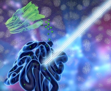
Photosynthesis provides energy for the vast majority of life on Earth. But chlorophyll, the green pigment that plants use to harvest sunlight, is relatively inefficient. To enable humans to capture more of the sun’s energy than natural photosynthesis can, scientists have taught bacteria to cover themselves in tiny, highly efficient solar panels to produce useful compounds.

“Rather than rely on inefficient chlorophyll to harvest sunlight, I’ve taught bacteria how to grow and cover their bodies with tiny semiconductor nanocrystals,” says Kelsey K. Sakimoto, Ph.D., who carried out the research in the lab of Peidong Yang, Ph.D. “These nanocrystals are much more efficient than chlorophyll and can be grown at a fraction of the cost of manufactured solar panels.”

Humans increasingly are looking to find alternatives to fossil fuels as sources of energy and feedstocks for chemical production. Many scientists have worked to create artificial photosynthetic systems to generate renewable energy and simple organic chemicals using sunlight. Progress has been made, but the systems are not efficient enough for commercial production of fuels and feedstocks.

Research in Yang’s lab at the University of California, Berkeley, where Sakimoto earned his Ph.D., focuses on harnessing inorganic semiconductors that can capture sunlight to organisms such as bacteria that can then use the energy to produce useful chemicals from carbon dioxide and water. “The thrust of research in my lab is to essentially ‘supercharge’ nonphotosynthetic bacteria by providing them energy in the form of electrons from inorganic semiconductors, like cadmium sulfide, that are efficient light absorbers,” Yang says. “We are now looking for more benign light absorbers than cadmium sulfide to provide bacteria with energy from light.”

Sakimoto worked with a naturally occurring, nonphotosynthetic bacterium, Moorella thermoacetica, which, as part of its normal respiration, produces acetic acid from carbon dioxide (CO2). Acetic acid is a versatile chemical that can be readily upgraded to a number of fuels, polymers, pharmaceuticals and commodity chemicals through complementary, genetically engineered bacteria.

When Sakimoto fed cadmium and the amino acid cysteine, which contains a sulfur atom, to the bacteria, they synthesized cadmium sulfide (CdS) nanoparticles, which function as solar panels on their surfaces. The hybrid organism, M. thermoacetica-CdS, produces acetic acid from CO2, water and light. “Once covered with these tiny solar panels, the bacteria can synthesize food, fuels and plastics, all using solar energy,” Sakimoto says. “These bacteria outperform natural photosynthesis.”

The bacteria operate at an efficiency of more than 80 percent, and the process is self-replicating and self-regenerating, making this a zero-waste technology. “Synthetic biology and the ability to expand the product scope of CO2 reduction will be crucial to poising this technology as a replacement, or one of many replacements, for the petrochemical industry,” Sakimoto says.

So, do the inorganic-biological hybrids have commercial potential? “I sure hope so!” he says. “Many current systems in artificial photosynthesis require solid electrodes, which is a huge cost. Our algal biofuels are much more attractive, as the whole CO2-to-chemical apparatus is self-contained and only requires a big vat out in the sun.” But he points out that the system still requires some tweaking to tune both the semiconductor and the bacteria. He also suggests that it is possible that the hybrid bacteria he created may have some naturally occurring analog. “A future direction, if this phenomenon exists in nature, would be to bioprospect for these organisms and put them to use,” he says.

For more insight into the work, check out Dexter Johnson’s Aug. 22, 2017 posting on his Nanoclast blog (on the IEEE [Institute of Electrical and Electronics Engineers] website),

“It’s actually a natural, overlooked feature of their biology,” explains Sakimoto in an e-mail interview with IEEE Spectrum. “This bacterium has a detoxification pathway, meaning if it encounters a toxic metal, like cadmium, it will try to precipitate it out, thereby detoxifying it. So when we introduce cadmium ions into the growth medium in which M. thermoacetica is hanging out, it will convert the amino acid cysteine into sulfide, which precipitates out cadmium as cadmium sulfide. The crystals then assemble and stick onto the bacterium through normal electrostatic interactions.”

I’ve just excerpted one bit, there’s more in Dexter’s posting.

Drink your spinach juice—illuminate your guts

Contrast agents used for magnetic resonance imaging, x-ray imaging, ultrasounds, and other imaging technologies are not always kind to the humans ingesting them. So, scientists at the University at Buffalo (also known as the State University of New York at Buffalo) have developed a veggie juice that does the job according to a July 11, 2016 news item on Nanowerk (Note: A link has been removed),

The pigment that gives spinach and other plants their verdant color may improve doctors’ ability to examine the human gastrointestinal tract.

That’s according to a study, published in the journal Advanced Materials (“Surfactant-Stripped Frozen Pheophytin Micelles for Multimodal Gut Imaging”), which describes how chlorophyll-based nanoparticles suspended in liquid are an effective imaging agent for the gut.

The University of Buffalo has provided an illustration of the work,

A new UB-led study suggests that chlorophyll-based nanoparticles are an effective imaging agent for the gut. The medical imaging drink, developed to diagnose and treat gastrointestinal illnesses, is made of concentrated chlorophyll, the pigment that makes spinach green. Photo illustration credit: University at Buffalo.

A new UB-led study suggests that chlorophyll-based nanoparticles are an effective imaging agent for the gut. The medical imaging drink, developed to diagnose and treat gastrointestinal illnesses, is made of concentrated chlorophyll, the pigment that makes spinach green. Photo illustration credit: University at Buffalo.

A July 11, 2016 University at Buffalo (UB) news release (also on EurekAlert) by Cory Nealon, which originated the news item, expands on the theme,

“Our work suggests that this spinach-like, nanoparticle juice can help doctors get a better look at what’s happening inside the stomach, intestines and other areas of the GI tract,” says Jonathan Lovell, PhD, assistant professor in the Department of Biomedical Engineering, a joint program between UB’s School of Engineering and Applied Sciences and the Jacobs School of Medicine and Biomedical Sciences at UB, and the study’s corresponding author.

To examine the gastrointestinal tract, doctors typically use X-rays, magnetic resonance imaging or ultrasounds, but these techniques are limited with respect to safety, accessibility and lack of adequate contrast, respectively.

Doctors also perform endoscopies, in which a tiny camera attached to a thin tube is inserted into the patient’s body. While effective, this procedure is challenging to perform in the small intestine, and it can cause infections, tears and pose other risks.

The new study, which builds upon Lovell’s previous medical imaging research, is a collaboration between researchers at UB and the University of Wisconsin-Madison. It focuses on Chlorophyll a, a pigment found in spinach and other green vegetables that is essential to photosynthesis.

In the laboratory, researchers removed magnesium from Chlorophyll a, a process which alters the pigment’s chemical structure to form another edible compound called pheophytin. Pheophytin plays an important role in photosynthesis, acting as a gatekeeper that allows electrons from sunlight to enter plants.

Next, they dissolved pheophytin in a solution of soapy substances known as surfactants. The researchers were then able to remove nearly all of the surfactants, leaving nearly pure pheophytin nanoparticles.

The drink, when tested in mice, provided imaging of the gut in three modes: photoacoustic imaging, fluorescence imaging and positron emission tomography (PET). (For PET, the researchers added to the drink Copper-64, an isotope of the metal that, in small amounts, is harmless to the human body.)

Additional studies are needed, but the drink has commercial potential because it:

·         Works in different imaging techniques.

·         Moves stably through the gut.

·         And is naturally consumed in the human diet already.

In lab tests, mice excreted 100 percent of the drink in photoacoustic and fluorescence imaging, and nearly 93 percent after the PET test.

“The veggie juice allows for techniques that are not commonly used today by doctors for imaging the gut like photoacoustic, PET, and fluorescence,” Lovell says. “And part of the appeal is the safety of the juice.”

Here’s a link to and a citation for the paper,

Surfactant-Stripped Frozen Pheophytin Micelles for Multimodal Gut Imaging by Yumiao Zhang, Depeng Wang, Shreya Goel, Boyang Sun, Upendra Chitgupi, Jumin Geng, Haiyan Sun, Todd E. Barnhart, Weibo Cai, Jun Xia, and Jonathan F. Lovell. Advanced Materials DOI: 10.1002/adma.201602373 Version of Record online: 11 JUL 2016

© 2016 WILEY-VCH Verlag GmbH & Co. KGaA, Weinheim

This paper is behind a paywall.

Observing nanostructures in attosecond time

German scientists have observed a phenomenon (light-matter interaction) that exists for attoseconds. (For anyone unfamiliar with that scale, micro = a millionth, nano = a billionth, pico = a trillionth, femto = a quadrillionth, and atto = a quintillionth.)  A May 31, 2016 news item on Nanowerk announces the work (Note: A link has been removed),

Physicists of the Laboratory for Attosecond Physics at the Max Planck Institute of Quantum Optics and the Ludwig-Maximilians-Universität Munich in collaboration with scientists from the Friedrich-Alexander-Universität Erlangen-Nürnberg have observed a light-matter phenomenon in nano-optics, which lasts only attoseconds (“Attosecond nanoscale near-field sampling”).

Here’s an illustration of the work,

When laser light interacts with a nanoneedle (yellow), electromagnetic near-fields are formed at its surface. A second laser pulse (purple) emits an electron (green) from the needle, permitting to characterize the near-fields. Image: Christian Hackenberger

When laser light interacts with a nanoneedle (yellow), electromagnetic near-fields are formed at its surface. A second laser pulse (purple) emits an electron (green) from the needle, permitting to characterize the near-fields.
Image: Christian Hackenberger

A May 31, 2016 Max Planck Institute of Quantum Optics press release (also on EurekAlert) by Thorsten Naeser, which originated the news item, describes the phenomenon and the work in more detail,

The interaction between light and matter is of key importance in nature, the most prominent example being photosynthesis. Light-matter interactions have also been used extensively in technology, and will continue to be important in electronics of the future. A technology that could transfer and save data encoded on light waves would be 100.000-times faster than current systems. A light-matter interaction which could pave the way to such light-driven electronics has been investigated by scientists from the Laboratory for Attosecond Physics (LAP) at the Ludwig-Maximilians-Universität (LMU) and the Max Planck Institute of Quantum Optics (MPQ), in collaboration with colleagues from the Chair for Laser Physics at the Friedrich-Alexander-Universität Erlangen-Nürnberg. The researchers sent intense laser pulses onto a tiny nanowire made of gold. The ultrashort laser pulses excited vibrations of the freely moving electrons in the metal. This resulted in electromagnetic ‘near-fields’ at the surface of the wire. The near-fields oscillated with a shift of a few hundred attoseconds with respect to the exciting laser field (one attosecond is a billionth of a billionth of a second). This shift was measured using attosecond light pulses which the scientists subsequently sent onto the nanowire.

When light illuminates metals, it can result in curious things in the microcosm at the surface. The electromagnetic field of the light excites vibrations of the electrons in the metal. This interaction causes the formation of ‘near-fields’ – electromagnetic fields localized close to the surface of the metal.

How near-fields behave under the influence of light has now been investigated by an international team of physicists at the Laboratory for Attosecond Physics of the Ludwig-Maximilians-Universität and the Max Planck Institute of Quantum Optics in close collaboration with scientists of the Chair for Laser Physics at the Friedrich-Alexander-Universität Erlangen-Nürnberg.

The researchers sent strong infrared laser pulses onto a gold nanowire. These laser pulses are so short that they are composed of only a few oscillations of the light field. When the light illuminated the nanowire it excited collective vibrations of the conducting electrons surrounding the gold atoms. Through these electron motions, near-fields were created at the surface of the wire.

The physicists wanted to study the timing of the near-fields with respect to the light fields. To do this they sent a second light pulse with an extremely short duration of just a couple of hundred attoseconds onto the nanostructure shortly after the first light pulse. The second flash released individual electrons from the nanowire. When these electrons reached the surface, they were accelerated by the near-fields and detected. Analysis of the electrons showed that the near-fields were oscillating with a time shift of about 250 attoseconds with respect to the incident light, and that they were leading in their vibrations. In other words: the near-field vibrations reached their maximum amplitude 250 attoseconds earlier than the vibrations of the light field.

“Fields and surface waves at nanostructures are of central importance for the development of lightwave-electronics. With the demonstrated technique they can now be sharply resolved.”, explained Prof. Matthias Kling, the leader of the team carrying out the experiments in Munich.

The experiments pave the way towards more complex studies of light-matter interaction in metals that are of interest in nano-optics and the light-driven electronics of the future. Such electronics would work at the frequencies of light. Light oscillates a million billion times per second, i.e. with petahertz frequencies – about 100.000 times faster than electronics available at the moment. The ultimate limit of data processing could be reached.

Here’s a link to and a citation for the paper,

Attosecond nanoscale near-field sampling by B. Förg, J. Schötz, F. Süßmann, M. Förster, M. Krüger, B. Ahn, W. A. Okell, K. Wintersperger, S. Zherebtsov, A. Guggenmos, V. Pervak, A. Kessel, S. A. Trushin, A. M. Azzeer, M. I. Stockman, D. Kim, F. Krausz, P. Hommelhoff, & M. F. Kling.  Nature Communications 7, Article number: 11717  doi:10.1038/ncomms11717 Published 31 May 2016

This paper is open access.

Split some water molecules and save solar and wind (energy) for a future day

Professor Ted Sargent’s research team at the University of Toronto has a developed a new technique for saving the energy harvested by sun and wind farms according to a March 28, 2016 news item on Nanotechnology Now,

We can’t control when the wind blows and when the sun shines, so finding efficient ways to store energy from alternative sources remains an urgent research problem. Now, a group of researchers led by Professor Ted Sargent at the University of Toronto’s Faculty of Applied Science & Engineering may have a solution inspired by nature.

The team has designed the most efficient catalyst for storing energy in chemical form, by splitting water into hydrogen and oxygen, just like plants do during photosynthesis. Oxygen is released harmlessly into the atmosphere, and hydrogen, as H2, can be converted back into energy using hydrogen fuel cells.

Discovering a better way of storing energy from solar and wind farms is “one of the grand challenges in this field,” Ted Sargent says (photo above by Megan Rosenbloom via flickr) Courtesy: University of Toronto

Discovering a better way of storing energy from solar and wind farms is “one of the grand challenges in this field,” Ted Sargent says (photo above by Megan Rosenbloom via flickr) Courtesy: University of Toronto

A March 24, 2016 University of Toronto news release by Marit Mitchell, which originated the news item, expands on the theme,

“Today on a solar farm or a wind farm, storage is typically provided with batteries. But batteries are expensive, and can typically only store a fixed amount of energy,” says Sargent. “That’s why discovering a more efficient and highly scalable means of storing energy generated by renewables is one of the grand challenges in this field.”

You may have seen the popular high-school science demonstration where the teacher splits water into its component elements, hydrogen and oxygen, by running electricity through it. Today this requires so much electrical input that it’s impractical to store energy this way — too great proportion of the energy generated is lost in the process of storing it.

This new catalyst facilitates the oxygen-evolution portion of the chemical reaction, making the conversion from H2O into O2 and H2 more energy-efficient than ever before. The intrinsic efficiency of the new catalyst material is over three times more efficient than the best state-of-the-art catalyst.

Details are offered in the news release,

The new catalyst is made of abundant and low-cost metals tungsten, iron and cobalt, which are much less expensive than state-of-the-art catalysts based on precious metals. It showed no signs of degradation over more than 500 hours of continuous activity, unlike other efficient but short-lived catalysts. …

“With the aid of theoretical predictions, we became convinced that including tungsten could lead to a better oxygen-evolving catalyst. Unfortunately, prior work did not show how to mix tungsten homogeneously with the active metals such as iron and cobalt,” says one of the study’s lead authors, Dr. Bo Zhang … .

“We invented a new way to distribute the catalyst homogenously in a gel, and as a result built a device that works incredibly efficiently and robustly.”

This research united engineers, chemists, materials scientists, mathematicians, physicists, and computer scientists across three countries. A chief partner in this joint theoretical-experimental studies was a leading team of theorists at Stanford University and SLAC National Accelerator Laboratory under the leadership of Dr. Aleksandra Vojvodic. The international collaboration included researchers at East China University of Science & Technology, Tianjin University, Brookhaven National Laboratory, Canadian Light Source and the Beijing Synchrotron Radiation Facility.

“The team developed a new materials synthesis strategy to mix multiple metals homogeneously — thereby overcoming the propensity of multi-metal mixtures to separate into distinct phases,” said Jeffrey C. Grossman, the Morton and Claire Goulder and Family Professor in Environmental Systems at Massachusetts Institute of Technology. “This work impressively highlights the power of tightly coupled computational materials science with advanced experimental techniques, and sets a high bar for such a combined approach. It opens new avenues to speed progress in efficient materials for energy conversion and storage.”

“This work demonstrates the utility of using theory to guide the development of improved water-oxidation catalysts for further advances in the field of solar fuels,” said Gary Brudvig, a professor in the Department of Chemistry at Yale University and director of the Yale Energy Sciences Institute.

“The intensive research by the Sargent group in the University of Toronto led to the discovery of oxy-hydroxide materials that exhibit electrochemically induced oxygen evolution at the lowest overpotential and show no degradation,” said University Professor Gabor A. Somorjai of the University of California, Berkeley, a leader in this field. “The authors should be complimented on the combined experimental and theoretical studies that led to this very important finding.”

Here’s a link to and a citation for the paper,

Homogeneously dispersed, multimetal oxygen-evolving catalysts by Bo Zhang, Xueli Zheng, Oleksandr Voznyy, Riccardo Comin, Michal Bajdich, Max García-Melchor, Lili Han, Jixian Xu, Min Liu, Lirong Zheng, F. Pelayo García de Arquer, Cao Thang Dinh, Fengjia Fan, Mingjian Yuan, Emre Yassitepe, Ning Chen, Tom Regier, Pengfei Liu, Yuhang Li, Phil De Luna, Alyf Janmohamed, Huolin L. Xin, Huagui Yang, Aleksandra Vojvodic, Edward H. Sargent. Science  24 Mar 2016: DOI: 10.1126/science.aaf1525

This paper is behind a paywall.

100 percent efficiency transporting the energy of sunlight from receptors to reaction centers

Genetic engineering has been combined with elements of quantum physics to find a better way of transferring the energy derived from sunlight from the receptors to the reaction centers (i.e., photosynthesis). From an Oct. 15, 2015 news item on Nanowerk,

Nature has had billions of years to perfect photosynthesis, which directly or indirectly supports virtually all life on Earth. In that time, the process has achieved almost 100 percent efficiency in transporting the energy of sunlight from receptors to reaction centers where it can be harnessed — a performance vastly better than even the best solar cells.

One way plants achieve this efficiency is by making use of the exotic effects of quantum mechanics — effects sometimes known as “quantum weirdness.” These effects, which include the ability of a particle to exist in more than one place at a time [superposition], have now been used by engineers at MIT to achieve a significant efficiency boost in a light-harvesting system.

Surprisingly, the MIT [Massachusetts Institute of Technology] researchers achieved this new approach to solar energy not with high-tech materials or microchips — but by using genetically engineered viruses.

An Oct. 15, 2015 MIT news release (also on EurekAlert), which originated the news item, recounts an exciting tale of interdisciplinary work and an international collaboration,

This achievement in coupling quantum research and genetic manipulation, described this week in the journal Nature Materials, was the work of MIT professors Angela Belcher, an expert on engineering viruses to carry out energy-related tasks, and Seth Lloyd, an expert on quantum theory and its potential applications; research associate Heechul Park; and 14 collaborators at MIT and in Italy.

Lloyd, a professor of mechanical engineering, explains that in photosynthesis, a photon hits a receptor called a chromophore, which in turn produces an exciton — a quantum particle of energy. This exciton jumps from one chromophore to another until it reaches a reaction center, where that energy is harnessed to build the molecules that support life.

But the hopping pathway is random and inefficient unless it takes advantage of quantum effects that allow it, in effect, to take multiple pathways at once and select the best ones, behaving more like a wave than a particle.

This efficient movement of excitons has one key requirement: The chromophores have to be arranged just right, with exactly the right amount of space between them. This, Lloyd explains, is known as the “Quantum Goldilocks Effect.”

That’s where the virus comes in. By engineering a virus that Belcher has worked with for years, the team was able to get it to bond with multiple synthetic chromophores — or, in this case, organic dyes. The researchers were then able to produce many varieties of the virus, with slightly different spacings between those synthetic chromophores, and select the ones that performed best.

In the end, they were able to more than double excitons’ speed, increasing the distance they traveled before dissipating — a significant improvement in the efficiency of the process.

The project started from a chance meeting at a conference in Italy. Lloyd and Belcher, a professor of biological engineering, were reporting on different projects they had worked on, and began discussing the possibility of a project encompassing their very different expertise. Lloyd, whose work is mostly theoretical, pointed out that the viruses Belcher works with have the right length scales to potentially support quantum effects.

In 2008, Lloyd had published a paper demonstrating that photosynthetic organisms transmit light energy efficiently because of these quantum effects. When he saw Belcher’s report on her work with engineered viruses, he wondered if that might provide a way to artificially induce a similar effect, in an effort to approach nature’s efficiency.

“I had been talking about potential systems you could use to demonstrate this effect, and Angela said, ‘We’re already making those,'” Lloyd recalls. Eventually, after much analysis, “We came up with design principles to redesign how the virus is capturing light, and get it to this quantum regime.”

Within two weeks, Belcher’s team had created their first test version of the engineered virus. Many months of work then went into perfecting the receptors and the spacings.

Once the team engineered the viruses, they were able to use laser spectroscopy and dynamical modeling to watch the light-harvesting process in action, and to demonstrate that the new viruses were indeed making use of quantum coherence to enhance the transport of excitons.

“It was really fun,” Belcher says. “A group of us who spoke different [scientific] languages worked closely together, to both make this class of organisms, and analyze the data. That’s why I’m so excited by this.”

While this initial result is essentially a proof of concept rather than a practical system, it points the way toward an approach that could lead to inexpensive and efficient solar cells or light-driven catalysis, the team says. So far, the engineered viruses collect and transport energy from incoming light, but do not yet harness it to produce power (as in solar cells) or molecules (as in photosynthesis). But this could be done by adding a reaction center, where such processing takes place, to the end of the virus where the excitons end up.

MIT has produced a video explanation of the work,

Here’s a link to and a citation for the paper,

Enhanced energy transport in genetically engineered excitonic networks by Heechul Park, Nimrod Heldman, Patrick Rebentrost, Luigi Abbondanza, Alessandro Iagatti, Andrea Alessi, Barbara Patrizi, Mario Salvalaggio, Laura Bussotti, Masoud Mohseni, Filippo Caruso, Hannah C. Johnsen, Roberto Fusco, Paolo Foggi, Petra F. Scudo, Seth Lloyd, & Angela M. Belcher. Nature Materials (2015) doi:10.1038/nmat4448 Published online 12 October 2015

This paper is behind a paywall.

Superposition in biological processes

Applying the concept of superposition to photosynthesis and olfaction is not the first thought that would have occurred to me on stumbling across the European Union’s PAPETS project (Phonon-Assisted Processes for Energy Transfer and Sensing). Thankfully, a July 9, 2015 news item on Nanowerk sets the record straight (Note: A link has been removed),

Quantum physics is helping researchers to better understand photosynthesis and olfaction.

Can something be for instance in two different places at the same time? According to quantum physics, it can. More precisely, in line with the principle of ‘superposition’, a particle can be described as being in two different states simultaneously.

While it may sound like voodoo to the non-expert, superposition is based on solid science. Researchers in the PAPETS project are exploring this and other phenomena on the frontier between biology and quantum physics. Their goal is to determine the role of vibrational dynamics in photosynthesis and olfaction.

A July 7, 2015 research news article on the CORDIS website, which originated the news item, further explains (Note: A link has been removed),

Quantum effects in a biological system, namely in a photosynthetic complex, were first observed by Greg Engel and collaborators in 2007, in the USA. These effects were reproduced in different laboratories at a temperature of around -193 degrees Celsius and subsequently at ambient temperature.

‘What’s surprising and exciting is that these quantum effects have been observed in biological complexes, which are large, wet and noisy systems,’ says PAPETS project coordinator, Dr. Yasser Omar, researcher at Instituto de Telecomunicações and professor at Universidade de Lisboa [Portugal]. ‘Superposition is fragile and we would expect it to be destroyed by the environment.’

Superposition contributes to more efficient energy transport. An exciton, a quantum quasi-particle carrying energy, can travel faster along the photosynthetic complex due to the fact that it can exist in two states simultaneously. When it comes to a bifurcation it need not choose left or right. It can proceed down both paths simultaneously.

‘It’s like a maze,’ says Dr. Omar. ‘Only one door leads to the exit but the exciton can probe both left and right at the same time. It’s more efficient.’

Dr. Omar and his colleagues believe that a confluence of factors help superposition to be effected and maintained, namely the dynamics of the vibrating environment, whose role is precisely what the PAPETS project aims to understand and exploit.

Theory and experimentation meet

The theories being explored by PAPETS are also tested in experiments to validate them and gain further insights. To study quantum transport in photosynthesis, for example, researchers shoot fast laser pulses into biological systems. They then observe interference along the transport network, a signature of wavelike phenomena.

‘It’s like dropping stones into a lake,’ explains Dr. Omar. ‘You can then see whether the waves that are generated grow bigger or cancel each other when they meet.’

Applications: more efficient solar cells and odour detection

While PAPETS is essentially an exploratory project, it is generating insights that could have practical applications. PAPETS’ researchers are getting a more fundamental understanding of how photosynthesis works and this could result in the design of much more efficient solar cells.

Olfaction, the capacity to recognise and distinguish different odours, is another promising area. Experiments focus on the behaviour of Drosophila flies. So far, researchers suspect that the tunnelling of electrons associated to the internal vibrations of a molecule may be a signature of odour. Dr. Omar likens this tunnelling to a ping-pong ball resting in a bowl that goes through the side of the bowl to appear outside it.

This work could have applications in the food, water, cosmetics or drugs industries. Better artificial odour sensing could be used to detect impurities or pollution, for example.

‘Unlike seeing, hearing or touching, the sense of smell is difficult to reproduce artificially with high efficacy,’ says Dr. Omar.

The PAPETS project, involving 7 partners, runs from September 2014 to August 2016 and has a budgeted EU contribution funding of EUR 1.8 million.

You can find out more about PAPETS here. In the meantime, I found the other partners in the project (in addition to Portugal), from the PAPETS Partners webpage (Note: Links have been removed),

– Controlled Quantum Dynamics Group, Universität Ulm (UULM), Germany. PI: Martin Plenio and Susana Huelga.
– Biophysics Research Group, Vrije Universiteit Amsterdam (VUA), Netherlands. PI: Rienk van Grondelle and Roberta Croce.
– Department of Chemical Sciences, Università degli Studi di Padova (UNIPD), Italy. PI: Elisabetta Collini.
– Biomedical Sciences Research Centre “Alexander Fleming” (FLEMING), Athens, Greece. PI: Luca Turin and Efthimios M. Skoulakis.
– Biological Physics and Complex Systems Group, Centre National de la Recherche Scientifique (CNRS), Orléans, France. PI: Francesco Piazza.
– Quantum Physics of Biomolecular Processes, University College London (UCL), UK. PI: Alexandra Olaya-Castro.

Viewing a photosynthesis subsystem in a near-natural state

[downloaded from http://www.desy.de/infos__services/presse/pressemeldungen/@@news-view?id=9383]

Molecular structure of photosystem II, which arranges itself in rows. Credit: Martin Bommer/HU Berlin [downloaded from http://www.desy.de/infos__services/presse/pressemeldungen/@@news-view?id=9383]

Apparently, this image represents a near-natural state for a photosynthesis subsystem called, Photosynthesis II. Here’s more from a Nov. 4, 2014 news item on Nanowerk (Note: A link has been removed),

Photosynthesis is one of the most important processes in nature. The complex method with which all green plants harvest sunlight and thereby produce the oxygen in our air is, however, still not fully understood. Researchers using DESY’s X-ray light source PETRA III have examined a photosynthesis subsystem in a near-natural state. According to the scientists led by Privatdozentin Dr. Athina Zouni from the Humboldt University (HU) Berlin, the X-ray experiments on what is known as photosystem II reveal, for example, yet unknown structures. Their results are published in the scientific journal Structure (“Native-like Photosystem II Superstructure at 2.44 Å Resolution through Detergent Extraction from the Protein Crystal”). The technology utilised could also be of interest for analysing other biomolecules.

A Nov. 4, 2014 DESY (Deutsches Elektronen-Synchrotron) press release, which originated the news item, describes some of the issues with studying ‘photosynthetic machinery’,

Photosystem II forms part of the photosynthetic machinery where water, with the help of sunlight, is split into hydrogen and oxygen. As one of the membrane proteins, it sits in the cell membrane. Membrane proteins are a large and vital group of biomolecules that are, for example, important in addressing a variety of medical issues. In order to decode the protein structure and reveal details on how biomolecules function, researchers use the very bright and short-wave X-rays of PETRA III and other similar facilities. Small crystals, however, must initially be grown from these biomolecules.

“The structure of single molecules cannot be directly seen even with the brightest X-rays,” explains co-author and DESY researcher Dr. Anja Burkhardt of Measuring Station P11, where the experiments were carried out. “In a crystal, however, a multitude of these molecules are arranged in a highly symmetrical fashion. Thus the signal, resulting from X-ray diffraction of these molecules, is amplified. The molecular structure can then be calculated from the diffraction images.”

In addition to these difficulties the scientists were also grappling with this problem (from the press release),

Biomolecules – and especially membrane proteins – cannot easily be compelled into crystal form as it is contrary to their natural state. Preparing suitable samples is therefore a crucial step in the whole analysis process. For instance, photosystem II must be first separated from the membrane, where it is bound to numerous small fat molecules (lipids). Researchers use special detergents for this purpose, such as those also principally found in soap. The catch: instead of lipids, the biomolecules are now surrounded by detergents, which may make the crystals spongy under certain conditions, thus exacerbating the analysis.

“What we want is to come as close as possible to nature,” stresses Zouni. The closer the proteins in the crystal are to their natural state, the better the results.

The press release describes how the team solved the problem,

“The trick was to use a detergent that strongly differs from the lipids in composition and structure,” explains the researcher.

Before examining the biomolecular crystals using X-rays, a portion of the water is extracted and replaced by an anti-freeze. The crystals are usually frozen for the experiments because the high-energy X-ray doesn’t damage them so quickly in the frozen state. During this process, the researchers would like to avoid ice formation.

“The dehydration process removed not only the water in our samples, but also completely removed the detergent, something we didn’t expect,” says Zouni.“Our samples are closer to the natural state than what has been reported before.”

Consequently, the investigation’s spatial resolution increased from about 0.6 nanometres (a millionth of a millimetre) to 0.244 nanometres. This is not, in fact, the highest resolution ever achieved in a photosystem II study, but the analysis shows that the photosystem II proteins are arranged within the crystals as pairs of rows, something that also occurs in the natural environment.

This latest development builds on previous research according to the press release,

Electron microscope investigations by Professor Egbert Boekema’s group at the University of Groningen in the Netherlands had already shown the photosystems’ crystal like arrangement in the natural membrane — a kind of tiny solar cell. Electron microscopy could better recognize connections using direct observation of the native membrane while X-ray crystallography could reveal the smallest details.

The press release ends with how the latest work could have an impact on further research,

“We placed the structural data over the electron microscope images – they matched precisely,” says Zouni. The investigation also revealed structures that were invisible before. “We can see exactly where the bonds to the lipids are located,” the scientist explains. The more the researchers discover about photosystem II, the better they understand exactly how it functions.

The procedure of using a new detergent, however, is not only interesting in terms of photosystem II. “The method can potentially be applied to many membrane proteins,” stresses Zouni. In the future, many biomolecules could maybe examined in a more natural state than ever before.

Here’s a link to and a citation for the paper from Zouni’s team,

Native-like Photosystem II Superstructure at 2.44 Å Resolution through Detergent Extraction from the Protein Crystal by Julia Hellmich, Martin Bommer, Anja Burkhardt, Mohamed Ibrahim, Jan Kern, Alke Meents, Frank Müh, Holger Dobbek, and Athina Zouni. Structure Volume 22, Issue 11, p1607–1615, 4 November 2014  DOI: http://dx.doi.org/10.1016/j.str.2014.09.007

This paper is open access.

ETA Nov. 6, 2014: On the off chance the links to the Nanowerk news item or DESY press release do not yield results, you may be able to find the DESY Nov. 5, 2014 news release here on EurekAlert.

Researchers find ‘dark state’ transforms light and could lead to better solar energy harvesting

Researchers at the University of Toronto (Canada) and the University of Glasgow (Scotland) have observed a dark state in caretinoids that is helps plants harvest solar energy (photosynthesis) more efficiently. From the April 4, 2013 news release on EurekAlert,

Pigments found in plants and purple bacteria employed to provide protection from sun damage do more than just that. Researchers from the University of Toronto and University of Glasgow have found that they also help to harvest light energy during photosynthesis.

Carotenoids, the same pigments which give orange color to carrots and red to tomatoes, are often found together in plants with chlorophyll pigments that harvest solar energy. Their main function is photoprotection when rays of light from the sun are the most intense. However, a new study published in Science this week shows how they capture blue/green light and pass the energy on to chlorophylls, which absorb red light.

“This is an example of how nature exploits subtleties that we would likely overlook if we were designing a solar energy harvester,” says Greg Scholes, the D.J. LeRoy Distinguished Professor in the Department of Chemistry at the University of Toronto and lead author of the study.

Advanced optical probes using femtosecond lasers enable light harvesting processes to be examined in exquisite detail. Anticlockwise from top right: Purple bacteria and the structure of the light harvesting complex that gives these cells their distinctive purple colour. This special protein incorporates molecules of bacteriochlorophyll and carotenoid to capture the energy from sunlight. The lower part of the figure shows the protein data recorded from two-dimensional laser spectroscopy. (Illustration:  Credit: Evgeny Ostroumov Courtesy: University of Toronto

Advanced optical probes using femtosecond lasers enable light harvesting processes to be examined in exquisite detail. Anticlockwise from top right: Purple bacteria and the structure of the light harvesting complex that gives these cells their distinctive purple colour. This special protein incorporates molecules of bacteriochlorophyll and carotenoid to capture the energy from sunlight. The lower part of the figure shows the protein data recorded from two-dimensional laser spectroscopy. (Illustration:
Credit: Evgeny Ostroumov Courtesy: University of Toronto

The April 4, 2013 University of Toronto news release, which originated the EurekAlert news release, provides some details about the research,

A series of experiments showed that a special “dark state” of the carotenoid – a hidden level not used for light absorption at all – acts as a mediator to help pass the energy it absorbs very efficiently to a chlorophyll pigment.

The researchers performed broadband two-dimensional electronic spectroscopy – a technique used to measure the electronic structure and its dynamics in atoms and molecules – on light-harvesting proteins from purple bacteria. The aim was to characterize in more detail the whole sequence of quantum mechanical states of carotenoids that capture light and channel energy to bacteriochlorophyll molecules. The data revealed a signature of a special state in this sequence that was predicted decades earlier, and sought ever since. The results point to this state’s role in mediating energy flow from carotenoid to bacteriochlorophyll.

“It is utterly counter-intuitive that a state not participating in light absorption is used in this manner,” says Scholes. “It is amazing that nature uses so many aspects of a whole range of quantum mechanical states in carotenoid molecules, moreover, and puts those states to use in such diverse ways.”

The other significant aspect of the work is that the existence of these dark states has been speculated for decades and that the report by Scholes and his colleagues is the clearest evidence to date of their existence.

The implications of this observation (from the University of Toronto news release),

“The energy transfer processes in natural light-harvesting systems have been intensively studied for the last 60 years, yet certain details of the underlying mechanisms remain controversial. Our work really clears up this particular mystery,” says Richard Cogdell, the Hooker Professor of Botany at the University of Glasgow, co-author of the report.

“It makes us look differently at the potential of molecules as building blocks,” Scholes says. “Just imagine one molecule, a carotenoid, that can be used to harvest light, photoprotect, convert to a ‘safety valve’ in bright light to dissipate excitations, or even be employed as a heat transducer by purple bacteria such as are found in the black hole on the island of San Andros in the Bahamas.”

The University of Glasgow also issued a news release about this work on April 5, 2013.

The quantum mechanics of photosynthesis

Thankfully, Jared Sagoff included a description of photosynthesis (I’ve long since forgotten the mechanics of the process) in his May 21, 2012 article, Scientists uncover a photosynthetic puzzle, on the US Dept. of Energy’s Argonne National Laboratory website. From Sagoff’s article, here’s the photosynthesis  description along with a description of the quantum effect the scientists observed,

While different species of plants, algae and bacteria have evolved a variety of different mechanisms to harvest light energy, they all share a feature known as a photosynthetic reaction center. Pigments and proteins found in the reaction center help organisms perform the initial stage of energy conversion.

These pigment molecules, or chromophores, are responsible for absorbing the energy carried by incoming light. After a photon hits the cell, it excites one of the electrons inside the chromophore. As they observed the initial step of the process, Argonne scientists saw something no one had observed before: a single photon appeared to excite different chromophores simultaneously.

Here’s a gorgeous image of a leaf provided with the article,

I was aware that scientists are working at hard at duplicating photosynthesis but until reading this upcoming excerpt from Sagoff’s article, I had not appreciated the dimensions of the problem,

The result of the study could significantly influence efforts by chemists and nanoscientists to create artificial materials and devices that can imitate natural photosynthetic systems. Researchers still have a long way to go before they will be able to create devices that match the light harvesting efficiency of a plant.

One reason for this shortcoming, Tiede [Argonne biochemist David Tiede] explained, is that artificial photosynthesis experiments have not been able to replicate the molecular matrix that contains the chromophores. “The level that we are at with artificial photosynthesis is that we can make the pigments and stick them together, but we cannot duplicate any of the external environment,” he said.  “The next step is to build in this framework, and then these kinds of quantum effects may become more apparent.”

Because the moment when the quantum effect occurs is so short-lived – less than a trillionth of a second – scientists will have a hard time ascertaining biological and physical rationales for their existence in the first place. [emphasis mine] “It makes us wonder if they are really just there by accident, or if they are telling us something subtle and unique about these materials,” Tiede said. “Whatever the case, we’re getting at the fundamentals of the first step of energy conversion in photosynthesis.”

Thanks to Nanowerk for the May 24, 2012 news item which drew this article to my attention.