Monthly Archives: September 2014

Silicene in Saskatchewan (Canada)

There’s some very exciting news coming out of the province of Saskatchewan (Canada) about silicene, a material some view as a possible rival to graphene (although that’s problematic according to my Jan. 12, 2014 posting) while others (US National Argonne Laboratory) challenge its existence (my Aug. 1,  2014 posting).

The researchers in Saskatchewan seem quite confident in silicene’s existence according to a Sept. 9, 2014 news item on phys.org,

“Once a device becomes too small it falls prey to the strange laws of the quantum world,” says University of Saskatchewan researcher Neil Johnson, who is using the Canadian Light Source synchrotron to help develop the next generation of computer materials. Johnson is a member of Canada Research Chair Alexander Moewes’ group of graduate students studying the nature of materials using synchrotron radiation.

His work focuses on silicene, a recent and exciting addition to the class of two-dimensional materials. Silicene is made up of an almost flat hexagonal pattern of silicon atoms. Every second atom in each hexagonal ring is slightly lifted, resulting in a buckled sheet that looks the same from the top or the bottom.

A Sept. 9, 2014 Canadian Light Source news release, which originated the news item, provides background as to how Johnson started studying silicene and some details about the work,

In 2012, mere months before Johnson began to study silicene, it was discovered and first created by the research group of Prof. Guy Le Lay of Aix-Marseille University, using silver as a base for the thin film. The Le Lay group is the world-leader in silicene growth, and taught Johnson and his colleagues how to make it at the CLS themselves.

“I read the paper when the Le Lay announced they had made silicene, and within three or four months, Alex had arranged for us to travel down to the Advanced Light Source with these people who had made it for the first time,” says Johnson. It was an exciting collaboration for the young physicist.

“This paper had already been cited over a hundred times in a matter of months. It was a major paper, and we were going to measure this new material that no one had really started doing experiments on yet.”

The most pressing question facing silicene research was its potential as a semiconductor. Today, most electronics use silicon as a switch, and researchers looking for new materials to manage quantum effects in computing could easily use the 2-D version if it was also semiconducting.

Calculations had shown that because of the special buckling of silicene, it would have what’s called a Dirac cone – a special electronic structure that could allow researchers to tune the band gap, or the energy space between electron levels. The band gap is what makes a semiconductor: if the space is too small, the material is simply a conductor. Too large, and there is no conduction at all.

Since silicene has only ever been made on a silver base, the materials community also wondered if silicene would maintain its semiconducting properties in this condition. Though its atomic structure is slightly different than freestanding silicene, it was still predicted to have a band gap. However, silver is a metal, which may make the silicene act as a metal as well.

No one really knew how silicene would behave on its silver base.

To adapt the Le Lay group’s silicene-growing process to the equipment at the CLS took several days of work. Though their team had succeeded in silicene synthesis at the Advanced Light Source at Berkeley lab, they had no way to keep those samples under vacuum to prevent them from oxygen damage. Thanks to the work of fellow beamteam members Drs. David Muir and Israel Perez, samples grown at the CLS could be produced, transported and measured in a matter of hours without ever leaving a vacuum chamber.

Johnson grew the silicene sheets at the Resonant Elastic and Inelastic X-ray Scattering (REIXS), beamline, then transferred them in a vacuum to the XAS/XES endstation for analysis. Finally, Johnson could find the answer to the silicene question.

“I didn’t really know what to expect until I saw the XAS and XES on the same energy scale, and I thought to myself, that looks like a metal,” says Johnson.

And while that result is unfortunate for those searching for a new computing wonder material, it does provide some vital information to that search.

“Our result does help to guide the hunt for 2-D silicon in the future, suggesting that metallic substrates should be avoided at all costs,” Johnson explains. “We’re hopeful that we can grow a similar structure on other substrates, ideally ones that leave the semiconducting nature of silicene intact.”

That work is already in process, with Johnson and his colleagues planning to explore three other growing bases this summer, along with multilayers and nanoribbons of silicene.

Like the Dutch researchers in the Jan. 12, 2014 posting, Johnson finds that silicene is not serious competition for graphene (as regards to its electrical properties), but he does not challenge its existence. He does note problems with the silver substrate although he comes to a different conclusion than did the Argonne National Laboratory researchers (Aug. 1,  2014 posting).

Here’s a link to and a citation for Johnson’s paper,

The Metallic Nature of Epitaxial Silicene Monolayers on Ag(111) by Neil W. Johnson, Patrick Vogt, Andrea Resta, Paola De Padova, Israel Perez, David Muir, Ernst Z. Kurmaev, Guy Le Lay, and Alexander Moewes. Advanced Functional Materials Volume 24, Issue 33, pages 5253–5259, September 3, 2014 DOI: 10.1002/adfm.201400769 Article first published online: 10 JUN 2014

This paper is behind a paywall.
Flexible, graphene-based display: first ever?

It seems like there’s been a lot of discussion about flexible displays, graphene or not, over the years so the announcement of the first graphene-based flexible display might seem a little anticlimactic. That’s one of the problems with the technology and science communities. Sometimes there’s so much talk about an idea or concept that by the time it becomes reality people think it’s already been done and is not news.

So, kudos to the folks at the University of Cambridge who have been working on this development for a long time. From a Sept. 10, 2014 news release on EurekAlert,

The partnership between the two organisations combines the graphene expertise of the Cambridge Graphene Centre (CGC), with the transistor and display processing steps that Plastic Logic has already developed for flexible electronics. This prototype is a first example of how the partnership will accelerate the commercial development of graphene, and is a first step towards the wider implementation of graphene and graphene-like materials into flexible electronics.

The new prototype is an active matrix electrophoretic display, similar to the screens used in today’s e-readers, except it is made of flexible plastic instead of glass. In contrast to conventional displays, the pixel electronics, or backplane, of this display includes a solution-processed graphene electrode, which replaces the sputtered metal electrode layer within Plastic Logic’s conventional devices, bringing product and process benefits.

Graphene is more flexible than conventional ceramic alternatives like indium-tin oxide (ITO) and more transparent than metal films. The ultra-flexible graphene layer may enable a wide range of products, including foldable electronics. Graphene can also be processed from solution bringing inherent benefits of using more efficient printed and roll-to-roll manufacturing approaches.

The new 150 pixel per inch (150 ppi) backplane was made at low temperatures (less than 100°C) using Plastic Logic’s Organic Thin Film Transistor (OTFT) technology. The graphene electrode was deposited from solution and subsequently patterned with micron-scale features to complete the backplane.

For this prototype, the backplane was combined with an electrophoretic imaging film to create an ultra-low power and durable display. Future demonstrations may incorporate liquid crystal (LCD) and organic light emitting diodes (OLED) technology to achieve full colour and video functionality. Lightweight flexible active-matrix backplanes may also be used for sensors, with novel digital medical imaging and gesture recognition applications already in development.

“We are happy to see our collaboration with Plastic Logic resulting in the first graphene-based electrophoretic display exploiting graphene in its pixels’ electronics,” said Professor Andrea Ferrari, Director of the Cambridge Graphene Centre. “This is a significant step forward to enable fully wearable and flexible devices. This cements the Cambridge graphene-technology cluster and shows how an effective academic-industrial partnership is key to help move graphene from the lab to the factory floor.”

As an example of how long this development has been in the works, I have a Nov. 7, 2011 posting about a University of Cambridge stretchable, electronic skin produced by what was then the university’s Nokia Research Centre. That ‘skin’ was a big step forward to achieving a phone/device/flexible display (the Morph), wrappable around your wrist, first publicized in 2008 as I noted in a March 30, 2010 posting.

According to the news release, there should be some more news soon,

This joint effort between Plastic Logic and the CGC was also recently boosted by a grant from the UK Technology Strategy Board, within the ‘realising the graphene revolution’ initiative. This will target the realisation of an advanced, full colour, OELD based display within the next 12 months.

My colleague Dexter Johnson has offered some business-oriented insight into this development at Cambridge in his Sept. 9, 2014 posting on the Nanoclast blog on the IEEE (Institute of Electrical and Electronics Engineers) website (Note: Links have been removed),

In the UK’s concerted efforts to become a hub for graphene commercialization, one of the key partnerships between academic research and industry has been the one between the Cambridge Graphene Centre located at the University of Cambridge and a number of companies, including Nokia, Dyson, BaE systems, Philips and Plastic Logic. The last on this list, Plastic Logic, was spun out originally from the University of Cambridge in 2000. However, since its beginnings it has required a $200 million investment from RusNano to keep itself afloat back in 2011 for a time called Mountain View, California, home.

The post is well worth reading for anyone interested in the twists and turns of graphene commercialization in the UK.

Nanorobotic approach to studying how skin falls apart

Scientists have combined robotic techniques with atomic force microscopy to achieve understanding of how skin falls apart at the nanoscale. From a Sept. 11, 2014 news item on Azonano,

University at Buffalo researchers and colleagues studying a rare, blistering disease have discovered new details of how autoantibodies destroy healthy cells in skin. This information provides new insights into autoimmune mechanisms in general and could help develop and screen treatments for patients suffering from all autoimmune diseases, estimated to affect 5-10 percent of the U.S. population.

“Our work represents a unique intersection between the fields of biology and engineering that allowed for entirely new investigational strategies applied to the study of clinical disease,” says Animesh A. Sinha, MD, PhD, Rita M. and Ralph T. Behling Professor and chair of the Department of Dermatology in the UB School of Medicine and Biomedical Sciences and senior author on the study.

A Sept. 9, 2014 University of Buffalo news release by Ellen Goldbaum (also on EurekAlert dated Sept. 10, 2014), which originated the news item, describes the condition and the research in more detail,

PV [Pemphigus Vulgaris] results in the often painful blistering of the skin and mucous membranes. Generally treated with corticosteroids and other immunosuppressive agents, the condition is life-threatening if untreated.

Sinha’s research team, in collaboration with scientists at Michigan State University, describe the use of atomic force microscopy (AFM), a technique originally developed to study nonbiological materials, to look at cell junctions and how they rupture, a process called acantholysis.

“It has been very difficult to study cell junctions, which maintain the skin’s barrier function by keeping cells attached to each other,” says Sinha. “These junctions, micron-sized spots on cell membranes, are very complex molecular structures. Their small size has made them resistant to detailed investigation.”

Sinha’s interest lies in determining what destroys those junctions in Pemphigus Vulgaris.

“We haven’t understood why some antibodies generated by the condition cause blisters and why other antibodies it generates do not,” says Sinha.

By studying the connections between skin cells using AFM and other techniques that probe cells at the nanoscale, Sinha and his colleagues report that pathogenic antibodies change structural and functional properties of skin cells in distinct ways.

“Our data suggest a new model for the action of autoantibodies in which there are two steps or ‘hits’ in the development of lesions,” says Sinha. “The first hit results in the initial separation of cells but only the pathogenic antibodies drive further intracellular changes that lead to the breaking of the cell junction and blistering.”

The researchers examined the cells using AFM, which requires minimal sample preparation and provides three-dimensional images of cell surfaces.

The AFM tip acts like a little probe, explains Sinha. When tapped against a cell, it sends back information regarding the cell’s mechanical properties, such as thickness, elasticity, viscosity and electrical potential.

“We combined existing and novel nanorobotic techniques with AFM, including a kind of nanodissection, where we physically detached cells from each other at certain points so that we could test what that did to their mechanical and biological functions,” Sinha adds.

Those data were then combined with information about functional changes in cell behavior to develop a nanomechanical profile, or phenotype, for specific cellular states.

He also envisions that this kind of nanomechanical phenotyping should allow for the development of predictive models for cellular behavior for any kind of cell.

“Ultimately, in the case of autoimmunity, we should be able to use these techniques as a high-throughput assay to screen hundreds or thousands of compounds that might block the effects of autoantibodies and identify novel agents with therapeutic potential in given individuals,” says Sinha.  “Such strategies aim to advance us toward a new era of personalized medicine”.

I found some more information about the nanorobotics technique, mentioned in the news release, in the researchers’ paper (Note: A link has been removed),

Nanorobotic surgery

AFM-based nanorobotics enables accurate and convenient sample manipulation and drug delivery. This capability was used in the current study to control the AFM tip position over the intercellular junction area, and apply vertical indentation forces, so that bundles of intercellular adhesion structures can be dissected precisely with an accuracy of less than 100 nm in height. We used a tip sharp enough (2 nm in tip apex diameter) to penetrate the cell membrane and the intermediate filaments. It has been shown that intermediate filaments have extremely high tensile strength by in vitro AFM stretching [19]. Thus, the vertical force and moving speed of the AFM cantilever (0.06 N/m in vertical spring constant) was controlled at a vertical force of 5 nN at an indentation speed of 0.1 µm/s to guarantee the rupture of the filament and to partially dissect cell adhesion structures between two neighboring cells.

For those who want to know more, here’s a link to and a citation for the paper,

Nanorobotic Investigation Identifies Novel Visual, Structural and Functional Correlates of Autoimmune Pathology in a Blistering Skin Disease Model by Kristina Seiffert-Sinha, Ruiguo Yang, Carmen K. Fung, King W. Lai, Kevin C. Patterson, Aimee S. Payne, Ning Xi, Animesh A. Sinha. PLOSONE Published: September 08, 2014 DOI: 10.1371/journal.pone.0106895

This is an open access paper.

Biosensing devices from Scotland

The timing for Deborah Rowe’s article in the Guardian newspaper is fascinating. Rowe is writing about nanoscale biosensors developed at the University of Edinburgh, research published in Dec. 2013, while her piece, published Sept. 9, 2014, appears less than 10 days before Scotland’s vote (Sept. 18, 2014) on the question of whether or not it should be independent. Also interesting, the published paper is available as open access until the end of Sept. 2014, which seems like a strategic time period to give open access to your paper.

That said, this is an exciting piece of research if you’re particularly interested in biosensors and ways to produce them more cheaply and at a higher volume (from Rowe’s Sept. 9, 2014 article),

An interdisciplinary research team from the Schools of Engineering and Chemistry at the University of Edinburgh (in association with Nanoflex Ltd), has overcome some of the constraints associated with conventional nano-scale electrode arrays, to develop the first precision-engineered nanoelectrode array system with the promise of high-volume and low-cost.*

Such miniaturised electrode arrays have the potential to provide a faster and more sensitive response to, for example, biomolecules than current biosensors. This would make them invaluable components in the increasingly sensitive devices being developed for biomedical sensing and electrochemical applications.

Rowe goes on to describe the researchers’ Microsquare Nanoband Edge Electrode (MNEE) array technology in lucid and brief detail. For those who want more, here’s a link to and a citation for the paper,

Nanoscale electrode arrays produced with microscale lithographic techniques for use in biomedical sensing applications by Jonathan G. Terry, Ilka Schmüser, Ian Underwood, Damion K. Corrigan, Neville J. Freeman, Andrew S. Bunting, Andrew R. Mount, Anthony J. Walton. IET Nanobiotechnology, Volume 7, Issue 4, December 2013, p. 125 – 134
DOI:  10.1049/iet-nbt.2013.0049 , Print ISSN 1751-8741, Online ISSN 1751-875X Published Oct. 29, 2013

Given the timing of the Guardian article and the availability of the paper for free access, I was moved to find information about the funding agencies, from the researchers’ IET paper,

Support from the Scottish Funding Council (SFC) is acknowledged through the Edinburgh Research Partnership in engineering and mathematics (ERPem) and the Edinburgh and St Andrews Chemistry (EaStCHEM) initiatives, along with knowledge transfer funding. Support from the Engineering and Physical Sciences Research Council (EPSRC) of the UK through the IeMRC (Smart Microsystems – FS/01/02/10) Grant is acknowledged. Ilka Schmüser thanks the EPSRC and the University of Edinburgh for financial support.

And, there was this from Rowe’s article,

The work is part of a larger R&D programme on the development of smart sensors at the University of Edinburgh. It involves staff and students from the Schools of Engineering and Chemistry thus providing the required broad set of skills and experience. The resulting MNEE technology is currently being commercialised by Nanoflex Ltd.

So, the funding comes from Scottish and UK sources and the company which is commercializing the MNEE is located in the North West of England in the  Sci-Tech Daresbury Campus (from the company’s LinkedIn page). This certainly illustrates how entwined the Scottish and UK science scenes are entwined as is the commercialization process.

I last mentioned Scotland, science, and the independence vote in a July 8, 2014 posting which covers some of the ‘pro’ and ‘con’ thinking at the time.

Buckydiamondoids steer electron flow

One doesn’t usually think about buckyballs (Buckminsterfullerenes) and diamondoids as being together in one molecule but that has not stopped scientists from trying to join them and, in this case, successfully. From a Sept. 9, 2014 news item on ScienceDaily,

Scientists have married two unconventional forms of carbon — one shaped like a soccer ball, the other a tiny diamond — to make a molecule that conducts electricity in only one direction. This tiny electronic component, known as a rectifier, could play a key role in shrinking chip components down to the size of molecules to enable faster, more powerful devices.

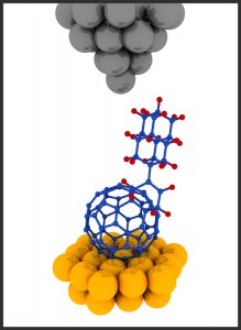
Here’s an illustration the scientists have provided,

Illustration of a buckydiamondoid molecule under a scanning tunneling microscope (STM). In this study the STM made images of the buckydiamondoids and probed their electronic properties.

Illustration of a buckydiamondoid molecule under a scanning tunneling microscope (STM). In this study the STM made images of the buckydiamondoids and probed their electronic properties.

A Sept. 9, 2014 Stanford University news release by Glenda Chui (also on EurekAlert), which originated the news item, provides some information about this piece of international research along with background information on buckyballs and diamondoids (Note: Links have been removed),

“We wanted to see what new, emergent properties might come out when you put these two ingredients together to create a ‘buckydiamondoid,'” said Hari Manoharan of the Stanford Institute for Materials and Energy Sciences (SIMES) at the U.S. Department of Energy’s SLAC National Accelerator Laboratory. “What we got was basically a one-way valve for conducting electricity – clearly more than the sum of its parts.”

The research team, which included scientists from Stanford University, Belgium, Germany and Ukraine, reported its results Sept. 9 in Nature Communications.

Many electronic circuits have three basic components: a material that conducts electrons; rectifiers, which commonly take the form of diodes, to steer that flow in a single direction; and transistors to switch the flow on and off. Scientists combined two offbeat ingredients – buckyballs and diamondoids – to create the new diode-like component.

Buckyballs – short for buckminsterfullerenes – are hollow carbon spheres whose 1985 discovery earned three scientists a Nobel Prize in chemistry. Diamondoids are tiny linked cages of carbon joined, or bonded, as they are in diamonds, with hydrogen atoms linked to the surface, but weighing less than a billionth of a billionth of a carat. Both are subjects of a lot of research aimed at understanding their properties and finding ways to use them.

In 2007, a team led by researchers from SLAC and Stanford discovered that a single layer of diamondoids on a metal surface can emit and focus electrons into a tiny beam. Manoharan and his colleagues wondered: What would happen if they paired an electron-emitting diamondoid with another molecule that likes to grab electrons? Buckyballs are just that sort of electron-grabbing molecule.

Details are then provided about this specific piece of research (from the Stanford news release),

For this study, diamondoids were produced in the SLAC laboratory of SIMES researchers Jeremy Dahl and Robert Carlson, who are world experts in extracting the tiny diamonds from petroleum. The diamondoids were then shipped to Germany, where chemists at Justus-Liebig University figured out how to attach them to buckyballs.

The resulting buckydiamondoids, which are just a few nanometers long, were tested in SIMES laboratories at Stanford. A team led by graduate student Jason Randel and postdoctoral researcher Francis Niestemski used a scanning tunneling microscope to make images of the hybrid molecules and measure their electronic behavior. They discovered that the hybrid is an excellent rectifier: The electrical current flowing through the molecule was up to 50 times stronger in one direction, from electron-spitting diamondoid to electron-catching buckyball, than in the opposite direction. This is something neither component can do on its own.

While this is not the first molecular rectifier ever invented, it’s the first one made from just carbon and hydrogen, a simplicity researchers find appealing, said Manoharan, who is an associate professor of physics at Stanford. The next step, he said, is to see if transistors can be constructed from the same basic ingredients.

“Buckyballs are easy to make – they can be isolated from soot – and the type of diamondoid we used here, which consists of two tiny cages, can be purchased commercially,” he said. “And now that our colleagues in Germany have figured out how to bind them together, others can follow the recipe. So while our research was aimed at gaining fundamental insights about a novel hybrid molecule, it could lead to advances that help make molecular electronics a reality.”

Other research collaborators came from the Catholic University of Louvain in Belgium and Kiev Polytechnic Institute in Ukraine. The primary funding for the work came from U.S. the Department of Energy Office of Science (Basic Energy Sciences, Materials Sciences and Engineering Divisions).

Here’s a link to and a citation for the paper,

Unconventional molecule-resolved current rectification in diamondoid–fullerene hybrids by Jason C. Randel, Francis C. Niestemski,    Andrés R. Botello-Mendez, Warren Mar, Georges Ndabashimiye, Sorin Melinte, Jeremy E. P. Dahl, Robert M. K. Carlson, Ekaterina D. Butova, Andrey A. Fokin, Peter R. Schreiner, Jean-Christophe Charlier & Hari C. Manoharan. Nature Communications 5, Article number: 4877 doi:10.1038/ncomms5877 Published 09 September 2014

This paper is open access. The scientists provided not only a standard illustration but a pretty picture of the buckydiamondoid,

Caption: An international team led by researchers at SLAC National Accelerator Laboratory and Stanford University joined two offbeat carbon molecules -- diamondoids, the square cages at left, and buckyballs, the soccer-ball shapes at right -- to create "buckydiamondoids," center. These hybrid molecules function as rectifiers, conducting electrons in only one direction, and could help pave the way to molecular electronic devices. Credit: Manoharan Lab/Stanford University

Caption: An international team led by researchers at SLAC National Accelerator Laboratory and Stanford University joined two offbeat carbon molecules — diamondoids, the square cages at left, and buckyballs, the soccer-ball shapes at right — to create “buckydiamondoids,” center. These hybrid molecules function as rectifiers, conducting electrons in only one direction, and could help pave the way to molecular electronic devices.
Credit: Manoharan Lab/Stanford University

OCSiAL will not be acquiring Zyvex

The world’s largest nanotechnology business: OCSiAl and its Zyvex acquisition as my June 23, 2014 post was titled is no longer true as per a Sept. 10, 2014 news item on Nanowerk,

Zyvex Technologies and OCSiAl today announced that a previously reported acquisition has been terminated. In June, the companies announced that Zyvex was to be acquired and would operate as the Zyvex Technologies division of OCSiAl. This decision does not affect future plans for cooperation between the companies.

Curiously Zyvex does not have a news release on its website about this latest turn of events although there is this Sept. 9, 2014 Zyvex news release on the Dayton [Ohio, US] Business Journal website, which appears to have originated the Nanowerk news item,

Zyvex Chairman Jim Von Ehr said, “When we started talking with OCSiAl earlier this year, we saw synergies in combining, but as we went along, it became apparent that we could better serve our customers and employees by remaining independent. We look forward to a continued relationship with OCSiAl across a number of areas, but as separate companies. The advanced technology and class-leading products offered by each company will continue to be independently available for commercial applications.”

About Zyvex Technologies
Zyvex was founded in 1997 as the first company solely focused on nanotechnology. Zyvex successfully introduced products to a variety of industries, from semiconductors to sporting goods, and received significant acclaim for its advances in commercializing molecular nanotechnology. More information can be found at www.zyvextech.com.

About OCSiAl
OCSiAl is the creator of a leading technology for the mass industrial production of single wall carbon nanotubes, redefining the market in terms of price and quality. … More information can be found at www.ocsial.com.

OCSiAL does have a Sept. 9, 2014 news release saying much the same as the Zyvex news release but offering a* quote from their Chief Executive Officer (CEO),

Max Atanassov, CEO of OCSiAl LLC said “Cancelling the deal was our mutual decision – we found it to be the best option. What is essential is that we continue to cooperate and see prospective opportunities in our partnership”.

The termination of the deal will not influence OCSiAl’s strategy and further plans. The company will continue to offer top-quality single wall carbon nanotubes (SWCNT) at industrial scale and specially designed universal nanomodifiers for various industries, including polymers, composite materials, elastomers, lithium-ion batteries and transparent conductive films.

And so OCSiAl loses its claim to being the world’s largest nanotechnology company. These are interesting times.

*’a’ added on Dec. 30, 2015.

Images of Nanotechnology competition in New Zealand

The deadline is Oct. 31, 2014 (Hallowe’en), which is on Friday this year. The competition, the third annual,  is for researchers and students based in New Zealand. Here’s more from the MacDiarmid Institute for Advanced Materials and Nanotechnology’s Images of Nanotechnology competition webpage,

Entries are now open for the third Images of Nanotechnology Competition to find the best NZ images from nanotechnology research. An exhibition of selected images will be held in Nelson [New Zealand] in February, in conjunction with the AMN7 conference (http://www.amn-7.com/) and $2000 in prizes will be awarded, courtesy of the MacDiarmid Institute.

Up to three entries can be submitted through the entry form on the website:
http://www.canterbury.ac.nz/nanoevents/compform.shtml

The best images will be displayed in the Nelson Provincial Museum, for four weeks in February 2015. The deadline for entries is 31 October 2014, and any entry received before 10 October will be eligible to be chosen as the poster image for the exhibition.

Please think about how you would explain your images to a lay audience, and have a description prepared when you submit your image(s). These descriptions may be used on the labels next to images that are shown in the Gallery. You should explain the image as though you are explaining it to a non-scientist friend – in the past we have found that many descriptions are far too technical and in fact it would be very help to try your description out on a friend before submitting it. We encourage you to also submit a few supporting images that might help a viewer understand how your image(s) were created.

Entries are encouraged from any researcher or research student based in NZ. Please do let all your colleagues, students and friends know about the competition.

The competition entry form can be found on the University of Canterbury’s Images of Nanotechnology competition webpage,

A competition to find the best NZ images from nanotechnology research.

  • 1st prize – $1000
  • 2nd prize – $700
  • 3rd prize – $300

Deadline for receipt of images is 5pm on Friday 31 October 2014.

All entries and supporting images must be added sequentially (i.e. one at a time) by returning to this form. Please note that there is a 10Mb file limit. Larger images can be submitted on a CD to “Images of Nanotechnology”, Main Office, level 7, Department of Physics and Astronomy, University of Canterbury, Christchurch 8140. If you submit images on a CD you must print out one copy of this form for each image submitted and send the signed copy with your CD.

By submitting this entry I confirm that the entries are my own work. I understand and agree to abide by the rules of the competition. I agree to allow these works to be published online and to be displayed to the public.

Here’s some more information about the AMN7 (Advanced Materials and Nanotechnology) conference being held Feb. 8 – 12, 2015 in Nelson, New Zealand,

Earlybird registration closes on October 31.  Please click here to register for AMN-7.

On behalf of the MacDiarmid Institute for Advanced Materials and Nanotechnology I would like to extend you a warm invitation to join us in Nelson for AMN-7 in February 2015.  AMN-7 is the seventh in our biennial series of meetings that focus on the latest research on advanced materials and nanotechnology.  This event will continue the best traditions of previous events, which include a range of high-impact plenary presentations, cutting-edge invited and contributed talks, interactive poster presentations and convivial social events.  The intimate scale of AMN conferences and the broad interests of fellow delegates offer many opportunities for networking and interdisciplinary discussions.

The venue of AMN-7 – the city of Nelson – has special significance for New Zealand science as it is the birthplace of Ernest Rutherford, the Nobel Prize winner for chemistry in 1908. The Rutherford Hotel will serve as the main conference venue.  Nelson’s excellent climate, beaches, mountains and lakes make it an attractive destination.  And it would be remiss of me not to mention the swag of local wineries and craft breweries.

I hope you’re able to join us in Nelson in 2015.

Shane Telfer
AMN-7 Conference Chair

For anyone curious about the organization which puts on this conference, from the MacDiarmid Institute’s About Us webpage (Note: Links have been removed),

The MacDiarmid Institute for Advanced Materials and Nanotechnology is a national network of New Zealand’s leading scientists, leveraging strength across the country and internationally. We build materials and devices from atoms and molecules, developing and applying cutting edge techniques in physics, chemistry and engineering. We capture our diversity to create benefit and build strength.

We partner with New Zealand businesses to take our innovative new technologies to export markets in sectors as diverse as health, electronics, food and fashion. We train entrepreneurial and socially-aware young scientists, many of whom go on to work in industry or start their own companies, in a culture of excellence and collaboration.

Through sharing the results of our scientific research with the public and with Government, we are inspiring researchers and working to generate a nationwide culture change where science and innovation are celebrated as the keys to New Zealand’s future prosperity.

While the Institute is hosted by Victoria University of Wellington, our Investigators work throughout New Zealand. Named after Alan MacDiarmid, whose curiosity and determination saw him awarded the Nobel Prize in Chemistry, the MacDiarmid Institute was New Zealand’s first Centre of Research Excellence.

Good luck!

Tibetan Buddhist singing bowls inspire more efficient solar cells

There’s no mention as to whether or not Dr Niraj Lal practices any form of meditation or how he came across Tibetan Buddhist singing bowls but somehow he was inspired by them when studying for his PhD at Cambridge University (UK). From a Sept. 8, 2014 news item by Niall Byrne for physorg.com,

The shape of a centuries-old Buddhist singing bowl has inspired a Canberra scientist to re-think the way that solar cells are designed to maximize their efficiency.

Dr Niraj Lal, of the Australian National University,  found during his PhD at the University of Cambridge, that small nano-sized versions of Buddhist singing bowls resonate with light in the same way as they do with sound, and he’s applied this shape to solar cells to increase their ability to capture more light and convert it into electricity.

A Sept. ?, 2014 news release from Australian science communication company, Science in Public, fills in a few more details without any mention of Lal’s meditation practices, should he have any,

“Current standard solar panels lose a large amount of light-energy as it hits the surface, making the panels’ generation of electricity inefficient,” says Niraj. “But if the cells are singing bowl-shaped, then the light bounces around inside the cell for longer”.

Normally used in meditation, music, and relaxation, Buddhist singing bowls make a continuous harmonic ringing sound when the rim of the metal bowl is vibrated with a wooden or other utensil.

During his PhD, Niraj discovered that his ‘nanobowls’ manipulated light by creating a ‘plasmonic’ resonance, which quadrupled the laboratory solar cell’s efficiency compared to a similarly made flat solar cell.

Now, Niraj and his team aim to change all that by applying his singing-bowl discovery to tandem solar cells: a technology that has previously been limited to aerospace applications.

In research which will be published in the November issue of IEEE Journal of Photonics, Niraj and his colleagues have shown that by layering two different types of solar panels on top of each other in tandem, the efficiency of flat rooftop solar panels can achieve 30 per cent—currently, laboratory silicon solar panels convert only 25 per cent of light into electricity, while commercial varieties convert closer to 20 per cent.

The tandem cell design works by absorbing a sunlight more effectively —each cell is made from a different material so that it can ‘see’ a different light wavelength.

“To a silicon solar cell, a rainbow just looks like a big bit of red in the sky—they don’t ‘see’ the blue, green or UV light—they convert all light to electricity as if it was red ,” says Niraj. “But when we put a second cell on top, which ‘sees’ the blue part of light, but allows the red to pass through to the ‘red-seeing’ cell below, we can reach a combined efficiency of more than 30 percent.”

Niraj and a team at ANU are now looking at ways to super-charge the tandem cell design by applying the Buddhist singing bowl shape to further increase efficiency.

“If we can make a solar cell that ‘sees’ more colours and  keeps the right light in the right layers, then we could increase efficiency even further,” says Niraj.

“Every extra percent in efficiency saves you thousands of dollars over the lifetime of the panel,” says Niraj. “Current roof-top solar panels have been steadily increasing in efficiency, which has been a big driver of the fourfold drop in the price for these panels over the last five years.”

More importantly, says Niraj, greater efficiency will allow solar technology to compete with fossil fuels and meet the challenges of climate change and access.

“Electricity is also one of the most enabling technologies we have ever seen, and linking people in rural areas around the world to electricity is one of the most powerful things we can do.”

At the end of the Science in Public news release there’s mention of a science communication competition,

Niraj was a 2014 national finalist of FameLab Australia. FameLab is a global science communication competition for early-career scientists. His work is supported by the Australian Research Council and ARENA – the Australian Renewable Energy Agency.

About FameLab

In 2014, the British Council and Fresh Science have joined forces to bring FameLab to Australia.

FameLab Australia will offer specialist science media training and, ultimately, the chance for early-career researchers to pitch their research at the FameLab International Grand Final in the UK at The Times Cheltenham Science Festival from 3 to 5 June 2014.

FameLab is an international communication competition for scientists, including engineers and mathematicians. Designed to inspire and motivate young researchers to actively engage with the public and with potential stakeholders, FameLab is all about finding the best new voices of science and engineering across the world.

Founded in 2005 by The Times Cheltenham Science Festival, FameLab, working in partnership with the British Council, has already seen more than 5,000 young scientists and engineers participate in over 23 different countries — from Hong Kong to South Africa, USA to Egypt.

Now, FameLab comes to Australia in a landmark collaboration with the British Council and Fresh Science — Australia’s very own science communication competition.

For more information about FameLab Australia, head to www.famelab.org.au

You can find out more about Australia’s Fresh Science here.

Getting back to Dr. Lal, here’s a video he made about his work and where he demonstrates a Tibetan Buddhist singing bowl (this is a very low tech video and the sound quality isn’t great),

Here’s a link to and a citation for Lal’s most recent paper,

Optics and Light Trapping for Tandem Solar Cells on Silicon by Lal, N.N.; White, T.P. ; and Catchpole, K.R. Photovoltaics, IEEE Journal of  (Volume:PP ,  Issue: 99) Page(s): 1 – 7 ISSN : 2156-3381 DOI: 10.1109/JPHOTOV.2014.2342491 Published online 19 August 2014

The paper is behind a paywall but there is open access to Lal’s 2012 University of Cambridge PhD thesis on his approach,

Enhancing solar cells with plasmonic nanovoids by Lal, Niraj Narsey
URI: http://www.dspace.cam.ac.uk/handle/1810/243864 Date:2012-07-03

Hap;y reading!

Simon Fraser University – Bioelectronics course: Week 1*

Last night (Sept. 8, 2014) I started teaching a course called, Bioelectronics, Medical Imaging and Our Bodies for Simon Fraser University’s (SFU) Continuing Studies programme and found that students wanted a copy of the slides from the first night. Unfortunately, SFU does not have a system in place for continuing studies instructors to make their course materials available online to students, so, at the end of this post you will find a link to my Week One PowerPoint slides.

For those who may be mildly curious, here’s a description of the course and of what I was covering on the first night (from SFU’s course description webpage),

Advances in understanding cells, enzymes and proteins will soon allow remaking “life”—and spare body parts—on our terms. How do we extract the best of technology and human intelligence to make breakthroughs in understanding the human body? How will “next generation” medical technologies alter organizational and societal landscapes? Should we halt advances to digest the consequences of technological developments? Or, hoping everything will work out, pursue new frontiers?

We will attempt to answer such questions in discussing the biotechnology used today. And we will look into the future to see how our bodies will be imaged, diagnosed, and fixed. This course requires no technical or scientific background, but an open mind and curiosity will prove helpful.

Week 1: Can the Blind Really See? The Future of Prosthetics

More and more, bioelectronics and/or other emerging technologies are being integrated into materials meant to repair or replace body parts. One example of this is the artificial retina. Others include synthetic tracheas, prosthetic hands and prosthetic legs.

Week 1_Artificial eye

Assuming that students may want slides from subsequent classes, I will be posting more materials over the next five weeks (the length of the course).

ETA Sept. 16, 2014: My ‘notes’ for this week consisting largely of brief heads designed to remind me of the content to be found by clicking the link directly after the head:

Week 1

*’Week 1 added to head on Sept. 16, 2014.

The Danes get more from their marijuana

A Sept. 8, 2014 news item on ScienceDaily features work at the University of Copenhagen where scientists are researching a new method for reducing consumption of drugs such as adrenaline and cannabis when used therapeutically,

About 40% of all medicines used today work through the so-called “G protein-coupled receptors.” These receptors react to changes in the cell environment, for example, to increased amounts of chemicals like cannabis, adrenaline or the medications we take and are therefore of paramount importance to the pharmaceutical industry.

“There is a lot of attention on research into “G protein-coupled receptors,” because they have a key roll in recognizing and binding different substances. Our new method is of interest to the industry because it can contribute to faster and cheaper drug development,” explains Professor Dimitrios Stamou, who heads the Nanomedicine research group at the Nano-Science Center, where the method has been developed. …

A Sept. 8, 2014 University of Copenhagen news release on EurekAlert, which originated the news item, provides a little more detail,

The new method will reduce dramatically the use of precious membrane protein samples. Traditionally, you test a medicinal substance by using small drops of a sample containing the protein that the medicine binds to. If you look closely enough however, each drop is composed of thousands of billions of small nano-containers containing the isolated proteins. Until now, it has been assumed that all of these nano-containers are identical. But it turns out this is not the case and that is why researchers can use a billion times smaller samples for testing drug candidates than hitherto.

“We have discovered that each one of the countless nano-containers is unique. Our method allows us to collect information about each individual nano-container. We can use this information to construct high-throughput screens, where you can, for example, test how medicinal drugs bind G protein-coupled receptors”, explains Signe Mathiasen, who is first author of the paper describing the screening method in Nature Methods. Signe Mathiasen has worked on developing a screening method over the last four years at the University of Copenhagen, where she wrote her PhD thesis research project under the supervision of Professor Stamou.

Although the title doesn’t betray its marijuana orientation, this is a link to and a citation for the researchers’ work,

Nanoscale high-content analysis using compositional heterogeneities of single proteoliposomes by Signe Mathiasen, Sune M Christensen, Juan José Fung, Søren G F Rasmussen, Jonathan F Fay, Sune K Jorgensen, Salome Veshaguri, David L Farrens, Maria Kiskowski, Brian Kobilka, & Dimitrios Stamou. Nature Methods 11, 931–934 (2014) doi:10.1038/nmeth.3062 Published online 03 August 2014

This paper is behind a paywall.