Tag Archives: regenerative medicine

Canada’s strength in regenerative medicine

Urgh! I will scream if I see the phrase “Canada punches above its weight” or some variant thereof one more time. Please! Stop the madness! The latest culprit is the Canadian Council of Academies in the title for its March 9, 2017 news release on EurekAlert,

Canada continues to punch above its weight in the field of regenerative medicine

A new workshop report, Building on Canada’s Strengths in Regenerative Medicine, released today [March 9, 2017] by the Council of Canadian Academies (CCA), confirms that Canadian researchers continue to be recognized as scientific leaders in the field of regenerative medicine and stem cell science.

“Overall, the evidence shows that Canadian research in regenerative medicine continues to be strong,” said Dr. Janet Rossant, FRSC, Chair of the Workshop Steering Committee and President and Scientific Director of the Gairdner Foundation. “While Canadian research is both of high quality and highly cited, it is our collaborative culture, enhanced by our national networks that keeps Canada leading in this field.”

Since the discovery of stem cells in the early 1960s by Canadian scientists Drs. James Till and Ernest McCulloch, significant advancements in regenerative medicine have followed, many by Canadian researchers and practitioners. The appeal of regenerative medicine lies in its curative approach. It replaces or regenerates human cells, tissues, or organs to restore or establish normal function using stem cells. A well-known example of regenerative medicine is the use of bone marrow transplants for leukemia. Although Canada has been historically strong in the field of regenerative medicine, experts caution that we must not lose momentum.

“Canada has been a leader in the field of regenerative medicine for decades, but maintaining this excellence requires ongoing efforts including continued stable and strategic investment in researchers, collaborative networks, and infrastructure,” Dr. Rossant notes. “Several countries are investing heavily in regenerative medicine and stem cell science. Canada has a real opportunity to stay ahead of the curve and remain at the forefront of this field, but it will require us to harness key opportunities now.” [emphasis mine]

The workshop report identifies several opportunities to strengthen the regenerative medicine community in Canada. Opportunities identified as particularly promising focus on:

* formalizing the coordination among regenerative medicine initiatives and key players to speak with one voice on common priorities;

* establishing long-term and stable support for current networks, including those focused on commercialization, to help address the so-called “valley of death” that exists when translating research discoveries to clinical and industry settings;

* enhancing coordination and alignment between the federal regulatory system and provincial healthcare systems; and

* supporting existing manufacturing infrastructure and growing the regenerative medicine industry in Canada to provide jobs for highly-skilled personnel while also benefiting the Canadian economy.

The workshop participants also considered several specific opportunities such as:

* enhancing coordination of Canada’s regenerative medicine clinical trial sites to enable sharing of best practices related to funding, design, and recruitment;

* continued support for cross-training programs to ensure future generations of Canadian researchers have wide-ranging skills suited to the multidisciplinary nature of regenerative medicine;

* new incentives that encourage partnerships between research institutions and industry; and

* increasing efforts related to public engagement and outreach.

“Sometimes becoming excellent is easier than maintaining excellence,” said Dr. Eric M. Meslin, FCAHS, President and CEO of the Council of Canadian Academies. “This is why taking stock of Canada’s place in the regenerative medicine landscape at a point in time is important, especially where the science is moving quickly; it helps those in the field understand the opportunities and will contribute to the ongoing policy discussion in Canada.”

This report was released a few weeks in advance of the federal budget (due tomorrow Wednesday, March 22, 2017). That’s a coincidence, yes?  Interestingly, the 2017 iteration is supposed to be an ‘innovation’ budget, i.e.. designed to stimulate the tech sector if a March 20, 2017 article by David Cochrane for CBC (Canadian Broadcasting Corporation) news online is to be believed. Nowhere in the article is there any mention of regenerative medicine or science, for that matter.

You can download the full report (60 pp.) from the Building on Canada’s Strengths in Regenerative Medicine webpage on the CCA website.
Antibiotic synthetic spider silk

I have a couple of questions, what is ‘click’ chemistry and how does a chance meeting lead to a five-year, interdisciplinary research project on synthetic spider silk? From a Jan. 4, 2017 news item on ScienceDaily,

A chance meeting between a spider expert and a chemist has led to the development of antibiotic synthetic spider silk.

After five years’ work an interdisciplinary team of scientists at The University of Nottingham has developed a technique to produce chemically functionalised spider silk that can be tailored to applications used in drug delivery, regenerative medicine and wound healing.

The Nottingham research team has shown for the first time how ‘click-chemistry’ can be used to attach molecules, such as antibiotics or fluorescent dyes, to artificially produced spider silk synthesised by E.coli bacteria. The research, funded by the Biotechnology and Biological Sciences Research Council (BBSRC) is published today in the online journal Advanced Materials.

A Jan. 3, 2016 University of Nottingham press release (also on EurekAlert), which originated the news item, provides a few more details about ‘click’ chemistry (not enough for me) and more information about the research,

The chosen molecules can be ‘clicked’ into place in soluble silk protein before it has been turned into fibres, or after the fibres have been formed. This means that the process can be easily controlled and more than one type of molecule can be used to ‘decorate’ individual silk strands.

Nottingham breakthrough

In a laboratory in the Centre of Biomolecular Sciences, Professor Neil Thomas from the School of Chemistry in collaboration with Dr Sara Goodacre from the School of Life Sciences, has led a team of BBSRC DTP-funded PhD students starting with David Harvey who was then joined by Victor Tudorica, Leah Ashley and Tom Coekin. They have developed and diversified this new approach to functionalising ‘recombinant’ — artificial — spider silk with a wide range of small molecules.

They have shown that when these ‘silk’ fibres are ‘decorated’ with the antibiotic levofloxacin it is slowly released from the silk, retaining its anti-bacterial activity for at least five days.

Neil Thomas, a Professor of Medicinal and Biological Chemistry, said: “Our technique allows the rapid generation of biocompatible, mono or multi-functionalised silk structures for use in a wide range of applications. These will be particularly useful in the fields of tissue engineering and biomedicine.”

Remarkable qualities of spider silk

Spider silk is strong, biocompatible and biodegradable. It is a protein-based material that does not appear to cause a strong immune, allergic or inflammatory reaction. With the recent development of recombinant spider silk, the race has been on to find ways of harnessing its remarkable qualities.

The Nottingham research team has shown that their technique can be used to create a biodegradable mesh which can do two jobs at once. It can replace the extra cellular matrix that our own cells generate, to accelerate growth of the new tissue. It can also be used for the slow release of antibiotics.

Professor Thomas said: “There is the possibility of using the silk in advanced dressings for the treatment of slow-healing wounds such as diabetic ulcers. Using our technique infection could be prevented over weeks or months by the controlled release of antibiotics. At the same time tissue regeneration is accelerated by silk fibres functioning as a temporary scaffold before being biodegraded.”

The medicinal properties of spider silk recognised for centuries.

The medicinal properties of spider silk have been recognised for centuries but not clearly understood. The Greeks and Romans treated wounded soldiers with spider webs to stop bleeding. It is said that soldiers would use a combination of honey and vinegar to clean deep wounds and then cover the whole thing with balled-up spider webs.

There is even a mention in Shakespeare’s Midsummer Night’s Dream: “I shall desire you of more acquaintance, good master cobweb,” the character ‘Bottom’ said. “If I cut my finger, I shall make bold of you.”

The press release goes on to describe the genesis of the project and how this multidisciplinary team was formed in more detail,

The idea came together at a discipline bridging university ‘sandpit’ meeting five years ago. Dr Goodacre says her chance meeting at that event with Professor Thomas proved to be one of the most productive afternoons of her career.

Dr Goodacre, who heads up the SpiderLab in the School of Life Sciences, said: “I got up at that meeting and showed the audience a picture of some spider silk. I said ‘I want to understand how this silk works, and then make some.’

“At the end of the session Neil came up to me and said ‘I think my group could make that.’ He also suggested that there might be more interesting ‘tweaks’ one could make so that the silk could be ‘decorated’ with different, useful, compounds either permanently or which could be released over time due to a change in the acidity of the environment.”

The approach required the production of the silk proteins in a bacterium where an amino acid not normally found in proteins was included. This amino acid contained an azide group which is widely used in ‘click’ reactions that only occur at that position in the protein. It was an approach that no-one had used before with spider silk — but the big question was — would it work?

Dr Goodacre said: “It was the start of a fascinating adventure that saw a postdoc undertake a very preliminary study to construct the synthetic silks. He was a former SpiderLab PhD student who had previously worked with our tarantulas. Thanks to his ground work we showed we could produce the silk proteins in bacteria. We were then joined by David Harvey, a new PhD student, who not only made the silk fibres, incorporating the unusual amino acid, but also decorated it and demonstrated its antibiotic activity. He has since extended those first ideas far beyond what we had thought might be possible.”

David Harvey’s work is described in this paper but Professor Thomas and Dr Goodacre say this is just the start. There are other joint SpiderLab/Thomas lab students working on uses for this technology in the hope of developing it further.

David Harvey, the lead author on this their first paper, has just been awarded his PhD and is now a postdoctoral researcher on a BBSRC follow-on grant so is still at the heart of the research. His current work is focused on driving the functionalised spider silk technology towards commercial application in wound healing and tissue regeneration.

Here’s a link to and a citation for the paper,

Antibiotic Spider Silk: Site-Specific Functionalization of Recombinant Spider Silk Using “Click” Chemistry by David Harvey, Philip Bardelang, Sara L. Goodacre, Alan Cockayne, and Neil R. Thomas. Advanced Materials DOI: 10.1002/adma.201604245 Version of Record online: 28 DEC 2016

© 2016 WILEY-VCH Verlag GmbH & Co. KGaA, Weinheim

This paper is behind a paywall.

I imagine Mr. Cockayne’s name has led to much teasing over the years. People who have names with that kind of potential tend to either change them or double down and refuse to compromise.

Better technique for growing organoids taking them from the lab to the clinic

A Nov. 16, 2016 École Polytechnique Fédérale de Lausanne (EPFL) press release (also on EurekAlert) describes a new material for growing organoids,

Organoids are miniature organs that can be grown in the lab from a person’s stem cells. They can be used to model diseases, and in the future could be used to test drugs or even replace damaged tissue in patients. But currently organoids are very difficult to grow in a standardized and controlled way, which is key to designing and using them. EPFL scientists have now solved the problem by developing a patent-pending “hydrogel” that provides a fully controllable and tunable way to grow organoids. …

Organoids need a 3D scaffold

Growing organoids begins with stem cells — immature cells that can grow into any cell type of the human body and that play key roles in tissue function and regeneration. To form an organoid, the stem cells are grown inside three-dimensional gels that contain a mix of biomolecules that promote stem cell renewal and differentiation.

The role of these gels is to mimic the natural environment of the stem cells, which provides them with a protein- and sugar-rich scaffold called the “extracellular matrix”, upon which the stem cells build specific body tissues. The stem cells stick to the extracellular matrix gel, and then “self-organize” into miniature organs like retinas, kidneys, or the gut. These tiny organs retain key aspects of their real-life biology, and can be used to study diseases or test drugs before moving on to human trials.

But the current gels used for organoid growth are derived from mice, and have problems. First, it is impossible to control their makeup from batch to batch, which can cause stem cells to behave inconsistently. Second, their biochemical complexity makes them very difficult to fine-tune for studying the effect of different parameters (e.g. biological molecules, mechanical properties, etc.) on the growth of organoids. Finally, the gels can carry pathogens or immunogens, which means that they are not suitable for growing organoids to be used in the clinic.

A hydrogel solution

The lab of Matthias Lütolf at EPFL’s Institute of Bioengineering has developed a synthetic “hydrogel” that eschews the limitations of conventional, naturally derived gels. The patent-pending gel is made of water and polyethylene glycol, a substance used widely today in various forms, from skin creams and toothpastes to industrial applications and, as in this case, bioengineering.

Nikolce Gjorevski, the first author of the study, and his colleagues used the hydrogel to grow stem cells of the gut into a miniature intestine. The functional hydrogel was not only a goal in and of itself, but also a means to identify the factors that influence the stem cells’ ability to expand and form organoids. By carefully tweaking the hydrogel’s properties, they discovered that separate stages of the organoid formation process require different mechanical environments and biological components.

One such factor is a protein called fibronectin, which helps the stem cells attach to the hydrogel. Lütolf’s lab found that this attachment itself is immensely important for growing organoids, as it triggers a whole host of signals to the stem cell that tell it to grow and build an intestine-like structure. The researchers also discovered an essential role for the mechanical properties, i.e. the physical stiffness, of the gel in regulating intestinal stem cell behavior, shedding light on how cells are able to sense, process and respond to physical stimuli. This insight is particularly valuable – while the influence of biochemical signals on stem cells is well-understood, the effect of physical factors has been more mysterious.

Because the hydrogel is man-made, it is easy to control its chemical composition and key properties, and ensure consistency from batch to batch. And because it is artificial, it does not carry any risk of infection or triggering immune responses. As such, it provides a means of moving organoids from basic research to actual pharmaceutical and clinical applications in the future.

Lütolf’s lab is now researching other types of stem cells in order to extend the capacities of their hydrogel into other tissues.

Here’s a link to and a citation for the paper,

Designer matrices for intestinal stem cell and organoid culture by Nikolce Gjorevski, Norman Sachs, Andrea Manfrin, Sonja Giger, Maiia E. Bragina, Paloma Ordóñez-Morán, Hans Clevers, & Matthias P. Lutolf.  Nature (2016) doi:10.1038/nature20168 Published online 16 November 2016

This paper is behind a paywall.

Brain and machine as one (machine/flesh)

The essay on brains and machines becoming intertwined is making the rounds. First stop on my tour was its Oct. 4, 2016 appearance on the Mail & Guardian, then there was its Oct. 3, 2016 appearance on The Conversation, and finally (moving forward in time) there was its Oct. 4, 2016 appearance on the World Economic Forum website as part of their Final Frontier series.

The essay was written by Richard Jones of Sheffield University (mentioned here many times before but most recently in a Sept. 4, 2014 posting). His book ‘Soft Machines’ provided me with an important and eminently readable introduction to nanotechnology. He is a professor of physics at the University of Sheffield and here’s more from his essay (Oct. 3, 2016 on The Conversation) about brains and machines (Note: Links have been removed),

Imagine a condition that leaves you fully conscious, but unable to move or communicate, as some victims of severe strokes or other neurological damage experience. This is locked-in syndrome, when the outward connections from the brain to the rest of the world are severed. Technology is beginning to promise ways of remaking these connections, but is it our ingenuity or the brain’s that is making it happen?

Ever since an 18th-century biologist called Luigi Galvani made a dead frog twitch we have known that there is a connection between electricity and the operation of the nervous system. We now know that the signals in neurons in the brain are propagated as pulses of electrical potential, whose effects can be detected by electrodes in close proximity. So in principle, we should be able to build an outward neural interface system – that is to say, a device that turns thought into action.

In fact, we already have the first outward neural interface system to be tested in humans. It is called BrainGate and consists of an array of micro-electrodes, implanted into the part of the brain concerned with controlling arm movements. Signals from the micro-electrodes are decoded and used to control the movement of a cursor on a screen, or the motion of a robotic arm.

A crucial feature of these systems is the need for some kind of feedback. A patient must be able to see the effect of their willed patterns of thought on the movement of the cursor. What’s remarkable is the ability of the brain to adapt to these artificial systems, learning to control them better.

You can find out more about BrainGate in my May 17, 2012 posting which also features a video of a woman controlling a mechanical arm so she can drink from a cup coffee by herself for the first time in 15 years.

Jones goes on to describe the cochlear implants (although there’s no mention of the controversy; not everyone believes they’re a good idea) and retinal implants that are currently available. Jones notes this (Note Links have been removed),

The key message of all this is that brain interfaces now are a reality and that the current versions will undoubtedly be improved. In the near future, for many deaf and blind people, for people with severe disabilities – including, perhaps, locked-in syndrome – there are very real prospects that some of their lost capabilities might be at least partially restored.

Until then, our current neural interface systems are very crude. One problem is size; the micro-electrodes in use now, with diameters of tens of microns, may seem tiny, but they are still coarse compared to the sub-micron dimensions of individual nerve fibres. And there is a problem of scale. The BrainGate system, for example, consists of 100 micro-electrodes in a square array; compare that to the many tens of billions of neurons in the brain. The fact these devices work at all is perhaps more a testament to the adaptability of the human brain than to our technological prowess.

Scale models

So the challenge is to build neural interfaces on scales that better match the structures of biology. Here, we move into the world of nanotechnology. There has been much work in the laboratory to make nano-electronic structures small enough to read out the activity of a single neuron. In the 1990s, Peter Fromherz, at the Max Planck Institute for Biochemistry, was a pioneer of using silicon field effect transistors, similar to those used in commercial microprocessors, to interact with cultured neurons. In 2006, Charles Lieber’s group at Harvard succeeded in using transistors made from single carbon nanotubes – whiskers of carbon just one nanometer in diameter – to measure the propagation of single nerve pulses along the nerve fibres.

But these successes have been achieved, not in whole organisms, but in cultured nerve cells which are typically on something like the surface of a silicon wafer. It’s going to be a challenge to extend these methods into three dimensions, to interface with a living brain. Perhaps the most promising direction will be to create a 3D “scaffold” incorporating nano-electronics, and then to persuade growing nerve cells to infiltrate it to create what would in effect be cyborg tissue – living cells and inorganic electronics intimately mixed.

I have featured Charles Lieber and his work here in two recent posts: ‘Bionic’ cardiac patch with nanoelectric scaffolds and living cells on July 11, 2016 and Long-term brain mapping with injectable electronics on Sept. 22, 2016.

For anyone interested in more about the controversy regarding cochlear implants, there’s this page on the Brown University (US) website. You might also want to check out Gregor Wolbring (professor at the University of Calgary) who has written extensively on the concept of ableism (links to his work can be found at the end of this post). I have excerpted from an Aug. 30, 2011 post the portion where Gregor defines ‘ableism’,

From Gregor’s June 17, 2011 posting on the FedCan blog,

The term ableism evolved from the disabled people rights movements in the United States and Britain during the 1960s and 1970s.  It questions and highlights the prejudice and discrimination experienced by persons whose body structure and ability functioning were labelled as ‘impaired’ as sub species-typical. Ableism of this flavor is a set of beliefs, processes and practices, which favors species-typical normative body structure based abilities. It labels ‘sub-normative’ species-typical biological structures as ‘deficient’, as not able to perform as expected.

The disabled people rights discourse and disability studies scholars question the assumption of deficiency intrinsic to ‘below the norm’ labeled body abilities and the favoritism for normative species-typical body abilities. The discourse around deafness and Deaf Culture would be one example where many hearing people expect the ability to hear. This expectation leads them to see deafness as a deficiency to be treated through medical means. In contrast, many Deaf people see hearing as an irrelevant ability and do not perceive themselves as ill and in need of gaining the ability to hear. Within the disabled people rights framework ableism was set up as a term to be used like sexism and racism to highlight unjust and inequitable treatment.

Ableism is, however, much more pervasive.

You can find out more about Gregor and his work here: http://www.crds.org/research/faculty/Gregor_Wolbring2.shtml or here:
https://www.facebook.com/GregorWolbring.

Stronger more robust nanofibers for everything from bulletproof vests to cellular scaffolds (tissue engineering)

This work on a new technique for producing nanofibers comes from Harvard University’s School of Engineering and Applied Sciences and the Wyss Institute for Biologically Inspired Engineering (also at Harvard University). From an Oct. 10, 2016 news item on phys.org,

Fibrous materials—known for their toughness, durability and pliability—are used in everything from bulletproof vests to tires, filtration systems and cellular scaffolds for tissue engineering and regenerative medicine.

The properties of these materials are such that the smaller the fibers are, the stronger and tougher they become. But making certain fibers very small has been an engineering challenge.

Now, researchers from the Harvard John A. Paulson School of Engineering and Applied Sciences (SEAS) and the Wyss Institute for Biologically Inspired Engineering at Harvard have developed a new method to make and collect nanofibers and control their size and morphology. This could lead to stronger, more durable bulletproof vests and armor and more robust cellular scaffolding for tissue repair.

An Oct. 7, 2016 Harvard University press release by Leah Burrows, which originated the news item, describes the research in more detail (Note: A link has been removed),

Nanofibers are smaller than one micrometer in diameter.  Most nanofiber production platforms rely on dissolving polymers in a solution, which then evaporates as the fiber forms.

Rotary Jet-Spinning (RJS), the technique developed by Kit Parker’s Disease Biophysics Group, works likes a cotton candy machine. Parker is Tarr Family Professor of Bioengineering and Applied Physics at SEAS and a Core Member of the Wyss Institute. A liquid polymer solution is loaded into a reservoir and pushed out through a tiny opening by centrifugal force as the device spins. As the solution leaves the reservoir, the solvent evaporates and the polymers solidify and elongate into small, thin fibers.

“This advance is important because it allows us to manufacture ballistic protection that is much lighter, more flexible and more functional than what is available today,” said Parker, who in addition to his Harvard role is a lieutenant colonel in the United States Army Reserve and was motivated by his own combat experiences in Afghanistan. “Not only could it save lives but for the warfighter, it also could help reduce the repetitive injury motions that soldiers, sailors, marines and airmen have suffered over the last 15 years of the war on terror.”

“Rotary Jet-Spinning is great for most polymer fibers you want to make,” said Grant Gonzalez, a graduate student at SEAS and first author of the paper.  “However, some fibers require a solvent that doesn’t evaporate easily. Para-aramid, the polymer used in Kevlar® for example, is dissolved in sulfuric acid, which doesn’t evaporate off. The solution just splashes against the walls of the device without forming fibers.”

Nanofibers are smaller than one micrometer in diameter.  Most nanofiber production platforms rely on dissolving polymers in a solution, which then evaporates as the fiber forms.

Rotary Jet-Spinning (RJS), the technique developed by Kit Parker’s Disease Biophysics Group, works likes a cotton candy machine. Parker is Tarr Family Professor of Bioengineering and Applied Physics at SEAS and a Core Member of the Wyss Institute. A liquid polymer solution is loaded into a reservoir and pushed out through a tiny opening by centrifugal force as the device spins. As the solution leaves the reservoir, the solvent evaporates and the polymers solidify and elongate into small, thin fibers.

“This advance is important because it allows us to manufacture ballistic protection that is much lighter, more flexible and more functional than what is available today,” said Parker, who in addition to his Harvard role is a lieutenant colonel in the United States Army Reserve and was motivated by his own combat experiences in Afghanistan. “Not only could it save lives but for the warfighter, it also could help reduce the repetitive injury motions that soldiers, sailors, marines and airmen have suffered over the last 15 years of the war on terror.”

“Rotary Jet-Spinning is great for most polymer fibers you want to make,” said Grant Gonzalez, a graduate student at SEAS and first author of the paper.  “However, some fibers require a solvent that doesn’t evaporate easily. Para-aramid, the polymer used in Kevlar® for example, is dissolved in sulfuric acid, which doesn’t evaporate off. The solution just splashes against the walls of the device without forming fibers.”

Other methods, such as electrospinning, which uses an electric field to pull the polymer into a thin fiber, also have poor results with Kevlar and other polymers such as alginate used for tissue scaffolding and DNA.

The Harvard team overcame these challenges by developing a wet-spinning platform, which uses the same principles as the RJS system but relies on precipitation rather than evaporation to separate the solvent from the polymer.

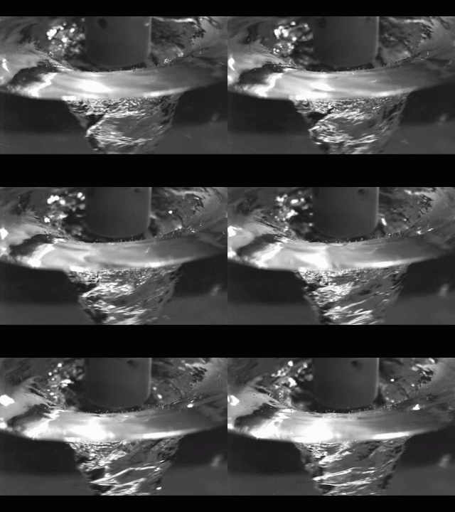
In this system, called immersion Rotary Jet-Spinning (iRJS), when the polymer solution shoots out of the reservoir, it first passes through an area of open air, where the polymers elongate and the chains align. Then the solution hits a liquid bath that removes the solvent and precipitates the polymers to form solid fibers. Since the bath is also spinning — like water in a salad spinner — the nanofibers follow the stream of the vortex and wrap around a rotating collector at the base of the device.

Using this system, the team produced Nylon, DNA, alginate and ballistic resistant para-aramid nanofibers. The team could tune the fiber’s diameter by changing the solution concentration, the rotational speed and the distance the polymer traveled from the reservoir to the bath.

“By being able to modulate fiber strength, we can create a cellular scaffold that can mimic skeleton muscle and native tissues,” said Gonzalez.  “This platform could enable us to create a wound dressing out of alginate material or seed and mature cells on scaffolding for tissue engineering.”

Because the fibers were collected by a spinning vortex, the system also produced well-aligned sheets of nanofibers, which is important for scaffolding and ballistic resistant materials.

This is the ‘candy floss’ technique at work,

Rotary Jet-Spinning (RJS) works likes a cotton candy machine. A liquid polymer solution is loaded into a reservoir and pushed out through a tiny opening by centrifugal force as the device spins. As the solution leaves the reservoir, the solvent evaporates and the polymers solidify and elongate into small, thin fibers. Courtesy: Harvard University

Rotary Jet-Spinning (RJS) works likes a cotton candy machine. A liquid polymer solution is loaded into a reservoir and pushed out through a tiny opening by centrifugal force as the device spins. As the solution leaves the reservoir, the solvent evaporates and the polymers solidify and elongate into small, thin fibers. Courtesy: Harvard University

Here’s a link to and a citation for the paper,

Production of Synthetic, Para-Aramid and Biopolymer Nanofibers by Immersion Rotary Jet-Spinning by Grant M. Gonzalez, Luke A. MacQueen, Johan U. Lind, Stacey A. Fitzgibbons, Christophe O. Chantre, Isabelle Huggler, Holly M. Golecki, Josue A. Goss, Kevin Kit Parker. Macromolecular Materials and Engineering DOI: 10.1002/mame.201600365 Version of Record online: 7 OCT 2016

© 2016 WILEY-VCH Verlag GmbH & Co. KGaA, Weinheim

This paper is behind a paywall.

Long-term brain mapping with injectable electronics

Charles Lieber and his team at Harvard University announced a success with their work on injectable electronics last year (see my June 11, 2015 posting for more) and now they are reporting on their work with more extensive animal studies according to an Aug. 29, 2016 news item on psypost.org,

Scientists in recent years have made great strides in the quest to understand the brain by using implanted probes to explore how specific neural circuits work.

Though effective, those probes also come with their share of problems as a result of rigidity. The inflammation they produce induces chronic recording instability and means probes must be relocated every few days, leaving some of the central questions of neuroscience – like how the neural circuits are reorganized during development, learning and aging- beyond scientists’ reach.

But now, it seems, things are about to change.

Led by Charles Lieber, The Mark Hyman Jr. Professor of Chemistry and chair of the Department of Chemistry and Chemical Biology, a team of researchers that included graduate student Tian-Ming Fu, postdoctoral fellow Guosong Hong, graduate student Tao Zhou and others, has demonstrated that syringe-injectable mesh electronics can stably record neural activity in mice for eight months or more, with none of the inflammation

An Aug. 29, 2016 Harvard University press release, which originated the news item, provides more detail,

“With the ability to follow the same individual neurons in a circuit chronically…there’s a whole suite of things this opens up,” Lieber said. “The eight months we demonstrate in this paper is not a limit, but what this does show is that mesh electronics could be used…to investigate neuro-degenerative diseases like Alzheimer’s, or processes that occur over long time, like aging or learning.”

Lieber and colleagues also demonstrated that the syringe-injectable mesh electronics could be used to deliver electrical stimulation to the brain over three months or more.

“Ultimately, our aim is to create these with the goal of finding clinical applications,” Lieber said. “What we found is that, because of the lack of immune response (to the mesh electronics), which basically insulates neurons, we can deliver stimulation in a much more subtle way, using lower voltages that don’t damage tissue.”

The possibilities, however, don’t end there.

The seamless integration of the electronics and biology, Lieber said, could open the door to an entirely new class of brain-machine interfaces and vast improvements in prosthetics, among other fields.

“Today, brain-machine interfaces are based on traditional implanted probes, and there has been some impressive work that’s been done in that field,” Lieber said. “But all the interfaces rely on the same technique to decode neural signals.”

Because traditional rigid implanted probes are invariably unstable, he explained, researchers and clinicians rely on decoding what they call the “population average” – essentially taking a host of neural signals and applying complex computational tools to determine what they mean.

Using tissue-like mesh electronics, by comparison, researchers may be able to read signals from specific neurons over time, potentially allowing for the development of improved brain-machine interfaces for prosthetics.

“We think this is going to be very powerful, because we can identify circuits and both record and stimulate in a way that just hasn’t been possible before,” Lieber said. “So what I like to say is: I think therefore it happens.”

Lieber even held out the possibility that the syringe-injectable mesh electronics could one day be used to treat catastrophic injuries to the brain and spinal cord.

“I don’t think that’s science-fiction,” he said. “Other people may say that will be possible through, for example, regenerative medicine, but we are pursuing this from a different angle.

“My feeling is that this is about a seamless integration between the biological and the electronic systems, so they’re not distinct entities,” he continued. “If we can make the electronics look like the neural network, they will work together…and that’s where you want to be if you want to exploit the strengths of both.”

In the 2015 posting, Lieber was discussing cyborgs, here he broaches the concept without using the word, “… seamless integration between the biological and the electronic systems, so they’re not distinct entities.”

Here’s a link to and a citation for the paper,

Stable long-term chronic brain mapping at the single-neuron level by Tian-Ming Fu, Guosong Hong, Tao Zhou, Thomas G Schuhmann, Robert D Viveros, & Charles M Lieber. Nature Methods (2016) doi:10.1038/nmeth.3969 Published online 29 August 2016

This paper is behind a paywall.

Better blood vessel growth for regenerative medicine?

If the organs and tissues grown in labs are to be successfully transplanted into bodies, then growing the blood vessels needed to maintain them becomes very important. A May 24, 2016 news item on ScienceDaily describes a new technique for the growing the vessels,

Growing tissues and organs in the lab for transplantation into patients could become easier after scientists discovered an effective way to produce three-dimensional networks of blood vessels, vital for tissue survival yet a current stumbling block in regenerative medicine.

In addition the technique to grow the blood vessels in a 3D scaffold cuts down on the risk of transplant rejection because it uses cells from the patient. It was developed by researchers from the University of Bath’s Department of Pharmacy and Pharmacology, working with colleagues at Bristol Heart Institute.

A May 24 (?), 2016 University of Bath (UK) press release, which originated the news item, expands on the theme (Note: Links have been removed),

So far the shortage of adequate patient-derived scaffolds that can support blood vessel growth has been a major limitation for regenerative medicine and tissue engineering.

Other methods only allow limited formation of small blood vessels such as capillaries, which makes tissue less likely to successfully transplant into a patient. In addition other methods of tissue growth require the use of animal products, unnecessary in this technique which uses human platelet lysate gel (hPLG) and endothelial progenitor cells (EPCs) – a type of cell which helps maintain blood vessel walls.

Dr Giordano Pula, Lecturer in Pharmacology at the University of Bath and head of the research team making the discovery, said: “A major challenge in tissue engineering and regenerative medicine is providing the new tissue with a network of blood vessels, and linking this to the patient’s existing blood supply; this is vital for the tissue’s survival and integration with adjacent tissues.

Dr Paul De Bank, Senior Lecturer in Pharmaceutics at the University of Bath and co-author of the paper, said: “By embedding EPCs in a gel derived from platelets, both of which can be isolated from the patient’s blood, we have demonstrated the formation of a network of small vessels. What is more, the gel contains a number of different growth factors which can induce existing blood vessels to infiltrate the gel and form connections with the new structures. Combining tissue-specific cells with this EPC-containing gel offers the potential for the formation of fully vascularised, functional tissues or organs, which integrate seamlessly with the patient.

“This discovery has the potential to accelerate the development of regenerative medicine applications.”

Professor Peter Weissberg, Medical Director of the British Heart Foundation, said: “Over a half a million people in the UK are living with heart failure, a disabling condition which can leave people unable to carry out everyday activities such as climbing the stairs or even walking to the shops. This regenerative research brings the British Heart Foundation’s goal to mend a broken heart and beat heart failure one step closer.

“All living tissues, including new heart muscle, need a blood supply. One of the fundamental goals of regenerative medicine is to find ways to grow a new blood supply from scratch. Previous attempts at this using human cells and synthetic scaffolds have met with only limited success.

“The beauty of this new approach is that components of a person’s own blood could be manipulated to create a scaffold on which new blood vessels could grow. This increases the likelihood that the new tissue will be integrated into the patient’s body which, if proven successful with more research, could improve the lives of people affected by heart failure.”

Here’s a link to and a citation for the paper,

Platelet lysate gel and endothelial progenitors stimulate microvascular network formation in vitro: tissue engineering implications by Tiago M. Fortunato, Cristina Beltrami, Costanza Emanueli, Paul A. De Bank & Giordano Pula. Scientific Reports 6, Article number: 25326 (2016)  doi:10.1038/srep25326 Published online: 04 May 2016

This is an open access paper.

One of the criticisms of Paolo Macchiarini’s work with synthetic tracheas centered around blood supply to the cells (from my April 19, 2016 posting; it was part 1 of a 2-part series),

This ground-breaking achievement consisted of bringing to life a dead windpipe from a donor, by putting it in a plastic box, a so-called ‘bioreactor’ together with bone marrow fluid (stem cells). A few weeks later, I [Pierre Delaere*]  wrote a letter to The Lancet, pointing out:

“The main drawback of the proposed reconstruction is the lack of an intrinsic blood supply to the trachea. We know that a good blood supply is the first requirement in all other tissue and organ transplantations. Therefore, the reported success of this technique is questionable” (correspondence by Delaere and Hermans, Lancet 2009).

The excerpt you’ve just seen features part of an open letter Pierre Delaere (a long time Macchiarini critic), published in Leonid Schneider’s blog ‘For Better Science’ in an April 2, 2016 posting.

Getting back to Bath, this is exciting stuff and I hope the research is reproducible.

Growing complex skin tissue—complete with hair follicles and sebaceous glands

A laboratory in Japan has managed to grow complex skin tissue according to an April 2, 2016 RIKEN (Japan) press release (also on EurekAlert but dated April 1, 2016),

Using reprogrammed iPS cells, scientists from the RIKEN Center for Developmental Biology (CDB) in Japan have, along with collaborators from Tokyo University of Science and other Japanese institutions, successfully grown complex skin tissue–complete with hair follicles and sebaceous glands–in the laboratory. They were then able to implant these three-dimensional tissues into living mice, and the tissues formed proper connections with other organ systems such as nerves and muscle fibers. This work opens a path to creating functional skin transplants for burn and other patients who require new skin.

Research into bioengineered tissues has led to important achievements in recent years–with a number of different tissue types being created–but there are still obstacles to be overcome. In the area of skin tissue, epithelial cells have been successfully grown into implantable sheets, but they did not have the proper appendages–the oil-secreting and sweat glands–that would allow them to function as normal tissue.

To perform the work, published in Science Advances, the researchers took cells from mouse gums and used chemicals to transform them into stem cell-like iPS cells. In culture, the cells properly developed into what is called an embryoid body (EB)?a three-dimensional clump of cells that partially resembles the developing embryo in an actual body. The researchers created EBs from iPS cells using Wnt10b signaling and then implanted multiple EBs into immune-deficient mice, where they gradually changed into differentiated tissue, following the pattern of an actual embryo. Once the tissue had differentiated, the scientists transplanted them out of those mice and into the skin tissue of other mice, where the tissues developed normally as integumentary tissue?the tissue between the outer and inner skin that is responsible for much of the function of the skin in terms of hair shaft eruption and fat excretion. Critically, they also found that the implanted tissues made normal connections with the surrounding nerve and muscle tissues, allowing it to function normally.

One important key to the development was that treatment with Wnt10b, a signaling molecule, resulted in a larger number of hair follicles, making the bioengineered tissue closer to natural tissue.

According to Takashi Tsuji of the RIKEN Center for Developmental Biology, who led the study, “Up until now, artificial skin development has been hampered by the fact that the skin lacked the important organs, such as hair follicles and exocrine glands, which allow the skin to play its important role in regulation. With this new technique, we have successfully grown skin that replicates the function of normal tissue. We are coming ever closer to the dream of being able to recreate actual organs in the lab for transplantation, and also believe that tissue grown through this method could be used as an alternative to animal testing of chemicals.”

Here’s a link to and a citation for the paper,

Bioengineering a 3D integumentary organ system from iPS cells using an in vivo transplantation model by Ryoji Takagi, Junko Ishimaru, Ayaka Sugawara, Koh-ei Toyoshima, Kentaro Ishida, Miho Ogawa, Kei Sakakibara, Kyosuke Asakawa, Akitoshi Kashiwakura, Masamitsu Oshima, Ryohei Minamide, Akio Sato, Toshihiro Yoshitake, Akira Takeda, Hiroshi Egusa, and Takashi Tsuji. Science Advances  01 Apr 2016: Vol. 2, no. 4, e1500887 DOI: 10.1126/sciadv.1500887

This appears to be an open access paper.

3D microtopographic scaffolds for transplantation and generation of reprogrammed human neurons

Should this technology prove successful once they start testing on people, the stated goal is to use it for the treatment of human neurodegenerative disorders such as Parkinson’s disease.  But, I can’t help wondering if they might also consider constructing an artificial brain.

Getting back to the 3D scaffolds for neurons, a March 17, 2016 US National Institutes of Health (NIH) news release (also on EurekAlert), makes the announcement,

National Institutes of Health-funded scientists have developed a 3D micro-scaffold technology that promotes reprogramming of stem cells into neurons, and supports growth of neuronal connections capable of transmitting electrical signals. The injection of these networks of functioning human neural cells — compared to injecting individual cells — dramatically improved their survival following transplantation into mouse brains. This is a promising new platform that could make transplantation of neurons a viable treatment for a broad range of human neurodegenerative disorders.

Previously, transplantation of neurons to treat neurodegenerative disorders, such as Parkinson’s disease, had very limited success due to poor survival of neurons that were injected as a solution of individual cells. The new research is supported by the National Institute of Biomedical Imaging and Bioengineering (NIBIB), part of NIH.

“Working together, the stem cell biologists and the biomaterials experts developed a system capable of shuttling neural cells through the demanding journey of transplantation and engraftment into host brain tissue,” said Rosemarie Hunziker, Ph.D., director of the NIBIB Program in Tissue Engineering and Regenerative Medicine. “This exciting work was made possible by the close collaboration of experts in a wide range of disciplines.”

The research was performed by researchers from Rutgers University, Piscataway, New Jersey, departments of Biomedical Engineering, Neuroscience and Cell Biology, Chemical and Biochemical Engineering, and the Child Health Institute; Stanford University School of Medicine’s Institute of Stem Cell Biology and Regenerative Medicine, Stanford, California; the Human Genetics Institute of New Jersey, Piscataway; and the New Jersey Center for Biomaterials, Piscataway. The results are reported in the March 17, 2016 issue of Nature Communications.

The researchers experimented in creating scaffolds made of different types of polymer fibers, and of varying thickness and density. They ultimately created a web of relatively thick fibers using a polymer that stem cells successfully adhered to. The stem cells used were human induced pluripotent stem cells (iPSCs), which can be readily generated from adult cell types such as skin cells. The iPSCs were induced to differentiate into neural cells by introducing the protein NeuroD1 into the cells.

The space between the polymer fibers turned out to be critical. “If the scaffolds were too dense, the stem cell-derived neurons were unable to integrate into the scaffold, whereas if they are too sparse then the network organization tends to be poor,” explained Prabhas Moghe, Ph.D., distinguished professor of biomedical engineering & chemical engineering at Rutgers University and co-senior author of the paper. “The optimal pore size was one that was large enough for the cells to populate the scaffold but small enough that the differentiating neurons sensed the presence of their neighbors and produced outgrowths resulting in cell-to-cell contact. This contact enhances cell survival and development into functional neurons able to transmit an electrical signal across the developing neural network.”

To test the viability of neuron-seeded scaffolds when transplanted, the researchers created micro-scaffolds that were small enough for injection into mouse brain tissue using a standard hypodermic needle. They injected scaffolds carrying the human neurons into brain slices from mice and compared them to human neurons injected as individual, dissociated cells.

The neurons on the scaffolds had dramatically increased cell-survival compared with the individual cell suspensions. The scaffolds also promoted improved neuronal outgrowth and electrical activity. Neurons injected individually in suspension resulted in very few cells surviving the transplant procedure.

Human neurons on scaffolds compared to neurons in solution were then tested when injected into the brains of live mice. Similar to the results in the brain slices, the survival rate of neurons on the scaffold network was increased nearly 40-fold compared to injected isolated cells. A critical finding was that the neurons on the micro-scaffolds expressed proteins that are involved in the growth and maturation of neural synapses–a good indication that the transplanted neurons were capable of functionally integrating into the host brain tissue.

The success of the study gives this interdisciplinary group reason to believe that their combined areas of expertise have resulted in a system with much promise for eventual treatment of human neurodegenerative disorders. In fact, they are now refining their system for specific use as an eventual transplant therapy for Parkinson’s disease. The plan is to develop methods to differentiate the stem cells into neurons that produce dopamine, the specific neuron type that degenerates in individuals with Parkinson’s disease. The work also will include fine-tuning the scaffold materials, mechanics and dimensions to optimize the survival and function of dopamine-producing neurons, and finding the best mouse models of the disease to test this Parkinson’s-specific therapy.

Here’s a link to and a citation for the paper,

Generation and transplantation of reprogrammed human neurons in the brain using 3D microtopographic scaffolds by Aaron L. Carlson, Neal K. Bennett, Nicola L. Francis, Apoorva Halikere, Stephen Clarke, Jennifer C. Moore, Ronald P. Hart, Kenneth Paradiso, Marius Wernig, Joachim Kohn, Zhiping P. Pang, & Prabhas V. Moghe. Nature Communications 7, Article number: 10862  doi:10.1038/ncomms10862 Published 17 March 2016

This paper is open access.

Feasibility of printing ear, bone, and muscle structures

Over ten years ago I attended a show at the Vancouver (Canada) Art Gallery titled ‘Massive Change’ where I saw part of a nose or ear being grown in a petri dish (the work was from an Israeli laboratory) and that was my introduction to tissue engineering. For anyone who’s been following the tissue engineering story, 3D printers have sped up the growth process considerably. More recently, researchers at Wake Forest Baptist Medical Center (North Carolina, US) have announced another step forward for growing organs and body parts, from a Feb. 15, 2016 Wake Forest Baptist Medical Center news release on EurekAlert,

Using a sophisticated, custom-designed 3D printer, regenerative medicine scientists at Wake Forest Baptist Medical Center have proved that it is feasible to print living tissue structures to replace injured or diseased tissue in patients.

Reporting in Nature Biotechnology, the scientists said they printed ear, bone and muscle structures. When implanted in animals, the structures matured into functional tissue and developed a system of blood vessels. Most importantly, these early results indicate that the structures have the right size, strength and function for use in humans.

“This novel tissue and organ printer is an important advance in our quest to make replacement tissue for patients,” said Anthony Atala, M.D., director of the Wake Forest Institute for Regenerative Medicine (WFIRM) and senior author on the study. “It can fabricate stable, human-scale tissue of any shape. With further development, this technology could potentially be used to print living tissue and organ structures for surgical implantation.”

With funding from the Armed Forces Institute of Regenerative Medicine, a federally funded effort to apply regenerative medicine to battlefield injuries, Atala’s team aims to implant bioprinted muscle, cartilage and bone in patients in the future.

Tissue engineering is a science that aims to grow replacement tissues and organs in the laboratory to help solve the shortage of donated tissue available for transplants. The precision of 3D printing makes it a promising method for replicating the body’s complex tissues and organs. However, current printers based on jetting, extrusion and laser-induced forward transfer cannot produce structures with sufficient size or strength to implant in the body.

The Integrated Tissue and Organ Printing System (ITOP), developed over a 10-year period by scientists at the Institute for Regenerative Medicine, overcomes these challenges. The system deposits both bio-degradable, plastic-like materials to form the tissue “shape” and water-based gels that contain the cells. In addition, a strong, temporary outer structure is formed. The printing process does not harm the cells.

A major challenge of tissue engineering is ensuring that implanted structures live long enough to integrate with the body. The Wake Forest Baptist scientists addressed this in two ways. They optimized the water-based “ink” that holds the cells so that it promotes cell health and growth and they printed a lattice of micro-channels throughout the structures. These channels allow nutrients and oxygen from the body to diffuse into the structures and keep them live while they develop a system of blood vessels.

It has been previously shown that tissue structures without ready-made blood vessels must be smaller than 200 microns (0.007 inches) for cells to survive. In these studies, a baby-sized ear structure (1.5 inches) survived and showed signs of vascularization at one and two months after implantation.

“Our results indicate that the bio-ink combination we used, combined with the micro-channels, provides the right environment to keep the cells alive and to support cell and tissue growth,” said Atala.

Another advantage of the ITOP system is its ability to use data from CT and MRI scans to “tailor-make” tissue for patients. For a patient missing an ear, for example, the system could print a matching structure.

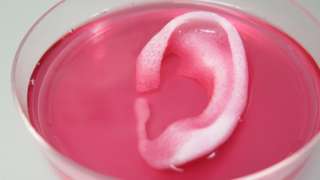
Several proof-of-concept experiments demonstrated the capabilities of ITOP. To show that ITOP can generate complex 3D structures, printed, human-sized external ears were implanted under the skin of mice. Two months later, the shape of the implanted ear was well-maintained and cartilage tissue and blood vessels had formed.

To demonstrate the ITOP can generate organized soft tissue structures, printed muscle tissue was implanted in rats. After two weeks, tests confirmed that the muscle was robust enough to maintain its structural characteristics, become vascularized and induce nerve formation.

And, to show that construction of a human-sized bone structure, jaw bone fragments were printed using human stem cells. The fragments were the size and shape needed for facial reconstruction in humans. To study the maturation of bioprinted bone in the body, printed segments of skull bone were implanted in rats. After five months, the bioprinted structures had formed vascularized bone tissue.

Ongoing studies will measure longer-term outcomes.

###

The research was supported, in part, by grants from the Armed Forces Institute of Regenerative Medicine (W81XWH-08-2-0032), the Telemedicine and Advanced Technology Research Center at the U.S. Army Medical Research and Material Command (W81XWH-07-1-0718) and the Defense Threat Reduction Agency (N66001-13-C-2027).

(Sometimes the information about the funding agencies is almost as interesting as the research.) Here’s a link to and a citation for the paper,

A 3D bioprinting system to produce human-scale tissue constructs with structural integrity by Hyun-Wook Kang, Sang Jin Lee, In Kap Ko, Carlos Kengla, James J Yoo, & Anthony Atala. Nature Biotechnology (2016)  doi:10.1038/nbt.3413 Published online 15 February 2016

This paper is behind a paywall.

As you can see, despite being printed, this latest ear is also spending time in a dish,

WakeBaptistEar

Courtesy: Wake Forest Baptist Medical Center