Tag Archives: tissue engineering

Better blood vessel growth for regenerative medicine?

If the organs and tissues grown in labs are to be successfully transplanted into bodies, then growing the blood vessels needed to maintain them becomes very important. A May 24, 2016 news item on ScienceDaily describes a new technique for the growing the vessels,

Growing tissues and organs in the lab for transplantation into patients could become easier after scientists discovered an effective way to produce three-dimensional networks of blood vessels, vital for tissue survival yet a current stumbling block in regenerative medicine.

In addition the technique to grow the blood vessels in a 3D scaffold cuts down on the risk of transplant rejection because it uses cells from the patient. It was developed by researchers from the University of Bath’s Department of Pharmacy and Pharmacology, working with colleagues at Bristol Heart Institute.

A May 24 (?), 2016 University of Bath (UK) press release, which originated the news item, expands on the theme (Note: Links have been removed),

So far the shortage of adequate patient-derived scaffolds that can support blood vessel growth has been a major limitation for regenerative medicine and tissue engineering.

Other methods only allow limited formation of small blood vessels such as capillaries, which makes tissue less likely to successfully transplant into a patient. In addition other methods of tissue growth require the use of animal products, unnecessary in this technique which uses human platelet lysate gel (hPLG) and endothelial progenitor cells (EPCs) – a type of cell which helps maintain blood vessel walls.

Dr Giordano Pula, Lecturer in Pharmacology at the University of Bath and head of the research team making the discovery, said: “A major challenge in tissue engineering and regenerative medicine is providing the new tissue with a network of blood vessels, and linking this to the patient’s existing blood supply; this is vital for the tissue’s survival and integration with adjacent tissues.

Dr Paul De Bank, Senior Lecturer in Pharmaceutics at the University of Bath and co-author of the paper, said: “By embedding EPCs in a gel derived from platelets, both of which can be isolated from the patient’s blood, we have demonstrated the formation of a network of small vessels. What is more, the gel contains a number of different growth factors which can induce existing blood vessels to infiltrate the gel and form connections with the new structures. Combining tissue-specific cells with this EPC-containing gel offers the potential for the formation of fully vascularised, functional tissues or organs, which integrate seamlessly with the patient.

“This discovery has the potential to accelerate the development of regenerative medicine applications.”

Professor Peter Weissberg, Medical Director of the British Heart Foundation, said: “Over a half a million people in the UK are living with heart failure, a disabling condition which can leave people unable to carry out everyday activities such as climbing the stairs or even walking to the shops. This regenerative research brings the British Heart Foundation’s goal to mend a broken heart and beat heart failure one step closer.

“All living tissues, including new heart muscle, need a blood supply. One of the fundamental goals of regenerative medicine is to find ways to grow a new blood supply from scratch. Previous attempts at this using human cells and synthetic scaffolds have met with only limited success.

“The beauty of this new approach is that components of a person’s own blood could be manipulated to create a scaffold on which new blood vessels could grow. This increases the likelihood that the new tissue will be integrated into the patient’s body which, if proven successful with more research, could improve the lives of people affected by heart failure.”

Here’s a link to and a citation for the paper,

Platelet lysate gel and endothelial progenitors stimulate microvascular network formation in vitro: tissue engineering implications by Tiago M. Fortunato, Cristina Beltrami, Costanza Emanueli, Paul A. De Bank & Giordano Pula. Scientific Reports 6, Article number: 25326 (2016)  doi:10.1038/srep25326 Published online: 04 May 2016

This is an open access paper.

One of the criticisms of Paolo Macchiarini’s work with synthetic tracheas centered around blood supply to the cells (from my April 19, 2016 posting; it was part 1 of a 2-part series),

This ground-breaking achievement consisted of bringing to life a dead windpipe from a donor, by putting it in a plastic box, a so-called ‘bioreactor’ together with bone marrow fluid (stem cells). A few weeks later, I [Pierre Delaere*]  wrote a letter to The Lancet, pointing out:

“The main drawback of the proposed reconstruction is the lack of an intrinsic blood supply to the trachea. We know that a good blood supply is the first requirement in all other tissue and organ transplantations. Therefore, the reported success of this technique is questionable” (correspondence by Delaere and Hermans, Lancet 2009).

The excerpt you’ve just seen features part of an open letter Pierre Delaere (a long time Macchiarini critic), published in Leonid Schneider’s blog ‘For Better Science’ in an April 2, 2016 posting.

Getting back to Bath, this is exciting stuff and I hope the research is reproducible.
Combining chitosan, agarose, and protein gelatine with clay nanotubes to create scaffolds for tissue engineering

Russian scientists have published work on clay nanotube-bipolymer composite scaffolds according to an April 29, 2016 news item on ScienceDaily,

Scientists combined three biopolymers, chitosan and agarose (polysaccharides), and a protein gelatine, as the materials to produce tissue engineering scaffolds and demonstrated the enhancement of mechanical strength (doubled pick load), higher water uptake and thermal properties in chitosan-gelatine-agarose hydrogels doped with halloysite [a clay mineral and a naturally occurring nanotube].

An April 29, 2016 Kazan Federal University (Russia) press release on EurekAlert, which originated the news item, provides more detail and context,

The fabrication of a prototype tissue having functional properties close to the natural ones is crucial for effective transplantation. Tissue engineering scaffolds are typically used as supports which allow cells to form tissue-like structures essentially required for the correct functioning of the cells under the conditions close to the three-dimensional tissue.

Chitosan, a natural biodegradable and chemically versatile biopolymer, has been effectively used in antibacterial, antifungal, anti-tumour and immunostimulating formulations. To overcome the disadvantages of pure chitosan scaffolds such as mechanical fragility and low biological resistance, chitosan scaffolds are typically doped with other supporting compounds which allow for mechanical strengthening, thus yielding ?omposite biologically resistant scaffolds.

Agarose is a galactose-based backbone polysaccharide isolated from red algae, having remarkable mechanical properties which are useful in the design of tissue engineering scaffolds.

Gelatine is formed from collagen by hydrolysis (breaking the triple-helix structure into single-strand molecules) and has a number of advantages over its precursor. It is less immunogenic compared with collagen and it retains informational signal sequences promoting cell adhesion, migration, differentiation and proliferation.

The surface irregularities of the scaffold pores due to the insoluble nanosized components promote the best adhesion of the cells on scaffold materials, while the nanoparticle fillers increase the composites’ strength. Thus, researchers doped halloysite nanotubes into a chitosan-agarose-gelatine matrix to design the implantable 3D cell scaffolds.

The resulting scaffolds demonstrate the shape memory upon deformation and have the porous structure suitable for cell adhesion and proliferation which is essential for artificial tissue fabrication. Macroscopic observations have confirmed that all the samples of scaffolds exhibited the sponge-like behaviour with the shape memory and shape reconstitution after deformation both in wet and dry states.

The swelling experiments indicated that the addition of halloysite can greatly improve the hydrophilicity and wetting of composite scaffolds. The incorporation of halloysite nanotubes into the scaffolds increases the water uptake and subsequently improves the biocompatibility. The intrinsic properties of halloysite nanotubes can be used for further improving the biocompatibility of scaffolds by the loading and sustained release of different bioactive compounds. This opens the prospect for fabrication of scaffolds with defined properties for directed differentiation of cells on matrixes due to gradual release of differentiation factors.

Experiments on two types of human cancer cells (A549 and Hep3B) show that in vitro cell adhesion and proliferation on the nanocomposites occur without changes in viability and cytoskeleton formation.

Further in vivo biocompatibility and biodegradability evaluation in rats has confirmed that the scaffolds promote the formation of novel blood vessels around the implantation sites. The scaffolds show excellent resorption within six weeks after implantation in rats. Neo-vascularization observed in newly formed connective tissue placed near the scaffold allows for the complete restoration of blood flow.

The results obtained indicate that the halloysite doped scaffolds are biocompatible as demonstrated both in vitro and in vivo. In addition, they confirm the great potential of chitosan-agarose-gelatine nanocomposite porous scaffolds doped with halloysite in tissue engineering with potential for sustained nanotube drug delivery.

For anyone interested about drug delivery and nanoparticles, there’s some interesting research profiled in my April 27, 2016 posting which describes how very few nanoparticles are actually delivered to specific sites.

Getting back to the regular program, here’s a link to and a citation for the paper on scaffolds and clay nanotubes,

Clay nanotube–biopolymer composite scaffolds for tissue engineering by Ekaterina A. Naumenko, Ivan D. Guryanov, Raghuvara Yendluri, Yuri M. Lvova, and Rawil F. Fakhrullin. Nanoscale, 2016,8, 7257-7271 DOI: 10.1039/C6NR00641H First published online 01 Mar 2016

This paper is behind a paywall.

Growing complex skin tissue—complete with hair follicles and sebaceous glands

A laboratory in Japan has managed to grow complex skin tissue according to an April 2, 2016 RIKEN (Japan) press release (also on EurekAlert but dated April 1, 2016),

Using reprogrammed iPS cells, scientists from the RIKEN Center for Developmental Biology (CDB) in Japan have, along with collaborators from Tokyo University of Science and other Japanese institutions, successfully grown complex skin tissue–complete with hair follicles and sebaceous glands–in the laboratory. They were then able to implant these three-dimensional tissues into living mice, and the tissues formed proper connections with other organ systems such as nerves and muscle fibers. This work opens a path to creating functional skin transplants for burn and other patients who require new skin.

Research into bioengineered tissues has led to important achievements in recent years–with a number of different tissue types being created–but there are still obstacles to be overcome. In the area of skin tissue, epithelial cells have been successfully grown into implantable sheets, but they did not have the proper appendages–the oil-secreting and sweat glands–that would allow them to function as normal tissue.

To perform the work, published in Science Advances, the researchers took cells from mouse gums and used chemicals to transform them into stem cell-like iPS cells. In culture, the cells properly developed into what is called an embryoid body (EB)?a three-dimensional clump of cells that partially resembles the developing embryo in an actual body. The researchers created EBs from iPS cells using Wnt10b signaling and then implanted multiple EBs into immune-deficient mice, where they gradually changed into differentiated tissue, following the pattern of an actual embryo. Once the tissue had differentiated, the scientists transplanted them out of those mice and into the skin tissue of other mice, where the tissues developed normally as integumentary tissue?the tissue between the outer and inner skin that is responsible for much of the function of the skin in terms of hair shaft eruption and fat excretion. Critically, they also found that the implanted tissues made normal connections with the surrounding nerve and muscle tissues, allowing it to function normally.

One important key to the development was that treatment with Wnt10b, a signaling molecule, resulted in a larger number of hair follicles, making the bioengineered tissue closer to natural tissue.

According to Takashi Tsuji of the RIKEN Center for Developmental Biology, who led the study, “Up until now, artificial skin development has been hampered by the fact that the skin lacked the important organs, such as hair follicles and exocrine glands, which allow the skin to play its important role in regulation. With this new technique, we have successfully grown skin that replicates the function of normal tissue. We are coming ever closer to the dream of being able to recreate actual organs in the lab for transplantation, and also believe that tissue grown through this method could be used as an alternative to animal testing of chemicals.”

Here’s a link to and a citation for the paper,

Bioengineering a 3D integumentary organ system from iPS cells using an in vivo transplantation model by Ryoji Takagi, Junko Ishimaru, Ayaka Sugawara, Koh-ei Toyoshima, Kentaro Ishida, Miho Ogawa, Kei Sakakibara, Kyosuke Asakawa, Akitoshi Kashiwakura, Masamitsu Oshima, Ryohei Minamide, Akio Sato, Toshihiro Yoshitake, Akira Takeda, Hiroshi Egusa, and Takashi Tsuji. Science Advances  01 Apr 2016: Vol. 2, no. 4, e1500887 DOI: 10.1126/sciadv.1500887

This appears to be an open access paper.

3D microtopographic scaffolds for transplantation and generation of reprogrammed human neurons

Should this technology prove successful once they start testing on people, the stated goal is to use it for the treatment of human neurodegenerative disorders such as Parkinson’s disease.  But, I can’t help wondering if they might also consider constructing an artificial brain.

Getting back to the 3D scaffolds for neurons, a March 17, 2016 US National Institutes of Health (NIH) news release (also on EurekAlert), makes the announcement,

National Institutes of Health-funded scientists have developed a 3D micro-scaffold technology that promotes reprogramming of stem cells into neurons, and supports growth of neuronal connections capable of transmitting electrical signals. The injection of these networks of functioning human neural cells — compared to injecting individual cells — dramatically improved their survival following transplantation into mouse brains. This is a promising new platform that could make transplantation of neurons a viable treatment for a broad range of human neurodegenerative disorders.

Previously, transplantation of neurons to treat neurodegenerative disorders, such as Parkinson’s disease, had very limited success due to poor survival of neurons that were injected as a solution of individual cells. The new research is supported by the National Institute of Biomedical Imaging and Bioengineering (NIBIB), part of NIH.

“Working together, the stem cell biologists and the biomaterials experts developed a system capable of shuttling neural cells through the demanding journey of transplantation and engraftment into host brain tissue,” said Rosemarie Hunziker, Ph.D., director of the NIBIB Program in Tissue Engineering and Regenerative Medicine. “This exciting work was made possible by the close collaboration of experts in a wide range of disciplines.”

The research was performed by researchers from Rutgers University, Piscataway, New Jersey, departments of Biomedical Engineering, Neuroscience and Cell Biology, Chemical and Biochemical Engineering, and the Child Health Institute; Stanford University School of Medicine’s Institute of Stem Cell Biology and Regenerative Medicine, Stanford, California; the Human Genetics Institute of New Jersey, Piscataway; and the New Jersey Center for Biomaterials, Piscataway. The results are reported in the March 17, 2016 issue of Nature Communications.

The researchers experimented in creating scaffolds made of different types of polymer fibers, and of varying thickness and density. They ultimately created a web of relatively thick fibers using a polymer that stem cells successfully adhered to. The stem cells used were human induced pluripotent stem cells (iPSCs), which can be readily generated from adult cell types such as skin cells. The iPSCs were induced to differentiate into neural cells by introducing the protein NeuroD1 into the cells.

The space between the polymer fibers turned out to be critical. “If the scaffolds were too dense, the stem cell-derived neurons were unable to integrate into the scaffold, whereas if they are too sparse then the network organization tends to be poor,” explained Prabhas Moghe, Ph.D., distinguished professor of biomedical engineering & chemical engineering at Rutgers University and co-senior author of the paper. “The optimal pore size was one that was large enough for the cells to populate the scaffold but small enough that the differentiating neurons sensed the presence of their neighbors and produced outgrowths resulting in cell-to-cell contact. This contact enhances cell survival and development into functional neurons able to transmit an electrical signal across the developing neural network.”

To test the viability of neuron-seeded scaffolds when transplanted, the researchers created micro-scaffolds that were small enough for injection into mouse brain tissue using a standard hypodermic needle. They injected scaffolds carrying the human neurons into brain slices from mice and compared them to human neurons injected as individual, dissociated cells.

The neurons on the scaffolds had dramatically increased cell-survival compared with the individual cell suspensions. The scaffolds also promoted improved neuronal outgrowth and electrical activity. Neurons injected individually in suspension resulted in very few cells surviving the transplant procedure.

Human neurons on scaffolds compared to neurons in solution were then tested when injected into the brains of live mice. Similar to the results in the brain slices, the survival rate of neurons on the scaffold network was increased nearly 40-fold compared to injected isolated cells. A critical finding was that the neurons on the micro-scaffolds expressed proteins that are involved in the growth and maturation of neural synapses–a good indication that the transplanted neurons were capable of functionally integrating into the host brain tissue.

The success of the study gives this interdisciplinary group reason to believe that their combined areas of expertise have resulted in a system with much promise for eventual treatment of human neurodegenerative disorders. In fact, they are now refining their system for specific use as an eventual transplant therapy for Parkinson’s disease. The plan is to develop methods to differentiate the stem cells into neurons that produce dopamine, the specific neuron type that degenerates in individuals with Parkinson’s disease. The work also will include fine-tuning the scaffold materials, mechanics and dimensions to optimize the survival and function of dopamine-producing neurons, and finding the best mouse models of the disease to test this Parkinson’s-specific therapy.

Here’s a link to and a citation for the paper,

Generation and transplantation of reprogrammed human neurons in the brain using 3D microtopographic scaffolds by Aaron L. Carlson, Neal K. Bennett, Nicola L. Francis, Apoorva Halikere, Stephen Clarke, Jennifer C. Moore, Ronald P. Hart, Kenneth Paradiso, Marius Wernig, Joachim Kohn, Zhiping P. Pang, & Prabhas V. Moghe. Nature Communications 7, Article number: 10862  doi:10.1038/ncomms10862 Published 17 March 2016

This paper is open access.

Feasibility of printing ear, bone, and muscle structures

Over ten years ago I attended a show at the Vancouver (Canada) Art Gallery titled ‘Massive Change’ where I saw part of a nose or ear being grown in a petri dish (the work was from an Israeli laboratory) and that was my introduction to tissue engineering. For anyone who’s been following the tissue engineering story, 3D printers have sped up the growth process considerably. More recently, researchers at Wake Forest Baptist Medical Center (North Carolina, US) have announced another step forward for growing organs and body parts, from a Feb. 15, 2016 Wake Forest Baptist Medical Center news release on EurekAlert,

Using a sophisticated, custom-designed 3D printer, regenerative medicine scientists at Wake Forest Baptist Medical Center have proved that it is feasible to print living tissue structures to replace injured or diseased tissue in patients.

Reporting in Nature Biotechnology, the scientists said they printed ear, bone and muscle structures. When implanted in animals, the structures matured into functional tissue and developed a system of blood vessels. Most importantly, these early results indicate that the structures have the right size, strength and function for use in humans.

“This novel tissue and organ printer is an important advance in our quest to make replacement tissue for patients,” said Anthony Atala, M.D., director of the Wake Forest Institute for Regenerative Medicine (WFIRM) and senior author on the study. “It can fabricate stable, human-scale tissue of any shape. With further development, this technology could potentially be used to print living tissue and organ structures for surgical implantation.”

With funding from the Armed Forces Institute of Regenerative Medicine, a federally funded effort to apply regenerative medicine to battlefield injuries, Atala’s team aims to implant bioprinted muscle, cartilage and bone in patients in the future.

Tissue engineering is a science that aims to grow replacement tissues and organs in the laboratory to help solve the shortage of donated tissue available for transplants. The precision of 3D printing makes it a promising method for replicating the body’s complex tissues and organs. However, current printers based on jetting, extrusion and laser-induced forward transfer cannot produce structures with sufficient size or strength to implant in the body.

The Integrated Tissue and Organ Printing System (ITOP), developed over a 10-year period by scientists at the Institute for Regenerative Medicine, overcomes these challenges. The system deposits both bio-degradable, plastic-like materials to form the tissue “shape” and water-based gels that contain the cells. In addition, a strong, temporary outer structure is formed. The printing process does not harm the cells.

A major challenge of tissue engineering is ensuring that implanted structures live long enough to integrate with the body. The Wake Forest Baptist scientists addressed this in two ways. They optimized the water-based “ink” that holds the cells so that it promotes cell health and growth and they printed a lattice of micro-channels throughout the structures. These channels allow nutrients and oxygen from the body to diffuse into the structures and keep them live while they develop a system of blood vessels.

It has been previously shown that tissue structures without ready-made blood vessels must be smaller than 200 microns (0.007 inches) for cells to survive. In these studies, a baby-sized ear structure (1.5 inches) survived and showed signs of vascularization at one and two months after implantation.

“Our results indicate that the bio-ink combination we used, combined with the micro-channels, provides the right environment to keep the cells alive and to support cell and tissue growth,” said Atala.

Another advantage of the ITOP system is its ability to use data from CT and MRI scans to “tailor-make” tissue for patients. For a patient missing an ear, for example, the system could print a matching structure.

Several proof-of-concept experiments demonstrated the capabilities of ITOP. To show that ITOP can generate complex 3D structures, printed, human-sized external ears were implanted under the skin of mice. Two months later, the shape of the implanted ear was well-maintained and cartilage tissue and blood vessels had formed.

To demonstrate the ITOP can generate organized soft tissue structures, printed muscle tissue was implanted in rats. After two weeks, tests confirmed that the muscle was robust enough to maintain its structural characteristics, become vascularized and induce nerve formation.

And, to show that construction of a human-sized bone structure, jaw bone fragments were printed using human stem cells. The fragments were the size and shape needed for facial reconstruction in humans. To study the maturation of bioprinted bone in the body, printed segments of skull bone were implanted in rats. After five months, the bioprinted structures had formed vascularized bone tissue.

Ongoing studies will measure longer-term outcomes.

###

The research was supported, in part, by grants from the Armed Forces Institute of Regenerative Medicine (W81XWH-08-2-0032), the Telemedicine and Advanced Technology Research Center at the U.S. Army Medical Research and Material Command (W81XWH-07-1-0718) and the Defense Threat Reduction Agency (N66001-13-C-2027).

(Sometimes the information about the funding agencies is almost as interesting as the research.) Here’s a link to and a citation for the paper,

A 3D bioprinting system to produce human-scale tissue constructs with structural integrity by Hyun-Wook Kang, Sang Jin Lee, In Kap Ko, Carlos Kengla, James J Yoo, & Anthony Atala. Nature Biotechnology (2016)  doi:10.1038/nbt.3413 Published online 15 February 2016

This paper is behind a paywall.

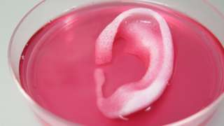
As you can see, despite being printed, this latest ear is also spending time in a dish,

WakeBaptistEar

Courtesy: Wake Forest Baptist Medical Center

Brushing your way to nanofibres

The scientists are using what looks like a hairbrush to create nanofibres ,

Figure 2: Brush-spinning of nanofibers. (Reprinted with permission by Wiley-VCH Verlag)) [downloaded from http://www.nanowerk.com/spotlight/spotid=41398.php]

Figure 2: Brush-spinning of nanofibers. (Reprinted with permission by Wiley-VCH Verlag)) [downloaded from http://www.nanowerk.com/spotlight/spotid=41398.php]

A Sept. 23, 2015 Nanowerk Spotlight article by Michael Berger provides an in depth look at this technique (developed by a joint research team of scientists from the University of Georgia, Princeton University, and Oxford University) which could make producing nanofibers for use in scaffolds (tissue engineering and other applications) more easily and cheaply,

Polymer nanofibers are used in a wide range of applications such as the design of new composite materials, the fabrication of nanostructured biomimetic scaffolds for artificial bones and organs, biosensors, fuel cells or water purification systems.

“The simplest method of nanofiber fabrication is direct drawing from a polymer solution using a glass micropipette,” Alexander Tokarev, Ph.D., a Research Associate in the Nanostructured Materials Laboratory at the University of Georgia, tells Nanowerk. “This method however does not scale up and thus did not find practical applications. In our new work, we introduce a scalable method of nanofiber spinning named touch-spinning.”

James Cook in a Sept. 23, 2015 article for Materials Views provides a description of the technology,

A glass rod is glued to a rotating stage, whose diameter can be chosen over a wide range of a few centimeters to more than 1 m. A polymer solution is supplied, for example, from a needle of a syringe pump that faces the glass rod. The distance between the droplet of polymer solution and the tip of the glass rod is adjusted so that the glass rod contacts the polymer droplet as it rotates.

Following the initial “touch”, the polymer droplet forms a liquid bridge. As the stage rotates the bridge stretches and fiber length increases, with the diameter decreasing due to mass conservation. It was shown that the diameter of the fiber can be precisely controlled down to 40 nm by the speed of the stage rotation.

The method can be easily scaled-up by using a round hairbrush composed of 600 filaments.

When the rotating brush touches the surface of a polymer solution, the brush filaments draw many fibers simultaneously producing hundred kilometers of fibers in minutes.

The drawn fibers are uniform since the fiber diameter depends on only two parameters: polymer concentration and speed of drawing.

Returning to Berger’s Spotlight article, there is an important benefit with this technique,

As the team points out, one important aspect of the method is the drawing of single filament fibers.

These single filament fibers can be easily wound onto spools of different shapes and dimensions so that well aligned one-directional, orthogonal or randomly oriented fiber meshes with a well-controlled average mesh size can be fabricated using this very simple method.

“Owing to simplicity of the method, our set-up could be used in any biomedical lab and facility,” notes Tokarev. “For example, a customized scaffold by size, dimensions and othermorphologic characteristics can be fabricated using donor biomaterials.”

Berger’s and Cook’s articles offer more illustrations and details.

Here’s a link to and a citation for the paper,

Touch- and Brush-Spinning of Nanofibers by Alexander Tokarev, Darya Asheghal, Ian M. Griffiths, Oleksandr Trotsenko, Alexey Gruzd, Xin Lin, Howard A. Stone, and Sergiy Minko. Advanced Materials DOI: 10.1002/adma.201502768ViewFirst published: 23 September 2015

This paper is behind a paywall.

Faster, cheaper, pseudo-organs (also known as organoids)

There’ve been any number of ‘organoid’ stories recently, here and elsewhere. This one is special due to a quasi extra-cellular matrix (cells have a type of skeletal structure known as an extra-cellular matrix or ECM). From a Sept. 11, 2015 news item on Azonano,

Scientists have developed a new technique that produces a user friendly, low cost, tissue-engineered pseudo-organ. The chip-based model produces a faithful mimic of the in vivo liver inside a scalable fluid-handling device, demonstrating proof of principle for toxicology tests and opening up potential use in drug testing and personalised medicine.

The work was done by researchers based at the Wake Forest Institute for Regenerative Medicine and the Virginia Tech-Wake Forest University School of Biomedical Engineering and Sciences. They created a device architecture within which were a series of 3D liver cell constructs enclosed in a biopolymer that closely mimics the extra-cellular matrix (ECM). Surrounding the printed cells with this ECM – which the body uses to support cells in the liver – makes this model a more realistic model of the cells in vivo.

A Sept. 10, 2015 Institute of Physics (IOP) press release, which originated the news item, provides more details about the technology,

The technique uses photopatterning to produce defined 3D constructs in a microfluidic system to probe the construct quickly. “It’s basically scaled-down pluming” explains Adam Hall, an author on the paper. “This paper describes fairly hefty devices – a few mm – but we’re working to scale this down considerably.”

Collaboration proved to be the key to success; “The challenges were not too significant once Adam and I merged our areas of expertise.” adds Aleksander Skardal, another author on the paper. “With his background in devices and microfabrication, and my background in biomaterials and biofabrication, the two technologies integrated rather well.”

The 3D construct device offers a new tool in the development of drug treatments. At present, 2D testing in vitro doesn’t replicate the activity of the cells, and until now 3D systems have not provided adequate interactions of cells with the ECM, or offered particularly high-throughput testing.

This is where the combination of technologies has proven vital. “3D constructs are less effective if you can’t probe them quickly” continues Hall. “And without some important task, microfluidics are just a fun party trick.”

The researchers were also happy how quickly the techniques fell into place.

“The first time we attempted to perform the in situ photopatterning – it just worked” says Skardal. “Science isn’t always that easy, so we knew we might be onto something.”

“Yes – this was one of those rare occasions where things seemed to fall into place” adds Hall.

The researchers are now working to reduce the size of the system allowing for multiple constructs that could be tested individually. This would open potential usage in drug testing and personalised medicine.

“Imagine being able to put, for example, tumor cells from a patient on a chip and test different drug cocktails on them” they conclude. “You could determine the effectiveness and side effects of different treatments on an individual basis without endangering the patient.”

Here’s a link to and a citation for the paper,

In situ patterned micro 3D liver constructs for parallel toxicology testing in a fluidic device by Aleksander Skardal, Mahesh Devarasetty, Shay Soker, and Adam R Hall. Biofabrication, Volume 7, Number 3 DOI: 10.1088/1758-5090/7/3/032001 Published 11 September 2015

© 2015 IOP Publishing Ltd

This is an open access paper.

People for the Ethical Treatment of Animals (PETA) and a grant for in vitro nanotoxicity testing

This grant seems to have gotten its start at a workshop held at the US Environmental Protection Agency (EPA) in Washington, D.C., Feb. 24-25, 2015 as per this webpage on the People for Ethical Treatment of Animals (PETA) International Science Consortium Limited website,

The invitation-only workshop included experts from different sectors (government, industry, academia and NGO) and disciplines (in vitro and in vivo inhalation studies of NMs, fibrosis, dosimetry, fluidic models, aerosol engineering, and regulatory assessment). It focused on the technical details for the development and preliminary assessment of the relevance and reliability of an in vitro test to predict the development of pulmonary fibrosis in cells co-cultured at the air-liquid interface following exposure to aerosolized multi-walled carbon nanotubes (MWCNTs). During the workshop, experts made recommendations on cell types, exposure systems, endpoints and dosimetry considerations required to develop the in vitro model for hazard identification of MWCNTs.

The method is intended to be included in a non-animal test battery to reduce and eventually replace the use of animals in studies to assess the inhalation toxicity of engineered NMs. The long-term vision is to develop a battery of in silico and in vitro assays that can be used in an integrated testing strategy, providing comprehensive information on biological endpoints relevant to inhalation exposure to NMs which could be used in the hazard ranking of substances in the risk assessment process.

A September 1, 2015 news item on Azonano provides an update,

The PETA International Science Consortium Ltd. announced today the winners of a $200,000 award for the design of an in vitro test to predict the development of lung fibrosis in humans following exposure to nanomaterials, such as multi-walled carbon nanotubes.

Professor Dr. Barbara Rothen-Rutishauser of the Adolphe Merkle Institute at the University of Fribourg, Switzerland and Professor Dr. Vicki Stone of the School of Life Sciences at Heriot-Watt University, Edinburgh, U.K. will jointly develop the test method. Professor Rothen-Rutishauser co-chairs the BioNanomaterials research group at the Adolphe Merkle Institute, where her research is focused on the study of nanomaterial-cell interactions in the lung using three-dimensional cell models. Professor Vicki Stone is the Director of the Nano Safety Research Group at Heriot-Watt University and the Director of Toxicology for SAFENANO.

The Science Consortium is also funding MatTek Corporation for the development of a three-dimensional reconstructed primary human lung tissue model to be used in Professors Rothen-Rutishauser and Stone’s work. MatTek Corporation has extensive expertise in manufacturing human cell-based, organotypic in vitro models for use in regulatory and basic research applications. The work at MatTek will be led by Dr. Patrick Hayden, Vice President of Scientific Affairs, and Dr. Anna Maione, head of MatTek’s airway models research group.

I was curious about MatTek Corporation and found this on company’s About Us webpage,

MatTek Corporation was founded in 1985 by two chemical engineering professors from MIT. In 1991 the company leveraged its core polymer surface modification technology into the emerging tissue engineering market.

MatTek Corporation is at the forefront of tissue engineering and is a world leader in the production of innovative 3D reconstructed human tissue models. Our skin, ocular, and respiratory tissue models are used in regulatory toxicology (OECD, EU guidelines) and address toxicology and efficacy concerns throughout the cosmetics, chemical, pharmaceutical and household product industries.

EpiDerm™, MatTek’s first 3D human cell based in vitro model, was introduced in 1993 and became an immediate technical and commercial success.

I wish them good luck in their research on developing better ways to test toxicity.

Job posting (post doc in tissue engineering [organ-on-a-chip]) for the Istituto Italiano di Technologia

Here’s the posting (deadline is July 19, 2015),

Istituto Italiano di Tecnologia (IIT), Genova, Italy (http://www.iit.it) is a private law Foundation, created with special Government Law no. 269 dated September 30th 2003 with the objective of promoting Italy’s technological development and higher education in science and technology. Research at IIT is carried out in highly innovative scientific fields with state-of-the-art technology.

A post-doc position to develop “Organs-on-Chips” is available in the Laboratory of Nanotechnology for Precision Medicine at IIT.

Candidates should have a PhD in Tissue Engineering or closely related fields and an excellent publication record and should be highly motivated to work in an interdisciplinary team.

The candidate will work on the development of microfluidic-based organs-on-chips.

These microchips will be used to recapitulate the microarchitecture and functions of living organs and pathological tissues such as cancer and will possibly form an accurate alternative to traditional animal testing and enable high-throughput screening of drugs and nanomedicines.

The candidate should have:

  • strong skills in tissue engineering as well as in molecular, cellular and in vivo tumor biology;
  • documented experience in primary cell culture and analysis;
  • excellent oral and written communication skills in English and the ability to work both independently and as part of a multidisciplinary team.

Interested applicants should contact directly Dr. Paolo Decuzzi ( paolo.decuzzi@iit.it) for any informal queries.

For a formal application  please send CV, list of publications with Impact Factor and names and email addresses of 2 referees to applications@iit.it

Please apply by July 19, 2015 quoting “Post doc position in Tissue Engineering” in the mail subject. [emphasis mine]

In order to comply with Italian law (art. 23 of Privacy Law of the Italian Legislative Decree n. 196/03), the candidate is kindly asked to give his/her consent to allow Istituto Italiano di Tecnologia to process his/her personal data.

We inform you that the information you provide will be solely used for the purpose of evaluating and selecting candidates in order to meet the requirements of Istituto Italiano di Tecnologia.

Your data will be processed by Istituto Italiano di Tecnologia, with its headquarters in Genoa, Via Morego 30, acting as the Data Holder, using computer and paper-based means, observing the rules on the protection of personal data, including those relating to the security of data, and they will not be communicated to thirds.

Please also note that, pursuant to art.7 of Legislative Decree 196/2003, you may exercise your rights at any time as a party concerned by contacting the Data Holder.

Istituto Italiano di Tecnologia is an Equal Opportunity Employer that actively seeks diversity in the workforce.

Don’t forget when preparing your application, should you be living on the West Coast of Canada or the US (not sure about Mexico as its coast veers east somewhat), Italy is +9 hours . This means you’d best get your application submitted by 3 pm PST on July 19, 2015.

Synthesizing nerve tissues with 3D printers and cellulose nanocrystals (CNC)

There are lots of stories about bioprinting and tissue engineering here and I think it’s time (again) for one which one has some good, detailed descriptions and, bonus, it features cellulose nanocrystals (CNC) and graphene. From a May 13, 2015 news item on Azonano,

The printer looks like a toaster oven with the front and sides removed. Its metal frame is built up around a stainless steel circle lit by an ultraviolet light. Stainless steel hydraulics and thin black tubes line the back edge, which lead to an inner, topside box made of red plastic.

In front, the metal is etched with the red Bio Bot logo. All together, the gray metal frame is small enough to fit on top of an old-fashioned school desk, but nothing about this 3D printer is old school. In fact, the tissue-printing machine is more like a sci-fi future in the flesh—and it has very real medical applications.

Researchers at Michigan Technological University hope to use this newly acquired 3D bioprinter to make synthesized nerve tissue. The key is developing the right “bioink” or printable tissue. The nanotechnology-inspired material could help regenerate damaged nerves for patients with spinal cord injuries, says Tolou Shokuhfar, an assistant professor of mechanical engineering and biomedical engineering at Michigan Tech.

Shokuhfar directs the In-Situ Nanomedicine and Nanoelectronics Laboratory at Michigan Tech, and she is an adjunct assistant professor in the Bioengineering Department and the College of Dentistry at the University of Illinois at Chicago.

In the bioprinting research, Shokuhfar collaborates with Reza Shahbazian-Yassar, the Richard and Elizabeth Henes Associate Professor in the Department of Mechanical Engineering-Engineering Mechanics at Michigan Tech. Shahbazian-Yassar’s highly interdisciplinary background on cellulose nanocrystals as biomaterials, funded by the National Science Foundation’s (NSF) Biomaterials Program, helped inspire the lab’s new 3D printing research. “Cellulose nanocrystals with extremely good mechanical properties are highly desirable for bioprinting of scaffolds that can be used for live tissues,” says Shahbazian-Yassar. [emphases mine]

A May 11, 2015 Michigan Technological University (MTU) news release by Allison Mills, which originated the news item, explains the ‘why’ of the research,

“We wanted to target a big issue,” Shokuhfar says, explaining that nerve regeneration is a particularly difficult biomedical engineering conundrum. “We are born with all the nerve cells we’ll ever have, and damaged nerves don’t heal very well.”

Other facilities are trying to address this issue as well. Many feature large, room-sized machines that have built-in cell culture hoods, incubators and refrigeration. The precision of this equipment allows them to print full organs. But innovation is more nimble at smaller scales.

“We can pursue nerve regeneration research with a simpler printer set-up,” says Shayan Shafiee, a PhD student working with Shokuhfar. He gestures to the small gray box across the lab bench.

He opens the red box under the top side of the printer’s box. Inside the plastic casing, a large syringe holds a red jelly-like fluid. Shafiee replenishes the needle-tipped printer, pulls up his laptop and, with a hydraulic whoosh, he starts to print a tissue scaffold.

The news release expands on the theme,

At his lab bench in the nanotechnology lab at Michigan Tech, Shafiee holds up a petri dish. Inside is what looks like a red gummy candy, about the size of a half-dollar.

Here’s a video from MTU illustrating the printing process,

Back to the news release, which notes graphene could be instrumental in this research,

“This is based on fractal geometry,” Shafiee explains, pointing out the small crenulations and holes pockmarking the jelly. “These are similar to our vertebrae—the idea is to let a nerve pass through the holes.”

Making the tissue compatible with nerve cells begins long before the printer starts up. Shafiee says the first step is to synthesize a biocompatible polymer that is syrupy—but not too thick—that can be printed. That means Shafiee and Shokuhfar have to create their own materials to print with; there is no Amazon.com or even a specialty shop for bioprinting nerves.

Nerves don’t just need a biocompatible tissue to act as a carrier for the cells. Nerve function is all about electric pulses. This is where Shokuhfar’s nanotechnology research comes in: Last year, she was awarded a CAREER grant from NSF for her work using graphene in biomaterials research. [emphasis mine] “Graphene is a wonder material,” she says. “And it has very good electrical conductivity properties.”

The team is extending the application of this material for nerve cell printing. “Our work always comes back to the question, is it printable or not?” Shafiee says, adding that a successful material—a biocompatible, graphene-bound polymer—may just melt, mush or flat out fail under the pressure of printing. After all, imagine building up a substance more delicate than a soufflé using only the point of a needle. And in the nanotechnology world, a needlepoint is big, even clumsy.

Shafiee and Shokuhfar see these issues as mechanical obstacles that can be overcome.

“It’s like other 3D printers, you need a design to work from,” Shafiee says, adding that he will tweak and hone the methodology for printing nerve cells throughout his dissertation work. He is also hopeful that the material will have use beyond nerve regeneration.

This looks like a news release designed to publicize work funded at MTU by the US National Science Foundation (NSF) which is why there is no mention of published work.

One final comment regarding cellulose nanocrystals (CNC). They have also been called nanocrystalline cellulose (NCC), which you will still see but it seems CNC is emerging as the generic term. NCC has been trademarked by CelluForce, a Canadian company researching and producing CNC (or if you prefer, NCC) from forest products.