Tag Archives: University of Wisconsin-Madison

Your tires generate energy that can be harvested

One day, this new work from the University of Wisconsin-Madison could help cut gas expenditures for your car and other motorized vehicles dependent on fossil fuels. A June 29, 2015 news item on Nanowerk describes the research (Note: A link has been removed),

A group of University of Wisconsin-Madison engineers and a collaborator from China have developed a nanogenerator that harvests energy from a car’s rolling tire friction.

An innovative method of reusing energy, the nanogenerator ultimately could provide automobile manufacturers a new way to squeeze greater efficiency out of their vehicles.

The researchers reported their development, which is the first of its kind, in a paper published May 6, 2015, in the journal Nano Energy (“Single-electrode triboelectric nanogenerator for scavenging friction energy from rolling tires”).

A June 29, 2015 University of Wisconsin-Madison news release (also on EurekAlert), which originated the news item, provides more details (Note: Links have been removed),

Xudong Wang, the Harvey D. Spangler fellow and an associate professor of materials science and engineering at UW-Madison, and his PhD student Yanchao Mao have been working on this device for about a year.

The nanogenerator relies on the triboelectric effect to harness energy from the changing electric potential between the pavement and a vehicle’s wheels. The triboelectric effect is the electric charge that results from the contact or rubbing together of two dissimilar objects.

Wang says the nanogenerator provides an excellent way to take advantage of energy that is usually lost due to friction.

“The friction between the tire and the ground consumes about 10 percent of a vehicle’s fuel,” he says. “That energy is wasted. So if we can convert that energy, it could give us very good improvement in fuel efficiency.”

The nanogenerator relies on an electrode integrated into a segment of the tire. When this part of the tire surface comes into contact with the ground, the friction between those two surfaces ultimately produces an electrical charge-a type of contact electrification known as the triboelectric effect.

During initial trials, Wang and his colleagues used a toy car with LED lights to demonstrate the concept. They attached an electrode to the wheels of the car, and as it rolled across the ground, the LED lights flashed on and off. The movement of electrons caused by friction was able to generate enough energy to power the lights, supporting the idea that energy lost to friction can actually be collected and reused.

“Regardless of the energy being wasted, we can reclaim it, and this makes things more efficient,” Wang says. “I think that’s the most exciting part of this, and is something I’m always looking for: how to save the energy from consumption.”

The researchers also determined that the amount of energy harnessed is directly related to the weight of a car, as well as its speed. Therefore the amount of energy saved can vary depending on the vehicle-but Wang estimates about a 10-percent increase in the average vehicle’s gas mileage given 50-percent friction energy conversion efficiency.

“There’s big potential with this type of energy,” Wang says. “I think the impact could be huge.”

Here’s a link to and a citation for the paper,

Single-electrode triboelectric nanogenerator for scavenging friction energy from rolling tires by Yanchao Mao, Dalong Geng, Erjun Liang, & Xudong Wang. Nano Energy Volume 15, July 2015, Pages 227–234 doi:10.1016/j.nanoen.2015.04.026

This paper is behind a paywall.

Boosting chip speeds with graphene

There’s a certain hysteria associated with chip speeds as engineers and computer scientists try to achieve the ever improved speed times that consumers have enjoyed for some decades. The question looms, is there some point at which we can no longer improve the speed? Well, we haven’t reached that point yet according to a June 18, 2015 news item on Nanotechnology Now,

Stanford engineers find a simple yet clever way to boost chip speeds: Inside each chip are millions of tiny wires to transport data; wrapping them in a protective layer of graphene could boost speeds by up to 30 percent. [emphasis mine]

A June 16, 2015 Stanford University news release by Tom Abate (also on EurekAlert but dated June 17, 2015), which originated the news item, describes how computer chips are currently designed and the redesign which yields more speed,

A typical computer chip includes millions of transistors connected with an extensive network of copper wires. Although chip wires are unimaginably short and thin compared to household wires both have one thing in common: in each case the copper is wrapped within a protective sheath.

For years a material called tantalum nitride has formed protective layer in chip wires.

Now Stanford-led experiments demonstrate that a different sheathing material, graphene, can help electrons scoot through tiny copper wires in chips more quickly.

Graphene is a single layer of carbon atoms arranged in a strong yet thin lattice. Stanford electrical engineer H.-S. Philip Wong says this modest fix, using graphene to wrap wires, could allow transistors to exchange data faster than is currently possible. And the advantages of using graphene would become greater in the future as transistors continue to shrink.

Wong led a team of six researchers, including two from the University of Wisconsin-Madison, who will present their findings at the Symposia of VLSI Technology and Circuits in Kyoto, a leading venue for the electronics industry.

Ling Li, a graduate student in electrical engineering at Stanford and first author of the research paper, explained why changing the exterior wrapper on connecting wires can have such a big impact on chip performance.

It begins with understanding the dual role of this protective layer: it isolates the copper from the silicon on the chip and also serve to conduct electricity.

On silicon chips, the transistors act like tiny gates to switch electrons on or off. That switching function is how transistors process data.

The copper wires between the transistors transport this data once it is processed.

The isolating material–currently tantalum nitride–keeps the copper from migrating into the silicon transistors and rendering them non-functional.

Why switch to graphene?

Two reasons, starting with the ceaseless desire to keep making electronic components smaller.

When the Stanford team used the thinnest possible layer of tantalum nitride needed to perform this isolating function, they found that the industry-standard was eight times thicker than the graphene layer that did the same work.

Graphene had a second advantage as a protective sheathing and here it’s important to differentiate how this outer layer functions in chip wires versus a household wires.

In house wires the outer layer insulates the copper to prevent electrocution or fires.

In a chip the layer around the wires is a barrier to prevent copper atoms from infiltrating the silicon. Were that to happen the transistors would cease to function. So the protective layer isolates the copper from the silicon

The Stanford experiment showed that graphene could perform this isolating role while also serving as an auxiliary conductor of electrons. Its lattice structure allows electrons to leap from carbon atom to carbon atom straight down the wire, while effectively containing the copper atoms within the copper wire.

These benefits–the thinness of the graphene layer and its dual role as isolator and auxiliary conductor–allow this new wire technology to carry more data between transistors, speeding up overall chip performance in the process.

In today’s chips the benefits are modest; a graphene isolator would boost wire speeds from four percent to 17 percent, depending on the length of the wire. [emphasis mine]

But as transistors and wires continue to shrink in size, the benefits of the ultrathin yet conductive graphene isolator become greater. [emphasis mine] The Stanford engineers estimate that their technology could increase wire speeds by 30 percent in the next two generations

The Stanford researchers think the promise of faster computing will induce other researchers to get interested in wires, and help to overcome some of the hurdles needed to take this proof of principle into common practice.

This would include techniques to grow graphene, especially growing it directly onto wires while chips are being mass-produced. In addition to his University of Wisconsin collaborator Professor Michael Arnold, Wong cited Purdue University Professor Zhihong Chen. Wong noted that the idea of using graphene as an isolator was inspired by Cornell University Professor Paul McEuen and his pioneering research on the basic properties of this marvelous material. Alexander Balandin of the University of California-Riverside has also made contributions to using graphene in chips.

“Graphene has been promised to benefit the electronics industry for a long time, and using it as a copper barrier is perhaps the first realization of this promise,” Wong said.

I gather they’ve decided to highlight the most optimistic outcomes.

Tiny, electrically conductive 3D-printed chair made from cellulose

Sweden’s Chalmers University of Technology researchers have just announced that they’ve printed a very small 3D chair with electrical properties using cellulose nanomaterials. From a June 17, 2015 news item on Nanowerk,

A group of researchers at Chalmers University of Technology have managed to print and dry three-dimensional objects made entirely by cellulose for the first time with the help of a 3D-bioprinter. They also added carbon nanotubes to create electrically conductive material. The effect is that cellulose and other raw material based on wood will be able to compete with fossil-based plastics and metals in the on-going additive manufacturing revolution, which started with the introduction of the 3D-printer.

Here’s the 3D-printed chair,

The tiny chair made of cellulose is a demonstrational object, printed using the 3D bioprinter at Chalmers University of Technology. Photo: Peter Widing

The tiny chair made of cellulose is a demonstrational object, printed using the 3D bioprinter at Chalmers University of Technology. Photo: Peter Widing

A June 17, 2015 Chalmers University of Technology press release (also on EurekAlert*), which originated the news item, describes the problem with printing from cellulose nanomaterials and how it was solved,

The difficulty using cellulose in additive manufacturing is that cellulose does not melt when heated. Therefore, the 3D printers and processes designed for printing plastics and metals cannot be used for materials like cellulose. The Chalmers researchers solved this problem by mixing cellulose nanofibrils in a hydrogel consisting of 95-99 percent water. The gel could then in turn be dispensed with high fidelity into the researchers’ 3D bioprinter, which was earlier used to produce scaffolds for growing cells, where the end application is patient-specific implants.

The next challenge was to dry the printed gel-like objects without them losing their three-dimensional shape.

“The drying process is critical,” Paul Gatenholm explains. “We have developed a process in which we freeze the objects and remove the water by different means as to control the shape of the dry objects. It is also possible to let the structure collapse in one direction, creating thin films.”

Furthermore, the cellulose gel was mixed with carbon nanotubes to create electrically conductive ink after drying. Carbon nanotubes conduct electricity, and another project at Wallenberg Wood Science Center aims at developing carbon nanotubes using wood.

Using the two gels together, one conductive and one non-conductive, and controlling the drying process, the researchers produced three-dimensional circuits, where the resolution increased significantly upon drying.

The two gels together provide a basis for the possible development of a wide range of products made by cellulose with in-built electric currents.

“Potential applications range from sensors integrated with packaging, to textiles that convert body heat to electricity, and wound dressings that can communicate with healthcare workers,” says Paul Gatenholm. “Our research group now moves on with the next challenge, to use all wood biopolymers, besides cellulose.”

The research findings are presented this week at the conference New Materials From Trees that takes place in Stockholm, Sweden, June 15-17 [2015].

The research team members are Ida Henriksson, Cristina de la Pena, Karl Håkansson, Volodymyr Kuzmenko and Paul Gatenholm at Chalmers University of Technology.

This research reminds me of another effort, a computer chip fashioned of cellulose nanofibrils (CNF) from the University of Wisconsin-Madison (mentioned in my May 27, 2015 post).

* EurekAlert link added June 18, 2015.

Wood chip/computer chip, a cellulose nanofibril development

I imagine researchers at the University of Wisconsin-Madison and the US Department of Agriculture Forest Products Laboratory (FPL) are hoping they have managed to create a wood-based computer chip that can be commercialized in the near future. From a May 26, 2015 news item on ScienceDaily,

Portable electronics — typically made of non-renewable, non-biodegradable and potentially toxic materials — are discarded at an alarming rate in consumers’ pursuit of the next best electronic gadget.

In an effort to alleviate the environmental burden of electronic devices, a team of University of Wisconsin-Madison researchers has collaborated with researchers in the Madison-based U.S. Department of Agriculture Forest Products Laboratory (FPL) to develop a surprising solution: a semiconductor chip made almost entirely of wood.

The research team, led by UW-Madison electrical and computer engineering professor Zhenqiang “Jack” Ma, described the new device in a paper published today (May 26, 2015) by the journal Nature Communications. The paper demonstrates the feasibility of replacing the substrate, or support layer, of a computer chip, with cellulose nanofibril (CNF), a flexible, biodegradable material made from wood.

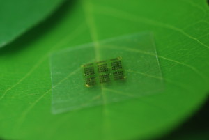
Here’s what the wood computer chip looks like,

A cellulose nanofibril (CNF) computer chip rests on a leaf. Photo: Yei Hwan Jung, Wisconsin Nano Engineering Device Laboratory

A cellulose nanofibril (CNF) computer chip rests on a leaf. Photo: Yei Hwan Jung, Wisconsin Nano Engineering Device Laboratory Courtesy University of Wisconsin-Madison

A May 25, 2015 University of Wisconsin-Madison news release by John Steeno, which originated the news item, provides more details,

“The majority of material in a chip is support. We only use less than a couple of micrometers for everything else,” Ma says. “Now the chips are so safe you can put them in the forest and fungus will degrade it. They become as safe as fertilizer.”

Zhiyong Cai, project leader for an engineering composite science research group at FPL, has been developing sustainable nanomaterials since 2009.

“If you take a big tree and cut it down to the individual fiber, the most common product is paper. The dimension of the fiber is in the micron stage,” Cai says. “But what if we could break it down further to the nano scale? At that scale you can make this material, very strong and transparent CNF paper.”

Working with Shaoqin “Sarah” Gong, a UW-Madison professor of biomedical engineering, Cai’s group addressed two key barriers to using wood-derived materials in an electronics setting: surface smoothness and thermal expansion.

“You don’t want it to expand or shrink too much. Wood is a natural hydroscopic material and could attract moisture from the air and expand,” Cai says. “With an epoxy coating on the surface of the CNF, we solved both the surface smoothness and the moisture barrier.”

Gong and her students also have been studying bio-based polymers for more than a decade. CNF offers many benefits over current chip substrates, she says.

“The advantage of CNF over other polymers is that it’s a bio-based material and most other polymers are petroleum-based polymers. Bio-based materials are sustainable, bio-compatible and biodegradable,” Gong says. “And, compared to other polymers, CNF actually has a relatively low thermal expansion coefficient.”

The group’s work also demonstrates a more environmentally friendly process that showed performance similar to existing chips. The majority of today’s wireless devices use gallium arsenide-based microwave chips due to their superior high-frequency operation and power handling capabilities. However, gallium arsenide can be environmentally toxic, particularly in the massive quantities of discarded wireless electronics.

Yei Hwan Jung, a graduate student in electrical and computer engineering and a co-author of the paper, says the new process greatly reduces the use of such expensive and potentially toxic material.

“I’ve made 1,500 gallium arsenide transistors in a 5-by-6 millimeter chip. Typically for a microwave chip that size, there are only eight to 40 transistors. The rest of the area is just wasted,” he says. “We take our design and put it on CNF using deterministic assembly technique, then we can put it wherever we want and make a completely functional circuit with performance comparable to existing chips.”

While the biodegradability of these materials will have a positive impact on the environment, Ma says the flexibility of the technology can lead to widespread adoption of these electronic chips.

“Mass-producing current semiconductor chips is so cheap, and it may take time for the industry to adapt to our design,” he says. “But flexible electronics are the future, and we think we’re going to be well ahead of the curve.”

Here’s a link to and a citation for the paper,

High-performance green flexible electronics based on biodegradable cellulose nanofibril paper by Yei Hwan Jung, Tzu-Hsuan Chang, Huilong Zhang, Chunhua Yao, Qifeng Zheng, Vina W. Yang, Hongyi Mi, Munho Kim,    Sang June Cho, Dong-Wook Park, Hao Jiang, Juhwan Lee,    Yijie Qiu, Weidong Zhou, Zhiyong Cai, Shaoqin Gong, & Zhenqiang Ma. Nature Communications 6, Article number: 7170 doi:10.1038/ncomms8170 Published 26 May 2015

This paper is open access.

US Dept. of Agriculture awards $3.8M for nanotechnology research grants

I wonder just how much funding the US Dept. of Agriculture (USDA) is devoting to nanotechnology this year (2015). I first came across an announcement of $23M in the body of a news item about Zinkicide (my April 7, 2015 posting),

Found in Florida orchards in 2005, a citrus canker, citrus greening, poses a serious threat to the US state’s fruit industry. An April 2, 2105 news item on phys.org describes a possible solution to the problem,

Since it was discovered in South Florida in 2005, the plague of citrus greening has spread to nearly every grove in the state, stoking fears among growers that the $10.7 billion-a-year industry may someday disappear.

Now the U.S. Department of Agriculture has awarded the University of Florida a $4.6 million grant aimed at testing a potential new weapon in the fight against citrus greening: Zinkicide, a bactericide invented by a nanoparticle researcher at the University of Central Florida.

An April 29, 2015 article by Diego Flammini for Farm.com describes the latest USDA nanotechnology funding announcement,

In an effort to increase America’s food security, nutrition, food safety and environmental protection, the United States Department of Agriculture’s (USDA) National Institute of Food and Agriculture (NIFA) announced $3.8 million in nanotechnology research grants.

Flammini lists three of the eight recipients,

University of Georgia
With $496,192, the research team will develop different sensors that are able to detect fungal pathogens in crops. The project will also develop a smartphone app for farmers to have so they can access their information whenever necessary.

Rutgers University
The school will use its $450,000 to conduct a nationwide survey about nanotechnology and gauge consumer beliefs about it and its relationship to health. Among the specifics it will touch on is the use of visuals to communicate nanotechnology.

University of Massachusetts
The researchers will concentrate their $444,200 on developing a platform to detect pathogens in food that is better than the current methods.

A full list of the recipients can be found in the April 27, 2015 USDA news release featuring the $3.8M in awards,

  • The University of Georgia, Athens, Ga., $496,192
  • University of Iowa, Iowa City, Iowa., $496,180
  • University of Kentucky Research Foundation, Lexington, Ky., $450,000
  • University of Massachusetts, Amherst, Mass., $444,200
  • North Dakota State University, Fargo, N.D., $149,714
  • Rutgers University, New Brunswick. N.J., $450,000
  • Pennsylvania State University, University Park, University Park, Pa., $447,788
  • West Virginia University, Morgantown, W. Va., $496,168
  • University of Wisconsin-Madison, Madison, Wis., $450,100

You can find more details about the awards in this leaflet featuring the USDA project descriptions for the eight recipients.

See-through medical sensors from the University of Wisconsin-Madison

This is quite the week for see-through medical devices based on graphene. A second team has developed a transparent sensor which could allow scientists to make observations of brain activity that are now impossible, according to an Oct. 20, 2014 University of Wisconsin-Madison news release (also on EurekAlert),

Neural researchers study, monitor or stimulate the brain using imaging techniques in conjunction with implantable sensors that allow them to continuously capture and associate fleeting brain signals with the brain activity they can see.

However, it’s difficult to see brain activity when there are sensors blocking the view.

“One of the holy grails of neural implant technology is that we’d really like to have an implant device that doesn’t interfere with any of the traditional imaging diagnostics,” says Justin Williams, the Vilas Distinguished Achievement Professor of biomedical engineering and neurological surgery at UW-Madison. “A traditional implant looks like a square of dots, and you can’t see anything under it. We wanted to make a transparent electronic device.”

The researchers chose graphene, a material gaining wider use in everything from solar cells to electronics, because of its versatility and biocompatibility. And in fact, they can make their sensors incredibly flexible and transparent because the electronic circuit elements are only 4 atoms thick—an astounding thinness made possible by graphene’s excellent conductive properties. “It’s got to be very thin and robust to survive in the body,” says Zhenqiang (Jack) Ma, the Lynn H. Matthias Professor and Vilas Distinguished Achievement Professor of electrical and computer engineering at UW-Madison. “It is soft and flexible, and a good tradeoff between transparency, strength and conductivity.”

Drawing on his expertise in developing revolutionary flexible electronics, he, Williams and their students designed and fabricated the micro-electrode arrays, which—unlike existing devices—work in tandem with a range of imaging technologies. “Other implantable micro-devices might be transparent at one wavelength, but not at others, or they lose their properties,” says Ma. “Our devices are transparent across a large spectrum—all the way from ultraviolet to deep infrared.”

The transparent sensors could be a boon to neuromodulation therapies, which physicians increasingly are using to control symptoms, restore function, and relieve pain in patients with diseases or disorders such as hypertension, epilepsy, Parkinson’s disease, or others, says Kip Ludwig, a program director for the National Institutes of Health neural engineering research efforts. “Despite remarkable improvements seen in neuromodulation clinical trials for such diseases, our understanding of how these therapies work—and therefore our ability to improve existing or identify new therapies—is rudimentary.”

Currently, he says, researchers are limited in their ability to directly observe how the body generates electrical signals, as well as how it reacts to externally generated electrical signals. “Clear electrodes in combination with recent technological advances in optogenetics and optical voltage probes will enable researchers to isolate those biological mechanisms. This fundamental knowledge could be catalytic in dramatically improving existing neuromodulation therapies and identifying new therapies.”

The advance aligns with bold goals set forth in President Barack Obama’s BRAIN (Brain Research through Advancing Innovative Neurotechnologies) Initiative. Obama announced the initiative in April 2013 as an effort to spur innovations that can revolutionize understanding of the brain and unlock ways to prevent, treat or cure such disorders as Alzheimer’s and Parkinson’s disease, post-traumatic stress disorder, epilepsy, traumatic brain injury, and others.

The UW-Madison researchers developed the technology with funding from the Reliable Neural-Interface Technology program at the Defense Advanced Research Projects Agency.

While the researchers centered their efforts around neural research, they already have started to explore other medical device applications. For example, working with researchers at the University of Illinois-Chicago, they prototyped a contact lens instrumented with dozens of invisible sensors to detect injury to the retina; the UIC team is exploring applications such as early diagnosis of glaucoma.

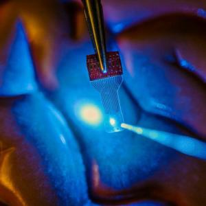
Here’s an image of the see-through medical implant,

Caption: A blue light shines through a clear implantable medical sensor onto a brain model. See-through sensors, which have been developed by a team of University of Wisconsin Madison engineers, should help neural researchers better view brain activity. Credit: Justin Williams research group

Caption: A blue light shines through a clear implantable medical sensor onto a brain model. See-through sensors, which have been developed by a team of University of Wisconsin Madison engineers, should help neural researchers better view brain activity.
Credit: Justin Williams research group

Here’s a link to and a citation for the paper,

Graphene-based carbon-layered electrode array technology for neural imaging and optogenetic applications by Dong-Wook Park, Amelia A. Schendel, Solomon Mikael, Sarah K. Brodnick, Thomas J. Richner, Jared P. Ness, Mohammed R. Hayat, Farid Atry, Seth T. Frye, Ramin Pashaie, Sanitta Thongpang, Zhenqiang Ma, & Justin C. Williams. Nature Communications 5, Article number: 5258 doi:10.1038/ncomms6258 Published
20 October 2014

This is an open access paper.

DARPA (US Defense Advanced Research Projects Agency), which funds this work at the University of Wisconsin-Madison, has also provided an Oct. 20, 2014 news release (also published an an Oct. 27, 2014 news item on Nanowerk) describing this research from the military perspective, which may not be what you might expect. First, here’s a description of the DARPA funding programme underwriting this research, from DARPA’s Reliable Neural-Interface Technology (RE-NET) webpage,

Advancing technology for military uniforms, body armor and equipment have saved countless lives of our servicemembers injured on the battlefield.  Unfortunately, many of those survivors are seriously and permanently wounded, with unprecedented rates of limb loss and traumatic brain injury among our returning soldiers. This crisis has motivated great interest in the science of and technology for restoring sensorimotor functions lost to amputation and injury of the central nervous system. For a decade now, DARPA has been leading efforts aimed at ‘revolutionizing’ the state-of-the-art in prosthetic limbs, recently debuting 2 advanced mechatronic limbs for the upper extremity. These new devices are truly anthropomorphic and capable of performing dexterous manipulation functions that finally begin to approach the capabilities of natural limbs. However, in the absence of a high bandwidth, intuitive interface for the user, these limbs will never achieve their full potential in improving the quality of life for the wounded soldiers that could benefit from this advanced technology.

DARPA created the Reliable Neural-Interface Technology (RE-NET) program in 2010 to directly address the need for high performance neural interfaces to control dexterous functions made possible with advanced prosthetic limbs.  Specifically, RE-NET seeks to develop the technologies needed to reliably extract information from the nervous system, and to do so at a scale and rate necessary to control many degree-of-freedom (DOF) machines, such as high-performance prosthetic limbs. Prior to the DARPA RE-NET program, all existing methods to extract neural control signals were inadequate for amputees to control high-performance prostheses, either because the level of extracted information was too low or the functional lifetime was too short. However, recent technological advances create new opportunities to solve both of these neural-interface problems. For example, it is now feasible to develop high-resolution peripheral neuromuscular interfaces that increase the amount of information obtained from the peripheral nervous system.  Furthermore, advances in cortical microelectrode technologies are extending the durability of neural signals obtained from the brain, making it possible to create brain-controlled prosthetics that remain useful over the full lifetime of the patient.

Canada’s Situating Science in Fall 2014

Canada’s Situating Science cluster (network of humanities and social science researchers focused on the study of science) has a number of projects mentioned and in its Fall 2014 newsletter,

1. Breaking News
It’s been yet another exciting spring and summer with new developments for the Situating Science SSHRC Strategic Knowledge Cluster team and HPS/STS [History of Philosophy of Science/Science and Technology Studies] research. And we’ve got even more good news coming down the pipeline soon…. For now, here’s the latest.

1.1. New 3 yr. Cosmopolitanism Partnership with India and Southeast Asia
We are excited to announce that the Situating Science project has helped to launch a new 3 yr. 200,000$ SSHRC Partnership Development Grant on ‘Cosmopolitanism and the Local in Science and Nature’ with institutions and scholars in Canada, India and Singapore. Built upon relations that the Cluster has helped establish over the past few years, the project will closely examine the actual types of negotiations that go into the making of science and its culture within an increasingly globalized landscape. A recent workshop on Globalizing History and Philosophy of Science at the Asia Research Institute at the National University of Singapore helped to mark the soft launch of the project (see more in this newsletter).

ARI along with Manipal University, Jawaharlal Nehru University, University of King’s College, Dalhousie University, York University, University of Toronto, and University of Alberta, form the partnership from which the team will seek new connections and longer term collaborations. The project’s website will feature a research database, bibliography, syllabi, and event information for the project’s workshops, lecture series, summer schools, and artifact work. When possible, photos, blogs, podcasts and videos from events will be posted online as well. The project will have its own mailing list so be sure to subscribe to that too. Check it all out: www.CosmoLocal.org

2.1. Globalizing History and Philosophy of Science workshop in Singapore August 21-22 2014
On August 21 and 22, scholars from across the globe gathered at the Asia Research Institute at the National University of Singapore to explore key issues in global histories and philosophies of the sciences. The setting next to the iconic Singapore Botanical Gardens provided a welcome atmosphere to examine how and why globalizing the humanities and social studies of science generates intellectual and conceptual tensions that require us to revisit, and possibly rethink, the leading notions that have hitherto informed the history, philosophy and sociology of science.

The keynote by Sanjay Subrahmanyam (UCLA) helped to situate discussions within a larger issue of paradigms of civilization. Workshop papers explored commensurability, translation, models of knowledge exchange, indigenous epistemologies, commercial geography, translation of math and astronomy, transmission and exchange, race, and data. Organizer Arun Bala and participants will seek out possibilities for publishing the proceedings. The event partnered with La Trobe University and Situating Science, and it helped to launch a new 3 yr. Cosmopolitanism project. For more information visit: www.CosmoLocal.org

2.2. Happy Campers: The Summer School Experience

We couldn’t help but feel like we were little kids going to summer camp while our big yellow school bus kicked up dust driving down a dirt road on a hot summer’s day. In this case it would have been a geeky science camp. We were about to dive right into day-long discussions of key pieces from Science and Technology Studies and History and Philosophy of Science and Technology.

Over four and a half days at one of the Queen’s University Biology Stations at the picturesque Elbow Lake Environmental Education Centre, 18 students from across Canada explored the four themes of the Cluster. Each day targeted a Cluster theme, which was introduced by organizer Sergio Sismondo (Sociology and Philosophy, Queen’s). Daryn Lehoux (Classics, Queen’s) explained key concepts in Historical Epistemology and Ontology. Using references of the anti-magnetic properties of garlic (or garlic’s antipathy with the loadstone) from the ancient period, Lehoux discussed the importance and significance of situating the meaning of a thing within specific epistemological contexts. Kelly Bronson (STS, St. Thomas University) explored modes of science communication and the development of the Public Engagement with Science and Technology model from the deficit model of Public Understanding of Science and Technology during sessions on Science Communication and its Publics. Nicole Nelson (University of Wisconsin-Madison) explained Material Culture and Scientific/Technological Practices by dissecting the meaning of animal bodies and other objects as scientific artifacts. Gordon McOuat wrapped up the last day by examining the nuances of the circulation and translation of knowledge and ‘trading zones’ during discussions of Geographies and Sites of Knowledge.

2.3. Doing Science in and on the Oceans
From June 14 to June 17, U. King’s College hosted an international workshop on the place and practice of oceanography in celebration of the work of Dr. Eric Mills, Dalhousie Professor Emeritus in Oceanography and co-creator of the History of Science and Technology program. Leading ocean scientists, historians and museum professionals came from the States, Europe and across Canada for “Place and Practice: Doing Science in and on the Ocean 1800-2012”. The event successfully connected different generations of scholars, explored methodologies of material culture analysis and incorporated them into mainstream historical work. There were presentations and discussions of 12 papers, an interdisciplinary panel discussion with keynote lecture by Dr. Mills, and a presentation at the Maritime Museum of the Atlantic by Canada Science and Technology Museum curator, David Pantalony. Paper topics ranged from exploring the evolving methodology of oceanographic practice to discussing ways that the boundaries of traditional scientific writing have been transcended. The event was partially organized and supported by the Atlantic Node and primary support was awarded by the SSHRC Connection Grant.

2.4. Evidence Dead or Alive: The Lives of Evidence National Lecture Series

The 2014 national lecture series on The Lives of Evidence wrapped up on a high note with an interdisciplinary panel discussion of Dr. Stathis Psillos’ exploration of the “Death of Evidence” controversy and the underlying philosophy of scientific evidence. The Canada Research Chair in Philosophy of Science spoke at the University of Toronto with panelists from law, philosophy and HPS. “Evidence: Wanted Dead of Alive” followed on the heels of his talk at the Institute for Science, Society and Policy “From the ‘Bankruptcy of Science’ to the ‘Death of Evidence’: Science and its Value”.

In 6 parts, The Lives of Evidence series examined the cultural, ethical, political, and scientific role of evidence in our world. The series formed as response to the recent warnings about the “Death of Evidence” and “War on Science” to explore what was meant by “evidence”, how it is interpreted, represented and communicated, how trust is created in research, what the relationship is between research, funding and policy and between evidence, explanations and expertise. It attracted collaborations from such groups as Evidence for Democracy, the University of Toronto Evidence Working Group, Canadian Centre for Ethics in Public Affairs, Dalhousie University Health Law Institute, Rotman Institute of Philosophy and many more.

A December [2013] symposium, “Hype in Science”, marked the soft launch of the series. In the all-day public event in Halifax, leading scientists, publishers and historians and philosophers of science discussed several case studies of how science is misrepresented and over-hyped in top science journals. Organized by the recent winner of the Gerhard Herzberg Canada Gold Medal for Science and Engineering, Ford Doolittle, the interdisciplinary talks in “Hype” explored issues of trustworthiness in science publications, scientific authority, science communication, and the place of research in the broader public.

The series then continued to explore issues from the creation of the HIV-Crystal Meth connection (Cindy Patton, SFU), Psychiatric Research Abuse (Carl Elliott, U. Minnesota), Evidence, Accountability and the Future of Canadian Science (Scott Findlay, Evidence for Democracy), Patents and Commercialized Medicine (Jim Brown, UofT), and Clinical Trials (Joel Lexchin, York).

All 6 parts are available to view on the Situating Science YouTube channel.You can read a few blogs from the events on our website too. Some of those involved are currently discussing possibilities of following up on some of the series’ issues.

2.5. Other Past Activities and Events
The Frankfurt School: The Critique of Capitalist Culture (July, UBC)

De l’exclusion à l’innovation théorique: le cas de l’éconophysique ; Prosocial attitudes and patterns of academic entrepreneurship (April, UQAM)

Critical Itineraries Technoscience Salon – Ontologies (April, UofT)

Technologies of Trauma: Assessing Wounds and Joining Bones in Late Imperial China (April, UBC)

For more, check out: www.SituSci.ca

You can find some of the upcoming talks and the complete Fall 2014 Situating Science newsletter here.

About one week after receiving the newsletter, I got this notice (Sept. 11, 2014),

We are ecstatic to announce that the Situating Science SSHRC Strategic Knowledge Cluster is shortlisted for a highly competitive SSHRC Partnership Impact Award!

And what an impact we’ve had over the past seven years: Organizing and supporting over 20 conferences and workshops, 4 national lecture series, 6 summer schools, and dozens of other events. Facilitating the development of 4 new programs of study at partner institutions. Leveraging more than one million dollars from Nodal partner universities plus more than one million dollars from over 200 supporting and partnering organizations. Hiring over 30 students and 9 postdoctoral fellows. Over 60 videos and podcasts as well as dozens of student blogs and over 50 publications. Launching a new Partnership Development Grant between Canada, India and Southeast Asia. Developing a national consortium…And more!

The winners will be presented with their awards at a ceremony in Ottawa on Monday, November 3, 2014.

From the Sept. 11, 2014 Situating Science press release:

University of King’s College [Nova Scotia, Canada] professor Dr. Gordon McOuat has been named one of three finalists for the Social Sciences and Humanities Research Council of Canada’s (SSHRC) Partnership Award, one of five Impact Awards annually awarded by SSHRC.

Congratulations on the nomination and I wish Gordon McQuat and Situating Science good luck in the competition.

A labradoodle, gold nanoparticles, and cancer treatment for dogs and cats

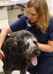
Here’s the labradoodle,

Caption: Dr. Shawna Klahn, an assistant professor of oncology at the Virginia-Maryland College of Veterinary Medicine, performs a checkup on "Grayton" four weeks after the animal's experimental cancer treatment involving gold nanoparticles and a targeted laser therapy. Credit: Virginia Tech

Caption: Dr. Shawna Klahn, an assistant professor of oncology at the Virginia-Maryland College of Veterinary Medicine, performs a checkup on “Grayton” four weeks after the animal’s experimental cancer treatment involving gold nanoparticles and a targeted laser therapy.
Credit: Virginia Tech

An Aug. 6, 2014 news item on Azonano outlines ‘Grayton’s’ story and how gold nanoparticles will factor in,

When Michael and Sandra Friedlander first came to the Virginia-Maryland College of Veterinary Medicine three years ago with their dog, Grayton, they learned some bad news: Grayton had nasal adenocarcinoma, a form of cancer with a short life expectancy.

“Most dogs with this form of cancer are with their owners no more than a few months after the diagnosis, but here Grayton is three years later,” said Michael Friedlander, who is the executive director of the Virginia Tech Carilion Research Institute and senior dean at the Virginia Tech Carilion School of Medicine.

No stranger to medical research, Friedlander was referred by Veterinary Teaching Hospital clinicians to an experimental treatment at the University of Florida called stereotactic radiation therapy, which delivers precise, high dosages of radiation to a tumor and can only be performed once.

“That shrunk the tumor down to almost nothing,” said Friedlander, who is also the associate provost for health sciences at Virginia Tech. “We knew when Grayton had the procedure that we couldn’t do it again, but now the cancer is back.”

An Aug. 4, 2014 Virginia Tech news release (also on EurekAlert) by Michael Sutphin, which originated the news item, explains what occasioned the release and how gold nanoparticles are being used in veterinary treatment for cancer,

Today [Aug. 4, 2014], the 11-year-old Labradoodle is the first patient at the Virginia-Maryland College of Veterinary Medicine in a new clinical trial that is testing the use of gold nanoparticles and a targeted laser treatment for solid tumors in dogs and cats. The study is one of several on new treatments for client-owned companion animals at the college. In January [2014], the college established the Veterinary Clinical Research Office to help facilitate this work.

“Clinical research at the veterinary college involves both primary research focused on advancing the treatment and diagnosis of veterinary diseases and translational research in which spontaneous diseases in animals can be used as models of human disease,” said Dr. Greg Daniel, head of the Department of Small Animal Clinical Sciences. “In the latter situation, we can provide our companion animal patients with treatment and diagnostic options that are not yet available in mainstream human medicine.”

Although medical researchers have tested gold nanoparticles with targeted laser treatments on human patients with some success, the treatment is still new to both human and veterinary medicine. The college is one of four current veterinary schools around the country testing the AuroLase therapy developed by Nanospectra Biosciences Inc., a startup company based in Houston, Texas. The others are Texas A&M University, the University of Wisconsin-Madison, and the University of Georgia.

Dr. Nick Dervisis, assistant professor of oncology in the Department of Small Animal Clinical Sciences, is leading the Nanospectra-funded study. Following a rhinoscopy performed on Grayton by Dr. David Grant, associate professor of internal medicine, Dervisis began the one-time, experimental therapy.

“The treatment involves two phases,” Dervisis said. “First, we infuse the patient with the gold nanoparticles. Although the nanoparticles distribute throughout the body, they tend to concentrate around blood vessels associated with tumors. Within 36 hours, they have cleared the bloodstream except for tumors. The gold nanoparticles are small enough to circulate freely in the bloodstream and become temporarily captured within the incomplete blood vessel walls common in solid tumors. Then, we use a non-ablative laser on the patient.”

Dervisis explained that a non-ablative laser is not strong enough to harm the skin or normal tissue, but “it does cause the remaining nanoparticles to absorb the laser energy and convert it into heat so that they damage the tumor cells.”

Like all clinical trials, the study involves many unknowns, including the treatment’s usefulness and effectiveness. One month after the AuroLase treatment, the nosebleeds that initially brought Grayton back to the Veterinary Teaching Hospital had stopped and Grayton has no other side effects.

“I’m delighted with the care and service that Grayton has received at the veterinary college,” said Friedlander, who explained that the treatment appears to be safe even though researchers do not know whether it is effective yet. “Grayton recently came with us on our annual vacation at the beach. We didn’t know if he would be able to come again, so it was great to have him with us swimming, catching fish and crabs, and doing what dogs do.”

Current clinical trials at the veterinary college range from the use of MRI to distinguish between benign and cancerous lymph nodes in dogs with oral melanoma, to a new chemotherapy drug for dogs with brain tumors, to the treatment of invasive skin cancer in horses with high-voltage, high-frequency electrical pulses. A complete list of current trials can be found at the college’s new clinical trials website.

Mindy Quigley, who oversees the college’s Veterinary Clinical Research Office, explained that veterinary trials, which follow a four-phase process and a variety of regulations similar to human medicine, have another layer of complexity that human trials do not.

“Variation among species means that a therapy that has proven safe and effective in, for example, humans or dogs, may not work for horses,” said Quigley, who comes to the college from the University of Edinburgh’s College of Medicine and Veterinary Medicine, where she helped set up a new neurology research clinic with funding from author J.K. Rowling. “Many veterinary clinical trials must therefore take therapies that have worked in one species and test them in other species with similar conditions. This is a necessary step to determine if a proposed treatment is safe and effective for our companion animals.”

Grayton may be the first companion animal in the AuroLase study at the veterinary college, but he certainly won’t be the last. Dervisis is continuing to enroll patients in the study and is seeking dogs and cats of a certain size with solid tumors who have not recently received radiation therapy or chemotherapy.

Interested parties can check this site for current clinical trials, including the Aurolase study,  being held by the Virginia-Maryland Regional College of Veterinary Medicine.

Nanojuice in your gut

A July 7, 2014 news item on Azonano features a new technique that could help doctors better diagnose problems in the intestines (guts),

Located deep in the human gut, the small intestine is not easy to examine. X-rays, MRIs and ultrasound images provide snapshots but each suffers limitations. Help is on the way.

University at Buffalo [State University of New York] researchers are developing a new imaging technique involving nanoparticles suspended in liquid to form “nanojuice” that patients would drink. Upon reaching the small intestine, doctors would strike the nanoparticles with a harmless laser light, providing an unparalleled, non-invasive, real-time view of the organ.

A July 5, 2014 University of Buffalo news release (also on EurekAlert) by Cory Nealon, which originated the news item, describes some of the challenges associated with medical imaging of small intestines,

“Conventional imaging methods show the organ and blockages, but this method allows you to see how the small intestine operates in real time,” said corresponding author Jonathan Lovell, PhD, UB assistant professor of biomedical engineering. “Better imaging will improve our understanding of these diseases and allow doctors to more effectively care for people suffering from them.”

The average human small intestine is roughly 23 feet long and 1 inch thick. Sandwiched between the stomach and large intestine, it is where much of the digestion and absorption of food takes place. It is also where symptoms of irritable bowel syndrome, celiac disease, Crohn’s disease and other gastrointestinal illnesses occur.

To assess the organ, doctors typically require patients to drink a thick, chalky liquid called barium. Doctors then use X-rays, magnetic resonance imaging and ultrasounds to assess the organ, but these techniques are limited with respect to safety, accessibility and lack of adequate contrast, respectively.

Also, none are highly effective at providing real-time imaging of movement such as peristalsis, which is the contraction of muscles that propels food through the small intestine. Dysfunction of these movements may be linked to the previously mentioned illnesses, as well as side effects of thyroid disorders, diabetes and Parkinson’s disease.

The news release goes on to describe how the researchers manipulated dyes that are usually unsuitable for the purpose of imaging an organ in the body,

Lovell and a team of researchers worked with a family of dyes called naphthalcyanines. These small molecules absorb large portions of light in the near-infrared spectrum, which is the ideal range for biological contrast agents.

They are unsuitable for the human body, however, because they don’t disperse in liquid and they can be absorbed from the intestine into the blood stream.

To address these problems, the researchers formed nanoparticles called “nanonaps” that contain the colorful dye molecules and added the abilities to disperse in liquid and move safely through the intestine.

In laboratory experiments performed with mice, the researchers administered the nanojuice orally. They then used photoacoustic tomography (PAT), which is pulsed laser lights that generate pressure waves that, when measured, provide a real-time and more nuanced view of the small intestine.

The researchers plan to continue to refine the technique for human trials, and move into other areas of the gastrointestinal tract.

Here’s an image of the nanojuice in the guts of a mouse,

The combination of "nanojuice" and photoacoustic tomography illuminates the intestine of a mouse. (Credit: Jonathan Lovell)

The combination of “nanojuice” and photoacoustic tomography illuminates the intestine of a mouse. (Credit: Jonathan Lovell)

This is an international collaboration both from a research perspective and a funding perspective (from the news release),

Additional authors of the study come from UB’s Department of Chemical and Biological Engineering, Pohang University of Science and Technology in Korea, Roswell Park Cancer Institute in Buffalo, the University of Wisconsin-Madison, and McMaster University in Canada.

The research was supported by grants from the National Institutes of Health, the Department of Defense and the Korean Ministry of Science, ICT and Future Planning.

Here’s a link to and a citation for the paper,

Non-invasive multimodal functional imaging of the intestine with frozen micellar naphthalocyanines by Yumiao Zhang, Mansik Jeon, Laurie J. Rich, Hao Hong, Jumin Geng, Yin Zhang, Sixiang Shi, Todd E. Barnhart, Paschalis Alexandridis, Jan D. Huizinga, Mukund Seshadri, Weibo Cai, Chulhong Kim, & Jonathan F. Lovell. Nature Nanotechnology (2014) doi:10.1038/nnano.2014.130 Published online 06 July 2014

This paper is behind a paywall.